Article

Three New Dipeptide and Two New Polyketide Derivatives from the Mangrove-Derived Fungus Talaromyces sp.: Antioxidant Activity of Two Isolated Substances

1
CAS Key Laboratory of Tropical Marine Bio-resources and Ecology/Guangdong Key Laboratory of Marine Materia Medica, South China Sea Institute of Oceanology, Chinese Academy of Sciences, Guangzhou 510301, China
2
University of Chinese Academy of Sciences, Beijing 100049, China
3
Guangxi Key Laboratory of Marine Drugs, Institute of Marine Drugs, Guangxi University of Chinese Medicine, Nanning 530200, China
4
National Institute of Fundamental Studies, Hantana Road, Kandy 200000, Sri Lanka
*
Author to whom correspondence should be addressed.
These authors contributed equally to this work.
Mar. Drugs 2024, 22(12), 559; https://doi.org/10.3390/md22120559 (registering DOI)
Submission received: 26 November 2024 / Revised: 8 December 2024 / Accepted: 11 December 2024 / Published: 14 December 2024

Abstract

:
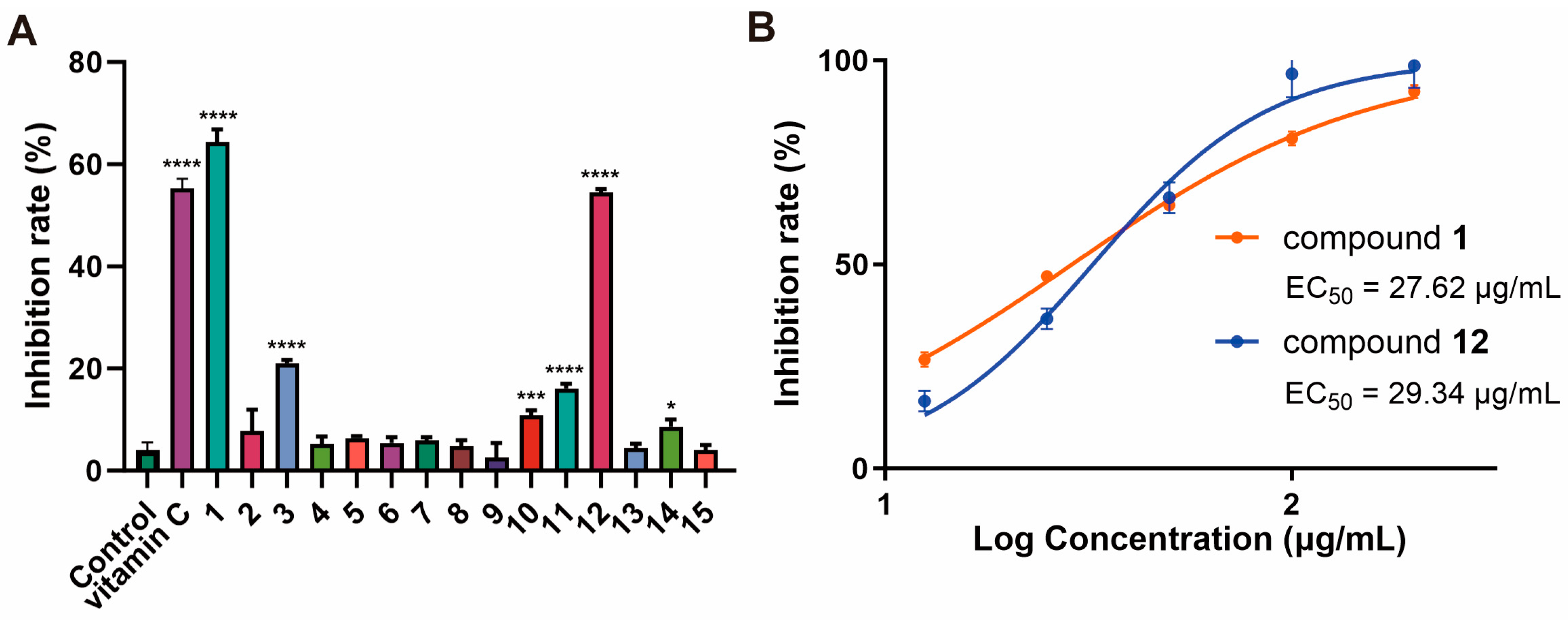
Five new metabolites, including three cyclic dipeptide derivatives (13) and two new polyketides (1011), together with nine known ones (49 and 1215), were isolated from the mangrove-sediments-derived fungus Talaromyces sp. SCSIO 41431. Their structures were determined using detailed NMR, MS spectroscopic analyses, and quantum chemical calculations. X-ray single-crystal diffraction analysis of 1 was described. Compounds 1315 demonstrated activity against Staphylococcus aureus, with MIC values ranging from 25 to 50 µg/mL. Compound 9 showed activity against Escherichia coli, Streptococcus suis, and Erysipelothrix rhusiopathiae, with an MIC value of 100 µg/mL. In addition, compounds 1 and 12 showed DPPH radical scavenging activity, with the EC50 of 27.62 and 29.34 µg/mL, compared to the positive control (ascorbic acid, EC50, 12.74 µg/mL).

1. Introduction

Mangroves, as highly productive ecosystems widely distributed along tropical and subtropical coasts, contain rich microbial resources [1,2]. A large number of structurally novel and biologically active natural products have been reported to be isolated from mangroves, making them one of the most important sources of marine natural products [3,4]. From January 2021 to May 2024, researchers isolated 165 novel secondary metabolites from 39 strains of fungi and 2 strains of actinomycetes from mangroves. Nearly half of these metabolites exhibited various biological activities, such as anti-inflammatory, antibacterial, cytotoxic, and antioxidant effects, thereby providing a substantial source of molecules for drug development [5].
Talaromyces sp. isolated from mangroves produce a wide range of biologically active secondary metabolites, mainly polyketides, alkaloids, and quinones [6,7,8,9]. Talaperoxides A-D produced by Talaromyces flavus, an endophytic fungus isolated from the mangrove plant Sonneratia apetala, were cytotoxic to five human cancer cells [10]. Talaromyone B produced by Talaromyces stipitatus SK-4, an endophytic fungus isolated from the mangrove plant Acanthus ilicifolius, showed anti-bacillus subtilis effects with a MIC value of 12.5 μg/mL [11].
In our study, five new metabolites (13 and 1011) (Figure 1) were isolated from the mangrove-sediment-derived fungus Talaromyces sp. SCSIO 41431. Herein, the specifics of the isolation, structural elucidation, and bioactive assessments of isolated compounds were reported.

2. Results and Discussion

2.1. Structural Determination

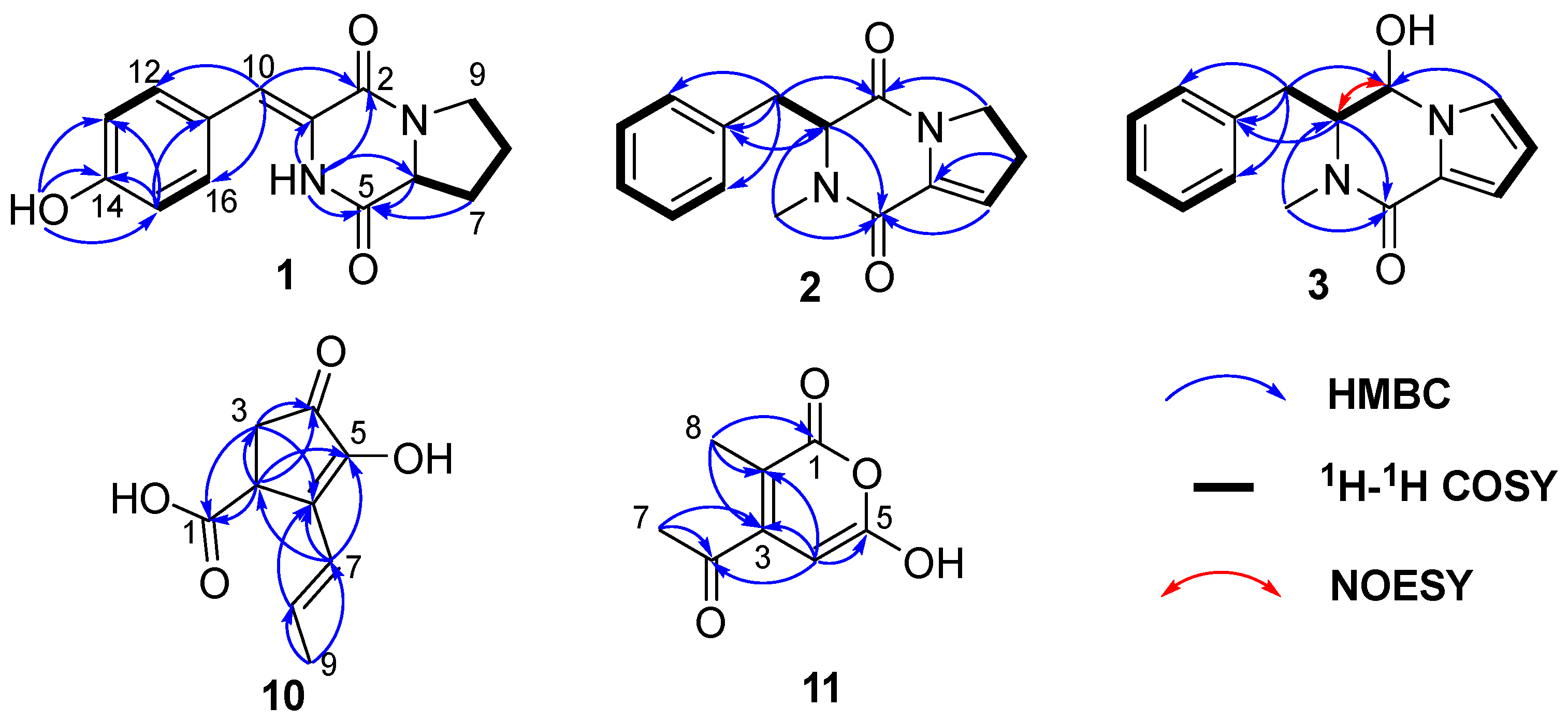
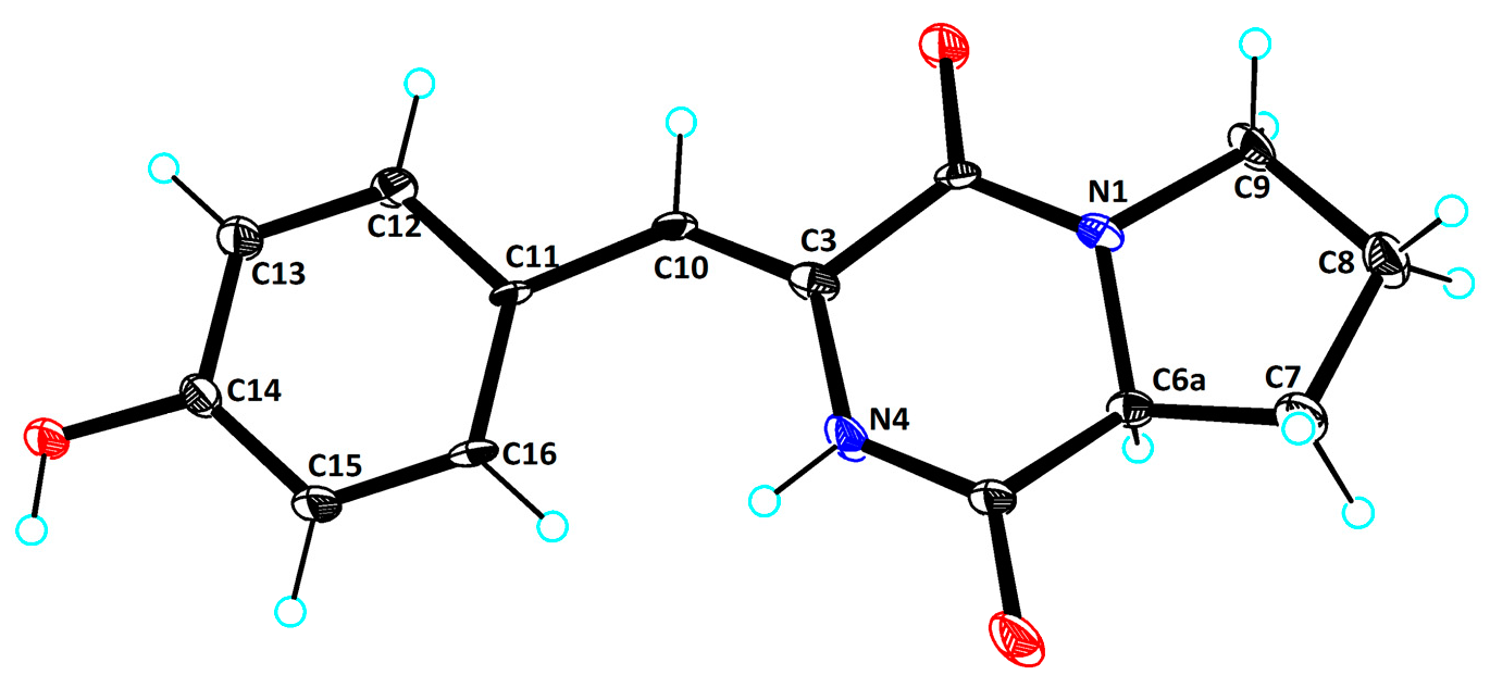
Compound 1 was isolated as green needles, and its molecular formula of C14H14N2O3 was determined via HRESIMS data at m/z 259.1078 [M+H]+ (calculated for C14H15N2O3+, 259.1077). One-dimensional NMR (Table 1) and HSQC spectra of 1 showed the presence of five aromatic/olefinic protons [δC/H 115.5/6.59 (CH-10), 131.1/7.38 (CH-12), 115.5/6.78 (CH-13, 15), 131.41/7.38 (CH-16)], one methine [δC/H 58.2/4.34 (CH-6)], three methylenes [δC/H 28.0/2.22, 1.88 (CH2-7), 21.7/1.88 (CH2-8), 45.1/3.51 (CH2-9)], and five nonprotonated sp2 carbons [δC 159.2 (C-2), 124.3 (C-3), 167.1 (C-5), 126.1 (C-11), 157.6 (C-14)]. Additionally, the 1H NMR revealed the presence of two reactive hydrogens: NH (9.83, s) and 14-OH (9.76, s). The data above indicated that compound 1 possessed a skeleton resembling a cyclic dipeptide, composed of tyrosine and proline. Compounds 1 and 7 exhibited structural similarities, with the distinction that compound 7 is composed of phenylalanine and proline, while in compound 1, the C-3 and C-13 positions of tyrosine form a Δ3,10 double bond. This finding was further supported by the 1H-1H COSY and HMBC correlations (Figure 2). The 1H-1H COSY correlations of H-12/H-13, H-15/H-16, along with the HMBC correlations of H-15/C-14, C-13, C-11, and H-10/C-12, C-16, C-2, confirmed the presence of the tyrosine moiety with a Δ3,10 double bond. Additionally, the 1H-1H COSY correlations of H-6/H2-7/H2-8/H2-9, and the HMBC correlations of H2-7/C-5, H-6/C-5, and NH/C-2, C-3, C-5, C-6, provided evidence for the presence of proline. Furthermore, to determine the absolute configuration of the compound, ECD calculations were performed for the 6S and 6R configurations of compound 1. The results showed that the experimental ECD curve fitted well with the calculated curve for 6S (Figure 3). Additionally, X-ray diffraction of 1 confirmed the accuracy of its planar structure and absolute configuration (Figure 4, CCDC 2401795). Consequently, compound 1 was identical to talarodipeptide A.
Compound 2 was isolated as a colourless oil, and the HRESIMS analysis determined its molecular formula to be C15H16N2O2, with an [M + H]+ value of m/z 257.1282 (calculated for C15H17N2O2+, 257.1285). The one-dimensional NMR (Table 1) showed that its structure was similar to that of compound 7, classifying it as a derivative of a cyclic dipeptide. Unlike compound 73,10 double bond) the position of the double bond in compound 26,7 double bond) was different. Compared to compound 1, compound 2 lacked the OH group at C-14 and the Δ3,10 double bond, as confirmed by the 1H-1H COSY correlations of H-12/H-13/H-14/H-15/H-16 and H-3/H2-10 (Figure 2). Additionally, compound 2 contained an N-CH3 group and a Δ6,7 double bond, as revealed by the HMBC correlations of H3-17/C-3, 5 and H2-8/C-6. By comparing calculated ECD with experimental ECD (Figure 3), the absolute configuration was determined to be 3R. Compound 2 is described here as a new natural product, designated talarodipeptide B.
Compound 3 was isolated as a brown oil, and its molecular formula was determined to be C15H16N2O2 using the HRESIMS, yielding an [M+H]+ value of m/z 257.1291 (calculated for C15H17N2O2+, 257.1285), with a total of 9 degrees of unsaturation. One-dimensional NMR (Table 1) indicated that its structure was similar to that of compound 2, classifying it as a derivative of a cyclic dipeptide. In contrast to compound 2, the C=O at C-2 in compound 3 had been reduced to an OH group and formed a Δ8, 9 double bond. This deduction was supported by the 1H-1H COSY correlations of H2-10/H-1/H-2 and H-7/H-8/H-9. The NOESY correlations between H-2 and H-3, along with the absence of NOESY signals between H-2 and H2-10, suggested that the relative configuration of compound 3 was 2R*, 3S*. Comparing calculated ECD (for 2R, 3S and 2S, 3R) with experimental ECD data, the absolute configuration of compound 3 was determined to be 2R, 3S (Figure 3), and it had been named talarodipeptide C.
Compound 10 was obtained as brown oil and had its molecular formula determined as C9H10O4 by HRESIMS m/z 181.0498 [M-H] (calculated for C9H9O4, 181.0506). The one-dimensional NMR (Table 2) and HSQC spectra indicated the presence of two olefinic protons (δC/H 123.8/6.49 and 131.1/6.15), one methine (δC/H 40.1/3.64), one methylene (δC/H 36.4/2.62, 2.28), one methyl (δC/H 19.0/1.84), one carboxyl group (δC 175.0), one carbonyl group (δC 200.2), and two nonprotonated sp2 carbons (δC 149.3 and 137.5). The HMBC correlations of H-2/C-1, 3, 4, 5 and H-3/C-1, 4, 6 (Figure 2) confirmed that the carboxyl group was attached to the five-membered ring at C-2, 3, 4, 5, and 6. The HMBC correlations of H-9/C-7, 8, H-8/C-6, and H-7/C-6, 5, 2 indicated the presence of a butene fragment at C-6. The coupling constant between H-7 and H-8 (15.9 Hz) suggested that the double bond was in the E configuration. Additionally, its optical rotation ([α ] D 25 −0.07) and ECD curve (Supporting Information) were close to zero, indicating the presence of a pair of enantiomers. The planar structure of compound 10 had been elucidated and named talaropolyketone A.
Compound 11 was obtained as brown solid and had its molecular formula determined as C8H8O4 by HRESIMS m/z 169.0494 [M+H]+ (calculated for C8H9O4+, 169.0495), indicating 5 degrees of unsaturation. One-dimensional NMR (Table 2) and HSQC spectra showed one olefinic proton (δC/H 104.8/6.85), two methyl groups (δC/H 25.6/2.14, 9.2/1.84), one ester carbonyl (δC 163.2), one ketone carbonyl (δC 191.1), and three nonprotonated olefinic carbons (δC 104.0, 151.7, 104.8, 164.2). The HMBC correlations of H-7/C-6, 3 indicate an acetyl group is attached at C-3. These data confirmed that compound 11 was a lactone within a six-membered ring. The HMBC correlations of H-8/C-1, 2, 3 showed that CH3-8 was connected to C-2. Additionally, the HMBC correlations of H-4/C-2, 3, 5, 6 confirmed that the olefinic proton is connected to C-3 and C-5. Therefore, the structure of compound 11 was determined and named talaropolyketone B.
Meanwhile, the other ten known compounds were identified as cyclo(D-6-Hyp-L-Phe) (4) [12], cyclo(l-6-Hyp-L-Phe) (5) [12], cyclo-(Pro-Phe) (6) [13], (Z)-3-benzylidene-2-methylhexahydropyrrolo [1,2] pyrazine-1,4-dione (7) [14], cyclo-(L-Ile-L-Pro) (8) [15], cyclo(D)-Pro-(D)-Leu (9) [15], alterlactone (12) [16], penicillide (13) [17], dehydroisopenicillide (14) [18], and 3′-O-Methyldehydroisopenicillide (15) [18], respectively, by comparing their NMR data (Supporting Information) to previous reports.

2.2. Bioactivity Assay

Compounds 115 were evaluated for their antibacterial (Escherichia coli, Staphylococcus aureus, Streptococcus suis, Erysipelothrix rhusiopathiae, Micrococcus luteus), antifungal (Colletotrichum acutatum, Curvularia australiensis, Fusarium oxysporum), phosphodiesterase 4 (PDE4) inhibitory, NF-κB inhibitory, and antioxidant activities. Compounds 1315 exhibited weak activities against Staphylococcus aureus, with the MIC values of 50, 50, and 25 µg/mL. Compound 9 exhibited activities against Escherichia coli, Streptococcus suis, and Erysipelothrix rhusiopathiae, with MIC values of 100 µg/mL. The obtained compounds showed no activity against fungi, PDE4, and NF-κB. In addition, compounds 1, 3, and 1012 at 50 μg/mL (vitamin C, 12.5 μg/mL) exhibited significant antioxidant activity, with compounds 1 and 12 showing DPPH radical scavenging rates of EC50 values of 27.62 and 29.34 µg/mL, respectively, compared to the positive control (vitamin C, with an EC50 of 12.74 µg/mL) (Figure 5).
Natural antioxidants can scavenge free radicals, and the development of novel antioxidants has significant implications for health and disease prevention [19]. This study found that dipeptides and polymer derivatives possess antioxidant activity, particularly emphasizing that the novel cyclic dipeptide compounds are worthy of further exploration. The structure–activity relationship (SAR) analysis revealed that the number and position of hydroxyl groups and double bonds significantly influence the intensity of antioxidant activity. Compared to compound 7, the presence of the 14-OH group and the Δ3,10 double bond in compound 1 significantly enhanced its antioxidant activity. Similarly, the hydroxyl group and Δ3,10 double bond in compound 3 increased its activity compared to compound 2. Likewise, compounds 10, 11, and 12 exhibit enhanced activity due to differences in the positions of their hydroxyl groups. Cyclic dipeptide compound 1 demonstrated the strongest activity, indicating that the tyrosine residue was an important moiety for radical scavenging activity. The presence of the tyrosine residue, which had electron/hydrogen donating capabilities, was a driving force for the dipeptide’s ability to scavenge free radicals. Additionally, adjacent residues were influenced by spatial effects, hydrophobicity, and hydrogen bonding, all of which could affect their activity [20]. Therefore, the novel cyclic dipeptides hold promise for development as new antioxidants.

3. Materials and Methods

3.1. General Experimental Procedures

Optical rotations were measured using an Anton Paar MPC500 polarimeter (Anton, Graz, Austria). UV spectra were recorded on a Shimadzu UV-2600 PC spectrometer (Shimadzu, Beijing, China), while IR spectra were determined with an IR Affinity-1 spectrometer (Shimadzu). ECD spectra were obtained using a Chirascan circular dichroism spectrometer (Applied Photophysics, Leatherhead Surrey, UK). High-resolution electrospray ionization mass spectrometry (HRESIMS) was performed on a Bruker maXis Q-TOF mass spectrometer (Bruker BioSpin International AG, Fällanden, Switzerland). NMR spectra were collected on a Quantum-I Plus 500 MHz spectrometer (Q-one Instrument Co., LTD, Wuhan, China), operating at 500 MHz for 1H NMR and 125 MHz for 13C NMR, with tetramethylsilane as the internal standard. Semipreparative high-performance liquid chromatography (HPLC) was conducted on a Hitachi Primaide with a DAD detector, using ODS columns (ChromCore 120 C18, 10 × 250 mm, 5 µm; YMC-pack ODS-A, 10 × 250 mm, 5 µm).

3.2. Fungal Material

The fungal strain SCSIO 41431 was isolated from the sediment sample of the mangrove in Gaoqiao Mangrove, Zhanjiang. It was stored in the CAS Key Laboratory of Tropical Marine Bioresources and Ecology, South China Sea Institute of Oceanology, Chinese Academy of Sciences, Guangzhou, China. The strain was designated Talaromyces sp. SCSIO 41431 was based on BLAST analysis of the ITS sequence (Supporting Information). Finally, the sequence was deposited in GenBank with the accession number PQ590239.

3.3. Fermentation and Extraction

The fungal strain Talaromyces sp. SCSIO 41431 was statically cultivated in potato dextrose broth (PDB) medium, followed by culture in 200 mL seed medium (1.5% malt extract, 2.0% sea salt) in 1 L Erlenmeyer flasks at 28 °C for 3 days on a rotary shaker at 180 rpm. A large-scale fermentation was then conducted at 26 °C for 28 days using a rice medium (200 g rice, 1% tryptone, 1.5% malt extract, 2% sea salt, 230 mL H2O) in 1 L flasks (60 flasks total) under static conditions. The entire fermented culture was extracted with ethyl acetate three times, yielding a brown extract weighing 180.5 g.

3.4. Isolation and Purification

The whole ethyl acetate extract was subjected to a silica gel vacuum liquid chromatography using a step gradient elution of petroleum ether (PE)-dichloromethane (DCM) (ν:ν 1:0, 1:1, 0:1), DCM-CH3OH (ν:ν 99:1, 97:3, 95:5, 90:10, 80:20, 50:50, 0:100), to yield 15 fractions (Frs. 1–15) in the light of TLC profiles. Fr. 8 was divided into 15 subfractions (Frs. 8-1–Fr. 8-15) by ODS silica gel eluting with CH3OH/H2O (5–100%). Fr. 8-2 was separated by semipreparative HPLC (38% CH3OH/H2O, 2.0 mL/min) to eight subfractions (Fr. 8-2-1–Fr. 8-2-8). Compounds 8 (16.4 mg, tR 13.0 min) and 9 (17.7 mg, tR 14.9 min) were further purified from Fr. 8-2-4 by semipreparative HPLC (18% CH3CN/H2O, 3.0 mL/min). Fr. 4-2-6 was separated by semipreparative HPLC (18% CH3CN/H2O, 3.0 mL/min) to gain compounds 5 (2.1 mg, tR 14.0 min), 4 (2.0 mg, tR 17.0 min), and 6 (67.4 mg, tR 19.4 min). Compound 2 (1.3 mg, tR 7.5 min) was further obtained from Fr. 8-7 by semipreparative HPLC (55% CH3OH/H2O, 3 mL/min). Compound 7 (25.0 mg, tR 7.0 min) was obtained from Fr. 8-8 by semipreparative HPLC (50% CH3CN /H2O, 3.0 mL/min). Compound 14 (6.3 mg, tR 19.5 min) and 15 (5.9 mg, tR 23.1 min) was obtained from Fr. 8-9 by semipreparative HPLC (75% CH3OH /H2O, 3.0 mL/min). Fr. 11 was divided into 16 subfractions (Frs. 11-1–Fr. 11-16) by ODS silica gel eluting with CH3OH/H2O (5–100%). Compound 11 (12.1 mg, tR 9.5 min) was separated from Fr. 11-5 by semipreparative HPLC (27% CH3CN/H2O, 0.04% formic acid, 3.0 mL/min). Compound 12 (8.2 mg, tR 20.2 min) and compound 3 (4.5 mg, tR 23.5 min) were further purified from Fr. 11-10 by semipreparative HPLC (61% CH3OH/H2O, 0.04% formic acid, 3.0 mL/min). Compound 13 (161.6 mg, tR 25.2 min) was further purified from Fr. 11-14 by semipreparative HPLC (61% CH3OH/H2O, 0.04% formic acid, 3.0 mL/min). Fr. 12 was divided into 15 subfractions (Frs. 12-1–Fr. 12-15) by ODS silica gel eluting with CH3OH/H2O (5–100%). Fr. 12-4 was separated by semipreparative HPLC (22% CH3CN/H2O, 3.0 mL/min) to gain compound 1 (11.3 mg, tR 10.6 min). Fr. 13-3 was separated by semipreparative HPLC (40% CH3CN/H2O, 3.0 mL/min) to gain compound 10 (3.0 mg, tR 7.2 min).

3.5. Spectroscopic Data of Compounds

Talaropolyketone A (1): green needles; [α ] D 25 +9.3 (c 0.1, CH3OH); UV (CH3OH) λmax (logε) 224 (3.81), 316 (3.88) nm; ECD (0.77 mM, CH3OH) λmaxε) 221 (+7.69), 246 (–1.95), 313 (+2.92); IR (film) νmax 3362, 2959, 2922, 1094, 1026, 677 cm−1; 1H and 13C NMR data, see Table 1; HRESIMS m/z 259.1078 [M+H]+ (calculated for C14H15N2O3+, 259.1077).
Talaropolyketone B (2): colourless oil; [α ] D 25 –6.9 (c 0.1, CH3OH); UV (CH3OH) λmax (logε) 200 (4.27), 242 (4.03) nm; ECD (0.78 mM, CH3OH) λmaxε) 245 (–6.13); IR (film) νmax 3387, 2963, 2943, 1682, 1634, 1402, 1045, 702 cm−1; 1H and 13C NMR data, see Table 1; HRESIMS m/z 257.1282 [M+H]+ (calculated for C15H17N2O2+, 257.1285).
Talaropolyketone C (3): brown oil; [α ] D 25 +6.1 (c 0.1, CH3OH); UV (CH3OH) λmax (logε) 200 (4.30), 274 (3.86) nm; ECD (0.78 mM, CH3OH) λmaxε) 201 (–5.45), 248 (+4.93); IR (film) νmax 3296, 2932, 1614, 1549, 1456, 1126, 745 cm−1; 1H and 13C NMR data, see Table 1; HRESIMS m/z 257.1291 [M+H]+ (calculated for C15H17N2O2+, 253.1285).
Talaropolyketone D (10): brown oil; [α ] D 25 -0.07 (c 0.1, CH3OH); UV (CH3OH) λmax (logε) 200 (3.72), 298 (3.63) nm; IR (film) νmax 3437, 2967, 2932, 1697, 1186, 1136, 974, 721 cm−1; 1H and 13C NMR data, see Table 2; HRESIMS m/z 181.0498 [M-H] (calculated for C9H9O4, 181.0506).
Talaropolyketone E (11): brown solid; UV (CH3OH) λmax (logε) 202 (3.80), 226 (4.05), 328 (3.59) nm; IR (film) νmax 3354, 1682, 1645, 1406, 1219, 1024, 750 cm−1; 1H and 13C NMR data, see Table 2; HRESIMS m/z 169.0494 [M+H]+ (calculated for C8H9O4+, 169.0495).

3.6. X-Ray Crystallographic Analysis

The crystallographic data for compound 1, obtained through slow evaporation in methanol, were collected using an XtaLAB PRO single-crystal diffractometer with Cu Kα radiation. The X-ray crystal structures were solved using SHELXS97, expanded via difference Fourier techniques, and refined through full-matrix least-squares calculations. Non-hydrogen atoms were refined anisotropically, while hydrogen atoms were fixed at calculated positions. The crystallographic data for these compounds have been deposited in the Cambridge Crystallographic Data Centre.
Crystal data for 1: C14H14N2O3 (M = 258.27 g/mol): monoclinic, space group P21/n (no. 14), a = 5.8882 (2) Å, b = 11.5552 (3) Å, c = 17.5786 (3) Å, β = 94.906 (2)°, V = 1191.65 (5) Å3,Z = 4, T = 99.97 (17) K, μ(Cu Kα) = 0.848 mm−1, Dcalc = 1.440 g/cm3, 5047 reflections measured (9.168° ≤ 2Θ ≤ 133.186°), 2084 unique (Rint = 0.0460, Rsigma = 0.0528) which were used in all calculations. The final R1 was 0.1370 (I > 2σ(I)) and wR2 was 0.3178 (all data) (Figure 4, CCDC 2401795).

3.7. ECD Computation Section

Compounds 13 were analyzed using Spartan’14, employing the MMFF molecular force field to conduct a conformational search for potential isomers. The most stable conformers, representing the top 5%, were then optimized in methanol solvent at the B3LYP/6-31G (d) level using Gaussian 09 (D.01, Pittsburgh, PA, USA). A TDDFT polarizable continuum model at the B3LYP/6-311G (d, p) level was utilized to calculate the optimized low-energy conformations. [21]. The calculated ECD spectra were generated using GaussView (6.0.16, Pittsburgh, PA, USA) with a half-bandwidth of 0.3 eV and weighted by Boltzmann distribution. Both the calculated ECD and experimental ECD spectra were plotted using Origin 2021.

3.8. Antioxidant Activity Assay

The obtained compounds were evaluated for its antioxidant activities against DPPH. The effect of the compounds on DPPH radical were estimated, as previously reported [22]. In summary, a methanolic DPPH solution was supplemented with compounds to achieve final concentrations of 12.5–200 μg/mL. The mixture was shaken and allowed to stand for 30 min at room temperature in the dark, after which the OD517 values were measured using a PerkinElmer Enspire Multi-mode detector (Thermo Scientific, Bremen, Germany). Vitamin C served as the positive control. The free radical scavenging rate (K%) was calculated from the OD517 values, and EC50 was determined using Origin 2021.

3.9. Antimicrobial Activity Assay

The antimicrobial activities against four bacteria (Escherichia coli, Staphylococcus aureus, Streptococcus suis, Erysipelothrix rhusiopathiae, Micrococcus luteus) and three fungi (Colletotrichum acutatum, Curvularia australiensis, Fusarium oxysporum) were evaluated in 96-well plates with a twofold serial dilution method described previously [23]. After 48 h of incubation, the optical density of the medium was measured at 600 nm (OD600) using a PerkinElmer Enspire Multi-mode detector. Incubation conditions were as follows: fungi PDB medium (Potato extract 0.4%, Glucose 2%), 26 °C, 48 h; Bacteria LB medium (Yeast extract 0.5%, Tryptone 1%, NaCl 0.5%), 26 °C, 48 h.

3.10. PDE4 and NF-κB Inhibitory Screening Assays

PDE4 inhibition screening test procedures, as previously reported using the phosphodiesterase scintillation proximity assay [22,24]. To assess the enzymatic activity of the catalytic domains, we used 3H-cAMP or 3H-cGMP as substrates. The experiments were performed in assay buffer containing Tris-HCl, MgCl2 or MnCl2, DTT, and 3H-cAMP or 3H-cGMP, and the protein concentration was set at 2 nM. The reaction was terminated after 15 min of incubation, and the products were separated by precipitation, while unreacted substrates retained in the supernatant. The radioactivity of the supernatant was measured using a liquid scintillation counter.
NF-κB inhibition screening test procedures, as previously reported using luciferase reporter gene assay [25]. RAW264.7 cells transfected with a luciferase reporter gene were cultured in 96-well plates and pretreated with test compounds (20 μM) and BAY11-7082 (NF-κB inhibitor, 5 μM, purchased from Sigma-Aldrich as a positive control) for 30 min, followed by stimulation of the cells with 5 μg/mL LPS for 8 h. At the end of the experiment, the cells were collected, and luciferase activity was measured using a luciferase assay system (Promega, Madison, WI, USA), and each sample was repeated three times.

4. Conclusions

In conclusion, five new metabolites (13 and 1011), together with ten known ones (49 and 1215), were isolated from the mangrove-sediments-derived fungus Talaromyces sp. SCSIO 41431. Their structures were determined by extensive spectroscopic analyses, ECD calculations, and X-ray single-crystal diffraction. Compounds 9 and 1315 exhibited weak activities against pathogenic bacteria. The obtained compounds showed no activity against PDE4 and NF-κB enzymes. Moreover, compounds 1 and 13 showed DPPH radical scavenging activity, with the EC50 of 27.62 and 29.34 µg/mL.

Supplementary Materials

The following supporting information can be downloaded at https://www.mdpi.com/article/10.3390/md22120559/s1: Figures S1–S43: The NMR, HRESIMS, UV, and IR spectra of 13 and 1011; physicochemical data of 49 and 1215; ITS sequence data of the strain.

Author Contributions

Data curation, Z.Z., J.C. and Y.C.; funding acquisition, X.Z.; investigation, X.L. and C.C.; project administration, Y.L. and X.Z.; writing—original draft, Z.Z. and J.C.; writing—review and editing, L.J. and X.Z. All authors have read and agreed to the published version of the manuscript.

Funding

This research was funded by the Special Project for Marine Economic Development of the Department of Natural Resources of Guangdong Province (GDNRC [2023]37), Guangdong Basic and Applied Basic Research Foundation (2024A1515010741), and the International Partnership Program of Chinese Academy of Sciences (059GJHZ2023104MI).

Institutional Review Board Statement

Not applicable.

Informed Consent Statement

Not applicable.

Data Availability Statement

Data are contained within the article and Supplementary Materials.

Acknowledgments

We are grateful to Z. Xiao, X. Zheng, A. Sun, Y. Zhang, and X. Ma in the analytical facility at SCSIO for recording spectroscopic data, and China-Sri Lanka Joint Center for Education & Research, Chinese Academy of Sciences.

Conflicts of Interest

The authors declare no conflicts of interest.

References

  1. Barbier, E.B.; Koch, E.W.; Silliman, B.R.; Hacker, S.D.; Wolanski, E.; Primavera, J.; Granek, E.F.; Polasky, S.; Aswani, S.; Cramer, L.A.; et al. Coastal Ecosystem-Based Management with Nonlinear Ecological Functions and Values. Science 2008, 319, 321–323. [Google Scholar] [CrossRef] [PubMed]
  2. Thatoi, H.; Behera, B.C.; Mishra, R.R.; Dutta, S.K. Biodiversity and Biotechnological Potential of Microorganisms from Mangrove Ecosystems: A Review. Ann. Microbiol. 2013, 63, 1–19. [Google Scholar] [CrossRef]
  3. Xu, J. Bioactive Natural Products Derived from Mangrove-Associated Microbes. RSC Adv. 2015, 5, 841–892. [Google Scholar] [CrossRef]
  4. Li, K.; Chen, S.; Pang, X.; Cai, J.; Zhang, X.; Liu, Y.; Zhu, Y.; Zhou, X. Natural Products from Mangrove Sediments-Derived Microbes: Structural Diversity, Bioactivities, Biosynthesis, and Total Synthesis. Eur. J. Med. Chem. 2022, 230, 114117. [Google Scholar] [CrossRef]
  5. Yu, Y.; Wang, Z.; Xiong, D.; Zhou, L.; Kong, F.; Wang, Q. New Secondary Metabolites of Mangrove-Associated Strains. Mar. Drugs 2024, 22, 372. [Google Scholar] [CrossRef]
  6. Liu, F.; Cai, X.-L.; Yang, H.; Xia, X.-K.; Guo, Z.-Y.; Yuan, J.; Li, M.-F.; She, Z.-G.; Lin, Y.-C. The Bioactive Metabolites of the Mangrove Endophytic Fungus Talaromyces sp. ZH-154 Isolated from Kandelia candel (L.) Druce. Planta Med. 2010, 76, 185–189. [Google Scholar] [CrossRef]
  7. Liu, H.; Chen, S.; Liu, W.; Liu, Y.; Huang, X.; She, Z. Polyketides with Immunosuppressive Activities from Mangrove Endophytic Fungus Penicillium sp. ZJ-SY2. Mar. Drugs 2016, 14, 217. [Google Scholar] [CrossRef] [PubMed]
  8. Li, J.; Chen, C.; Fang, T.; Wu, L.; Liu, W.; Tang, J.; Long, Y. New Steroid and Isocoumarin from the Mangrove Endophytic Fungus Talaromyces sp. SCNU-F0041. Molecules 2022, 27, 5766. [Google Scholar] [CrossRef]
  9. Nicoletti, R.; Salvatore, M.; Andolfi, A. Secondary Metabolites of Mangrove-Associated Strains of Talaromyces. Mar. Drugs 2018, 16, 12. [Google Scholar] [CrossRef] [PubMed]
  10. Li, H.; Huang, H.; Shao, C.; Huang, H.; Jiang, J.; Zhu, X.; Liu, Y.; Liu, L.; Lu, Y.; Li, M.; et al. Cytotoxic Norsesquiterpene Peroxides from the Endophytic Fungus Talaromyces flavus Isolated from the Mangrove Plant Sonneratia apetala. J. Nat. Prod. 2011, 74, 1230–1235. [Google Scholar] [CrossRef]
  11. Cai, R.; Chen, S.; Long, Y.; Li, C.; Huang, X.; She, Z. Depsidones from Talaromyces stipitatus SK-4, an Endophytic Fungus of the Mangrove Plant Acanthus Ilicifolius. Phytochem. Lett. 2017, 20, 196–199. [Google Scholar] [CrossRef]
  12. Park, Y.C.; Gunasekera, S.P.; Lopez, J.V.; McCarthy, P.J.; Wright, A.E. Metabolites from the Marine-Derived Fungus Chromocleista sp. Isolated from a Deep-Water Sediment Sample Collected in the Gulf of Mexico. J. Nat. Prod. 2006, 69, 580–584. [Google Scholar] [CrossRef] [PubMed]
  13. Sun, Y.; Wang, C.; Du, G.; Deng, W.; Yang, H.; Li, R.; Xu, Q.; Guo, Q. Two Nematicidal Compounds from Lysinimonas M4 against the Pine Wood Nematode, Bursaphelenchus xylophilus. Forests 2022, 13, 1191. [Google Scholar] [CrossRef]
  14. Lv, M.; Tan, M.; Lu, L.; Bao, S.; Guo, Z.; Deng, Z.; Zhou, K. Secondary Metabolites from Endophytes of Elaeocarpus Decipiens Hemsl. with Co-Cultivation Method. J. China Three Gorges Univ. Sci. 2018, 5, 2758. [Google Scholar]
  15. Guo, Z.K.; Wang, R.; Chen, F.X.; Liu, T.M.; Yang, M.Q. Bioactive Aromatic Metabolites from the Sea Urchin-Derived Actinomycete Streptomyces spectabilis Strain HDa1. Phytochem. Lett. 2018, 25, 132–135. [Google Scholar] [CrossRef]
  16. Aly, A.H.; Edrada-Ebel, R.; Indriani, I.D.; Wray, V.; Müller, W.E.G.; Totzke, F.; Zirrgiebel, U.; Schächtele, C.; Kubbutat, M.H.G.; Lin, W.H.; et al. Cytotoxic Metabolites from the Fungal Endophyte Alternaria sp. and Their Subsequent Detection in Its Host Plant Polygonum senegalense. J. Nat. Prod. 2008, 71, 972–980. [Google Scholar] [CrossRef] [PubMed]
  17. Jeon, H.; Shim, S.H. Chemical Constituents of the Endophyte Penicillium sp. Isolated from Artemisia princeps. Chem. Nat. Compd. 2020, 56, 122–124. [Google Scholar] [CrossRef]
  18. Xia, X.; Kim, S.; Liu, C.; Shim, S. Secondary Metabolites Produced by an Endophytic Fungus Pestalotiopsis sydowiana and Their 20S Proteasome Inhibitory Activities. Molecules 2016, 21, 944. [Google Scholar] [CrossRef]
  19. Gulcin, İ. Antioxidants and Antioxidant Methods: An Updated Overview. Arch. Toxicol. 2020, 94, 651–715. [Google Scholar] [CrossRef]
  20. Zheng, L.; Zhao, Y.; Dong, H.; Su, G.; Zhao, M. Structure-Activity Relationship of Antioxidant Dipeptides: Dominant Role of Tyr, Trp, Cys and Met Residues. J. Funct. Foods 2016, 21, 485–496. [Google Scholar] [CrossRef]
  21. Luo, X.; Lin, X.; Tao, H.; Wang, J.; Li, J.; Yang, B.; Zhou, X.; Liu, Y. Isochromophilones A-F, Cytotoxic Chloroazaphilones from the Marine Mangrove Endophytic Fungus Diaporthe sp. SCSIO 41011. J. Nat. Prod. 2018, 81, 934–941. [Google Scholar] [CrossRef] [PubMed]
  22. Chen, Y.; Cai, J.; Xia, Z.; Chen, C.; Liu, Y.; Jayasinghe, L.; Wang, X.; Zhou, X. New Bioactive Polyketides from the Mangrove-Derived Fungus Penicillium sp. SCSIO 41411. Mar. Drugs 2024, 22, 384. [Google Scholar] [CrossRef] [PubMed]
  23. Cai, J.; Wang, X.; Yang, Z.; Tan, Y.; Peng, B.; Liu, Y.; Zhou, X. Thiodiketopiperazines and Alkane Derivatives Produced by the Mangrove Sediment-Derived Fungus Penicillium ludwigii SCSIO 41408. Front. Microbiol. 2022, 13, 857041. [Google Scholar] [CrossRef] [PubMed]
  24. Cai, J.; Zhou, Q.; Qi, X.; Zhang, F.; Yang, J.; Chen, C.; Zhang, K.; Chen, Z.; Luo, H.-B.; Liu, Y.; et al. Discovery of Oxidized P-Terphenyls as Phosphodiesterase 4 Inhibitors from Marine-Derived Fungi. J. Nat. Prod. 2024, 87, 1808–1816. [Google Scholar] [CrossRef] [PubMed]
  25. Cai, J.; Gao, L.; Wang, Y.; Zheng, Y.; Lin, X.; Zhou, P.; Chen, C.; Liu, K.; Tang, L.; Liu, Y.; et al. Discovery of a Novel Anti-Osteoporotic Agent from Marine Fungus-Derived Structurally Diverse Sirenins. Eur. J. Med. Chem. 2024, 265, 116068. [Google Scholar] [CrossRef]
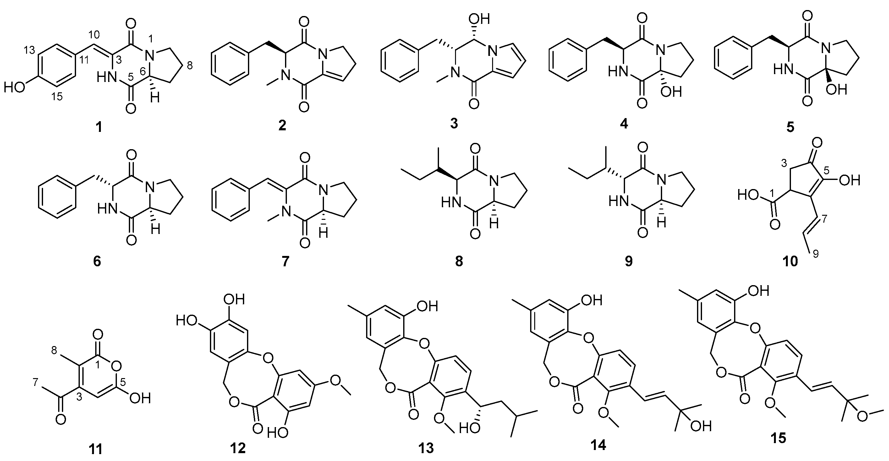
Figure 1. Structures of compounds 115.
Marinedrugs 22 00559 g001
Figure 2. Key HMBC, 1H-1H COSY, and NOESY correlations of 13 and 1011.
Marinedrugs 22 00559 g002
Figure 3. Experimental and calculated ECD spectra of compounds 13.
Marinedrugs 22 00559 g003
Figure 4. X-ray single-crystal diffraction of compound 1.
Marinedrugs 22 00559 g004
Figure 5. (A) DPPH radical scavenging activity of the compounds 115 (50 µg/mL). All experiments were performed at least three times. The data are presented as the mean ± SD of representative experiments. Statistical significance was determined with one-way ANOVA. * p < 0.05, *** p < 0.001, and **** p < 0.0001 were considered statistically significant. (B) Compounds 1 and 12 of DPPH scavenging activity of EC50.
Marinedrugs 22 00559 g005
Table 1. 1H (500 MHz) and 13C (125 MHz) NMR data of 13 in DMSO-d6.
Pos.123
δC TypeδH (J in Hz)δC TypeδH (J in Hz)δC TypeδH (J in Hz)
2159.2, C 77.0, CH5.68, d (3.7)165.5, C
3124.3, C 63.7, CH3.92, dt (9.0, 3.7)62.8, CH4.37, t (4.4)
5167.1, C 158.1, C 158.8, C
658.2, CH4.34, m122.9, C 127.0, C
728.0, CH22.20, m;
1.88, overlapped
120.3, CH6.60, dd (3.7, 1.6)114.4, CH5.40, t (7.7)
821.7, CH21.88, overlapped108.9, CH6.15, dd (3.7, 2.7)28.6, CH22.04, m
945.1, CH23.51, m112.2, CH6.99, dd (2.7, 1.6)59.8, CH23.23, m
10115.5, CH6.59, s33.3, CH22.56, m;
3.08, dd (13.6, 5.2)
30.7, CH23.04, dd (13.8, 3.8);
3.15, dd (13.8, 5.1)
11126.1, C 138.0, C 134.8, C
12131.1, CH7.38, d (8.3)129.4, CH7.16, d (6.8)129.7, CH7.03, m
13115.5, CH6.78, d (8.4)128.3, CH7.28, d (7.4)128.1, CH7.21, overlapped
14157.6, C 126.3, CH7.21, m126.9, CH7.21, overlapped
15115.5, CH6.78, d (8.4)128.3, CH7.28, d (7.4)128.1, CH7.21, overlapped
16131.1, CH7.38, d (8.3)129.4, CH7.16, d (6.8)129.7, CH7.03, m
17 32.9, CH32.54, s32.3, CH32.97, s
NH 9.83, s NH9.79, s
14-OH 9.76, s
Table 2. 1H (500 MHz) and 13C (125 MHz) NMR data of 1011 in DMSO-d6.
Pos.1011
δC TypeδH (J in Hz)δC TypeδH (J in Hz)
1175.0, C 163.2, C
240.1, CH3.64, d (7.0)104.0, C
336.4, CH22.62, dd (18.4, 7.0);
2.28, d (18.4)
151.7, C
4200.2, C 104.8, CH6.85, s
5149.3, C 164.2, C
6137.5, C 191.1, C
7123.8, CH6.49, d (15.9)25.6, CH32.14, s
8131.2, CH6.15, dq (15.9, 6.8)9.2, CH31.84, s
919.0, CH31.84, d (6.8)
5-OH 9.78, s
Disclaimer/Publisher’s Note: The statements, opinions and data contained in all publications are solely those of the individual author(s) and contributor(s) and not of MDPI and/or the editor(s). MDPI and/or the editor(s) disclaim responsibility for any injury to people or property resulting from any ideas, methods, instructions or products referred to in the content.