
Marine Microorganism Molecules as Potential Anti-Inflammatory Therapeutics


Abstract
1. Introduction
2. The Link between the Inflammation and CIDs
2.1. Inflammatory Pathways
2.2. Therapeutic Strategies to Target Inflammation

| Macro-Organisms | ||||||
|---|---|---|---|---|---|---|
| Organisms | Classification (Phylum) | Species | Type of Molecules | Molecules | Target/Mode of Action | Ref(s). |
| Sponge | Porifera | Fasciospongia cavernosa | Terpene lactone | Cavernolide | TNF-α, NO, and PGE2 inhibition in RAW 264.7 cells | [19] |
| Sponge | Porifera | Dysidea spp. | Sesquiterpene | Dysidotronic acid | TNF-α, IL-1, NO, PGE2 inhibition in RAW 264.7 cells | [20] |
| Sponge | Porifera | Plakortis spp. | α-exomethylene-γ-lactone | Plakolide A | iNOS inhibition in RAW 264.7 cells | [21] |
| Sponge | Porifera | Luffariella variabilis | Sesterterpene | Manoalide | Eicosanoids synthesis inhibition in human polymorphonuclear leukocytes | [22] |
| Caribbean sponge | Porifera | Cacospongia linteiformis | Sesterterpene | Cyclolinteinone | iNOS and COX-2 inhibition in LPS-stimulated J774 macrophages | [23] |
| Sponge | Porifera | Dysidea sp. and Petrosaspongia nigra | Merosesquiterpene & Sesterterpene | Bolinaquinone and petrosaspongiolide M | Protection against TNBS-induced colitis in BALB/c mice | [24] |
| Sponge | Porifera | Petrosia spp. | Polyacetylenes | Petrocortyne D, Petrocortyne E, Petrocortyne F, Petrocortyne G, Petrocortyne H | Inhibition of PLA2 activity in K-562 cell line | [25] |
| Sponge | Porifera | Petrosia spp. | Polyacetylenic alcohol | Petrocortyne A | TNF-α inhibition in LPS-activated RAW 264.7 and PMA/LPS-treated U937 cells and NO inhibition in LPS- or IFNγ-treated RAW 264.7 cells | [26] |
| Sponge | Porifera | Theonella swinhoe | Steroid | Solomonsterol A | Reduction in arthritic score in anti-type II collagen antibody-induced arthritis murine model | [27] |
| Sponge | Porifera | Geodia barretti | Alpha amino acids and derivatives | Barettin | TNF-α and IL-1β inhibition in LPS-stimulated THP-1 cells | [28] |
| Sponge | Porifera | Geodia barretti | Alkaloids | 6-bromoindole derivatives geobarettin B, 6-bromoindole derivatives geobarettin C, 6-bromoindole alkaloids 6-bromoconicamin, barettin | IL-12 p40 inhibition and IL-10 increasing in dendritic cells | [29] |
| Sponge | Porifera | Halichondria okadai | Alkaloid | Halichlorine | VCAM-1, ICAM-1, and E-selectin inhibition in LPS-stimulated aortic endothelial cells, inhibition of macrophage adhesion to cultured cell monolayers, an anti-inflammatory effect associated with NF-κB pathway | [30] |
| Sponge | Porifera | Stylissa | Alkaloid | Pyrrole alkaloid (10Z)-debromohymenialdisine | IL-1β, IL-6, TNF-α, iNOS, COX-2, NO and PGE2 inhibition in co-cultures of LPS-stimulated Caco-2 and THP-1 cells | [31] |
| Sponge | Porifera | Stylissa flabellata | Alkaloids | Stylissadine A, Stylissadine B | Antagonistic effect on P2X7 receptors in THP-1 cells | [32] |
| Soft coral | Cnidaria | Sinularia dissecta | Diterpene | Seco-sethukarailin | Inhibition of pro-inflammatory cytokines in bone marrow-derived dendritic cells | [33] |
| Soft coral | Cnidaria | Pseudopterogorgia elisabethae | Diterpenes | Pseudopterosin E, Pseudopterosin A | Reduction of PMA-induced mouse ear edema; PGE2 and LCT4 inhibition in zymosan-stimulated murine peritoneal macrophages | [34] |
| Soft coral | Cnidaria | Sinularia gibberosa | Steroid | Gibberoketosterol | Inhibition of pro-inflammatory iNOS and COX-2 proteins in LPS-stimulated RAW264.7 cells | [35] |
| Okinawan soft coral | Cnidaria | Sinularia spp. | Diterpenes | Norcembranolide and sinuleptolide | TNF-α and NO inhibition in LPS-stimulated RAW 264.7 cells | [36] |
| Soft coral | Cnidaria | Sinularia lochmodes | Sesquiterpene | Lochmolins A, Lochmolins B | Inhibition of COX-2 expression in LPS-activated RAW 264.7 cells | [37] |
| Lochmolins C | Inhibition of COX-2 expression in LPS-activated RAW 264.7 cells | [38] | ||||
| Lochmolins D | Inhibition of COX-2 expression in LPS-activated RAW 264.7 cells | [37] | ||||
| Soft coral | Cnidaria | Lemnalia cervicorni | Sesquiterpene | Lemnalol | Inhibition of iNOS and COX-2 expression in LPS-activated RAW 264.7 cells; inhibition of iNOS and COX-2 expression in carrageenan-activated rat paws | [39] |
| Soft coral | Cnidaria | Lemnalia flava | Sesquiterpene | Flavalin A | iNOS and COX-2 inhibition in RAW 264.7 cells | [40] |
| Soft coral | Cnidaria | Lobophytum crassum | Diterpenes | Crassumol E 1R,4R,2E,7E,11E-cembra-2,7,11-trien-4-ol | Inhibition of NF-κB activation in TNF-α-activated HepG2 cells | [41] |
| Diterpenes | Lobocrasol A, Lobocrasol B | Inhibition of NF-κB activation in TNF-α-activated HepG2 cells | [42] | |||
| Soft coral | Cnidaria | Scleronephthya gracillimum | Steroid | Sclerosteroid J | Inhibition of iNOS and COX-2 expression in LPS-activated RAW 264.7 cells | [43] |
| Octocoral | Cnidaria | Pseudopterogorgia acerosa | Diterpene | Pseudopterane | Inhibition of NO, TNF-α, IL-1β and IFNγ-induced protein production in LPS-activated peritoneal macrophages | [44] |
| Coral | Cnidaria | Rumphella antipathies (classification rhumphella antipathes Linnaeus 1758) | Sesquiterpene | Clovane compound 1 | Inhibition of superoxide anions generation and elastase release | [45] |
| Sesquiterpene | Clovane compound 2 | Inhibition of elastase release in fMLP/CB-activated human neutrophils | [45] | |||
| Sesquiterpene | Rumphellaone C | Inhibition of superoxide anion generation and elastase release in human neutrophils | [46] | |||
| Sesquiterpene | Rumphellol A | Inhibition of superoxide generation and elastase release in human neutrophils | [47] | |||
| Sesquiterpene | Rumpheloll B | |||||
| Coral | Cnidaria | Briareum excavatum | Diterpene | Excavatolide B | Inhibition of iNOS expression in carrageenan-activated rat paws | [48] |
| Coral | Cnidaria | Briareum excavatum | Diterpene | Excavatolide B | Inhibition of 12-O-tetradecanoylphorbol-13-acetate (TPA)-induced vascular permeability; inhibition of TPA-induced matrix metalloproteinase-9 expression in mouse skin; inhibition of IL-6 expression of LPS-activated mouse bone marrow-derived dendritic cells | [49] |
| Anemone | Cnidaria | Zoanthus kuroshio | Alkaloid | 5α-iodozoanthenamine | Anti-inflammatory effect on—neutrophils, reduction of superoxide anion generation, and elastase by cells | [50] |
| Anemone | Cnidaria | Zoanthus pulchellus | Alkaloids | 3-hydroxinorzoanthamine Norzoanthine Roanthamine | ROS and NO inhibition in LPS-stimulated BV-2 cells | [51] |
| Starfish | Echinodermata | Marthasterias glacialis | Steroid | Ergosta-7,22-dien-3-ol | Inhibition of iNOS protein level in LPS-activated RAW 264.7 cells | [52] |
| Starfish | Echinodermata | Astropecten polycanthus | Steroid | Steroid compound 5 | Inhibition of IL-12 p40, IL-6, and TNF-α production in LPS-activated mice bone marrow-derived dendritic cells | [53] |
| Starfish | Echinodermata | Asterias amurensis | Fatty acid | Fatty acids | Inhibition of the expression of inflammatory genes via NF-κB and MAPK pathways in LPS-stimulated RAW 264.7 cells | [54] |
| Starfish | Echinodermata | Marthasterias glacialis | Fatty acid | Cis 11-eicosenoic and cis 11,14 eicosadienoic acids | Inhibition of iNOS, COX-2, IκBα, and NF-κB gene expression in LPS-stimulated RAW 264.7 cells | [52] |
| Starfish | Echinodermata | Protoreaster nodosus | Steroid | Oxygenated steroid derivatives | IL-12 p40, IL-6, and TNF-α inhibition in bone marrow-derived dendritic cells | [55] |
| Starfish | Echinodermata | Protoreaster lincki | Steroids | Protolinckioside A, Protolinckioside B, Protolinckioside C, Protolinckioside D | Reduction of ROS formation and NO production in LPS-stimulated RAW 264.7 cells | [56] |
| Starfish | Echinodermata | Anthenea aspera | Steroid | Anthenoside O | [57] | |
| Starfish | Echinodermata | Pentaceraster regulus | Steroid | Pentareguloside C, Pentareguloside D, Pentareguloside E | Reduction of ROS formation and NO production in LPS-stimulated RAW 264.7 cells | [58] |
| Starfish | Echinodermata | Acanthaster planci | Pyrrole oligoglycoside | Plancipyrroside A, Plancipyrroside B | Reduction of ROS formation and NO production in LPS-stimulated RAW 264.7 cells | [59] |
| Starfish | Echinodermata | Asterina batheri | Pyrrole oligoglycoside | Astebatherioside B, Astebatherioside C, Astebatherioside D | IL-12 p40 inhibition in LPS-stimulated bone marrow-derived dendritic cells | [60] |
| Sea cucumber | Echinodermata | Holothuria grisea | Protein | Lectin | Inhibition of neutrophil migration to the peritoneal cavity in carrageenan-activated rats; reduction of myeloperoxidase activity in carrageenan-activated rats | [61] |
| Sea cucumber | Echinodermata | Apostichopus japonicus and Stichopus chloronotus | Sulfated polysaccharide | Fucosylated chondroitin sulfate | Reduction of neutrophil migration, inhibition of paw edema in carrageenan-induced paw edema in rats | [62] |
| Sea cucumber | Echinodermata | Isostichopus badionotus | Sulfated polysaccharide | Fucosylated chondroitin sulfate | Suppression of TPA-mediated up-regulation of TNF-α, IL-6, NF-κB, iNOS, IL-10, IL-11, COX-2 and STAT3 genes in mouse ear tissue | [63] |
| Sea cucumber | Echinodermata | Isostichopus badionotus | Sulfated polysaccharide | Fucoidan | Regulation of serum inflammatory cytokines (TNF-α, CRP, MIP-1, IL-1β, IL-6, and IL-10) and their mRNA expression, inactivation of JNK and IκB/NF-κB pathways | [64] |
| Sea cucumber | Echinodermata | Holothuria albiventer and Cucumaria frondosa | Sulfated polysaccharide | Sulfated fucan/FCS | Suppression of TNF-α and IL-6 production | [65] |
| Sea cucumber | Echinodermata | Holothuria tomasi | Triterpenes glycoside | Inhibition of IL-6, TNF-α levels in STZ-induced diabetic rats | [66] | |
| Sea cucumber | Echinodermata | Pearsonothuria graeffei | Triterpenes glycoside | Holothurin A and Echinoside A | Inhibition of IL-1β, TNF-α, IL-6 and infiltration of macrophages in obese mice via p-ERK/cPLA2/COX-1 pathway and reduction of the PGE2 levels | [67] |
| Sea cucumber | Echinodermata | Aspostichopus japonicus and Acaudina leucoprocta | Peptide | Oligopeptides | Downregulation of pro-inflammatory cytokines transcription, upregulation of anti-inflammatory cytokines, and inhibition of TLR4/MyD88/NF-κB signaling pathway | [68] |
| Sea cucumber | Echinodermata | Cucumaria frondosa | Fatty acid | Eicosapentaenoic acid | Inhibition of TNF-α, IL-6, and MCP1 expression, attenuation of macrophage infiltration in the liver in mice, attenuation of the phosphorylation of NF-κB in RAW 264.7 cells | [69] |
| Sea cucumber | Echinodermata | Cucumaria frondosa | Lipid | Frondanol | Reduction of inflammation-associated changes in the colon in mice, reduction of cytokine content at the protein and mRNA level | [70] |
| Sea cucumber | Echinodermata | Cucumaria frondosa | Lipid | Sphingolipids | Inhibition of pro-inflammatory cytokines IL-1β, IL-6 TNF-α and increasing anti-inflammatory IL-10 via inhibition of phosphorylation of JNK and translocation of NF-κB | [71] |
| Sea cucumber | Echinodermata | Cucumaria frondosa | Lipid | Frondaol A5 | Attenuation of circulating inflammatory cytokines and suppression of mRNA expression of inflammatory markers such as 5-LOX and FLAP | [72] |
| Sea urchins | Echinodermata | Scaphechinus mirabilis | Dark red pigment | EchA | Attenuation of macrophage activation and infiltration (neutrophils), inhibition of TNF-α and IFNγ in bleomycin-induced scleroderma mouse model | [73] |
| Sea urchins | Echinodermata | ? | Dark red pigment | EchA | Decreasing DIA, improvement of colon length and suppression of tissue damage, suppression of macrophage activation | [74] |
| Sea urchins | Echinodermata | ? | Dark red pigment | EchA | TNF-α and NF-κB inhibition in Lewis rats | [75] |
| Sea urchins | Echinodermata | Paracentrotus lividus | Dark red pigment | EchA | Potent stabilizing effect on the human red blood cells, suppression of the production of IL-6 and TNF-α in septic rats | [76] |
| Sea urchins | Echinodermata | Scaphechinus mirabilis | Pigment | Spinochrome A | Reduction of chronic inflammation in cotton-pellet granuloma rat model | [77] |
| Sea urchins | Echinodermata | Scaphechinus mirabilis | Pigment | Spinochrome B | [77] | |
| Sea urchins | Echinodermata | Echinometra mathaei, diadema savignyi, tripneustes gratilla and Toxopneustes pileolus | Pigment | Spinochromes | TNF-α inhibition in J774 macrophages | [78] |
| Sea urchins | Echinodermata | Echinometra mathaei, diadema savignyi, tripneustes gratilla and Toxopneustes pileolus | Pigment | EchA | ||
| Sea urchins | Echinodermata | Strongylocentrotus droebachiensis | Peptide | Centrocin 1 (CEN1HC-Br) | IL-12 p40, IL-6, IL-1β and TNF-α inhibition in THP-1 cells | [79,80] |
| Sea urchins | Echinodermata | Salmacis bicolor | Isochroman derived polyketide | Salmachroman | COX-2 and 5-LOX inhibition by using the 2, 7-dichlorofluorescein method | [81] |
| Sea urchins | Echinodermata | Salmacis bicolor | Polyoxygenated furanocembranoid derivatives | Salmacembrane A Salmacembrane B | COX-1, COX-2, and 5-LOX inhibition by the 2, 7-dichlorofluorescein method | [82] |
| Sea urchins | Echinodermata | Stomopneustes variolaris | Cembrane type of diterpene | 4-hydroxy-1-(16methoxyprop-16-en-15-yl)-8-methyl-21,22-dioxatricyclo[11.3.1.15,8]octadecane-3,19-dione | Inhibition of 5-LOX, COX-1 and COX-2 inhibition by the 2, 7-dichlorofluorescein method | [83] |
| Sea urchins | Echinodermata | Stomopneustes variolaris | Macrocyclic lactone | Stomopneulactones D | COX-2, 5-LOX, iNOS inhibition in RAW 264.7 cells | [84] |
| Sea urchins | Echinodermata | Brisaster latifrons | Sulfonic acid | (Z)-4-methylundeca-1,9-diene-6-sulfonic acid | Inhibition of proinflammatory cytokines by the inactivation of JNK/p38 MAPK and NF-kB pathways | [85] |
| Sea urchins | Echinodermata | Hemicentrotus pulcherrimus and Diadema setosum | Lipid | Hp-s1 ganglioside | Inhibition of iNOS, COX-2, and cytokines, downregulation of the NF-κB and JNK/P38 MAPK signaling pathway | [86] |
| Ascidian | Chordata | Aplidium orthium | Alkaloids | Alkaloid tubastrine, Orthidine A, Orthidine B, Orthidine C, Orthidine E, Orthidine F | Reduction of the superoxide synthesis in PMA-stimulated neutrophils in vitro and in in vivo models | [87] |
| Ascidian | Chordata | Aplidium spp. | Alkaloids | Ascidiathiazone A, Ascidiathiazone B | Reduction of the superoxide production by PMA-stimulated neutrophils in vitro and in vivo in murine gout model | [88] |
| Ascidian | Chordata | Pycnoclavella kottae | Alkaloid | Kottamide D | Reduction of superoxide synthesis by PMA and N-formylmethionyl-leucyl-phenylalanine (fMLP)-activated neutrophils in vitro | [89] |
| Red algae | Rhodophyta | Gracilaria opuntia | Alkaloid | Azocinyl morpholinone | Inhibition of the carrageenan-induced paw edema | [90] |
| Green algae | Chlorophyta | Enteromorpha prolifera | Chlorophyll | Pheophytin | Suppression of the production of superoxide anion in mouse macrophages | [91] |
| Green algae | Chlorophyta | Ulva lactuca | Sterol | 3-0-B-D-glucopyranosil-stigmata-5,25,-dien sterol | Topical anti-inflammatory activity in mouse edema | [92] |
| Green algae | Chlorophyta | Caulerpa racemosa | Alkaloid | Caulerpin//Sulfated polysaccharides | Inhibition of capsaicin-induced ear edema model and significant reduction of the number of recruited cells; reduction in neutrophil counts in the peritoneal cavity and paws of carrageenan-treated rats; reduction of edema volume in carrageenan and dextran-activated mouse paws | [93,94] |
| Green algae | Chlorophyta | Enteromorpha prolifera | Chlorophyll | Pheophytin A | Significant suppression of TPA-induced inflammatory reactions such as edema formation in BALB/c mouse ears | [91] |
| Green algae | Chlorophyta | Caulerpa mexicana | Sulfated polysaccharides | Sulfated polysaccharides | Reduction of edema volume and neutrophilic infiltration in carrageenan-activated raw paws; Reduction of edema volume in dextran and histamine-activated rat paws | [95] |
| Green algae | Chlorophyta | Caulerpa cupressoids | Protein | Lectin | Reduction of leukocyte counts and myeloperoxidase activity in rat temporomandibular joint synovial lavage fluid in zymosan-activated rats | [96] |
| Brown algae | Heterokontophyta | Ecklonia cava | Pholorotannin | Dieckol | Inhibition of NO, PGE2, and the expression of iNOS production in murine BV2 microglia | [97] |
| Brown algae | Heterokontophyta | Undaria pinnatifida | Fatty acid | Ω-3 polyunsaturated fatty acids | Inhibition of the mouse ear inflammation induced by PMA | [98] |
| Brown algae | Heterokontophyta | Laminaria japonica | Sulfated polysaccharide | Fucoidan | NO and IL-6 inhibition in Caco-2 cells | [99] |
| Brown algae | Heterokontophyta | Fucus vesiculosus | Sulfated polysaccharide | Fucoidan | Reduction of NO, PGE2, TNF-α and IL-1β production in RAW 264.7 cells | [100] |
| Microorganisms | ||||||
| Organisms | Classification (Phylum) | Species | Type of Molecules | Molecules | Target/Mode of Action | Ref(s). |
| Dinoflagellate (microalgae) | Dinoflagellata | Symbiodinium spp. | Amphoteric iminium | 6,6,6-tricyclic iminium ring and aryl sulfate moiety | Inhibition of the COX-2 activity in RAW 264.7 cells | [101] |
| Haptophyte (microalgae) | Haptophyta | Isochrysis galbana | Galactolipids | Monogalactosyldiacylglycerols Digalactosyldiacylglycerol | Inhibition of the synthesis of TNF-α, IL-1β, IL-6, IL-17 in THP-1 cells | [102] |
| Green microalgae | Chlorophyta | Chlorella vulgaris | Polyunsaturated fatty acid | Linoleic acid and α-linolenic | Inhibition of TNF-α, IL-6, PGE2, and NO production in RAW 264.7 cells | [103] |
| Red microalgae | Rhodophyta | Porphyridium cruentum | Fatty acids | Fatty acids | Inhibition of superoxide anion production by peritoneal leukocytes primed with PMA | [104] |
| Red microalgae | Rhodophyta | Porphyridium cruentum | Exopolysaccharide (EPS) | EPS | Inhibition of 77% of COX-2 in human keratinocytes and murine fibroblasts Balb/c-3T3 | [105] |
| Pigment | Phycoerythrin | Inhibition of COX-2 in human keratinocytes and murine fibroblasts Balb/c-3T3 | [105] | |||
| Cyanobacteria | Cyanobacteria | Spirulina subsalsa | Lipids (glycophospholipids, phospholipids) | Sulfoquinovosyl diacylglycerols, monogalactosylodiglycerides, cerebrosides; ceramides, phosphatidylcholines, phosphatidylethanolamines | Inhibitory effects on platelet-activating factor and thrombin-induced platelet aggregation | [106] |
| Cyanobacteria | Cyanobacteria | Lyngbya majuscula | Malyngamide | Malyngamide F acetate | Inhibition of the NO production in RAW 264.7 cells | [107] |
| Cyanobacteria | Cyanobacteria | Caldora sp. | Azirine | Dysidazirine carboxylic acid | Inhibition of the NO production by almost 50% at 50 µM in RAW 264.7 cells | [108] |
| Fungi | Ascomycota | Chaetomium globosum QEN-14 | Alkaloid | Chaetoglobosin Fex | Inhibition of TNF-α and IL-6 production in LPS-activated RAW 264.7 cells | [109] |
| Fungi | Ascomycota | Stachybotrys sp. HH1 ZSDS1F1-2 (isolated from a sponge from Xisha Island, China, in April 2012) | Xanthonne | Xanthone derivatives 3 (others), | Inhibition of COX-2 | [110] |
| Xanthone derivatives 4 (others), | ||||||
| Xanthone derivatives 11 (others) | ||||||
| Fungi | Ascomycota | Aspergillus spp. | Diketopiperazine alkaloids | 5-prenyl-dihydrovariecolorin F | Inhibition of iNOS and COX-2 activity, reduction of NO and PGE2 levels in LPS-stimulated RAW 264.7 and BV2 cells | [111] |
| Fungi | Ascomycota | Aspergillus spp. | Diketopiperazine alkaloids | 5-prenyl-dihydrorubrumazine A | ||
| Fungi | Ascomycota | Aspergillus sp. SF-6354 | Polyketide | TMC-256C1 | NO and PGE2 inhibition in LPS-activated BV2 cells | [112] |
| Fungi | Ascomycota | Aspergillus sp. SCSIO Ind09F01 | Polyketides | Diorcinol, Cordyol C, 3,7-dihydroxy-1,9-dimethyldibenzofuran | Inhibition of COX-2 (IC50 = 2.4–10.6 µM) | [113] |
| Fungi | Ascomycota | Aspergillus sp. SF-5974 and Aspergillus sp. SF-5976 | Polyketides | Cladosporin 8-0-α-ribofuranoside, Cladosporin, Asperentin 6-O-methyl ether Cladosporin 8-O-methyl ether, 4′-hydroxyasperentin, 5′-hydroxyasperentin | Inhibition of NO and PGE2 expression, (IC50 = 20–65 µM) in LPS-activated microglial cells | [114] |
| Fungi | Ascomycota | Aspergillus sp. SF-5044 | Polyketide | Asperlin | Inhibition of NO and PGE2 expression in LPS-activated murine macrophages | [115] |
| Fungi | Ascomycota | Aspergillus sp. | Peptide | Aurantiamide acetate | Inhibition of NO and PGE2 expression in LPS-activated BV2 cells | [116] |
| Fungi | Ascomycota | A.europaeus WZXY-SX-4-1 | Polyketides | Eurobenzophenone B, Euroxanthone A, 3-de-O-methylsulochrin, Yicathin B, Dermolutein, Methylemodin | Inhibition of NF-κB activation and NO expression in LPS-activated SW480 cells | [117] |
| Fungi | Ascomycota | Aspergillus sp. ZLO-1b14 | Terpenes | Aspertetranone A, Aspertetranone B, Aspertetranone C, Aspertetranone D | Inhibition of IL-6 expression in LPS-activated RAW 264.7 cells | [118] |
| Fungi | Ascomycota | A.sydowii J05B-7F-4 | Polyketide | Violaceol II, Cordyol E | Inhibition of NO (IC50 = 73 µM) expression in LPS-activated RAW 264.7 cells | [119] |
| Fungi | Ascomycota | A.niger SCSIO Jcsw6F30 | Polyketides | Aurasperone F, Aurasperone C, Asperpyrone A | Inhibition of COX-2 expression (IC50 = 11.1, 4.2, and 6.4 µM for F, C, and A, respectively) in LPS-activated RAW 264.7 cells | [120] |
| Fungi | Ascomycota | A. flocculosus 16D-1 | Alkaloids | Preussin C, Preussin D, Preussin E, Preussin F, Preussin G, Preussin H, Preussin I, Preussin J, Preussin K | Inhibition of IL-6 expression in LPS-activated THP-1 cells | [121] |
| Fungi | Ascomycota | A.versicolor | Alkaloids | Asperversiamide B, Asperversiamide C, Asperversiamide F, Asperversiamide G | Inhibition of iNOS expression in LPS-activated RAW 264.7 cells | [122] |
| Fungi | Ascomycota | A.terreus | Alkaloid | Luteoride E | Inhibition of NO in LPS-activated RAW 264.7 cells | [123] |
| Fungi | Ascomycota | A.terreus | Terpene | Lovastatin | Inhibition of NO production in LPS-activated RAW 264.7 cells | [123] |
| Fungi | Ascomycota | A.terreus CFCC 81836 | Terpene | Brasilanone A | Inhibition NO production in LPS-activated RAW 264.7 cells | [124] |
| Fungi | Ascomycota | A.terreus CFCC 81836 | Terpene | Brasilanone E | [124] | |
| Fungi | Ascomycota (phylum) | A.terreus | Polyketide | Versicolactone G | Inhibition of NO production (IC50 = 15.72 and 29.34 µM for G and A, respectively) in LPS-activated RAW 264.7 cells | [123] |
| Fungi | Ascomycota | A.terreus | Polyketide | Territrem A | ||
| Fungi | Ascomycota | A.terreus | Peptide | Methyl 3,4,5-trimethoxy-2-(2-(nicotinamido)benzamido)benzoate | Inhibition of NO production in LPS-activated RAW 264.7 cells | [123] |
| Fungi | Ascomycota | A. terreus (isolated from the coral Sarcophyton subviride) | Aliphatic alcohol | (3E,7E)-4,8-dimethyl-undecane-3,7-diene-1,11-diol, 14α-hydroxyergosta-4,7,22-triene-3,6-dione | Inhibition of NO expression in LPS-activated RAW 264.7 cells | [123] |
| Fungi | Ascomycota | Aspergillus sp. SCSIOW2 | Terpenes | Dihydrobipolaroxins B-D Dihydrobipolaroxin | NO inhibition in RAW 264.7 cells | [125] |
| Fungi | Ascomycota | Eurotium sp., SF-5989 | Alkaloid | Neoechinulin B | Inhibition of NO production in amyloid-β 1-42-activated BV-2 cells | [126] |
| Fungi | Ascomycota | Eurotium sp. SF-5989 | Polyketide | Flavoglaucin Isotecrahydroauroglaucin | Inhibition of NO and PGE2 expression in LPS-activated RAW 264.7 cells | [127] |
| Fungi | Ascomycota | Eurotium spp. | Indolic alkaloid | Neoechinulin A | Reduction of NO and PGE2 production by inhibiting iNOS and COX-2 expression and reduced the production of IL-1β, TNF-α in LPS-stimulated RAW 264.7 cells | [126] |
| Fungi | Ascomycota | Eurotium sp. SF-5989 | Alkaloid | Neocechinulin A | Inhibition of NO and PGE2 in LPS-stimulated RAW 264.7 macrophages | [126] |
| Fungi | Ascomycota | E.amstelodami | Polyketide | Asperflavin | Inhibition of 4.6% and 55.9% of NO and PGE2 expression, respectively, in LPS-activated RAW 264.7 cells | [128] |
| Fungi | Ascomycota | E.amstelodami | Polyketide | Questinol | Inhibition of 73% and 43.5% of NO and PGE2 expression, respectively, in LPS-stimulated RAW 264.7 cells | [129] |
| Fungi | Ascomycota | Penicillium sp. SF-5859 (isolated from a sponge) | Polyketides | Curvularin, (11R,15S)-11-hydroxycurvularin, (11S,15S)-11-hydroxycurvularin, (11R,15S)-11-methoxycurvularin, (11S,15S)-11-methoxycurvularin, (10E,15S)-10,11-dehydrocurvularin, (10Z,15S)-10,11-dehydrocurvularin | Inhibition of NO and PGE2 expression (IC50 values ranging from 1.9 to 18.7 µM) in LPS-stimulated RAW 264.7 cells | [130] |
| Fungi | Ascomycota | Graphostroma sp. MCCC 3A00421 | Terpene | Graphostromane F | Inhibition of NO in LPS-activated RAW 264.7 cells | [131] |
| Fungi | Ascomycota | Graphostroma sp. MCCC 3A00421 | Terpene | Khusinol B | Inhibition of NO expression in LPS-activated RAW 264.7 cells | [132] |
| Fungi | Ascomycota | P.chrysogenum SCSIO41001 | Alkaloid | Chrysamide C | Inhibition of IL-17 expression in mice T-cells | [133] |
| Fungi | Ascomycota | Penicillium sp. SF-5295 | Alkaloid | Viridicaol | Inhibition of NO and PGE2 expression in LPS-activated RAW 264.7 and in LPS-activated BV2 cells | [134] |
| Fungi Fungi | Ascomycota | Penicillium sp. | Alkaloids | Brevicompanine E, Brevicompanine H | Inhibition of NO production in LPS-activated RAW 264.7 cells | [135] |
| Fungi | Ascomycota | Penicillium sp. SF-5995 | Alkaloid | Methylpenicinoline | Inhibition of NO, PGE2, iNOS and COX-2 expression in LPS-induced RAW 264.7 cells and BV2 microglia | [136] |
| Fungi | Ascomycota | Penicillium sp. SF-5497 | Terpenes | 7-acetoxydehydroaustinol, Austinolide, 7-acetoxydehydroaustin, 11-hydroxyisoaustinone, 11-acetoxyisoaustinone | Inhibition of NO expression in LPS-activated BV-2 cells | [137] |
| Fungi | Ascomycota | Penicillium sp. SF 6013 | Terpenes | 2E,4Z-tanzawaic acid D, Tanzawaicacids A, Tanzawaicacids E | Inhibition of NO expression in LPS-activated RAW 264.7 cells | [138] |
| Fungi | Ascomycota | Penicillium sp. SF-5629 | Polyketide | Citrinin H1 | Inhibition of NO and prostaglandin E2 expression (IC50 = 8.1 and 8.0 µM) in LPS-activated BV2 cells | [139] |
| Fungi | Ascomycota | Penicillium sp. SF-5292 | Polyketide | Penicillospirone | Inhibition of NO and PGE2 expression (with IC50 values of 21.9–27.6 µM) in LPS-activated RAW 264.7 and BV2 cells | [134] |
| Fungi | Ascomycota | Penicillium sp. SF-5292 | Polyketide | Penicillinolide A | Inhibition of NO, PGE2, TNF-α, IL-1β, and IL-6 expression (IC50 = 20.47, 17.54, 8.63, 11.32, and 20.92 µM, respectively) in LPS-activated RAW 264.7 and BV2 cells | [140] |
| Fungi | Ascomycota | Penicillium sp. J05B-3-F-1 | Hexylitaconic acid derivatives | Methyl 8 -hydroxy-3-methoxycarbonyl-2-methylenenonanoate, (3S)-Methyl 9-hydroxy-3-methoxycarbonyl-2-methylenenonanoate | Inhibition of pro-inflammatory cytokines and NO expression in LPS-activated RAW 264.7 cells | [141] |
| Fungi | Ascomycota | P. atrovenetum | Terpene | Citreohybridonol | Anti-neuroinflammatory activity | [142] |
| Fungi | Ascomycota | P.steckii 108YD142 | Terpenes | Tanzawaic acid Q, Tanzawaic acid A, Tanzawaic acid C, Tanzawaic acid D, Tanzawaic acid K | Inhibition of NO expression in LPS-activated RAW 264.7 cells | [143] |
| Fungi | Ascomycota | P.paxililli | Polyketide | Pyrenocine A | Inhibition of TNF-α and PGE2 expression in LPS-activated RAW 264.7 cells | [144] |
| Fungi | Ascomycota | P.thomii KMM 4667 | Terpene | Thomimarine E | Inhibition of 22.5% of NO production in LPS-activated RAW 264.7 cells | [145] |
| Fungi | Ascomycota | P.thomii KMM 4667 | Polyketide | Guaiadiol A, 4,10,11 trihydroxyguaiane | Inhibition of 24.1% and 36.6% of NO production at 10 µM in LPS-activated RAW 264.7 cells | [145] |
| Fungi | Ascomycota | P.citrinum SYP-F-2720 | Peptide | (S)-2-(2-hydroxypropanamido)benzoic acid | Reduction of the inflammation in xylene-induced mouse ear edema model | [146] |
| Fungi | Ascomycota | Hypocreales sp. HLS-104 | Terpene | 1R,6R,7R,10S-10-hydroxy-4(5)-cadinen-3-one | Inhibition of NO expression in LPS-activated RAW 264.7 cells with Emax value of 26.46% at 1 µM | [147] |
| Fungi | Ascomycota | Hypocreales sp. HLS-104 | Polyketide | (R)-5,6-dihydro-6-pentyl-2H-pyran-2-one | ||
| Fungi | Ascomycota | F.heterosporum CNC-477 | Sesterpene polyol | Mangicol A | Inhibition of PMA-induced mouse ear edema assay | [148] |
| Fungi | Ascomycota | F.heterosporum CNC-477 | Sesterpene polyol | Mangicol B | ||
| Fungi | Basidiomycota | Chondrostereum sp. NTOU4196 | Sesquiterpenes | Chondroterpene A, Chondroterpene B, Chondroterpene H, Hirsutanol A, Chondrosterin A, Chondrosterin B | Inhibition of NO expression in LPS-activated BV-2 cells | [149] |
| Fungi | Ascomycota | Pleosporales sp. | Terpenes | Pleosporallin A, Pleosporallin B, Pleosporallin C | Inhibition of IL-6 expression in LPS-activated RAW 264.7 cells | [150] |
| Fungi | Ascomycota | Phoma sp. NTOU4195 | Polyketide | Phomaketides A-C, FR-111142 | Inhibition of NO expression (IC50 values ranging from 8.8 to 19.3 µM) in LPS-activated RAW 264.7 cells | [151] |
| Fungi | Ascomycota | Stachybotrys chartarum 952 | Terpenes | Stachybotrysin C, Stachybonoid F, Stachybotylactone | Inhibition of NO expression in LPS-activated RAW 264.7 cells | [152] |
| Fungi | Ascomycota | Leptosphaerulina chartarum 3608 | Polyketide | (4R,10S,4′S)-leptothalenone B | Inhibition of NO in LPS-activated RAW 264.7 cells (IC50 = 44.5 µM) | [153] |
| Fungi | Ascomycota | Glimastix sp. ZSDS1-F11 | Polyketides | Expansol A, Expansol B, Expansol C, Expansol D, Expansol E, Expansol F | Inhibition of COX-1 (IC50 = 5.3, 16.2, 30.2, 41 and 56.8 µM, for A, B, C, E, F respectively) and COX-2 (IC50 = 3.1, 5.6, 3, 5.1, 3.2 and 3.7 µM, for A, B, C, D, E, F, respectively) | [154] |
| Fungi | Ascomycota | Diaporthe sp. HLY-1 | Polyketide | Mycoepoxydiene | Inhibition of NO and TNF-α, IL-6, and IL-1β expression in LPS-activated macrophages | [155] |
| Fungi | Ascomycota | Aspergillus violaceofuscus | Peptides | Violaceotide A, diketopiperazine dimer | Inhibition of IL-10 expression in LPS-activated THP1 cells | [156] |
| Fungi | Ascomycota | Acremonium sp. | Peptide | Oxepinamide A | Inhibition of RTX-activated mouse ear edema assay | [157] |
| Fungi | Ascomycota | Alternaria sp. | Peptide | Alternaramide | Inhibition NO and PGE2 expression in LPS-activated RAW 264.7 and BV2 cells | [158] |
| Fungi | Ascomycota | Trichoderma citrinoviride (isolated from green alga Cladophora) | Sorbicillinoid | Trichodermanone C | Inhibitory effect on nitrite levels in LPS-activated J774A.1 macrophages | [159] |
| Fungi | Ascomycota | Paraconiothyrium sp. VK-13 | Polyketide | 1-(2,5-dihydroxyphenyl)-3-hydroxybutan-1-one, 1-(2,5-dihydroxyphenyl)-2-buten-1-one | Inhibition of NO and PGE2 expression in LPS-activated RAW 264.7 cells (IC50 = 3.9–12.5 µM). | [160] |
| Fungi | Basidiomycota | Cystobasidium larynges IV17-028 | Phenazine derivatives | 6-[1-(2-aminobenzoyloxy)ethyl]-1-phenazinecarboxylic acid, Saphenol, (R)-saphenic acid, Phenazine-1-carboxylic acid, 6-(1-hydroxyethyl)phenazine-1-carboxylic acid, 6-acetyl-phenazine-1-carboxylic acid | Inhibition of NO production in RAW 264.7 cells | [161] |
| Fungi | Ascomycota | Penicillium sp JF-55 (polyketide) | Phenylpropanoid | Penstyrylpyrone | Inhibition of NO, PGE2, TNF-α, IL-1β in LPS-activated murine peritoneal macrophages | [162] |
| Bacteria | Actinobacteria | Streptomyces spp. | Alkaloid | Actinoquinoline A | Inhibition of COX-1 and COX-2 | [163] |
| Actinoquinoline B | ||||||
| Bacteria | Actinobacteria | Streptomyces caniferus | Macrolide | Caniferolide A | Inhibition of NF-κB p65 translocation and pro-inflammatory cytokines expression in BV2 microglial cells | [164] |
| Bacteria | Actinobacteria | Nocardiopsis sp. | Macrolide | Fijiolide A | Reduction of TNF-α-induced NF-κB in human embryonic kidney cells 293 (IC50 = 0.57 µM) | [165] |
| Bacteria | Actinobacteria | Kocuria sp. strain AG5 | Exopolysaccharide | EPS5 | Inhibition of LOX-5 and COX-2 (IC50 = 15.39 ± 0.82 µg/mL and 28.06 ± 1.1 µg/mL, respectively) | [166] |
| Bacteria | Bacillota | Bacillus subtilis B5 | Macrolactin derivative | 7,13-epoxyl-macrolactin A; 7-O-2′E-butenoyl macrolactin A | Inhibition of inducible nitric oxide synthase (iNOS), interleukin-1β (IL-1β), and interleukin-6 (IL-6) expression in LPS-stimulated RAW 264.7 macrophages | [167] |
3. Marine Microorganisms vs. Macro-Organisms: Who Are the Actual Producers of Metabolites?
4. Challenges and Future Directions
5. Conclusions
Author Contributions
Funding
Data Availability Statement
Acknowledgments
Conflicts of Interest
References
- El-Gabalawy, H.; Guenther, L.C.; Bernstein, C.N. Epidemiology of immune-mediated inflammatory diseases: Incidence, prevalence, natural history, and comorbidities. J. Rheumatol. Suppl. 2010, 85, 2–10. [Google Scholar] [CrossRef] [PubMed]
- Furman, D.; Campisi, J.; Verdin, E.; Carrera-Bastos, P.; Targ, S.; Franceschi, C.; Ferrucci, L.; Gilroy, D.W.; Fasano, A.; Miller, G.W.; et al. Chronic inflammation in the etiology of disease across the life span. Nat. Med. 2019, 25, 1822–1832. [Google Scholar] [CrossRef]
- Castelli, M.S.; McGonigle, P.; Hornby, P.J. The pharmacology and therapeutic applications of monoclonal antibodies. Pharmacol. Res. Perspect. 2019, 7, e00535. [Google Scholar] [CrossRef]
- Alexander, M.; Luo, Y.; Raimondi, G.; O’shea, J.J.; Gadina, M. Jakinibs of All Trades: Inhibiting Cytokine Signaling in Immune-Mediated Pathologies. Pharmaceuticals 2021, 15, 48. [Google Scholar] [CrossRef] [PubMed]
- Choi, S.R.; Shin, A.; Ha, Y.J.; Lee, Y.J.; Lee, E.B.; Kang, E.H. Comparative risk of infections between JAK inhibitors versus TNF inhibitors among patients with rheumatoid arthritis: A cohort study. Arthritis Res. Ther. 2023, 25, 129. [Google Scholar] [CrossRef]
- Medzhitov, R. The spectrum of inflammatory responses. Science 2021, 374, 1070–1075. [Google Scholar] [CrossRef] [PubMed]
- Gong, T.; Liu, L.; Jiang, W.; Zhou, R. DAMP-sensing receptors in sterile inflammation and inflammatory diseases. Nat. Rev. Immunol. 2020, 20, 95–112. [Google Scholar] [CrossRef]
- Liu, T.; Zhang, L.; Joo, D.; Sun, S.C. NF-κB signaling in inflammation. Signal Transduct. Target. Ther. 2017, 2, 17023. [Google Scholar] [CrossRef]
- Laurindo, L.F.; Santos, A.R.D.O.D.; Carvalho, A.C.A.D.; Bechara, M.D.; Guiguer, E.L.; Goulart, R.D.A.; Vargas Sinatora, R.; Araújo, A.C.; Barbalho, S.M. Phytochemicals and Regulation of NF-kB in Inflammatory Bowel Diseases: An Overview of In Vitro and In Vivo Effects. Metabolites 2023, 13, 96. [Google Scholar] [CrossRef]
- Awasthi, A.; Raju, M.B.; Rahman, M.A. Current Insights of Inhibitors of p38 Mitogen-Activated Protein Kinase in Inflammation. Med. Chem. 2021, 17, 555–575. [Google Scholar] [CrossRef] [PubMed]
- Liu, S.; Ma, H.; Zhang, H.; Deng, C.; Xin, P. Recent advances on signaling pathways and their inhibitors in rheumatoid arthritis. Clin. Immunol. 2021, 230, 108793. [Google Scholar] [CrossRef] [PubMed]
- Philips, R.L.; Wang, Y.; Cheon, H.; Kanno, Y.; Gadina, M.; Sartorelli, V.; Horvath, C.M.; Darnell, J.E.; Stark, G.R.; O’shea, J.J. The JAK-STAT pathway at 30: Much learned, much more to do. Cell 2022, 185, 3857–3876. [Google Scholar] [CrossRef] [PubMed]
- Malemud, C.J. The role of the JAK/STAT signal pathway in rheumatoid arthritis. Ther. Adv. Musculoskelet. Dis. 2018, 10, 117–127. [Google Scholar] [CrossRef] [PubMed]
- Prakash, A.V.; Park, I.-H.; Park, J.W.; Bae, J.P.; Lee, G.S.; Kang, T.J. NLRP3 Inflammasome as Therapeutic Targets in Inflammatory Diseases. Biomol. Ther. 2023, 31, 395–401. [Google Scholar] [CrossRef] [PubMed]
- Sparks, J.A.; Harrold, L.R.; Simon, T.A.; Wittstock, K.; Kelly, S.; Lozenski, K.; Khaychuk, V.; Michaud, K. Comparative effectiveness of treatments for rheumatoid arthritis in clinical practice: A systematic review. Semin. Arthritis Rheum. 2023, 62, 152249. [Google Scholar] [CrossRef] [PubMed]
- Cush, J.J. Rheumatoid Arthritis: Early Diagnosis and Treatment. Rheum. Dis. Clin. N. Am. 2022, 48, 537–547. [Google Scholar] [CrossRef] [PubMed]
- Kim, K.U.; Kim, J.; Kim, W.-H.; Min, H.; Choi, C.H. Treatments of inflammatory bowel disease toward personalized medicine. Arch. Pharm. Res. 2021, 44, 293–309. [Google Scholar] [CrossRef] [PubMed]
- Longhino, S.; Chatzis, L.G.; Dal Pozzolo, R.; Peretti, S.; Fulvio, G.; La Rocca, G.; Navarro Garcia, I.C.; Orlandi, M.; Quartuccio, L.; Baldini, C. Sjögren’s syndrome: One year in review 2023. Clin. Exp. Rheumatol. 2023, 41, 2343–2356. [Google Scholar] [CrossRef] [PubMed]
- Posadas, I.; Terencio, M.C.; De Rosa, S.; Payá, M. Cavernolide: A new inhibitor of huma, sPLA2 sharing unusual chemical features. Life Sci. 2000, 67, 3007–3014. [Google Scholar] [CrossRef]
- Posadas, I.; Terencio, M.C.; Giannini, C.; D’Auria, M.V.; Payá, M. Dysidotronic acid, a new sesquiterpenoid, inhibits cytokine production and the expression of nitric oxide synthase. Eur. J. Pharmacol. 2001, 415, 285–292. [Google Scholar] [CrossRef]
- Gunasekera, S.P.; Isbrucker, R.A.; Longley, R.E.; Wright, A.E.; Pomponi, S.A.; Reed, J.K. Plakolide a, a new gamma-lactone from the marine sponge Plakortis sp. J. Nat. Prod. 2004, 67, 110–111. [Google Scholar] [CrossRef]
- Cabré, F.; Carabaza, A.; Suesa, N.; García, A.M.; Rotllan, E.; Gómez, M.; Tost, D.; Mauleón, D.; Carganico, G. Effect of manoalide on human 5-lipoxygenase activity. Inflamm. Res. 1996, 45, 218–223. [Google Scholar] [CrossRef]
- D’Acquisto, F.; Lanzotti, V.; Carnuccio, R. Cyclolinteinone, a sesterterpene from sponge Cacospongia linteiformis, prevents inducible nitric oxide synthase and inducible cyclo-oxygenase protein expression by blocking nuclear factor-kappaB activation in J774 macrophages. Biochem. J. 2000, 346 Pt 3, 793–798. [Google Scholar] [CrossRef] [PubMed]
- Busserolles, J.; Payá, M.; D’Auria, M.V.; Gomez-Paloma, L.; Alcaraz, M.J. Protection against 2,4,6-trinitrobenzenesulphonic acid-induced colonic inflammation in mice by the marine products bolinaquinone and petrosaspongiolide M. Biochem. Pharmacol. 2005, 69, 1433–1440. [Google Scholar] [CrossRef]
- Shin, J.; Seo, Y.; Cho, K.W. Five new polyacetylenes from a sponge of the genus petrosia. J. Nat. Prod. 1998, 61, 1268–1273. [Google Scholar] [CrossRef]
- Hong, S.; Kim, S.H.; Rhee, M.H.; Kim, A.R.; Jung, J.H.; Chun, T.; Yoo, E.S.; Cho, J.Y. In vitro anti-inflammatory and pro-aggregative effects of a lipid compound, petrocortyne A, from marine sponges. Naunyn Schmiedebergs Arch. Pharmacol. 2003, 368, 448–456. [Google Scholar] [CrossRef]
- Mencarelli, A.; D’Amore, C.; Renga, B.; Cipriani, S.; Carino, A.; Sepe, V.; Perissutti, E.; D’Auria, M.V.; Zampella, A.; Distrutti, E.; et al. Solomonsterol A, a marine pregnane-X-receptor agonist, attenuates inflammation and immune dysfunction in a mouse model of arthritis. Mar. Drugs 2013, 12, 36–53. [Google Scholar] [CrossRef] [PubMed]
- Lind, K.F.; Hansen, E.; Østerud, B.; Eilertsen, K.-E.; Bayer, A.; Engqvist, M.; Leszczak, K.; Jørgensen, T.; Andersen, J.H. Antioxidant and anti-inflammatory activities of barettin. Mar. Drugs 2013, 11, 2655–2666. [Google Scholar] [CrossRef] [PubMed]
- Di, X.; Rouger, C.; Hardardottir, I.; Freysdottir, J.; Molinski, T.F.; Tasdemir, D.; Omarsdottir, S. 6-Bromoindole Derivatives from the Icelandic Marine Sponge Geodia barretti: Isolation and Anti-Inflammatory Activity. Mar. Drugs 2018, 16, 437. [Google Scholar] [CrossRef]
- Tsubosaka, Y.; Murata, T.; Yamada, K.; Uemura, D.; Hori, M.; Ozaki, H. Halichlorine reduces monocyte adhesion to endothelium through the suppression of nuclear factor-kappaB activation. J. Pharmacol. Sci. 2010, 113, 208–213. [Google Scholar] [CrossRef]
- Lee, S.M.; Kim, N.H.; Lee, S.; Kim, Y.N.; Heo, J.D.; Rho, J.R.; Jeong, E.J. (10Z)-Debromohymenialdisine from Marine Sponge Stylissa sp. Regulates Intestinal Inflammatory Responses in Co-Culture Model of Epithelial Caco-2 Cells and THP-1 Macrophage Cells. Molecules 2019, 24, 3394. [Google Scholar] [CrossRef] [PubMed]
- Buchanan, M.S.; Carroll, A.R.; Addepalli, R.; Avery, V.M.; Hooper, J.N.; Quinn, R.J. Natural products, stylissadines A and B, specific antagonists of the P2X7 receptor, an important inflammatory target. J. Org. Chem. 2007, 72, 2309–2317. [Google Scholar] [CrossRef] [PubMed]
- Nguyen, P.T.; Nguyen, H.N.; Nguyen, X.C.; Bui, H.T.; Tran, H.Q.; Nguyen, T.T.N.; Bui, T.T.L.; Yang, S.Y.; Choi, C.H.; Kim, S.; et al. Steroidal Constituents from the Soft Coral Sinularia dissecta and Their Inhibitory Effects on Lipopolysaccharide-Stimulated Production of Pro-inflammatory Cytokines in Bone Marrow-Derived Dendritic Cells. Bull. Korean Chem. Soc. 2013, 34, 949–952. [Google Scholar] [CrossRef]
- Mayer, A.M.; Jacobson, P.B.; Fenical, W.; Jacobs, R.S.; Glaser, K.B. Pharmacological characterization of the pseudopterosins: Novel anti-inflammatory natural products isolated from the Caribbean soft coral, Pseudopterogorgia elisabethae. Life Sci. 1998, 62, Pl401–Pl407. [Google Scholar] [CrossRef]
- Ahmed, A.F.; Hsieh, Y.-T.; Wen, Z.-H.; Wu, Y.-C.; Sheu, J.-H. Polyoxygenated sterols from the Formosan soft coral Sinularia gibberosa. J. Nat. Prod. 2006, 69, 1275–1279. [Google Scholar] [CrossRef] [PubMed]
- Takaki, H.; Koganemaru, R.; Iwakawa, Y.; Higuchi, R.; Miyamoto, T. Inhibitory Effect of Norditerpenes on LPS-Induced TNF-α Production from the Okinawan Soft Coral, Sinularia sp. Biol. Pharm. Bull. 2003, 26, 380–382. [Google Scholar]
- Tseng, Y.J.; Shen, K.P.; Lin, H.L.; Huang, C.Y.; Dai, C.F.; Sheu, J.H. Lochmolins A-G, new sesquiterpenoids from the soft coral Sinularia lochmodes. Mar. Drugs 2012, 10, 1572–1581. [Google Scholar] [CrossRef]
- Chen, K.J.; Tseng, C.-K.; Chang, F.-R.; Yang, J.-I.; Yeh, C.-C.; Chen, W.-C.; Wu, S.-F.; Chang, H.-W.; Lee, J.-C. Aqueous extract of the edible Gracilaria tenuistipitata inhibits hepatitis C viral replication via cyclooxygenase-2 suppression and reduces virus-induced inflammation. PLoS ONE 2013, 8, e57704. [Google Scholar] [CrossRef]
- Lee, H.P.; Huang, S.-Y.; Lin, Y.-Y.; Wang, H.-M.; Jean, Y.-H.; Wu, S.-F.; Duh, C.-Y.; Wen, Z.-H. Soft coral-derived lemnalol alleviates monosodium urate-induced gouty arthritis in rats by inhibiting leukocyte infiltration and iNOS, COX-2 and c-Fos protein expression. Mar. Drugs 2013, 11, 99–113. [Google Scholar] [CrossRef]
- Lu, Y.; Li, P.-J.; Hung, W.-Y.; Su, J.-H.; Wen, Z.-H.; Hsu, C.-H.; Dai, C.-F.; Chiang, M.Y.; Sheu, J.-H. Nardosinane sesquiterpenoids from the Formosan soft coral Lemnalia flava. J. Nat. Prod. 2011, 74, 169–174. [Google Scholar] [CrossRef] [PubMed]
- Cuong, N.X.; Thao, N.P.; Luyen, B.T.T.; Ngan, N.T.T.; Thuy, D.T.T.; Song, S.B.; Nam, N.H.; Van Kiem, P.; Kim, Y.H.; Van Minh, C. Cembranoid diterpenes from the soft coral Lobophytum crassum and their anti-inflammatory activities. Chem. Pharm. Bull. 2014, 62, 203–208. [Google Scholar] [CrossRef]
- Thao, N.P.; Luyen, B.T.T.; Ngan, N.T.T.; Song, S.B.; Cuong, N.X.; Nam, N.H.; Van Kiem, P.; Kim, Y.H.; Van Minh, C. New anti-inflammatory cembranoid diterpenoids from the Vietnamese soft coral Lobophytum crassum. Bioorg. Med. Chem. Lett. 2014, 24, 228–232. [Google Scholar] [CrossRef]
- Fang, H.Y.; Hsu, C.-H.; Chao, C.-H.; Wen, Z.-H.; Wu, Y.-C.; Dai, C.-F.; Sheu, J.-H. Cytotoxic and anti-inflammatory metabolites from the soft coral Scleronephthya gracillimum. Mar. Drugs 2013, 11, 1853–1865. [Google Scholar] [CrossRef] [PubMed]
- Gonzalez, Y.; Doens, D.; Santamaría, R.; Ramos, M.; Restrepo, C.M.; de Arruda, L.B.; Lleonart, R.; Gutiérrez, M.; Fernández, P.L. A pseudopterane diterpene isolated from the octocoral Pseudopterogorgia acerosa inhibits the inflammatory response mediated by TLR-ligands and TNF-alpha in macrophages. PLoS ONE 2013, 8, e84107. [Google Scholar] [CrossRef]
- Chung, H.M.; Wang, W.-H.; Hwang, T.-L.; Wu, Y.-C.; Sung, P.-J. Natural clovanes from the gorgonian coral Rumphella antipathies. Nat. Prod. Commun. 2013, 8, 1037–1040. [Google Scholar] [CrossRef]
- Chung, H.M.; Wang, W.H.; Hwang, T.L.; Li, J.J.; Fang, L.S.; Wu, Y.C.; Sung, P.J. Rumphellaones B and C, new 4,5-seco-caryophyllane sesquiterpenoids from Rumphellan antipathies. Molecules 2014, 19, 12320–12327. [Google Scholar] [CrossRef]
- Chung, H.M.; Wang, W.-H.; Hwang, T.-L.; Chen, J.-J.; Fang, L.-S.; Wen, Z.-H.; Wang, Y.-B.; Wu, Y.-C.; Sung, P.-J. Rumphellols A and B, new caryophyllene sesquiterpenoids from a Formosan gorgonian coral, Rumphella antipathies. Int. J. Mol. Sci. 2014, 15, 15679–15688. [Google Scholar] [CrossRef] [PubMed]
- Lin, Y.Y.; Lin, S.-C.; Feng, C.-W.; Chen, P.-C.; Su, Y.-D.; Li, C.-M.; Yang, S.-N.; Jean, Y.-H.; Sung, P.-J.; Duh, C.-Y.; et al. Anti-Inflammatory and Analgesic Effects of the Marine-Derived Compound Excavatolide B Isolated from the Culture-Type Formosan Gorgonian Briareum excavatum. Mar. Drugs 2015, 13, 2559–2579. [Google Scholar] [CrossRef]
- Wei, W.C.; Lin, S.-Y.; Chen, Y.-J.; Wen, C.-C.; Huang, C.-Y.; Palanisamy, A.; Yang, N.-S.; Sheu, J.-H. Topical application of marine briarane-type diterpenes effectively inhibits 12-O-tetradecanoylphorbol-13-acetate-induced inflammation and dermatitis in murine skin. J. Biomed. Sci. 2011, 18, 94. [Google Scholar] [CrossRef] [PubMed]
- Hsu, Y.M.; Chang, F.R.; Lo, I.W.; Lai, K.H.; El-Shazly, M.; Wu, T.Y.; Du, Y.C.; Hwang, T.L.; Cheng, Y.B.; Wu, Y.C. Zoanthamine-Type Alkaloids from the Zoanthid Zoanthus kuroshio Collected in Taiwan and Their Effects on Inflammation. J. Nat. Prod. 2016, 79, 2674–2680. [Google Scholar] [CrossRef]
- Guillen, P.O.; Gegunde, S.; Jaramillo, K.B.; Alfonso, A.; Calabro, K.; Alonso, E.; Rodriguez, J.; Botana, L.M.; Thomas, O.P. Zoanthamine Alkaloids from the Zoantharian Zoanthus cf. pulchellus and Their Effects in Neuroinflammation. Mar. Drugs 2018, 16, 242. [Google Scholar] [CrossRef] [PubMed]
- Pereira, D.M.; Correia-da-Silva, G.; Valentão, P.; Teixeira, N.; Andrade, P.B. Anti-inflammatory effect of unsaturated fatty acids and Ergosta-7,22-dien-3-ol from Marthasterias glacialis: Prevention of CHOP-mediated ER-stress and NF-kappaB activation. PLoS ONE 2014, 9, e88341. [Google Scholar] [CrossRef]
- Thao, N.P.; Cuong, N.X.; Luyen, B.T.T.; Van Thanh, N.; Nhiem, N.X.; Koh, Y.-S.; Ly, B.M.; Nam, N.H.; Van Kiem, P.; Van Minh, C.; et al. Anti-inflammatory asterosaponins from the starfish Astropecten monacanthus. J. Nat. Prod. 2013, 76, 1764–1770. [Google Scholar] [CrossRef]
- Monmai, C.; Go, S.H.; Shin, I.S.; You, S.; Kim, D.O.; Kang, S.; Park, W.J. Anti-Inflammatory Effect of Asterias amurensis Fatty Acids through NF-kappaB and MAPK Pathways against LPS-Stimulated RAW264.7 Cells. J. Microbiol. Biotechnol. 2018, 28, 1635–1644. [Google Scholar] [CrossRef]
- Thao, N.P.; Luyen, B.T.T.; Koo, J.E.; Kim, S.; Koh, Y.S.; Cuong, N.X.; Nam, N.H.; Van Kiem, P.; Kim, Y.H.; Van Minh, C. Anti-inflammatory components of the Vietnamese starfish Protoreaster nodosus. Biol. Res. 2015, 48, 12. [Google Scholar] [CrossRef]
- Malyarenko, T.V.; Kicha, A.A.; Kalinovsky, A.I.; Ivanchina, N.V.; Popov, R.S.; Pislyagin, E.A.; Menchinskaya, E.S.; Padmakumar, K.P.; Stonik, V.A. Four New Steroidal Glycosides, Protolinckiosides A-D, from the Starfish Protoreaster lincki. Chem. Biodivers. 2016, 13, 998–1007. [Google Scholar] [CrossRef]
- Malyarenko, T.V.; Kharchenko, S.D.; Kicha, A.A.; Ivanchina, N.V.; Dmitrenok, P.S.; Chingizova, E.A.; Pislyagin, E.A.; Evtushenko, E.V.; Antokhina, T.I.; Minh, C.V.; et al. Anthenosides L-U, Steroidal Glycosides with Unusual Structural Features from the Starfish Anthenea aspera. J. Nat. Prod. 2016, 79, 3047–3056. [Google Scholar] [CrossRef] [PubMed]
- Kicha, A.A.; Kalinovsky, A.I.; Ivanchina, N.V.; Malyarenko, T.V.; Dmitrenok, P.S.; Kuzmich, A.S.; Sokolova, E.V.; Stonik, V.A. Furostane Series Asterosaponins and Other Unusual Steroid Oligoglycosides from the Tropical Starfish Pentaceraster regulus. J. Nat. Prod. 2017, 80, 2761–2770. [Google Scholar] [CrossRef]
- Vien, L.T.; Hanh, T.T.H.; Huong, P.T.T.; Dang, N.H.; Van Thanh, N.; Lyakhova, E.; Cuong, N.X.; Nam, N.H.; Van Kiem, P.; Kicha, A.; et al. Pyrrole Oligoglycosides from the Starfish Acanthaster planci Suppress Lipopolysaccharide-Induced Nitric Oxide Production in RAW264.7 Macrophages. Chem. Pharm. Bull. 2016, 64, 1654–1657. [Google Scholar] [CrossRef]
- Thao, N.P.; Dat, L.D.; Ngoc, N.T.; Tu, V.A.; Hanh, T.T.H.; Huong, P.T.T.; Nhiem, N.X.; Tai, B.H.; Cuong, N.X.; Nam, N.H.; et al. Pyrrole and furan oligoglycosides from the starfish Asterina batheri and their inhibitory effect on the production of pro-inflammatory cytokines in lipopolysaccharide-stimulated bone marrow-derived dendritic cells. Bioorg. Med. Chem. Lett. 2013, 23, 1823–1827. [Google Scholar] [CrossRef] [PubMed]
- Moura Rda, M.; Aragão, K.S.; de Melo, A.A.; Carneiro, R.F.; Osório, C.B.; Luz, P.B.; de Queiroz, A.F.; Dos Santos, E.A.; de Alencar, N.M.; Cavada, B.S. Holothuria grisea agglutinin (HGA): The first invertebrate lectin with anti-inflammatory effects. Fundam. Clin. Pharmacol. 2013, 27, 656–668. [Google Scholar] [CrossRef] [PubMed]
- Mou, J.; Li, Q.; Qi, X.; Yang, J. Structural comparison, antioxidant and anti-inflammatory properties of fucosylated chondroitin sulfate of three edible sea cucumbers. Carbohydr. Polym. 2018, 185, 41–47. [Google Scholar] [CrossRef]
- Olivera-Castillo, L.; Grant, G.; Kantún-Moreno, N.; Barrera-Pérez, H.A.; Montero, J.; Olvera-Novoa, M.A.; Carrillo-Cocom, L.M.; Acevedo, J.J.; Puerto-Castillo, C.; Solís, V.M.; et al. A Glycosaminoglycan-Rich Fraction from Sea Cucumber Isostichopus badionotus Has Potent Anti-Inflammatory Properties In Vitro and In Vivo. Nutrients 2020, 12, 1698. [Google Scholar] [CrossRef] [PubMed]
- Wang, J.; Hu, S.; Jiang, W.; Song, W.; Cai, L.; Wang, J. Fucoidan from sea cucumber may improve hepatic inflammatory response and insulin resistance in mice. Int. Immunopharmacol. 2016, 31, 15–23. [Google Scholar] [CrossRef] [PubMed]
- Zhu, Q.; Lin, L.; Zhao, M. Sulfated fucan/fucosylated chondroitin sulfate-dominated polysaccharide fraction from low-edible-value sea cucumber ameliorates type 2 diabetes in rats: New prospects for sea cucumber polysaccharide based-hypoglycemic functional food. Int. J. Biol. Macromol. 2020, 159, 34–45. [Google Scholar] [CrossRef]
- El Barky, A.R.; Hussein, S.A.; Alm-Eldeen, A.A.; Hafez, Y.A.; Mohamed, T.M. Anti-diabetic activity of Holothuria thomasi saponin. Biomed. Pharmacother. 2016, 84, 1472–1487. [Google Scholar] [CrossRef]
- Chen, C.; Han, X.; Dong, P.; Li, Z.; Yanagita, T.; Xue, C.; Zhang, T.; Wang, Y. Sea cucumber saponin liposomes ameliorate obesity-induced inflammation and insulin resistance in high-fat-diet-fed mice. Food Funct. 2018, 9, 861–870. [Google Scholar] [CrossRef] [PubMed]
- Wan, H.; Han, J.; Tang, S.; Bao, W.; Lu, C.; Zhou, J.; Ming, T.; Li, Y.; Su, X. Comparisons of protective effects between two sea cucumber hydrolysates against diet induced hyperuricemia and renal inflammation in mice. Food Funct. 2020, 11, 1074–1086. [Google Scholar] [CrossRef]
- Tian, Y.; Liu, Y.; Xue, C.; Wang, J.; Wang, Y.; Xu, J.; Li, Z. The exogenous natural phospholipids, EPA-PC and EPA-PE, contribute to ameliorate inflammation and promote macrophage polarization. Food Funct. 2020, 11, 6542–6551. [Google Scholar] [CrossRef]
- Subramanya, S.B.; Chandran, S.; Almarzooqi, S.; Raj, V.; Al Zahmi, A.S.; Al Katheeri, R.A.; Al Zadjali, S.A.; Collin, P.D.; Adrian, T.E. Frondanol, a Nutraceutical Extract from Cucumaria frondosa, Attenuates Colonic Inflammation in a DSS-Induced Colitis Model in Mice. Mar. Drugs 2018, 16, 148. [Google Scholar] [CrossRef]
- Hu, S.; Wang, J.; Wang, J.; Xue, C.; Wang, Y. Long-chain bases from sea cucumber mitigate endoplasmic reticulum stress and inflammation in obesity mice. J. Food Drug Anal. 2017, 25, 628–636. [Google Scholar] [CrossRef] [PubMed]
- Janakiram, N.B.; Mohammed, A.; Bryant, T.; Lightfoot, S.; Collin, P.D.; Steele, V.E.; Rao, C.V. Improved innate immune responses by Frondanol A5, a sea cucumber extract, prevent intestinal tumorigenesis. Cancer Prev. Res. 2015, 8, 327–337. [Google Scholar] [CrossRef] [PubMed]
- Park, G.T.; Yoon, J.-W.; Yoo, S.-B.; Song, Y.-C.; Song, P.; Kim, H.-K.; Han, J.; Bae, S.-J.; Ha, K.-T.; Mishchenko, N.P.; et al. Echinochrome A Treatment Alleviates Fibrosis and Inflammation in Bleomycin-Induced Scleroderma. Mar. Drugs 2021, 19, 237. [Google Scholar] [CrossRef]
- Oh, S.J.; Seo, Y.; Ahn, J.-S.; Shin, Y.Y.; Yang, J.W.; Kim, H.K.; Han, J.; Mishchenko, N.P.; Fedoreyev, S.A.; Stonik, V.A.; et al. Echinochrome A Reduces Colitis in Mice and Induces In Vitro Generation of Regulatory Immune Cells. Mar. Drugs 2019, 17, 622. [Google Scholar] [CrossRef] [PubMed]
- Lennikov, A.; Kitaichi, N.; Noda, K.; Mizuuchi, K.; Ando, R.; Dong, Z.; Fukuhara, J.; Kinoshita, S.; Namba, K.; Ohno, S.; et al. Amelioration of endotoxin-induced uveitis treated with the sea urchin pigment echinochrome in rats. Mol. Vis. 2014, 20, 171–177. [Google Scholar]
- Sadek, S.A.; Hassanein, S.S.; Mohamed, A.S.; Soliman, A.M.; Fahmy, S.R. Echinochrome pigment extracted from sea urchin suppress the bacterial activity, inflammation, nociception, and oxidative stress resulted in the inhibition of renal injury in septic rats. J. Food Biochem. 2022, 46, e13729. [Google Scholar] [CrossRef] [PubMed]
- Hou, Y.; Carne, A.; McConnell, M.; Bekhit, A.A.; Mros, S.; Amagase, K.; Bekhit, A.E.-D.A. In vitro antioxidant and antimicrobial activities, and in vivo anti-inflammatory activity of crude and fractionated PHNQs from sea urchin (Evechinus chloroticus). Food Chem. 2020, 316, 126339. [Google Scholar] [CrossRef]
- Brasseur, L.; Hennebert, E.; Fievez, L.; Caulier, G.; Bureau, F.; Tafforeau, L.; Flammang, P.; Gerbaux, P.; Eeckhaut, I. The Roles of Spinochromes in Four Shallow Water Tropical Sea Urchins and Their Potential as Bioactive Pharmacological Agents. Mar. Drugs 2017, 15, 179. [Google Scholar] [CrossRef]
- Han, R.; Blencke, H.-M.; Cheng, H.; Li, C. The antimicrobial effect of CEN1HC-Br against Propionibacterium acnes and its therapeutic and anti-inflammatory effects on acne vulgaris. Peptides 2018, 99, 36–43. [Google Scholar] [CrossRef]
- Björn, C.; Håkansson, J.; Myhrman, E.; Sjöstrand, V.; Haug, T.; Lindgren, K.; Blencke, H.-M.; Stensvåg, K.; Mahlapuu, M. Anti-infectious and anti-inflammatory effects of peptide fragments sequentially derived from the antimicrobial peptide centrocin 1 isolated from the green sea urchin, Strongylocentrotus droebachiensis. AMB Express 2012, 2, 67. [Google Scholar] [CrossRef]
- Francis, P.; Chakraborty, K. An anti-inflammatory salmachroman from the sea urchin Salmacis bicolor: A prospective duel inhibitor of cyclooxygenase-2 and 5-lipoxygenase. Nat. Prod. Res. 2021, 35, 5102–5111. [Google Scholar] [CrossRef] [PubMed]
- Francis, P.; Chakraborty, K. Anti-inflammatory polyoxygenated furanocembranoids, salmacembranes A–B from the sea urchin Salmacis bicolor attenuate pro-inflammatory cyclooxygenases and lipoxygenase. Med. Chem. Res. 2020, 29, 2066–2076. [Google Scholar] [CrossRef]
- Francis, P.; Chakraborty, K. Antioxidant and anti-inflammatory cembrane-type diterpenoid from Echinoidea sea urchin Stomopneustes variolaris attenuates pro-inflammatory 5-lipoxygenase. Med. Chem. Res. 2020, 29, 656–664. [Google Scholar] [CrossRef]
- Chakraborty, K.; Francis, P. Stomopneulactone D from long-spined sea urchin Stomopneustes variolaris: Anti-inflammatory macrocylic lactone attenuates cyclooxygenase-2 expression in lipopolysaccharide-activated macrophages. Bioorg. Chem. 2020, 103, 104140. [Google Scholar] [CrossRef]
- Lee, D.S.; Cui, X.; Ko, W.; Kim, K.S.; Kim, I.C.; Yim, J.H.; An, R.B.; Kim, Y.C.; Oh, H. A new sulfonic acid derivative, (Z)-4-methylundeca-1,9-diene-6-sulfonic acid, isolated from the cold water sea urchin inhibits inflammatory responses through JNK/p38 MAPK and NF-kappaB inactivation in RAW 264.7. Arch. Pharm. Res. 2014, 37, 983–991. [Google Scholar] [CrossRef] [PubMed]
- Shih, J.H.; Tsai, Y.F.; Li, I.H.; Chen, M.H.; Huang, Y.S. Hp-s1 Ganglioside Suppresses Proinflammatory Responses by Inhibiting MyD88-Dependent NF-kappaB and JNK/p38 MAPK Pathways in Lipopolysaccharide-Stimulated Microglial Cells. Mar. Drugs 2020, 18, 496. [Google Scholar] [CrossRef] [PubMed]
- Pearce, A.N.; Chia, E.W.; Berridge, M.V.; Maas, E.W.; Page, M.J.; Harper, J.L.; Webb, V.L.; Copp, B.R. Orthidines A–E, tubastrine, 3,4-dimethoxyphenethyl-β-guanidine, and 1,14-sperminedihomovanillamide: Potential anti-inflammatory alkaloids isolated from the New Zealand ascidian Aplidium orthium that act as inhibitors of neutrophil respiratory burst. Tetrahedron 2008, 64, 5748–5755. [Google Scholar] [CrossRef]
- Pearce, A.N.; Chia, E.W.; Berridge, M.V.; Clark, G.R.; Harper, J.L.; Larsen, L.; Maas, E.W.; Page, M.J.; Perry, N.B.; Webb, V.L.; et al. Anti-inflammatory thiazine alkaloids isolated from the New Zealand ascidian Aplidium sp.: Inhibitors of the neutrophil respiratory burst in a model of gouty arthritis. J. Nat. Prod. 2007, 70, 936–940. [Google Scholar] [CrossRef]
- Appleton, D.R.; Page, M.J.; Lambert, G.; Berridge, M.V.; Copp, B.R. Kottamides A-D: Novel bioactive imidazolone-containing alkaloids from the New Zealand ascidian Pycnoclavella kottae. J. Org. Chem. 2002, 67, 5402–5404. [Google Scholar] [CrossRef]
- Makkar, F.; Chakraborty, K. Previously undescribed antioxidative azocinyl morpholinone alkaloid from red seaweed Gracilaria opuntia with anti-cyclooxygenase and lipoxygenase properties. Nat. Prod. Res. 2018, 32, 1150–1160. [Google Scholar] [CrossRef]
- Okai, Y.; Higashi-Okai, K. Potent anti-inflammatory activity of pheophytin a derived from edible green alga, Enteromorpha prolifera (Sujiao-nori). Int. J. Immunopharmacol. 1997, 19, 355–358. [Google Scholar] [CrossRef] [PubMed]
- Awad, N.E. Biologically active steroid from the green alga Ulva lactuca. Phytother. Res. 2000, 14, 641–643. [Google Scholar] [CrossRef] [PubMed]
- de Souza, E.T.; de Lira, D.P.; de Queiroz, A.C.; Silva, D.J.C.D.; de Aquino, A.B.; Campessato Mella, E.A.; Lorenzo, V.P.; De Miranda, G.E.C.; de Araujo-Junior, J.X.; de Oliveira Chaves, M.C.; et al. The antinociceptive and anti-inflammatory activities of caulerpin, a bisindole alkaloid isolated from seaweeds of the genus Caulerpa. Mar. Drugs 2009, 7, 689–704. [Google Scholar] [CrossRef] [PubMed]
- Ribeiro, N.A.; Abreu, T.M.; Chaves, H.V.; Bezerra, M.M.; Monteiro, H.S.A.; Jorge, R.J.B.; Benevides, N.M.B. Sulfated polysaccharides isolated from the green seaweed Caulerpa racemosa plays antinociceptive and anti-inflammatory activities in a way dependent on HO-1 pathway activation. Inflamm. Res. 2014, 63, 569–580. [Google Scholar] [CrossRef]
- Carneiro, J.G.; Rodrigues, J.A.G.; Vanderlei, E.d.S.O.; Souza, R.B.; Quinderé, A.L.G.; Coura, C.O.; de Araújo, I.W.F.; Chaves, H.V.; Bezerra, M.M.; Benevides, N.M.B. Peripheral antinociception and anti-inflammatory effects of sulphated polysaccharides from the alga Caulerpa mexicana. Basic Clin. Pharmacol. Toxicol. 2014, 115, 335–342. [Google Scholar] [CrossRef]
- da Conceicao Rivanor, R.L.; Chaves, H.V.; Val, D.R.D.; de Freitas, A.R.; Lemos, J.C.; Rodrigues, J.A.G.; Pereira, K.M.A.; de Araújo, I.W.F.; Bezerra, M.M.; Benevides, N.M.B. A lectin from the green seaweed Caulerpa cupressoides reduces mechanical hyper-nociception and inflammation in the rat temporomandibular joint during zymosan-induced arthritis. Int. Immunopharmacol. 2014, 21, 34–43. [Google Scholar] [CrossRef] [PubMed]
- Lee, J.-B.; Koizumi, S.; Hayashi, K.; Hayashi, T. Structure of rhamnan sulfate from the green alga Monostroma nitidum and its anti-herpetic effect. Carbohydr. Polym. 2010, 81, 572–577. [Google Scholar] [CrossRef]
- Khan, M.N.; Cho, J.-Y.; Lee, M.-C.; Kang, J.-Y.; Park, N.G.; Fujii, H.; Hong, Y.-K. Isolation of two anti-inflammatory and one pro-inflammatory polyunsaturated fatty acids from the brown seaweed Undaria pinnatifida. J. Agric. Food Chem. 2007, 55, 6984–6988. [Google Scholar] [CrossRef]
- Yang, H.S.; Haj, F.G.; Lee, M.; Kang, I.; Zhang, G.; Lee, Y. Laminaria japonica Extract Enhances Intestinal Barrier Function by Altering Inflammatory Response and Tight Junction-Related Protein in Lipopolysaccharide-Stimulated Caco-2 Cells. Nutrients 2019, 11, 1001. [Google Scholar] [CrossRef] [PubMed]
- Jeong, J.-W.; Hwang, S.J.; Han, M.H.; Lee, D.-S.; Yoo, J.S.; Choi, I.-W.; Cha, H.-J.; Kim, S.; Kim, H.-S.; Kim, G.-Y.; et al. Fucoidan inhibits lipopolysaccharide-induced inflammatory responses in RAW 264.7 macrophages and zebrafish larvae. Mol. Cell. Toxicol. 2017, 13, 405–417. [Google Scholar] [CrossRef]
- Kita, M.; Ohishi, N.; Washida, K.; Kondo, M.; Koyama, T.; Yamada, K.; Uemura, D. Symbioimine and neosymbioimine, amphoteric iminium metabolites from the symbiotic marine dinoflagellate Symbiodinium sp. Bioorg. Med. Chem. 2005, 13, 5253–5258. [Google Scholar] [CrossRef] [PubMed]
- de Los Reyes, C.; Ortega, M.J.; Rodríguez-Luna, A.; Talero, E.; Motilva, V.; Zubía, E. Molecular Characterization and Anti-inflammatory Activity of Galactosylglycerides and Galactosylceramides from the Microalga Isochrysis galbana. J. Agric. Food Chem. 2016, 64, 8783–8794. [Google Scholar] [CrossRef]
- Sibi, G.; Rabina, S. Inhibition of Pro-inflammatory Mediators and Cytokines by Chlorella Vulgaris Extracts. Pharmacogn. Res. 2016, 8, 118–122. [Google Scholar] [CrossRef]
- Bergé, J.P.; Debiton, E.; Dumay, J.; Durand, P.; Barthomeuf, C. In vitro anti-inflammatory and anti-proliferative activity of sulfolipids from the red alga Porphyridium cruentum. J. Agric. Food Chem. 2002, 50, 6227–6232. [Google Scholar] [CrossRef]
- Liberti, D.; Imbimbo, P.; Giustino, E.; D’elia, L.; Silva, M.; Barreira, L.; Monti, D.M. Shedding Light on the Hidden Benefit of Porphyridium cruentum Culture. Antioxidants 2023, 12, 337. [Google Scholar] [CrossRef] [PubMed]
- Shiels, K.; Tsoupras, A.; Lordan, R.; Zabetakis, I.; Murray, P.; Saha, S.K. Anti-inflammatory and antithrombotic properties of polar lipid extracts, rich in unsaturated fatty acids, from the Irish marine cyanobacterium Spirulina subsalsa. J. Funct. Foods 2022, 94, 105124. [Google Scholar] [CrossRef]
- Villa, F.A.; Lieske, K.; Gerwick, L. Selective MyD88-dependent pathway inhibition by the cyanobacterial natural product malyngamide F acetate. Eur. J. Pharmacol. 2010, 629, 140–146. [Google Scholar] [CrossRef]
- Gunasekera, S.P.; Kokkaliari, S.; Ratnayake, R.; Sauvage, T.; Dos Santos, L.A.; Luesch, H.; Paul, V.J. Anti-Inflammatory Dysidazirine Carboxylic Acid from the Marine Cyanobacterium Caldora sp. Collected from the Reefs of Fort Lauderdale, Florida. Molecules 2022, 27, 1717. [Google Scholar] [CrossRef]
- Dou, H.; Song, Y.; Liu, X.; Gong, W.; Li, E.; Tan, R.; Hou, Y. Chaetoglobosin Fex from the marine-derived endophytic fungus inhibits induction of inflammatory mediators via Toll-like receptor 4 signaling in macrophages. Biol. Pharm. Bull. 2011, 34, 1864–1873. [Google Scholar] [CrossRef] [PubMed]
- Qin, C.; Lin, X.; Lu, X.; Wan, J.; Zhou, X.; Liao, S.; Tu, Z.; Xu, S.; Liu, Y. Sesquiterpenoids and xanthones derivatives produced by sponge-derived fungus Stachybotry sp. HH1 ZSDS1F1-2. J. Antibiot. 2015, 68, 121–125. [Google Scholar] [CrossRef] [PubMed]
- Kwon, J.; Lee, H.; Ko, W.; Kim, D.-C.; Kim, K.-W.; Kwon, H.C.; Guo, Y.; Sohn, J.H.; Yim, J.H.; Kim, Y.-C.; et al. Chemical constituents isolated from Antarctic marine-derived Aspergillus sp. SF-5976 and their anti-inflammatory effects in LPS-stimulated RAW 264.7 and BV2 cells. Tetrahedron 2017, 73, 3905–3912. [Google Scholar] [CrossRef]
- Kim, D.C.; Cho, K.H.; Ko, W.; Yoon, C.S.; Sohn, J.H.; Yim, J.H.; Kim, Y.C.; Oh, H. Anti-Inflammatory and Cytoprotective Effects of TMC-256C1 from Marine-Derived Fungus Aspergillus sp. SF-6354 via up-Regulation of Heme Oxygenase-1 in Murine Hippocampal and Microglial Cell Lines. Int. J. Mol. Sci. 2016, 17, 529. [Google Scholar] [CrossRef]
- Tian, Y.; Qin, X.; Lin, X.; Kaliyaperumal, K.; Zhou, X.; Liu, J.; Ju, Z.; Tu, Z.; Liu, Y. Sydoxanthone C and acremolin B produced by deep-sea-derived fungus Aspergillus sp. SCSIO Ind09F01. J. Antibiot. 2015, 68, 703–706. [Google Scholar] [CrossRef] [PubMed]
- Kim, D.C.; Quang, T.H.; Ngan, N.T.T.; Yoon, C.S.; Sohn, J.H.; Yim, J.H.; Feng, Y.; Che, Y.; Kim, Y.C.; Oh, H. Dihydroisocoumarin Derivatives from Marine-Derived Fungal Isolates and Their Anti-inflammatory Effects in Lipopolysaccharide-Induced BV2 Microglia. J. Nat. Prod. 2015, 78, 2948–2955. [Google Scholar] [CrossRef]
- Lee, D.S.; Jeong, G.-S.; Li, B.; Lee, S.U.; Oh, H.; Kim, Y.-C. Asperlin from the marine-derived fungus Aspergillus sp. SF-5044 exerts anti-inflammatory effects through heme oxygenase-1 expression in murine macrophages. J. Pharmacol. Sci. 2011, 116, 283–295. [Google Scholar] [CrossRef]
- Yoon, C.S.; Kim, D.C.; Lee, D.S.; Kim, K.S.; Ko, W.; Sohn, J.H.; Yim, J.H.; Kim, Y.C.; Oh, H. Anti-neuroinflammatory effect of aurantiamide acetate from the marine fungus Aspergillus sp. SF-5921: Inhibition of NF-kappaB and MAPK pathways in lipopolysaccharide-induced mouse BV2 microglial cells. Int. Immunopharmacol. 2014, 23, 568–574. [Google Scholar] [CrossRef]
- Du, X.; Liu, D.; Huang, J.; Zhang, C.; Proksch, P.; Lin, W. Polyketide derivatives from the sponge associated fungus Aspergillus europaeus with antioxidant and NO inhibitory activities. Fitoterapia 2018, 130, 190–197. [Google Scholar] [CrossRef]
- Wang, Y.; Qi, S.; Zhan, Y.; Zhang, N.; Wu, A.A.; Gui, F.; Guo, K.; Yang, Y.; Cao, S.; Hu, Z.; et al. Aspertetranones A-D, Putative Meroterpenoids from the Marine Algal-Associated Fungus Aspergillus sp. ZL0-1b14. J. Nat. Prod. 2015, 78, 2405–2410. [Google Scholar] [CrossRef] [PubMed]
- Liu, S.; Wang, H.; Su, M.; Hwang, G.J.; Hong, J.; Jung, J.H. New metabolites from the sponge-derived fungus Aspergillus sydowii J05B-7F-4. Nat. Prod. Res. 2017, 31, 1682–1686. [Google Scholar] [CrossRef]
- Fang, W.; Lin, X.; Wang, J.; Liu, Y.; Tao, H.; Zhou, X. Asperpyrone-Type Bis-Naphtho-gamma-Pyrones with COX-2-Inhibitory Activities from Marine-Derived Fungus Aspergillus niger. Molecules 2016, 21, 941. [Google Scholar] [CrossRef] [PubMed]
- Gu, B.B.; Jiao, F.R.; Wu, W.; Jiao, W.H.; Li, L.; Sun, F.; Wang, S.P.; Yang, F.; Lin, H.W. Preussins with Inhibition of IL-6 Expression from Aspergillus flocculosus 16D-1, a Fungus Isolated from the Marine Sponge Phakellia fusca. J. Nat. Prod. 2018, 81, 2275–2281. [Google Scholar] [CrossRef] [PubMed]
- Li, H.; Sun, W.; Deng, M.; Zhou, Q.; Wang, J.; Liu, J.; Chen, C.; Qi, C.; Luo, Z.; Xue, Y.; et al. Asperversiamides, Linearly Fused Prenylated Indole Alkaloids from the Marine-Derived Fungus Aspergillus versicolor. J. Org. Chem. 2018, 83, 8483–8492. [Google Scholar] [CrossRef]
- Liu, M.; Sun, W.; Wang, J.; He, Y.; Zhang, J.; Li, F.; Qi, C.; Zhu, H.; Xue, Y.; Hu, Z.; et al. Bioactive secondary metabolites from the marine-associated fungus Aspergillus terreus. Bioorg. Chem. 2018, 80, 525–530. [Google Scholar] [CrossRef] [PubMed]
- Wu, Z.; Li, D.; Zeng, F.; Tong, Q.; Zheng, Y.; Liu, J.; Zhou, Q.; Li, X.-N.; Chen, C.; Lai, Y.; et al. Brasilane sesquiterpenoids and dihydrobenzofuran derivatives from Aspergillus terreus [CFCC 81836]. Phytochemistry 2018, 156, 159–166. [Google Scholar] [CrossRef]
- Wang, L.; Li, M.; Tang, J.; Li, X. Eremophilane Sesquiterpenes from a Deep Marine-Derived Fungus, Aspergillus sp. SCSIOW2, Cultivated in the Presence of Epigenetic Modifying Agents. Molecules 2016, 21, 473. [Google Scholar] [CrossRef]
- Kim, K.S.; Cui, X.; Lee, D.S.; Sohn, J.H.; Yim, J.H.; Kim, Y.C.; Oh, H. Anti-inflammatory effect of neoechinulin a from the marine fungus Eurotium sp. SF-5989 through the suppression of NF-small ka, CyrillicB and p38 MAPK Pathways in lipopolysaccharide-stimulated RAW264.7 macrophages. Molecules 2013, 18, 13245–13259. [Google Scholar] [CrossRef] [PubMed]
- Kim, K.S.; Cui, X.; Lee, D.-S.; Ko, W.; Sohn, J.H.; Yim, J.H.; An, R.-B.; Kim, Y.-C.; Oh, H. Inhibitory effects of benzaldehyde derivatives from the marine fungus Eurotium sp. SF-5989 on inflammatory mediators via the induction of heme oxygenase-1 in lipopolysaccharide-stimulated RAW264.7 macrophages. Int. J. Mol. Sci. 2014, 15, 23749–23765. [Google Scholar] [CrossRef]
- Yang, X.; Kang, M.-C.; Li, Y.; Kim, E.-A.; Kang, S.-M.; Jeon, Y.-J. Asperflavin, an Anti-Inflammatory Compound Produced by a Marine-Derived Fungus, Eurotium amstelodami. Molecules 2017, 22, 1823. [Google Scholar] [CrossRef] [PubMed]
- Yang, X.; Kang, M.-C.; Li, Y.; Kim, E.-A.; Kang, S.-M.; Jeon, Y.-J. Anti-inflammatory activity of questinol isolated from marine-derived fungus Eurotium amstelodami in lipopolysaccharide-stimulated RAW 264.7 macrophages. J. Microbiol. Biotechnol. 2014, 24, 1346–1353. [Google Scholar] [CrossRef]
- Ha, T.M.; Ko, W.; Lee, S.J.; Kim, Y.C.; Son, J.Y.; Sohn, J.H.; Yim, J.H.; Oh, H. Anti-Inflammatory Effects of Curvularin-Type Metabolites from a Marine-Derived Fungal Strain Penicillium sp. SF-5859 in Lipopolysaccharide-Induced RAW264.7 Macrophages. Mar. Drugs 2017, 15, 282. [Google Scholar] [CrossRef] [PubMed]
- Niu, S.; Xie, C.L.; Xia, J.M.; Luo, Z.H.; Shao, Z.; Yang, X.W. New anti-inflammatory guaianes from the Atlantic hydrotherm-derived fungus Graphostroma sp. MCCC 3A00421. Sci. Rep. 2018, 8, 530. [Google Scholar] [CrossRef]
- Niu, S.; Xie, C.-L.; Zhong, T.; Xu, W.; Luo, Z.-H.; Shao, Z.; Yang, X.-W. Sesquiterpenes from a deep-sea-derived fungus Graphostroma sp. MCCC 3A00421. Tetrahedron 2017, 73, 7267–7273. [Google Scholar] [CrossRef]
- Chen, S.; Wang, J.; Lin, X.; Zhao, B.; Wei, X.; Li, G.; Kaliaperumal, K.; Liao, S.; Yang, B.; Zhou, X.; et al. Chrysamides A-C, Three Dimeric Nitrophenyl trans-Epoxyamides Produced by the Deep-Sea-Derived Fungus Penicillium chrysogenum SCSIO41001. Org. Lett. 2016, 18, 3650–3653. [Google Scholar] [CrossRef]
- Ko, W.; Sohn, J.H.; Kim, Y.C.; Oh, H. Viridicatol from Marine-derived Fungal Strain Penicillium sp. SF-5295 Exerts Anti-inflammatory Effects through Inhibiting NF-κB Signaling Pathway on Lipopolysaccharide-induced RAW264.7 and BV2 Cells. Nat. Product. Sci. 2015, 21, 240–247. [Google Scholar] [CrossRef]
- Du, L.; Yang, X.; Zhu, T.; Wang, F.; Xiao, X.; Park, H.; Gu, Q. Diketopiperazine alkaloids from a deep ocean sediment derived fungus Penicillium sp. Chem. Pharm. Bull. 2009, 57, 873–876. [Google Scholar] [CrossRef]
- Kim, D.C.; Lee, H.S.; Ko, W.; Lee, D.S.; Sohn, J.H.; Yim, J.H.; Kim, Y.C.; Oh, H. Anti-inflammatory effect of methylpenicinoline from a marine isolate of Penicillium sp. (SF-5995): Inhibition of NF-kappaB and MAPK pathways in lipopolysaccharide-induced RAW264.7 macrophages and BV2 microglia. Molecules 2014, 19, 18073–18089. [Google Scholar] [CrossRef]
- Park, J.S.; Quang, T.H.; Yoon, C.-S.; Kim, H.J.; Sohn, J.H.; Oh, H. Furanoaustinol and 7-acetoxydehydroaustinol: New meroterpenoids from a marine-derived fungal strain Penicillium sp. SF-5497. J. Antibiot. 2018, 71, 557–563. [Google Scholar] [CrossRef] [PubMed]
- Quang, T.H.; Ngan, N.T.T.; Ko, W.; Kim, D.-C.; Yoon, C.-S.; Sohn, J.H.; Yim, J.H.; Kim, Y.-C.; Oh, H. Tanzawaic acid derivatives from a marine isolate of Penicillium sp. (SF-6013) with anti-inflammatory and PTP1B inhibitory activities. Bioorg. Med. Chem. Lett. 2014, 24, 5787–5791. [Google Scholar] [CrossRef] [PubMed]
- Ngan, N.T.; Quang, T.H.; Kim, K.-W.; Kim, H.J.; Sohn, J.H.; Kang, D.G.; Lee, H.S.; Kim, Y.-C.; Oh, H. Anti-inflammatory effects of secondary metabolites isolated from the marine-derived fungal strain Penicillium sp. SF-5629. Arch. Pharm. Res. 2017, 40, 328–337. [Google Scholar] [CrossRef]
- Lee, D.S.; Ko, W.; Quang, T.H.; Kim, K.-S.; Sohn, J.H.; Jang, J.-H.; Ahn, J.S.; Kim, Y.-C.; Oh, H. Penicillinolide A: A new anti-inflammatory metabolite from the marine fungus Penicillium sp. SF-5292. Mar. Drugs 2013, 11, 4510–4526. [Google Scholar] [CrossRef]
- Li, J.L.; Zhang, P.; Lee, Y.M.; Hong, J.; Yoo, E.S.; Bae, K.S.; Jung, J.H. Oxygenated hexylitaconates from a marine sponge-derived fungus Penicillium sp. Chem. Pharm. Bull. 2011, 59, 120–123. [Google Scholar] [CrossRef]
- Ozkaya, F.C.; Ebrahim, W.; Klopotowski, M.; Liu, Z.; Janiak, C.; Proksch, P. Isolation and X-ray structure analysis of citreohybridonol from marine-derived Penicillium atrovenetum. Nat. Prod. Res. 2018, 32, 840–843. [Google Scholar] [CrossRef]
- Shin, H.J.; Pil, G.B.; Heo, S.-J.; Lee, H.-S.; Lee, J.S.; Lee, Y.-J.; Lee, J.; Won, H.S. Anti-Inflammatory Activity of Tanzawaic Acid Derivatives from a Marine-Derived Fungus Penicillium steckii 108YD142. Mar. Drugs 2016, 14, 14. [Google Scholar] [CrossRef]
- Toledo, T.R.; Dejani, N.N.; Monnazzi, L.G.S.; Kossuga, M.H.; Berlinck, R.G.; Sette, L.D.; Medeiros, A.I. Potent anti-inflammatory activity of pyrenocine A isolated from the marine-derived fungus Penicillium paxilli Ma(G)K. Mediat. Inflamm. 2014, 2014, 767061. [Google Scholar] [CrossRef] [PubMed]
- Afiyatullov, S.S.; Leshchenko, E.V.; Sobolevskaya, M.P.; Antonov, A.S.; Denisenko, V.A.; Popov, R.S.; Khudyakova, Y.V.; Kirichuk, N.N.; Kuz’mich, A.S.; Pislyagin, E.A.; et al. New Thomimarine E from Marine Isolate of the Fungus Penicillium thomii. Chem. Nat. Compd. 2017, 53, 290–294. [Google Scholar] [CrossRef]
- Li, L.; Zhang, Y.; Li, Z.; Yu, Z.; Sun, T. Stereochemical Investigation of a Novel Biological Active Substance from the Secondary Metabolites of Marine Fungus Penicillium chrysogenum SYP-F-2720. J. Mex. Chem. Soc. 2017, 59, 53–58. [Google Scholar]
- Zhu, H.; Hua, X.-X.; Gong, T.; Pang, J.; Hou, Q.; Zhu, P. Hypocreaterpenes A and B, cadinane-type sesquiterpenes from a marine-derived fungus, Hypocreales sp. Phytochem. Lett. 2013, 6, 392–396. [Google Scholar] [CrossRef]
- Renner, M.K.; Jensen, P.R.; Fenical, W. Mangicols: Structures and biosynthesis of A new class of sesterterpene polyols from a marine fungus of the genus Fusarium. J. Org. Chem. 2000, 65, 4843–4852. [Google Scholar] [CrossRef] [PubMed]
- Hsiao, G.; Chi, W.C.; Pang, K.L.; Chen, J.J.; Kuo, Y.H.; Wang, Y.K.; Cha, H.J.; Chou, S.C.; Lee, T.H. Hirsutane-Type Sesquiterpenes with Inhibitory Activity of Microglial Nitric Oxide Production from the Red Alga-Derived Fungus Chondrostereum sp. NTOU4196. J. Nat. Prod. 2017, 80, 1615–1622. [Google Scholar] [CrossRef]
- Chen, C.J.; Zhou, Y.Q.; Liu, X.X.; Zhang, W.J.; Hu, S.S.; Lin, L.P.; Huo, G.M.; Jiao, R.H.; Tan, R.X.; Ge, H.M. Antimicrobial and anti-inflammatory compounds from a marine fungus Pleosporales sp. Tetrahedron Lett. 2015, 56, 6183–6189. [Google Scholar] [CrossRef]
- Lee, M.S.; Wang, S.W.; Wang, G.J.; Pang, K.L.; Lee, C.K.; Kuo, Y.H.; Cha, H.J.; Lin, R.K.; Lee, T.H. Angiogenesis Inhibitors and Anti-Inflammatory Agents from Phoma sp. NTOU4195. J. Nat. Prod. 2016, 79, 2983–2990. [Google Scholar] [CrossRef] [PubMed]
- Zhang, P.; Li, Y.; Jia, C.; Lang, J.; Niaz, S.I.; Li, J.; Yuan, J.; Yu, J.; Chen, S.; Liu, L. Antiviral and anti-inflammatory meroterpenoids: Stachybonoids A–F from the crinoid-derived fungus Stachybotrys chartarum 952. RSC Adv. 2017, 7, 49910–49916. [Google Scholar] [CrossRef]
- Zhang, P.; Jia, C.; Lang, J.; Li, J.; Luo, G.; Chen, S.; Yan, S.; Liu, L. Mono- and Dimeric Naphthalenones from the Marine-Derived Fungus Leptosphaerulina chartarum 3608. Mar. Drugs 2018, 16, 173. [Google Scholar] [CrossRef]
- Wang, J.F.; Qin, X.; Xu, F.Q.; Zhang, T.; Liao, S.; Lin, X.; Yang, B.; Liu, J.; Wang, L.; Tu, Z.; et al. Tetramic acid derivatives and polyphenols from sponge-derived fungus and their biological evaluation. Nat. Prod. Res. 2015, 29, 1761–1765. [Google Scholar] [CrossRef]
- Chen, Q.; Chen, T.; Li, W.; Zhang, W.; Zhu, J.; Li, Y.; Huang, Y.; Shen, Y.; Yu, C. Mycoepoxydiene inhibits lipopolysaccharide-induced inflammatory responses through the suppression of TRAF6 polyubiquitination [corrected]. PLoS ONE 2012, 7, e44890. [Google Scholar]
- Liu, J.; Gu, B.; Yang, L.; Yang, F.; Lin, H. New Anti-inflammatory Cyclopeptides from a Sponge-Derived Fungus Aspergillus violaceofuscus. Front. Chem. 2018, 6, 226. [Google Scholar] [CrossRef]
- Belofsky, G.N.; Anguera, M.; Jensen, P.R.; Fenical, W.; Köck, M. Oxepinamides A-C and fumiquinazolines H--I: Bioactive metabolites from a marine isolate of a fungus of the genus Acremonium. Chem. Eur. J. 2000, 6, 1355–1360. [Google Scholar] [CrossRef]
- Ko, W.; Sohn, J.H.; Jang, J.H.; Ahn, J.S.; Kang, D.G.; Lee, H.S.; Kim, J.S.; Kim, Y.C.; Oh, H. Inhibitory effects of alternaramide on inflammatory mediator expression through TLR4-MyD88-mediated inhibition of NF-small ka, CyrillicB and MAPK pathway signaling in lipopolysaccharide-stimulated RAW264.7 and BV2 cells. Chem. Biol. Interact. 2016, 244, 16–26. [Google Scholar] [CrossRef] [PubMed]
- Marra, R.; Nicoletti, R.; Pagano, E.; DellaGreca, M.; Salvatore, M.M.; Borrelli, F.; Lombardi, N.; Vinale, F.; Woo, S.L.; Andolfi, A. Inhibitory effect of trichodermanone C, a sorbicillinoid produced by Trichoderma citrinoviride associated to the green alga Cladophora sp., on nitrite production in LPS-stimulated macrophages. Nat. Prod. Res. 2019, 33, 3389–3397. [Google Scholar] [CrossRef]
- Quang, T.H.; Kim, D.C.; Van Kiem, P.; Van Minh, C.; Nhiem, N.X.; Tai, B.H.; Yen, P.H.; Thi Thanh Ngan, N.; Kim, H.J.; Oh, H. Macrolide and phenolic metabolites from the marine-derived fungus Paraconiothyrium sp. VK-13 with anti-inflammatory activity. J. Antibiot. 2018, 71, 826–830. [Google Scholar] [CrossRef]
- Lee, H.S.; Kang, J.S.; Choi, B.K.; Lee, H.S.; Lee, Y.J.; Lee, J.; Shin, H.J. Phenazine Derivatives with Anti-Inflammatory Activity from the Deep-Sea Sediment-Derived Yeast-Like Fungus Cystobasidium laryngis IV17-028. Mar. Drugs 2019, 17, 482. [Google Scholar] [CrossRef] [PubMed]
- Lee, D.S.; Jang, J.H.; Ko, W.; Kim, K.S.; Sohn, J.H.; Kang, M.S.; Ahn, J.S.; Kim, Y.C.; Oh, H. PTP1B inhibitory and anti-inflammatory effects of secondary metabolites isolated from the marine-derived fungus Penicillium sp. JF-55. Mar. Drugs 2013, 11, 1409–1426. [Google Scholar] [CrossRef] [PubMed]
- Hassan, H.M.; Boonlarppradab, C.; Fenical, W. Actinoquinolines A and B, anti-inflammatory quinoline alkaloids from a marine-derived Streptomyces sp., strain CNP975. J. Antibiot. 2016, 69, 511–514. [Google Scholar] [CrossRef]
- Alvariño, R.; Alonso, E.; Lacret, R.; Oves-Costales, D.; Genilloud, O.; Reyes, F.; Alfonso, A.; Botana, L.M. Caniferolide A, a Macrolide from Streptomyces caniferus, Attenuates Neuroinflammation, Oxidative Stress, Amyloid-Beta, and Tau Pathology in Vitro. Mol. Pharm. 2019, 16, 1456–1466. [Google Scholar] [CrossRef] [PubMed]
- Nam, S.-J.; Gaudêncio, S.P.; Kauffman, C.A.; Jensen, P.R.; Kondratyuk, T.P.; Marler, L.E.; Pezzuto, J.M.; Fenical, W. Fijiolides A and B, Inhibitors of TNF-α-Induced NFκB Activation, from a Marine-Derived Sediment Bacterium of the Genus Nocardiopsis. J. Nat. Prod. 2010, 73, 1080–1086. [Google Scholar] [CrossRef]
- Alshawwa, S.Z.; Alshallash, K.S.; Ghareeb, A.; Elazzazy, A.M.; Sharaf, M.; Alharthi, A.; Abdelgawad, F.E.; El-Hossary, D.; Jaremko, M.; Emwas, A.H.; et al. Assessment of Pharmacological Potential of Novel Exopolysaccharide Isolated from Marine Kocuria sp. Strain AG5: Broad-Spectrum Biological Investigations. Life 2022, 12, 1387. [Google Scholar] [CrossRef] [PubMed]
- Yan, X.; Zhou, Y.X.; Tang, X.X.; Liu, X.X.; Yi, Z.W.; Fang, M.J.; Wu, Z.; Jiang, F.Q.; Qiu, Y.K. Macrolactins from Marine-Derived Bacillus subtilis B5 Bacteria as Inhibitors of Inducible Nitric Oxide and Cytokines Expression. Mar. Drugs 2016, 14, 195. [Google Scholar] [CrossRef] [PubMed]
- Rotter, A.; Barbier, M.; Bertoni, F.; Bones, A.M.; Cancela, M.L.; Carlsson, J.; Carvalho, M.F.; Cegłowska, M.; Chirivella-Martorell, J.; Conk Dalay, M.; et al. The Essentials of Marine Biotechnology. Front. Mar. Sci. 2021, 8, 629629. [Google Scholar] [CrossRef]
- Bouchet, P.; Decock, W.; Lonneville, B.; Vanhoorne, B.; Vandepitte, L. Marine biodiversity discovery: The metrics of new species descriptions. Front. Mar. Sci. 2023, 10, 929989. [Google Scholar] [CrossRef]
- Dias, D.A.; Urban, S.; Roessner, U. A historical overview of natural products in drug discovery. Metabolites 2012, 2, 303–336. [Google Scholar] [CrossRef]
- Leal, M.C.; Puga, J.; Serodio, J.; Gomes, N.C.; Calado, R. Trends in the discovery of new marine natural products from invertebrates over the last two decades—Where and what are we bioprospecting? PLoS ONE 2012, 7, e30580. [Google Scholar] [CrossRef] [PubMed]
- Qian, P.Y.; Cheng, A.; Wang, R.; Zhang, R. Marine biofilms: Diversity, interactions and biofouling. Nat. Rev. Microbiol. 2022, 20, 671–684. [Google Scholar] [CrossRef] [PubMed]
- Dewapriya, P.; Kim, S.-K. Marine microorganisms: An emerging avenue in modern nutraceuticals and functional foods. Food Res. Int. 2014, 56, 115–125. [Google Scholar] [CrossRef]
- Carroll, A.R.; Copp, B.R.; Davis, R.A.; Keyzers, R.A.; Prinsep, M.R. Marine natural products. Nat. Prod. Rep. 2020, 37, 175–223. [Google Scholar] [CrossRef] [PubMed]
- Carroll, A.R.; Copp, B.R.; Davis, R.A.; Keyzers, R.A.; Prinsep, M.R. Marine natural products. Nat. Prod. Rep. 2023, 40, 275–325. [Google Scholar] [CrossRef] [PubMed]
- Lindequist, U. Marine-Derived Pharmaceuticals-Challenges and Opportunities. Biomol. Ther. 2016, 24, 561–571. [Google Scholar] [CrossRef]
- Sudek, S.; Lopanik, N.B.; Waggoner, L.E.; Hildebrand, M.; Anderson, C.; Liu, H.; Patel, A.; Sherman, D.H.; Haygood, M.G. Identification of the putative bryostatin polyketide synthase gene cluster from “Candidatus Endobugula sertula”, the uncultivated microbial symbiont of the marine bryozoan Bugula neritina. J. Nat. Prod. 2007, 70, 67–74. [Google Scholar] [CrossRef]
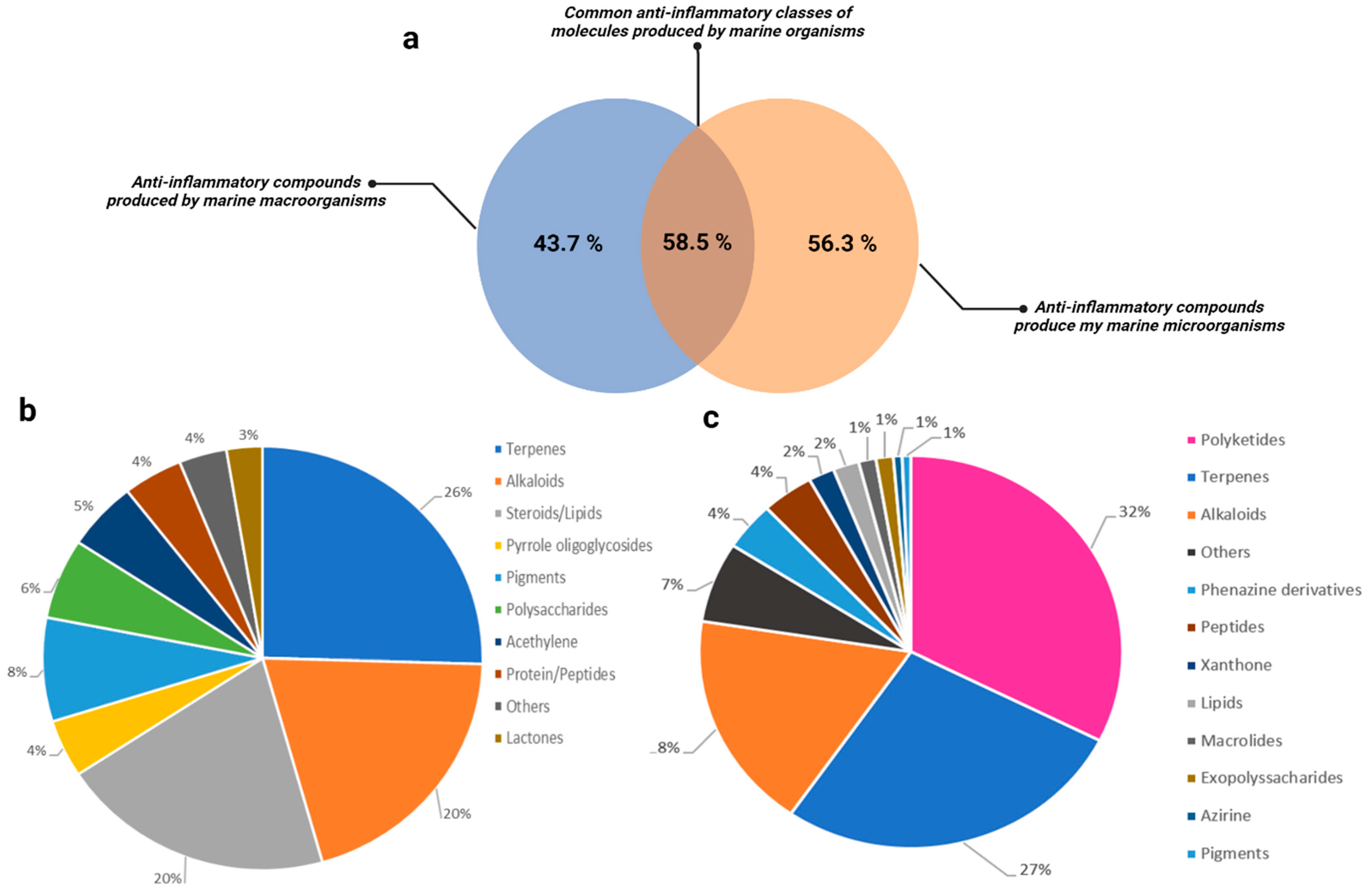


Disclaimer/Publisher’s Note: The statements, opinions and data contained in all publications are solely those of the individual author(s) and contributor(s) and not of MDPI and/or the editor(s). MDPI and/or the editor(s) disclaim responsibility for any injury to people or property resulting from any ideas, methods, instructions or products referred to in the content. |
© 2024 by the authors. Licensee MDPI, Basel, Switzerland. This article is an open access article distributed under the terms and conditions of the Creative Commons Attribution (CC BY) license (https://creativecommons.org/licenses/by/4.0/).
Share and Cite
Lasalo, M.; Jauffrais, T.; Georgel, P.; Matsui, M. Marine Microorganism Molecules as Potential Anti-Inflammatory Therapeutics. Mar. Drugs 2024, 22, 405. https://doi.org/10.3390/md22090405
Lasalo M, Jauffrais T, Georgel P, Matsui M. Marine Microorganism Molecules as Potential Anti-Inflammatory Therapeutics. Marine Drugs. 2024; 22(9):405. https://doi.org/10.3390/md22090405
Chicago/Turabian StyleLasalo, Malia, Thierry Jauffrais, Philippe Georgel, and Mariko Matsui. 2024. "Marine Microorganism Molecules as Potential Anti-Inflammatory Therapeutics" Marine Drugs 22, no. 9: 405. https://doi.org/10.3390/md22090405
APA StyleLasalo, M., Jauffrais, T., Georgel, P., & Matsui, M. (2024). Marine Microorganism Molecules as Potential Anti-Inflammatory Therapeutics. Marine Drugs, 22(9), 405. https://doi.org/10.3390/md22090405