Article

New Polyketides and a Ferroptosis Inhibitor from the Marine-Derived Fungus Diaporthe searlei CS-HF-1

1
Key Laboratory of Tropical Biological Resources of Ministry of Education, School of Pharmaceutical Sciences, Hainan University, Haikou 570228, China
2
Fujian Institute of Subtropical Botany, Xiamen 361006, China
3
College of Oceanology and Food Science, Quanzhou Normal University, Quanzhou 362000, China
4
School of Basic Medical Sciences, YiChun University, Yichun 336000, China
5
Xiamen Chenge Biotechnology Co., Ltd., Xiamen 361000, China
*
Authors to whom correspondence should be addressed.
These authors contributed equally to this work.
Mar. Drugs 2025, 23(10), 402; https://doi.org/10.3390/md23100402
Submission received: 19 August 2025 / Revised: 13 October 2025 / Accepted: 14 October 2025 / Published: 16 October 2025
(This article belongs to the Special Issue Bioactive Secondary Metabolites of Marine Fungi, 3rd Edition)

Abstract

As a driver of neurodegenerative disorders, ischemic injuries, and acute organ dysfunction, ferroptosis represents a therapeutic target, and its inhibition may provide novel therapies. In our ongoing efforts to discover ferroptosis inhibitors from fungal strains, chemical investigation of the strain Diaporthe searlei CS-HF-1 led to the isolation of four polyketide-derived alkaloids (13 and 17) and fourteen polyketides (416 and 18), including three new isoindolone derivatives (13), a new phthalide (4), a new butyrolactone derivative (10), and three new nonenolides (1113). The structures were determined by comprehensive spectroscopic analysis. The structures of 1, 2, and 10 were confirmed by comparison of experimental and calculated 13C NMR chemical shifts. The absolute configurations of compounds 10, 11, and 14 were assigned by ECD calculations, while those of 12 and 13 were assigned based on their biogenetic relationship with 14. Notably, compound 1 represents the first isoindolone featuring a primary amide group attached to the lactam nitrogen, while compound 2 is the first naturally occurring isoindolone dimer. These compounds were assessed for the anti-ferroptotic activity. As a result, asperlactone A (15) exhibited inhibition on RSL3-induced ferroptosis in HT22 cells with an EC50 of 11.3 ± 0.4 μM. Preliminary mechanistic study revealed that 15 attenuated lipid peroxidation, as evidenced by reduced MDA levels, elevated GSH content, and suppression of lipid radical generation. This study offers a new chemotype for the development of novel ferroptosis inhibitors.

1. Introduction

The genus Diaporthe, belonging to the family Diaporthaceae (order Diaporthales, class Sordariomycetes), is a large and taxonomically complex genus, with more than 1200 species recorded in the database Index Fungorum [1]. The genus was established in 1870, and its members have been discovered worldwide on a wide variety of terrestrial host plants and marine samples [1]. It is an outstanding genus of filamentous fungi that has been proven to be a prolific source of secondary metabolites [2,3]. In recent years, the metabolites of marine-derived Diaporthe species have attracted great attention, leading to the identification of a variety of new compounds [4,5,6,7,8,9,10,11,12,13,14,15,16,17,18,19,20,21,22,23,24,25], including polyketides (butyrolactone derivatives [4], oxygen-bridged cyclooctadiene derivatives [7], monomeric/dimeric/trimeric clavatol derivatives [6], chromone derivatives [9,16,17,21], mono or dimeric xanthones [11], highly oxygenated chloroazaphilone derivatives [19], anthraquinones [16], octaketides [18], and highly substituted phthalides [18]), monoterpenes (the rare thujanes diaporterpenes A–C [10] and acyclic monoterpenes diaporterpenes D–F [5]), sesquiterpenoids including drimane-type sesquiterpenoids [12,14,22], diterpenoids (diaporpenoid A and longidiacids A–B) [14,15], alkaloids (cytochalasins, chromeno[3,2-c]pyridines, and isoindolinones) [15,20], and meroterpenoids including chrodrimanins [12]. Their bioactivities mainly involve cytotoxic, anti-fibrotic, antifungal, antibacterial, antiviral, antioxidant, anti-inflammatory, anti-osteoclastogenesis, and enzyme inhibitory activities. Moreover, diaporthe H, a clavatol-dimer derivative, exhibited potent anti-fibrotic activity with an EC50 value of 3.5 μM while showing low cytotoxicity [6]. Mycoepoxydiene (A549, IC50 = 1.97 μM) and the cyclohexanone derivative 5,6-dihydroxy-3-(hydroxymethyl)-2,6-dimethylcyclohex-2-en-1-one (MDA-MB-231, IC50 = 2.54 μM) exhibited potent cytotoxicities [7]. Pestalotiopsone B, isolated from Diaporthe sp., displayed significant anti-influenza A virus activity against the A/Puerto Rico/8/34 (H1N1) strain (IC50 = 2.56 μM) [18]. Therefore, chemical study on Diaporthe spp. may yield novel/bioactive molecules.
Ferroptosis is an iron-dependent form of programmed cell death driven by lipid peroxidation [26]. It plays a significant role in pathologies, including neurodegenerative diseases, ischemic injuries, and cancer [27,28]. However, current inhibitors face clinical challenges such as poor metabolic stability and bioavailability. Given their structural diversity and biocompatibility, natural products are considered a promising source for developing next-generation ferroptosis inhibitors [29,30].
In our ongoing search for bioactive metabolites from fungal strains [31,32,33,34,35,36], especially ferroptosis inhibitors [31,32,33,34], the 1H NMR spectrum of the EtOAc extract of the strain Diaporthe searlei CS-HF-1 indicated a rich secondary metabolite profile (Figure S1). A literature review revealed that only a potent antibacterial dimeric xanthone (18, secalonic acid A) was isolated from this species [37]. Thus, a systematic chemical study of this strain was performed and yielded 18 compounds (Figure 1). Herein, we present the isolation, structural elucidation, and anti-ferroptotic activity of these compounds.

2. Results and Discussion

2.1. Structural Elucidation

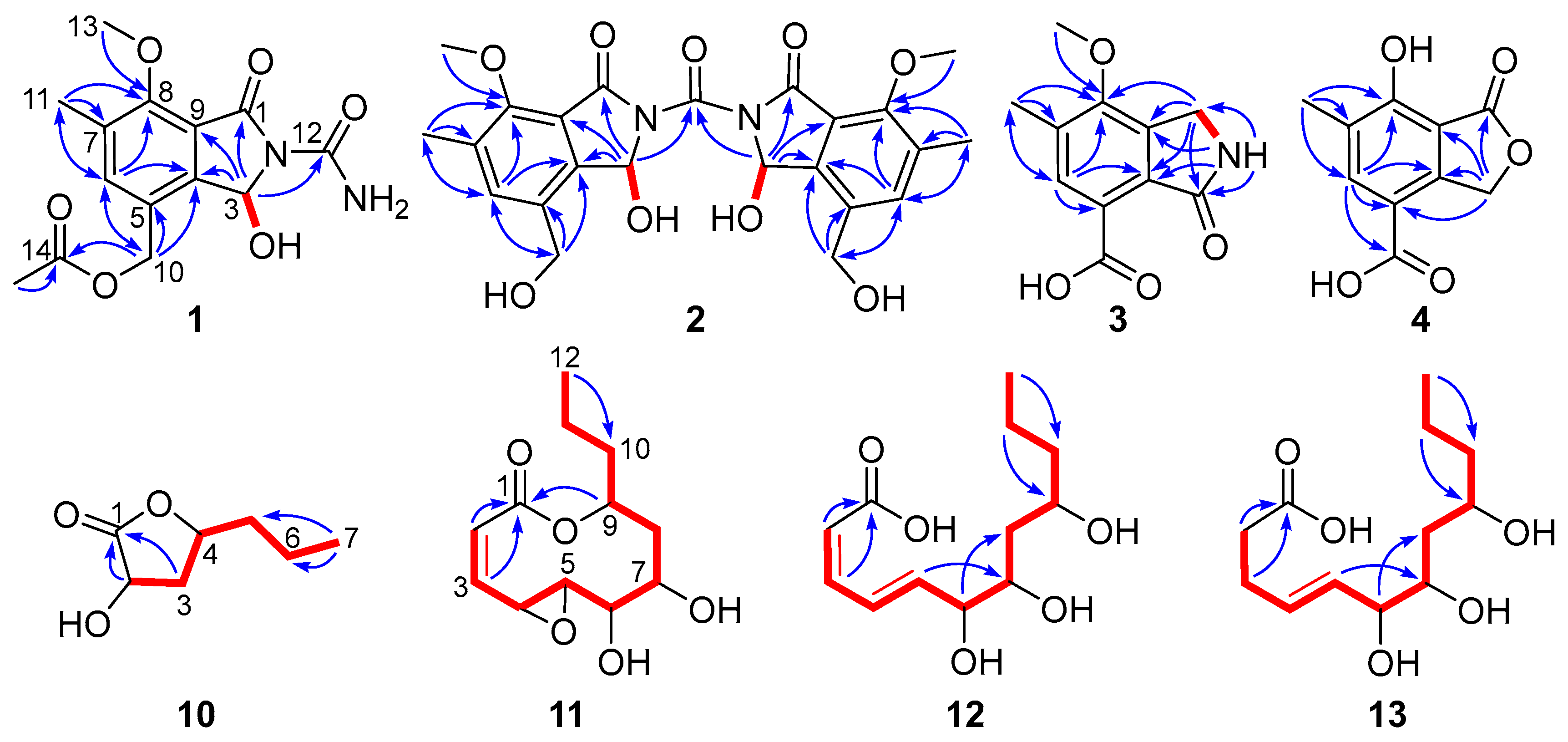
Compound 1 was obtained as a light-yellow oil. Its molecular formula, C14H16N2O6, was determined by the positive HRESIMS ion peak at m/z 331.0909 [M + Na]+ (calcd. 331.0901), indicating eight degrees of unsaturation. The 1H NMR spectrum (Table 1) showed signals for one methoxy (δH 3.93, s), one aromatic methyl (δH 2.28, s), one acetyl methyl (δH 2.06, s), one aromatic proton (δH 7.61, s), a pair of two coupled protons [δH 7.40 (1H, d, J = 10.0 Hz); 6.94 (1H, d, J = 10.0 Hz)], a broad singlet at δH 5.98 integrated for two protons, and two isolated and geminally coupled protons [δH 5.08 (1H, d, J = 12.7 Hz), 4.98 (1H, d, J = 12.7 Hz)] of an oxymethylene (which suggested spatial proximity to a methine stereocenter). The 13C NMR and HSQC spectra revealed a total of 14 carbons, which were assigned to three methyls (δC 61.7, 20.5, 15.2), including one methoxy, one oxymethylene carbon (δC 61.5), one oxymethine carbon (δC 81.5), six aromatic carbons for a pentasubstituted benzene ring (δC 156.0, 144.3, 137.4, 132.6, 126.4, 118.3), and three carbonyl carbons (δC 170.2, 166.3, and 156.9). With seven degrees of unsaturation assigned to three carbonyl groups and a benzene ring, the remaining one implied that compound 1 was bicyclic.
The aforementioned data closely resembled the structural features of the co-isolated analogue phomotone (6) [38], containing an identical 4-methoxy-3-methylbenzyloxy moiety. The notable distinctions were the presence of one additional carbonyl carbon (δC 156.9), an acetyl group (δH 2.06; δC 20.5, 170.2), and the upfield-shifted hydroxymethine carbon (1: δC 81.5; 6: δC 108.7). The chemical shifts of the hydroxylated methine (δH 6.94; δC 81.5) combined with the N-containing molecular formula indicated the replacement of the lactone moiety in 6 by a lactam unit in 1, suggesting an isoindolone derivative. In addition, the HMBC correlation from the oxymethylene protons H2-10 (δH 5.08, 4.98) to the acetyl carbonyl carbon C-14 (δC 170.2) linked the acetoxy group to C-10 (Figure 2). The residual elemental composition, after accounting for the aforementioned functionalities, was consistent with a primary amide group (-CONH2). The deduction was confirmed by the characteristic broad singlet for protons of primary amine at δH 5.98 (2H, br s) and the HMBC correlation from the hydroxymethine proton at δH 6.94 to the primary amine carbonyl carbon (δC 156.9). The structure of 1 was confirmed by comprehensive 2D NMR analysis (Figure 2). Compound 1, a polyketide-derived alkaloid, was an isoindolone derivative featuring a primary amine unit attached to the amide nitrogen atom.
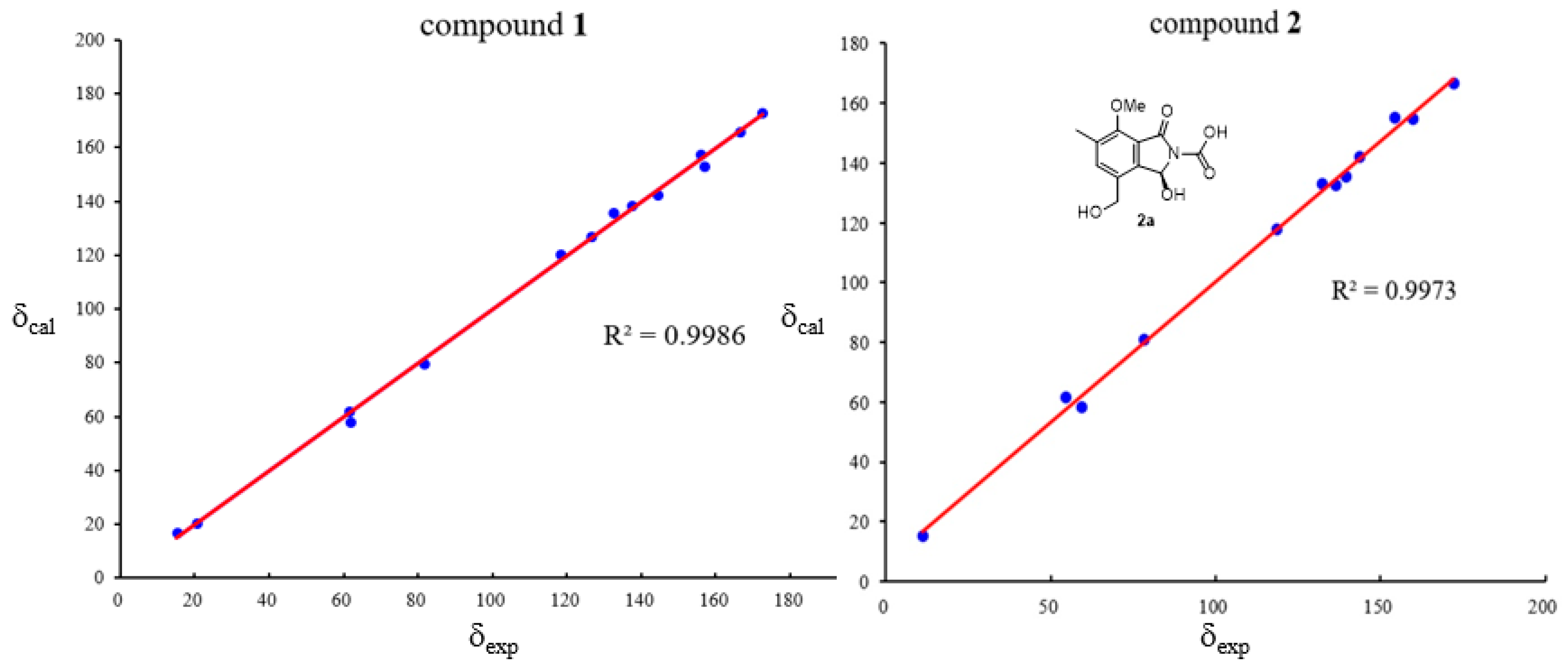
To confirm its structure, we conducted DFT calculations of the 13C NMR chemical shifts employing the GIAO method at the mPW1PW91/6-311+G(d,p) level using the conductor polarizable calculation model (CPCM) with dimethyl sulfoxide (DMSO) as solvent. The results revealed an excellent linear correlation (R2 = 0.9986) between the calculated and experimental chemical shifts (Figure 3 and Table S1), thereby providing strong evidence for the assigned structure. Compound 1 was optically inactive and gave a flat CD spectrum (200–400 nm), which indicated that 1 was racemic. Subsequent attempts to resolve the enantiomers of 1 on a Phenomenex Lux Cellulose-2 chiral column with the mobile phase CH3CN/H2O or n-hexane/isopropanol failed. Thus, compound 1 was characterized as a racemate and assigned the trivial name (±)-diasearamide A.
Compound 2 was assigned the molecular formula C23H24N2O9 based on positive HRESIMS data (m/z 495.1398 [M + Na]+, calcd. 495.1374) and negative HRESIMS data (m/z 471.1400 [M − H], calcd. 471.1409). The 1H NMR spectrum displayed signals for one aromatic singlet (δH 7.62, s), one aromatic methyl (δH 2.28, s), one methoxy (δH 3.91, s), two coupled protons [δH 7.71 (1H, d, J = 9.3 Hz); 6.93 (1H, d, J = 9.3 Hz)], one oxygenated methylene (δH 4.51, s), and one broad singlet for one proton at δH 5.30. The 13C NMR spectrum revealed only 12 carbons (Table 1), which were assigned with the aid of the HSQC spectrum to two methyl carbons (δC 61.6, 15.2), one oxymethylene carbon (δC 58.4), one oxymethine carbon (δC 80.9), six aromatic carbons for a pentasubstituted benzene ring (δC 154.8, 142.2, 135.5, 132.8, 132.4, 117.8), and two carbonyl carbons (δC 166.6, and 155.2). The observation of 12 carbons, inconsistent with the molecular formula C23H24N2O9, suggested a dimeric architecture comprising two identical C-11 subunits linked by a shared carbon atom. A detailed comparison of the NMR data of 1 and 2 indicated that the monomeric unit of 2 corresponded to a derivative of 1 lacking the acetyl and the amide groups. The two C11 subunits were linked by a carbonyl carbon based on the HMBC correlation from the oxymethine proton (δH 6.93) to the remaining carbonyl carbon at δC 155.2. The structure of 2 was secured by detailed 2D NMR analysis (Figure 2). To further confirm the structure, we performed computational predictions of the NMR chemical shifts of 2 with a simplified model molecule 2a using the GIAO method at the mPW1PW91/6-311+G(d,p) level with the conductor polarizable calculation model (CPCM) in DMSO. The calculated NMR chemical shifts (Figure 3 and Table S2) for 2a were in good agreement with the experimental 13C NMR data of 2 (R2 = 0.9979), which further supported the structure of 2. Since the monomers of the two fragments could have either the same or opposite absolute configurations, the chiral centers are depicted with wavy bonds. Compound 2 was named diasearamide B.
The molecular formula of diasearamide C (3) was determined to be C11H11NO4 based on analysis of the HRESIMS (m/z 220.0614 [M − H], calcd. 220.0615) and 13C NMR data. The 1H NMR spectrum exhibited signals for one methoxy (δH 3.80), one aromatic methyl (δH 2.32), one aromatic singlet (δH 7.66), one oxymethylene (δH 4.39), and one broad singlet assigned to an amide proton (δH 8.51). The 13C NMR and HSQC spectra indicated the presence of six aromatic carbons for a pentasubstituted benzene ring (δC 160.1, 143.5, 132.3, 128.1, 127.6, 121.8), one carbonyl carbon (δC 168.9), one methylene carbon (δC 45.4), and two methyl carbons (δC 61.3, 16.1), including one methoxy. These structural features were very similar to those of 1 and 2, indicating an isoindolone derivative with a similar carbon skeleton. The HMBC correlations from the methoxy protons to C-8 (δC 160.1) and from the aromatic methyl protons to C-6 (δC 127.6), C-7 (δC 132.3), C-8, and C-9 (δC 128.1) indicated these two groups were located at the same positions as those in 1 and 2. The structure of 3 was further determined by detailed 2D NMR analysis (Figure 2). Specifically, the HMBC correlations from the amide proton at δH 8.51 (H-2, br s) to C-3 (δC 168.9), C-4 (δC 143.5), and C-9 (δC 128.1) and from H2-1 (δH 4.39) to C-8 and C-7 positioned the methylene group at C-1 and the carbonyl carbon at C-3. Based on the molecular formula, the presence of a carboxyl group (δC 166.4) was required, and it must be attached to the remaining non-hydrogenated aromatic carbon C-5 (δC 121.8). Thus, the structure of 3 was determined as depicted and named diasearamide C.
Diasearlide acid (4), a light-yellow oil, was assigned the molecular formula C10H8O5 as determined by the negative HRESIMS ion at m/z 207.0296 ([M − H], calcd. 207.0299), suggesting seven degrees of unsaturation. The 1H and 13C NMR spectrum revealed the presence of one pentasubstituted benzene ring (δH 7.75; δC 165.9, 148.8, 130.2, 128.1, 115.1, 108.0), one methylene (δH 5.40; δC 70.4), one aromatic methyl (δH 2.23; δC 15.6), and two carbonyl groups (δC 171.3, 170.1). The benzene ring and the two carbonyl carbons accounted for six degrees of unsaturation; the remaining one suggested 4 to be bicyclic. These data indicated a phthalide derivative, structurally similar to the co-isolated convolvulol (7) [39]. The differences were the absence of the methoxy (δH 4.02) and the hydroxymethyl groups (δH 4.68). The HMBC correlations from the aromatic proton (δH 7.75) to C-4 (δC 148.8), C-5 (δC 115.1), and the carbonyl group (δC 170.1) indicated the replacement of the hydroxymethyl group in 7 by a carboxyl group. Based on the molecular formula, the methoxy group in 7 was replaced by a hydroxy group. Detailed analysis of the 2D NMR data confirmed the structure of 4 (Figure 2).
The molecular formula of compound 10 was determined to be C7H12O3 by HRESIMS (m/z 143.0710 [M − H], calcd. 143.0714), establishing an index of hydrogen deficiency of two. The 1H NMR spectrum of 10 showed signals for two oxygenated protons [δH 4.53 (1H, dd, J = 10.7, 8.8 Hz); 4.40 (1H, m)], a methyl triplet [δH 0.98 (3H, t, J = 7.2 Hz)], and several aliphatic protons. The 13C NMR and HSQC spectra indicated the presence of a carbonyl carbon (δC 179.4), two oxymethine carbons (δC 78.1, 69.5), three methylene carbons (δC 38.7, 38.6, 19.4), and a methyl carbon (δC 14.1). Since one of the two degrees of unsaturation was accounted for by one carbonyl carbon, the remaining one indicated that 10 was monocyclic. The gross structure was further established by HMBC and 1H-1H COSY correlations. Specifically, the 1H-1H COSY relationship of H-2 (δH 4.53)/H-3a (δH 2.66)/H-4 (δH 4.40)/H-5 (δH 1.63)/H2-6 (δH 1.47)/H3-7 (δH 0.98) defined a six-carbon spin system (CH-2/CH2-3/CH-4/CH2-5/CH2-6/CH3-7), the HMBC correlations from H-2 and H-3 to C-1 indicated the C6 unit was linked to the carbonyl carbon (δC 179.4). The remaining degree of unsaturation required that C-4 must be connected to the carbonyl carbon C-1 via one O-atom to form a lactone moiety. Since there was no NOESY correlation between H-2 and H-4, the relative configuration of 10 was assigned to be 2S* and 4S*, respectively. The structure of 10 was confirmed by matching its experimental 13C NMR data with the calculated shifts (R2 = 0.9991), demonstrating excellent consistency (Figure 4, Table S3). Further ECD calculation defined the absolute configuration of the two chiral centers to be 2S and 4S, respectively (Figure 4). Thus, the structure of compound 10 was determined to be (2S,4S)-2-hydroxy-4-propylbutyrolactone.
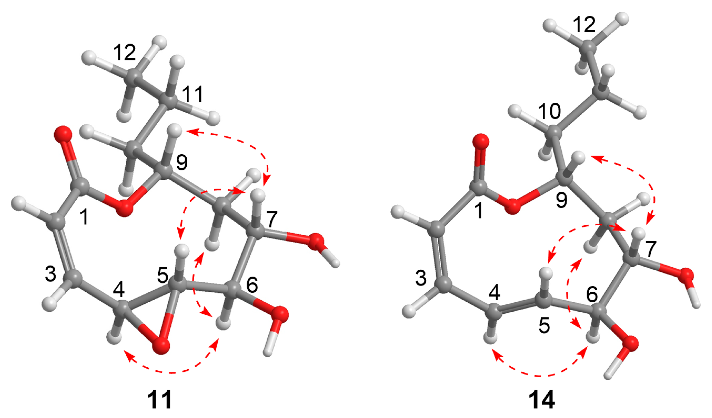
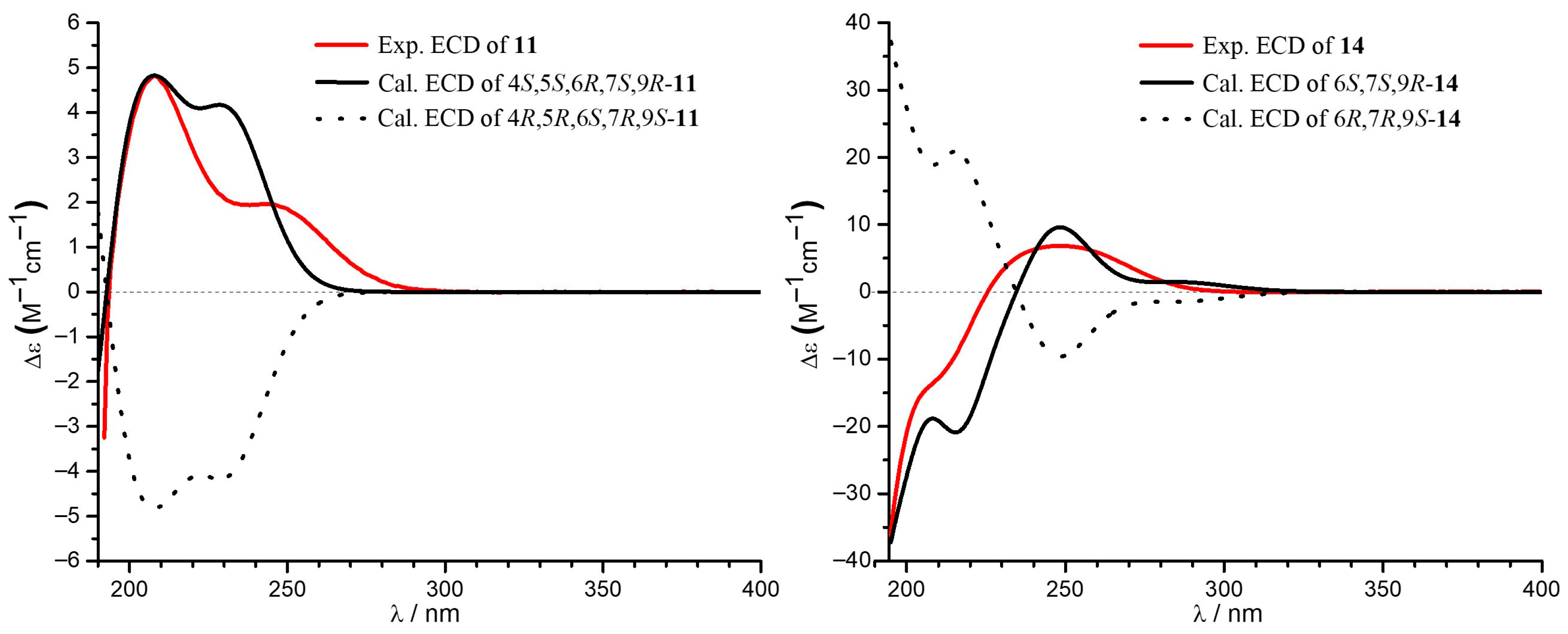
The molecular formula of 11 was established as C12H18O5 based on the HRESIMS (m/z 241.1065 [M − H], calcd. 241.1081), indicating the presence of one additional oxygen atom compared to the co-isolated xylarolide (14) [40]. The 1H NMR spectrum showed signals for two coupled olefinic protons of a cis-double bond [δH 6.52 (d, J = 11.0 Hz), 5.96 (d, J = 11.0, 1.5 Hz)], five oxygenated protons (δH 4.88, 3.75, 3.58, 3.03, 2.60), a methyl triplet [δH 0.95 (3H, s, J = 7.2 Hz)], and several aliphatic protons. The 13C NMR and HSQC spectra revealed 12 carbons, including one carbonyl carbon (δC 166.6), two olefinic carbons (δC 142.6, 126.0), five oxymethine carbons (δC 79.0, 76.3, 73.7, 62.6, 57.5), three methylene carbons (δC 41.6, 38.5, 19.3), and one methyl carbon (δC 14.3). These data (Table 2) showed high similarity to the co-isolated analogue xylarolide (14) except for the presence of two additional oxygenated carbon signals (δC 62.6 and 57.5) in 11 instead of signals for Δ3 in 14. This indicated that 11 was a 3,4-epoxy derivative of 14. The location of the epoxy ring was confirmed by the COSY relationships of H-2 (δH 5.96)/H-3 (δH 6.52)/H-4 (δH 3.75)/H-5 (δH 2.60), along with HMBC correlations from H-2 (δH 5.96) to C-4 (δC 57.5) and from H-3 (δH 6.52) to C-5 (δC 62.6) (Figure 2). The relative configuration was determined by a NOESY experiment (Figure 5). Specifically, the correlations from H-6 to H-4 and H-8b, from H-7 to H-5 and H-9, in association with the coupling constant J4,5 (2.0 Hz), indicated that the protons H-5, H-7, and H-9 were cofacial and H-4 and H-6 were in the opposite orientation. To determine the absolute configuration of 11, ECD calculation of 4S, 5S, 6R, 7S, 9R-11 was conducted using B3LYP/6-31+G(d,p) optimized geometries at the B3LYP/6-31+G(d,p) level with the solvation model based on density (SMD) in MeOH. The experimental ECD spectrum of 11 showed a similar ECD curve to the calculated curve for 4S, 5S, 6R, 7S, 9R-11 at around 220 nm (Figure 6). Compound 11 was named diasearolide A.
Compound 12 had the molecular formula C12H20O5 as determined by the HRESIMS (m/z 243.1236 [M − H], calcd. 243.1238), indicating three degrees of unsaturation. The NMR data indicated the presence of one carbonyl carbon (δC 169.7), one trans and one cis double bonds [δH 7.54 (1H, dd, J = 15.4, 11.5 Hz), 6.68 (1H, dd, J = 11.5, 11.4 Hz), 6.13 (1H, dd, J = 15.4, 6.4 Hz), 5.65 (1H, dd, J = 11.4 Hz); δC 145.4, 144.2, 128.8, 118.9], three oxymethines (δH 4.06, 3.82, 3.80; δC 76.7, 72.3, 68.8), three methylenes [δH 1.51 (2H, m), 1.43 (2H, m), 1.44 (1H, m), 1.37 (1H, m); δC 41.5, 41.2, 19.9), and one methyl [δH 0.93 (3H, t, J = 6.9 Hz); δC 14.4)]. These data closely resembled those of 14, with the sole discrepancy owing to the chemical shifts of the oxymethine CH-9 (12: δH 3.82, δC 68.8; 14: δH 4.86, δC 77.1). As three degrees of unsaturation were accounted for by the two double bonds and the carbonyl group, indicating 12 to be acyclic. Thus, compound 12 was the ring-opened derivative generated from the hydrolysis of the lactone moiety in compound 14. The structure of 12 was confirmed by detailed 2D NMR analysis (Figure 2). It should be noted that the relative configuration of 14 was originally determined to be 6S*, 7S*, and 9R* in the literature [40]. However, a subsequent total synthesis of the proposed structure (6S*, 7S*, 9R*)-14 yielded a product with NMR data that did not match those of the natural 14. This discrepancy led the authors to suggest a revision of the structure of xylarolide [41]. In our study, the structure of natural xylarolide was confirmed to be correct by detailed NMR analysis (including the key NOESY correlations in Figure 5) and by comparison with the co-isolated analogue asperlactone A (15) [42], whose structure was unequivocally confirmed by X-ray crystallography. The absolute configuration of 14 was also determined to be 6S, 7S, and 9R by comparison of the experimental ECD spectrum with the calculated ECD curves (Figure 6). The absolute configuration of C-6, C-7, and C-9 in 12 was determined to be the same as 14 based on biogenetic considerations and sharing almost identical NMR data.
Compound 13 had the molecular formula C12H22O5 as determined by the negative HRESIMS ion at m/z 245.1378 ([M − H], calcd. 245.1394), two mass units over that of 12. The NMR spectra of 13 showed similar structural features to 12, with obvious differences being the presence of two additional methylenes (δC 34.4 and 29.0) and the absence of one double bond. The aforementioned information indicated that 13 was the hydrogenated derivative of 12. The HMBC correlations from the extra methylene protons [δH 2.43 (2H, m) and 2.36 (2H, m)] to the sole carboxyl carbon (δC 176.2) indicated that the Δ2 in 12 was hydrogenated in 13. The structure of 13 was secured by 2D NMR analysis (Figure 2). The absolute configuration of the chiral centers in 13 was proposed to be identical to that of 12 based on biogenetic considerations.
Additionally, the co-isolated known compounds were determined to be dihydrogladiolic acid methyl lactal (5) [43], phomotone (6) [38], convolvulol (7) [39], 4,6-dihydroxy-5-methyl-1(3H)-isobenzofuranone (8) [44], 2,5-dimethylresorcin (9) [45], xylarolide (14) [40], asperlactone A (15) [42], phomolide D (16) [38], 4-hydroxy-3,6-dimethyl-2(1H)-pyridinone (17) [46], and secalonic acid A (18) [37] by comparing the NMR data and specific rotations with those in the literature.

2.2. Biological Activity Assessment

The isolated compounds were evaluated for their anti-ferroptotic and anti-inflammatory activities.

2.2.1. Anti-Ferroptotic Activity in an RSL3-Induced Ferroptosis Model in HT22 Cells

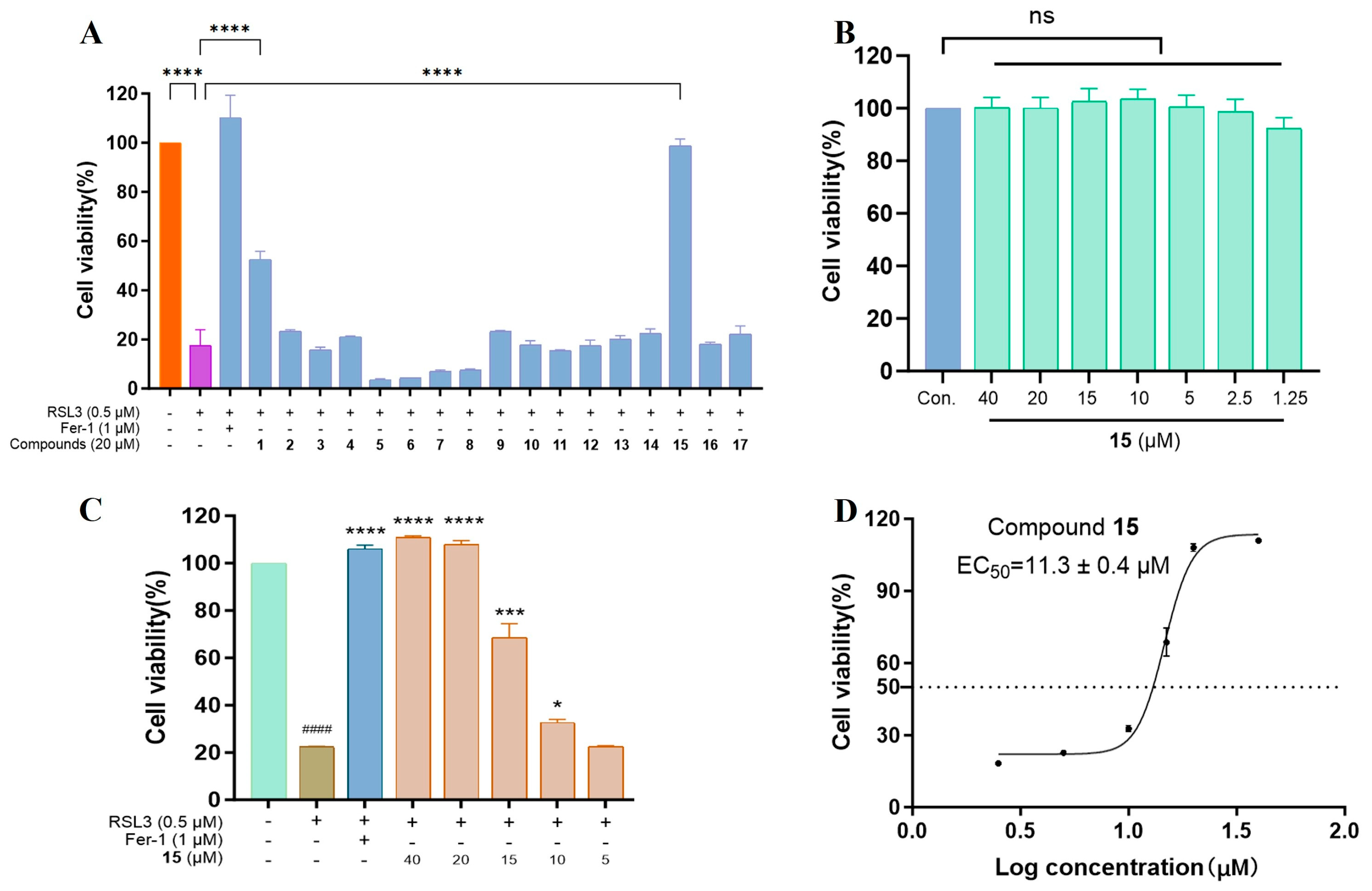
Compounds 117 were screened for their anti-ferroptotic activity (18 was excluded due to its known cytotoxicity) in an RSL3-induced ferroptosis model in HT22 cells. As shown in Figure 7A, treatment with 0.5 μM RSL3 reduced cell viability to roughly 20% relative to the untreated control. At a concentration of 20 μM, only compounds 1 and 15 exhibited obvious cytoprotection, restoring cell viability to 52% and 98%, respectively, whereas the remaining analogues showed negligible effects (<30% viability). Notably, 15 was non-cytotoxic to HT22 cells at concentrations up to 40 μM (Figure 7B). The potency of 15 was further assessed via a concentration–response study (5–40 μM), using ferrostatin-1 (Fer-1) as a positive control. As a result, compound 15 produced a dose-dependent increase in cell viability over the concentration range of 5–20 μM (Figure 7C), with an EC50 value of 11.3 ± 0.4 μM (Figure 7D).
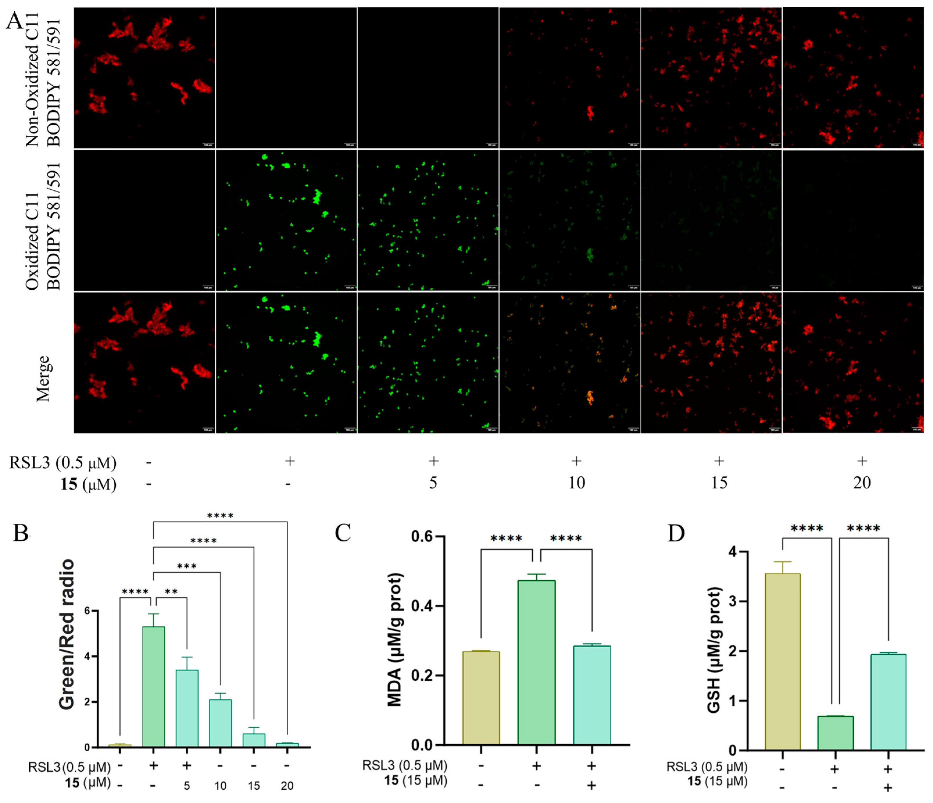
To elucidate the preliminary mechanism of action underlying the anti-ferroptotic activity of compound 15, we investigated its effect on key biomarkers of ferroptosis. Fluorescence imaging using C11-BODIPY581/591 fluorescent probe demonstrated that 15 concentration-dependently mitigated the pronounced lipid peroxidation induced by RSL3, with significant suppression observed at concentrations above 15 μM (Figure 8A,B). Consistent with the suppression of lipid peroxidation, compound 15 (15 μM) also attenuated the RSL3-induced elevation in MDA (Figure 8C) and reduction in GSH (Figure 8D). These results indicated that the protective effect of 15 is mediated through the inhibition of lipid peroxidation.
It is worth noting that 15 exhibited ferroptosis-inhibitory activity with a distinct scaffold that has not been previously reported among current known ferroptosis inhibitors. This study offers a new chemotype for the development of novel ferroptosis inhibitors.

2.2.2. Anti-Inflammatory Activity Against LPS-Activated NO Production in RAW264.7 Macrophages

These compounds were first assessed for cytotoxicity against RAW264.7 cells at 50 μM. Compounds demonstrating >90% cell viability (compounds 6, 7, 13, and 18 were excluded) were then assessed for inhibition of NO production in LPS-activated RAW264.7 macrophages, using the natural NO inhibitor quercetin as a reference (IC50 = 16 ± 1 μM). Only compounds 9 and 11 exhibited weak inhibitory effects, with 28.3% and 31.6% inhibition at 50 μM, respectively. The reduced activity of compound 14 compared to its epoxy derivative 11 suggested the epoxy moiety enhanced the inhibitory effect.

3. Experimental Section

3.1. General Experimental Procedure

Specific rotations were measured by an Autopol III automatic polarimeter (Rudolph Research Co., Ltd., Flanders, NJ, USA). Ultraviolet spectra were recorded on a UV-2600 spectrometer (Shimadzu Co., Kyoto, Japan). ECD spectra were obtained on an Applied Photophysics Chirascan spectrometer (Surrey, UK). The NMR spectra were acquired on a Bruker AVANCE III HD 400NMR spectrometer (Bruker, Fällanden, Switzerland) using solvent signals (Methanol-d4: δH 3.31/δC 49.0; DMSO-d6H 2.5/δC 39.52) as references. HRESIMS spectra were acquired on a Shimadzu LCMS-IT-TOF spectrometer (Shimadzu Co., Kyoto, Japan) equipped with an ESI source. Semi-preparative high-performance liquid chromatography (HPLC) was undertaken on a Shimadzu LC-6AD pump (Shimadzu Co., Kyoto, Japan) equipped with a UV detector, employing a YMC-Pack ODS-A HPLC (YMC Co., Ltd., Kyoto, Japan) column (250 mm × 10 mm, S-5 μm, 12 nm).

3.2. Fungal Strain and Identification

Fungus CS-HF-1 was isolated from the sediment at a depth of 0.6 m at Haikou Holiday Beach. The strain was identified as Diaporthe searlei through microscopic examination and internal transcribed spacer (ITS) sequencing. Its ITSsequence was deposited in GenBank under accession number PX129557, and the strain was preserved at the School of Pharmaceutical Sciences, Hainan University, China.

3.3. Fermentation, Extraction, and Isolation

The fermentation was carried out in 30 Fernbach flasks (500 mL), each containing 70 g of rice. Artificial seawater (90 mL) was added to each flask, and the contents were soaked for three hours before autoclaving at 15 psi for 30 min. After cooling to room temperature, each flask was inoculated with 3.0 mL of the spore inoculum and incubated at room temperature for 30 days. The fermentation was conducted in 100 Erlenmeyer flasks (500 mL), each containing 80 g of rice and 95 mL of artificial seawater. The contents were soaked for 1 h and then autoclaved for 20 min. Each flask was inoculated with 1.0 mL of the spore inoculum and incubated at room temperature (r. t.) for 30 days. The fermented material from all flasks was combined, soaked in EtOAc (5 L), and ultrasonically extracted for 1 h. After evaporation in vacuo, the EtOAc extract (14.3 g) was dissolved in methanol (50 mL) and 18 (200 mg) was collected as a precipitate. The mother liquor was further fractionated on an MCI gel column chromatography (CC) with MeOH/H2O (10:90 → 100:0) as eluent to yield seven fractions (F1–F7). Fraction F2 (2.7 g) was separated on an ODS silica gel CC (55 g) using a gradient elution of MeOH/H2O (10:90 → 60:40, 500 mL per gradient) to yield 6 subfractions (F2.1–F2.6). F2.1 (0.15 g) was purified by HPLC (YMC-Pack ODS-A column, 250 × 10 mm, S-5 μm, 12 nm) using MeOH/H2O (15:85, 2 mL/min) as the mobile phase to afford compound 17 (7.4 mg, Rt 29.0 min). Compounds 2 (2.8 mg) and 3 (3.1 mg) were obtained from fractions F2.3 (0.38 g) and F2.4 (1.37 g) by precipitation, respectively. The mother liquor from fraction F2.4 was further separated on an ODS silica gel CC using MeOH/H2O (10:90 → 60:40) as eluent to yield 5 subfractions (F2.4.1–F2.4.5). The subfraction F2.4.2 (419 mg) was separated by HPLC using MeOH/H2O (35:65) as the mobile phase to collect 7 subfractions (F2.4.2.1–F2.4.2.7). F2.4.2.2 (30 mg) was purified by HPLC using MeCN/H2O (17:83, 2 mL/min) as the mobile phase to afford compound 10 (2.0 mg, Rt 37.9 min). F2.4.2.3 (101 mg) was purified by HPLC using MeCN/H2O (16:84, 2 mL/min) as the mobile phase to afford compound 7 (2.5 mg, Rt 52.2 min) and a mixture, the latter was purified by HPLC using MeOH/H2O (31:69, 2 mL/min) to afford compound 8 (2.0 mg, Rt 42.0 min) and compound 15 (1.2 mg, Rt 47.2 min). F2.4.2.4 (87 mg) was purified by HPLC using MeCN/H2O (17:83, 2.5 mL/min) as the mobile phase to afford compound 13 (40.0 mg, Rt 30.9 min). F2.4.5 (332 mg) was separated on an ODS silica gel CC using a gradient elution of MeOH/H2O to yield 6 subfractions (F2.4.5.1–F2.4.5.6). F2.4.5.4 (94 mg) was purified by HPLC using MeOH/H2O (37:63, 2 mL/min) to afford compound 16 (8.2 mg, Rt 34.1 min). F2.4.5.5 (99 mg) was purified by HPLC using the solvent MeOH/H2O (37:63, 2 mL/min) to afford compounds 1 (5.3 mg, Rt 34.8 min), 4 (3.9 mg, Rt 51.2 min), 6 (6.2 mg, Rt 38.6 min), and 11 (7.2 mg, Rt 41.7 min). F2.4.5.6 (52 mg) was purified by HPLC using MeOH/H2O (35:65, 2 mL/min) as the mobile phase to afford compound 12 (10.3 mg, Rt 33.1 min). F2.5 (0.31 g) was purified by HPLC using MeOH/H2O (45:55, 2.5 mL/min) to afford compound 5 (7.1 mg, Rt 32.0 min). Fraction F4 (0.4 g) was purified on an ODS silica gel CC eluting with MeOH/H2O (20:80 → 60:40) to give compound 14 (128 mg). F5 (2.2 g) was separated on an ODS silica gel CC using a gradient elution of MeOH/H2O (10:90 → 30:70) to afford compound 9 (8.8 mg).
(±)–Diasearamide A (1): Light yellow oil; [α]25D 0 (c 0.1, MeOH); UV (MeOH) λmax (log ε) 210 (3.73), 291 (3.02) nm; 1H and 13C NMR data, see Table 1; HRESIMS m/z 331.0909 [M + Na]+ (calcd. for C14H16N2O6Na+, 331.0901); 343.0698 [M + Cl] (calcd. for C14H16N2O6Cl, 343.0702); 370.0885 [M + NO3] (calcd. for C14H16N3O9, 370.0892).
(±)–Diasearamide B (2): White powder; [α]25D 0 (c 0.1, MeOH); UV (MeOH) λmax (log ε) 209 (4.64), 240 (3.80), 297 (3.58) nm; 1H and 13C NMR data, see Table 1; HRESIMS m/z 495.1398 [M + Na]+ (calcd. for C23H24N2O9Na+, 495.1374); 471.1400 [M − H] (calcd. for C23H23N2O9, 471.1409); 507.1162 [M + Cl] (calcd. for C23H24N2O9Cl, 507.1176); 534.1338 [M + NO3] (calcd. for C23H24N2O9NO3, 534.1365).
Diasearamide C (3): White powder; UV (MeOH) λmax (log ε) 211 (4.33), 245 (3.80) nm; HRESIMS m/z 220.0614 [M − H] (calcd. for C11H10NO4, 220.0615).
Diasearlide acid (4): Light yellow oil; UV (MeOH) λmax (log ε) 218 (3.93), 263 (3.46), 294 (3.24) nm; 1H and 13C NMR data, see Table 1; HRESIMS m/z 207.0296 [M − H] (calcd. for C10H7O5, 207.0299).
(2S,4S)-2-Hydroxy-4-propylbutyrolactone (10): Colorless oil; [α]25D −25 (c 0.1, MeOH); UV (MeOH) λmax (log ε) 245, 289 nm; ECD (c 5.6 × 10⁻⁴ M, MeOH) λmax (De) 249 (+9.48) nm. 1H NMR (400 MHz, methanol-d4): δH 4.53 (1H, dd, J = 10.7, 8.8 Hz, H-2), 2.66 (1H, m, H-3a), 1.76 (1H, m, H-3b), 4.40 (1H, m, H-4), 1.70 (1H, m, H-5a), 1.63 (1H, m, H-5b), 1.47 (2H, m, H-6), 0.98 (3H, t, J = 7.2 Hz, H-7); δC 179.4 (C, C-1), 69.5 (CH, C-2), 38.7 (CH2, C-3), 78.1 (CH, C-4), 38.6 (CH2, C-5), 19.4 (CH2, C-6), 14.1 (CH3, C-7); HRESIMS m/z 143.0710 [M − H] (calcd. for C7H11O3, 143.0714).
Diasearolide A (11): Light yellow oil; [α]25D +33 (c 0.1, MeOH); UV (MeOH) λmax (log ε) 203 (3.84) nm; ECD (c 2.1 × 10⁻³ M, MeOH) λmax (Δε) 208 (+4.82), 242 (+1.96) nm. 1H and 13C NMR data, see Table 2; HRESIMS m/z 241.1065 [M − H] (calcd. for C12H17O5, 241.1081).
Diasearolide B (12): Light yellow oil; [α]25D –8 (c 0.1, MeOH); UV (MeOH) λmax (log ε) 254 (3.61) nm; 1H and 13C NMR data, see Table 2; HRESIMS m/z 243.1236 [M − H] (calcd. for C12H19O5, 243.1238).
Diasearolide C (13): Light yellow oil; [α]25D −9 (c 0.1, MeOH); UV (MeOH) λmax (log ε) 202 (3.43) nm; 1H and 13C NMR data, see Table 2; HRESIMS m/z 245.1378 [M − H] (calcd. for C12H21O5, 245.1394).

3.4. ECD and NMR Calculation

ECD calculation: Conformational analysis of 10 was performed via random searching in the Sybyl-X 2.0 version using the MMFF94S force field with an energy cutoff of 3 kcal/mol. The results showed four conformers (52.88%, 28.44%, 12.83%, 5.85%) for (2S, 4S)-10. The conformers of 11 and 14 were determined by analysis of the NOESY correlations and referencing to the X-Ray data of 15, followed by energy minimization. The conformers were optimized using density functional theory (DFT) at the B3LYP/6-31+g(d,p) level in MeOH using the solvation model density (SMD) by the GAUSSIAN 09 program. The energies, oscillator strengths, and rotational strengths (velocity) of the first 30 electronic excitations were calculated using the TDDFT methodology at the B3LYP/6-31+g(d,p) level using the SMD solvation model in MeOH. The ECD spectrum was simulated by the overlapping Gaussian function (half the bandwidth at 1/e peak height, σ = 0.3) [47]. To obtain the final spectra, the simulated spectra of the conformers were averaged according to the Boltzmann distribution theory and their relative Gibbs free energy (ΔG). Theoretical ECD spectra of the enantiomers were obtained by inverting the calculated ones. Comparison between calculated and experimental ECD spectra resolved the absolute configuration.
NMR calculation: The conformers (which were obtained via random searching in the Sybyl-X 2.0 version using the MMFF94S force field with an energy cutoff of 3 kcal/mol) of 1, 2a, and 10 were optimized using density functional theory (DFT) at the B3LYP/6-31g* level (Gaussian 09). The NMR shielding constants were calculated with the GIAO method at mPW1PW91/6-311+G(d,p) levels using the conductor polarizable calculation model (CPCM) in MeOH. The computational 13C NMR data were obtained by the linear regression analysis method in the literature [48].

3.5. Biological Study

The ferroptosis-related assays (cell culture, cell viability, MDA and GSH levels, and C11-BODIPY581/591 staining) were performed as described previously [31]. Additionally, the measurement of inhibitory activity against NO production followed the reported method [49].

Supplementary Materials

The following supporting information can be downloaded at: https://www.mdpi.com/article/10.3390/md23100402/s1, Figure S1: 1H NMR spectrum of the EtOAc extract of Diaporthe searlei CS-HF-1; Figures S2−S25, Figures S34−S57, and Figures S68−S75: 1D and 2D NMR, and HRESIMS spectra of 14 and 1013; Figures S26−S33 and Figures S58−S67: 1H and 13CNMR spectra of 59 and 1418; Tables S1–S3: Calculated NMR data of 1, 2a, and 10.

Author Contributions

Investigation, J.X., P.W., Y.Z., Q.L. and Y.C.; resources, W.X., W.L. and Z.C.; writing—original draft preparation, J.X. and Y.Z.; writing—review and editing, W.L. and Z.C.; project administration, W.L. and Z.C.; funding acquisition, Z.C. All authors have read and agreed to the published version of the manuscript.

Funding

This research was funded by grants from the National Natural Science Foundation of China (82460683 to Z.C.) and the Fundamental Research Funds for Hainan University (KYQD(ZR)-23061 to Z.C.).

Institutional Review Board Statement

Not applicable.

Data Availability Statement

Data is contained within the article and Supplementary Materials.

Conflicts of Interest

Wei Xu is employed by Xiamen Chenge Biotechnology Co., Ltd., All other authors declare that there are no potential conflicts of interest. Xiamen Chenge Biotechnology Co., Ltd. has no role in the study design, collection, analysis, interpretation of data, the writing of this article or the decision to submit it for publication.

References

  1. Xie, S.; Ma, X.; Wu, H.; Zang, R.; Li, H.; Liu, M.; Li, Q.; Ma, Q.; Guo, Y.; Zhang, M. The six whole mitochondrial genomes for the Diaporthe species: Features, evolution and phylogeny. IMA Fungus 2025, 16, e140572. [Google Scholar] [CrossRef]
  2. Xu, T.-C.; Lu, Y.-H.; Wang, J.-F.; Song, Z.-Q.; Hou, Y.-G.; Liu, S.-S.; Liu, C.-S.; Wu, S.-H. Bioactive secondary metabolites of the genus Diaporthe and anamorph Phomopsis from terrestrial and marine habitats and endophytes: 2010–2019. Microorganisms 2021, 9, 217. [Google Scholar] [CrossRef] [PubMed]
  3. Nagarajan, K.; Tong, W.-Y.; Leong, C.-R.; Tan, W.-N. Potential of endophytic Diaporthe sp. as a new source of bioactive compounds. J. Microbiol. Biotechnol. 2021, 31, 493–500. [Google Scholar] [CrossRef] [PubMed]
  4. Yuan, S.; Zhuang, S.; Qiao, Y.; Wu, Q.; Ma, Y.; Chen, S.; Liu, L.; Yan, Y.; Gao, Z. Metabolic shunting in marine-derived Diaporthe sp. SYSU-MS4722 yields diverse γ-butyrolactone derivatives with anti-inflammatory activity. J. Nat. Prod. 2025, 88, 1605–1615. [Google Scholar] [CrossRef]
  5. Yuan, S.; Zhuang, S.; Han, Y.; Wen, Y.; Chen, S.; Liu, L.; Lin, Q.; Yan, Y.; Gao, Z.; Wu, Q. Deletion of a polyketide synthase phoE induces monoterpenes production in ascidian-derived fungus Diaporthe sp. SYSU-MS4722. Front. Microbiol. 2025, 16, 1593027. [Google Scholar] [CrossRef] [PubMed]
  6. Ren, S.; Yan, Y.; Zhou, Y.; Han, Y.; Yuan, S.; Chen, J.; Guo, H.; Lin, Z.; Lin, Q.; Chen, S.; et al. Genome mining of nonenzymatic ortho-quinone methide-based pseudonatural products from ascidian-derived fungus Diaporthe sp. SYSU-MS4722. Bioorg. Chem. 2025, 154, 108081. [Google Scholar] [CrossRef]
  7. Yin, Y.; Chen, T.; Sun, B.; Li, J.; She, Z.; Hu, G.; Wang, B. Oxygen-bridged cyclooctadienes and other polyketides from the mangrove endophytic fungus Diaporthe sp. ZJHJYZ-1. J. Mol. Struct. 2024, 1302, 137534. [Google Scholar] [CrossRef]
  8. Yin, Y.; Yang, W.; Chen, T.; Tan, Q.; Zou, G.; Zang, Z.; Li, J.; Wang, B.; She, Z. Cytosporones W and X: Two mutually converting epimers from a mangrove endophytic fungus Diaporthe sp. ZJHJYZ-1. ACS Omega 2023, 8, 26628–26634. [Google Scholar] [CrossRef]
  9. Xing, D.-X.; Song, X.-S.; Pan, W.-C.; Cui, H.; Zhao, Z.-X. New chromone compounds from the marine derived fungus Diaporthe sp. XW12-1. Fitoterapia 2023, 164, 105384. [Google Scholar] [CrossRef]
  10. Zhai, G.; Chen, S.; Shen, H.; Guo, H.; Jiang, M.; Liu, L. Bioactive monoterpenes and polyketides from the ascidian-derived fungus Diaporthe sp. SYSU-MS4722. Mar. Drugs 2022, 20, 553. [Google Scholar] [CrossRef]
  11. Chen, S.; Guo, H.; Jiang, M.; Wu, Q.; Li, J.; Shen, H.; Liu, L. Mono- and dimeric xanthones with anti-glioma and anti-inflammatory activities from the ascidian-derived fungus Diaporthe sp. SYSU-MS4722. Mar. Drugs 2022, 20, 51. [Google Scholar] [CrossRef]
  12. Luo, X.-W.; Chen, C.-M.; Li, K.-L.; Lin, X.-P.; Gao, C.-H.; Zhou, X.-F.; Liu, Y.-H. Sesquiterpenoids and meroterpenoids from a mangrove derived fungus Diaporthe sp. SCSIO 41011. Nat. Prod. Res. 2021, 35, 282–288. [Google Scholar] [CrossRef]
  13. Hu, C.; Li, S.; Chen, Y.; Gao, X.; Liu, Z.; Zhang, W. Polyketides from the deep-sea-derived fungus Diaporthe phaseolorum FS459. Chin. J. Org. Chem. 2021, 41, 1591–1598. [Google Scholar] [CrossRef]
  14. Chen, Y.; Zou, G.; Yang, W.; Zhao, Y.; Tan, Q.; Chen, L.; Wang, J.; Ma, C.; Kang, W.; She, Z. Metabolites with anti-Inflammatory activity from the mangrove endophytic fungus Diaporthe sp. QYM12. Mar. Drugs 2021, 19, 56. [Google Scholar] [CrossRef]
  15. Liu, Z.; Chen, Y.; Li, S.; Wang, Q.; Hu, C.; Liu, H.; Zhang, W. Bioactive metabolites from the deep-sea-derived fungus Diaporthe longicolla FS429. Mar. Drugs 2020, 18, 381. [Google Scholar] [CrossRef] [PubMed]
  16. Niu, Z.; Chen, Y.; Guo, H.; Li, S.-N.; Li, H.-H.; Liu, H.-X.; Liu, Z.; Zhang, W. Cytotoxic polyketides from a deep-sea sediment derived fungus Diaporthe phaseolorum FS431. Molecules 2019, 24, 3062. [Google Scholar] [CrossRef] [PubMed]
  17. Guo, H.; Liu, Z.-M.; Chen, Y.-C.; Tan, H.-B.; Li, S.-N.; Li, H.-H.; Gao, X.-X.; Liu, H.-X.; Zhang, W.-M. Chromone-derived polyketides from the deep-sea fungus Diaporthe phaseolorum FS431. Mar. Drugs 2019, 17, 182. [Google Scholar] [CrossRef] [PubMed]
  18. Luo, X.; Yang, J.; Chen, F.; Lin, X.; Chen, C.; Zhou, X.; Liu, S.; Liu, Y. Structurally diverse polyketides from the mangrove-derived fungus Diaporthe sp. SCSIO 41011 with their anti-influenza a virus activities. Front. Chem. 2018, 6, 282. [Google Scholar] [CrossRef]
  19. Luo, X.; Lin, X.; Tao, H.; Wang, J.; Li, J.; Yang, B.; Zhou, X.; Liu, Y. Isochromophilones A-F, cytotoxic chloroazaphilones from the marine mangrove endophytic fungus Diaporthe sp. SCSIO 41011. J. Nat. Prod. 2018, 81, 934–941. [Google Scholar] [CrossRef]
  20. Cui, H.; Yu, J.; Chen, S.; Ding, M.; Huang, X.; Yuan, J.; She, Z. Alkaloids from the mangrove endophytic fungus Diaporthe phaseolorum SKS019. Bioorg. Med. Chem. Lett. 2017, 27, 803–807. [Google Scholar] [CrossRef]
  21. Cui, H.; Ding, M.; Huang, D.; Zhang, Z.; Liu, H.; Huang, H.; She, Z. Chroman-4-one and pyrano [4,3-b] chromenone derivatives from the mangrove endophytic fungus Diaporthe phaseolorum SKS019. RSC Adv. 2017, 7, 20128–20134. [Google Scholar] [CrossRef]
  22. Zang, L.Y.; Wei, W.; Guo, Y.; Wang, T.; Jiao, R.H.; Ng, S.W.; Tan, R.X.; Ge, H.M. Sesquiterpenoids from the mangrove-derived endophytic fungus Diaporthe sp. J. Nat. Prod. 2012, 75, 1744–1749. [Google Scholar] [CrossRef] [PubMed]
  23. Zhu, M.-X.; Hao, Y.; Yan, B.-Y.; Jin, Y.; Gao, Z.; Li, X.-W. Diverse secondary metabolites from the Ligia exotica-derived fungus Diaporthe melitensis HZ-1. Chem. Biodivers. 2025, 22, e202500919. [Google Scholar] [CrossRef] [PubMed]
  24. Ma, X.; Wei, X.; Xing, S.; Cheng, X.; Lu, J.; Yang, C.; Zhao, G.; Dai, Z.; Hou, S.; Liu, Y.; et al. Polyketide production in a mangrove-associated fungus Diaporthe goulteri, induced by chemical epigenetic modification. Nat. Prod. Res. 2024, 1–9. [Google Scholar] [CrossRef]
  25. Lu, H.; Feng, Y.; Lin, M.; Yu, X.; Liu, Y.; Tan, Y.; Luo, X. Discovery of polyketides and alkaloids from the mangrove endophytic fungus Diaporthe sp. and their anti-osteoclastogenic bioactivities. Chem. Biodivers. 2025, 22, e202500858. [Google Scholar] [CrossRef]
  26. Brown, A.R.; Hirschhorn, T.; Stockwell, B.R. Ferroptosis—Disease perils and therapeutic promise. Science 2024, 386, 848–849. [Google Scholar] [CrossRef]
  27. Stockwell, B.R.; Jiang, X.; Gu, W. Emerging mechanisms and disease relevance of ferroptosis. Trends Cell Biol. 2020, 30, 478–490. [Google Scholar] [CrossRef]
  28. Jiang, X.; Stockwell, B.R.; Conrad, M. Ferroptosis: Mechanisms, biology and role in disease. Nat. Rev. Mol. Cell Bio. 2021, 22, 266–282. [Google Scholar] [CrossRef]
  29. Li, H.; Puopolo, T.; Seeram, N.P.; Liu, C.; Ma, H. Anti-ferroptotic effect of cannabidiol in human skin keratinocytes characterized by data-independent acquisition-based proteomics. J. Nat. Prod. 2024, 87, 1493–1499. [Google Scholar] [CrossRef]
  30. Wang, Y.-Y.; Wang, Y.-S.; Li, J.-J.; Wan, Z.-Y.; Zhang, H. Unusual chaetoglobosins and a new type of ferroptosis inducer from an endophytic fungus Chaetomium sp. UJN-EF006. Bioorg. Chem. 2025, 158, 108342. [Google Scholar] [CrossRef]
  31. Zhang, Y.; Wang, H.; Xiao, J.; Xu, W.; Liu, W.; Luo, Z.; Wu, P.; Yang, X.; Zhang, Z.; Cheng, Z. Agathic acid derivatives including a novel ferroptosis inhibitor from the fungus Penicillium thomii. Bioorg. Chem. 2025, 164, 108867. [Google Scholar] [CrossRef] [PubMed]
  32. Zhang, Z.; Li, Y.; Wang, H.; Xu, W.; Wang, C.; Ma, H.; Zhong, F.; Ou, J.; Luo, Z.; Luo, H.-B.; et al. Ergone derivatives from the deep-sea-derived fungus Aspergillus terreus YPGA10 and 25,28-dihydroxyergone-induced apoptosis in human colon cancer SW620 Cells. J. Nat. Prod. 2024, 87, 1563–1573. [Google Scholar] [CrossRef] [PubMed]
  33. Han, S.; Ma, H.; Wu, Y.; Wang, C.; Li, Y.; Li, Q.; Cheng, Z. Andrastin-type meroterpenoids, α-pyrone polyketides, and sesquicarane derivatives from Penicillium sp., a fungus isolated from Pinus koraiensis seed. Phytochemistry 2024, 225, 114202. [Google Scholar] [CrossRef] [PubMed]
  34. Li, Y.; Shi, J.; Liu, R.; Liu, Y.; Liu, R.; Wu, Z.; Xu, W.; Ma, H.; Luo, H.-B.; Cheng, Z. Structure revisions of phenolic bisabolane sesquiterpenes and a ferroptosis inhibitor from the marine-derived fungus Aspergillus versicolor YPH93. J. Nat. Prod. 2023, 86, 830–841. [Google Scholar] [CrossRef]
  35. Li, Q.; Xu, W.; Fan, R.Z.; Zhang, J.; Li, Y.L.; Wang, X.W.; Han, S.Y.; Liu, W.; Pan, M.H.; Cheng, Z.B. Penithoketone and penithochromones A-L, polyketides from the deep-sea-derived fungus Penicillium thomii YPGA3. J. Nat. Prod. 2020, 83, 2679–2685. [Google Scholar] [CrossRef]
  36. Cheng, Z.B.; Li, Y.L.; Xu, W.; Liu, W.; Liu, L.J.; Zhu, D.G.; Kang, Y.; Luo, Z.H.; Li, Q. Three new cyclopiane-type diterpenes from a deep-sea derived fungus Penicillium sp. YPGA11 and their effects against human esophageal carcinoma cells. Bioorg. Chem. 2019, 91, 103129. [Google Scholar] [CrossRef]
  37. Dominique, S.; Alex, P.G.; Christiane, E.Y.; Dodehe, Y.; Adèle, K.N. Diversity of endophytic fungi isolated from the bark of Ceiba pentandra (L.) Gaertn., (Bombacaceae) and antibacterial potential of secalonic acid A produced by Diaporthe searlei EC 321. Chem. Biodivers. 2023, 20, e202301010. [Google Scholar] [CrossRef]
  38. Li, Y.-Y.; Wang, M.-Z.; Huang, Y.-J.; Shen, Y.-M. Secondary metabolites from Phomopsis sp. A123. Mycology 2010, 1, 254–261. [Google Scholar] [CrossRef]
  39. Tsantrizos, Y.S.; Ogilvie, K.K.; Watson, A.K. Phytotoxic metabolites of Phomopsis convolvulus, a host-specific pathogen of field bindweed. Can. J. Chem. 1992, 70, 2276. [Google Scholar] [CrossRef]
  40. Li, Y.-Y.; Hu, Z.-Y.; Shen, Y.-M. Two new cyclopeptides and one new nonenolide from Xylaria sp. 101. Nat. Prod. Commun. 2011, 6, 1843–1846. [Google Scholar] [CrossRef]
  41. Mopuri, S.R.; Palakodety, R.K. Total synthesis of the proposed structure of xylarolide. Tetrahedron Lett. 2019, 60, 150944. [Google Scholar] [CrossRef]
  42. Zhang, W.; Hao, L.; Qin, X.; Huang, J.; Yang, R.; Li, J.; Huang, X. A new lactone from mangrove endophytic fungus Aspergillus sp. GXNU-A9. Nat. Prod. Res. 2023, 37, 417–423. [Google Scholar] [CrossRef] [PubMed]
  43. Ichihara, A.; Sawamura, S.; Kawakami, Y.; Sakamura, S. Dihydrogladiolic acid, another phytotoxin from Phoma asparagi Sacc. Agric. Biol. Chem. 1985, 49, 1891. [Google Scholar]
  44. Fujita, M.; Yamada, M.; Nakajima, S.; Kawai, K.; Nagai, M. O-Methylation effect on the carbon-13 nuclear magnetic resonance signals of ortho-disubstituted phenols and its application to structure determination of new phthalides from Aspergillus silvaticus. Chem. Pharm. Bull. 1984, 32, 2622. [Google Scholar] [CrossRef]
  45. Waugh, T.M.; Masters, J.; Aliev, A.E.; Marson, C.M. Monocyclic quinone structure-activity patterns: Synthesis of catalytic inhibitors of topoisomerase ii with potent antiproliferative activity. ChemMedChem 2020, 15, 114–124. [Google Scholar] [CrossRef]
  46. Sharma, A.; Slaughter, A.; Jena, N.; Feng, L.; Kessl, J.J.; Fadel, H.J.; Malani, N.; Male, F.; Wu, L.; Poeschla, E.; et al. A new class of multimerization selective inhibitors of HIV-1 integrase. PLoS Pathog. 2014, 10, e1004171. [Google Scholar] [CrossRef]
  47. Stephens, P.J.; Harada, N. ECD cotton effect approximated by the Gaussian curve and other methods. Chirality 2010, 22, 229–233. [Google Scholar] [CrossRef]
  48. Lodewyk, M.W.; Siebert, M.R.; Tantillo, D.J. Computational prediction of 1H and 13C chemical shifts: A useful tool for natural product, mechanistic, and synthetic organic chemistry. Chem. Rev. 2012, 112, 1839–1862. [Google Scholar] [CrossRef]
  49. Cheng, Z.B.; Zhao, J.J.; Liu, D.; Proksch, P.; Zhao, Z.M.; Lin, W.H. Eremophilane-type sesquiterpenoids from an Acremonium sp. fungus isolated from deep-sea sediments. J. Nat. Prod. 2016, 79, 1035–1047. [Google Scholar] [CrossRef]
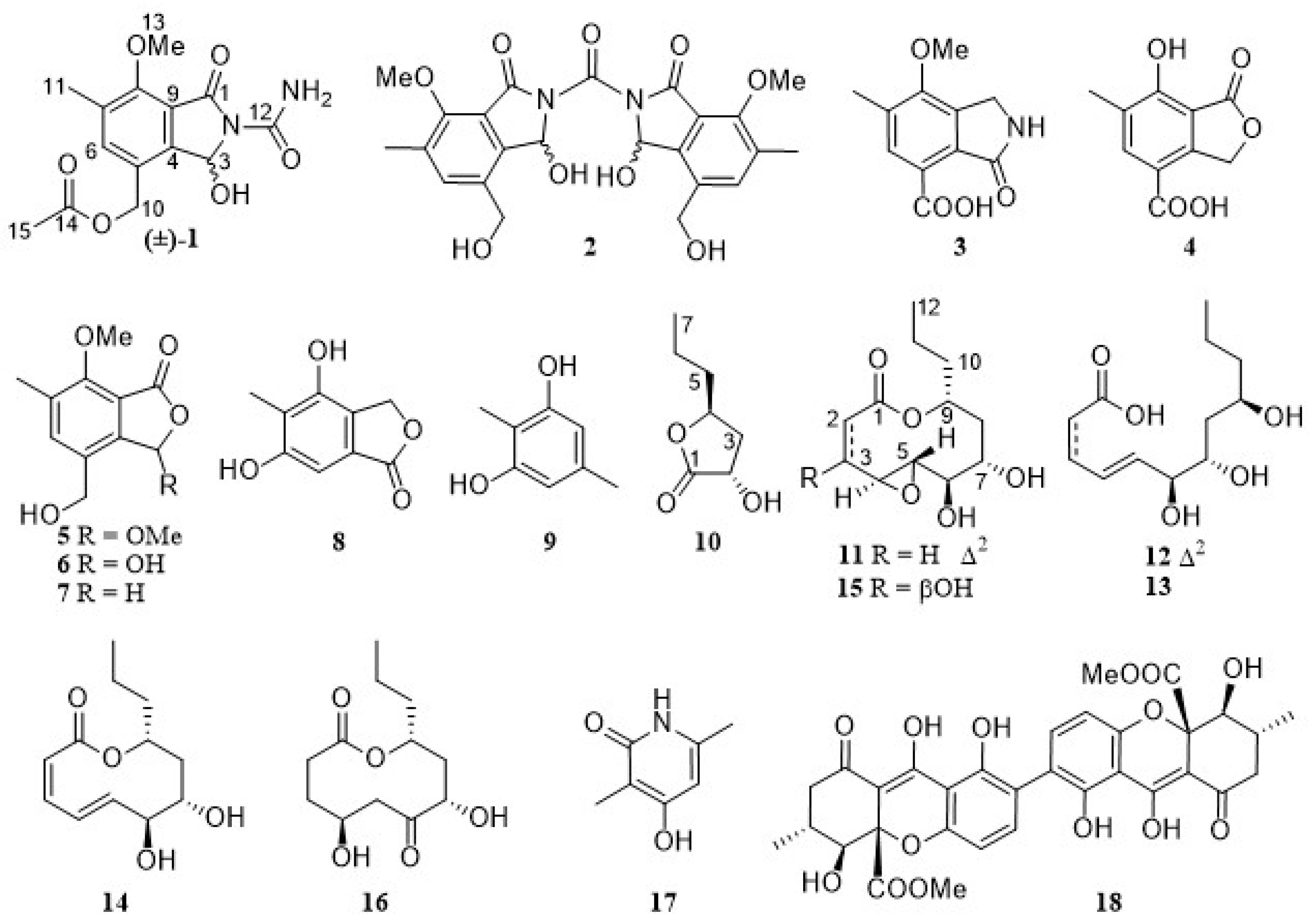
Figure 1. Structures of compounds 118 from the fungus Diaporthe searlei CS-HF-1.
Marinedrugs 23 00402 g001
Figure 2. Key COSY (Marinedrugs 23 00402 i001) and HMBC (Marinedrugs 23 00402 i002) correlations of 14 and 1013.
Marinedrugs 23 00402 g002
Figure 3. Regression analysis of experimental and calculated 13C NMR chemical shifts (ppm) of 1 and 2 (a simplified structure 2a was used).
Marinedrugs 23 00402 g003
Figure 4. (A) Regression analysis of experimental and calculated 13C NMR chemical values (MeOH) of 10; (B) experimental and calculated Electronic Circular Dichroism (ECD) in MeOH of 10.
Marinedrugs 23 00402 g004
Figure 5. The NOESY correlations of compounds 11 and 14.
Marinedrugs 23 00402 g005
Figure 6. Experimental and Calculated Electronic Circular Dichroism (ECD) Spectra of 11 and 14 in MeOH.
Marinedrugs 23 00402 g006
Figure 7. Compound 15 inhibited ferroptosis in HT22 cells. (A) Viability of HT22 cells treated with tested compounds (20 μM) and RSL3 (0.5 μM). (B) Viability of HT22 cells treated with 15 (1.25–40 μM) for 24 h in the absence of RSL3. (C,D) Dose-dependent protective effect of 15 (5–40 μM) in RSL3-induced HT22 cells. Data are presented as mean ± SD (n = 3). #### p < 0.0001 vs. nontreated cells; * p < 0.05, *** p < 0.001, and **** p < 0.0001 vs. RSL3. DMSO: vehicle control; Fer-1 (1 μM): positive control.
Marinedrugs 23 00402 g007
Figure 8. (A,B) C11 BODIPY fluorescence ratio (green/red) reflecting lipid peroxidation. (C) MDA levels; (D) GSH content; ** p < 0.01, *** p < 0.001, **** p < 0.0001 vs. RSL3.
Marinedrugs 23 00402 g008
Table 1. 1H (400 MHz) and 13C NMR (101 MHz) data of 14 and 1H NMR data of 58 (δ in ppm, J in Hz).
No.1 a2 a3 a4 a5 b6 b7 b8 b
δHδCδHδCδHδCδHδCδHδHδHδH
1 166.3, C 166.6, C4.39, br s45.4, CH2 171.3, C 5.20, s
2 8.51, br s
36.94, d (10.0)81.5, CH6.93, d (9.3)80.9, CH 168.9, C5.40, s70.4, CH26.40, s6.60, s5.28, s
4 144.3, C 142.2, C 143.5, C 148.8, C
5 126.4, C 132.8, C 121.8, C 115.1, C 6.78, s
67.61, s137.4, CH7.62, s135.5, CH7.66, s127.6, CH7.75, s130.2, CH7.60, s7.61, s7.58, s
7 132.6, C 132.4, C 132.3, C 128.1, C
8 156.0, C 154.8, C 160.1, C 165.9, C
9 118.3, C 117.8, C 128.1, C 108.0, C
105.08, d (12.7) 4.98, d (12.7)61.5, CH24.51, br s58.4, CH2 166.4, C 170.1, C 4.68, s
112.28, s15.2, CH32.28, s15.2, CH32.32, s16.1, CH32.23, s15.6, CH32.33, s2.33, s2.31, s2.16, s
12 156.9, C 155.2, C
133.93, s61.7, CH33.91, s61.6, CH33.80, s61.3, CH3 4.00, s4.00, s4.02, s
14 170.2, C
152.06, s20.5, CH3
NH25.98, s
3-OH7.40, d (10.0) 7.71, d (9.3)
3-OMe 3.58, s
10-OH 5.30, br s
a In DMSO-d6; b in methanol-d4.
Table 2. 1H (400 MHz) and 13C NMR (101 MHz) data of 1114 and 13C NMR data of 15 and 16 in methanol-d4H in ppm, J in Hz).
No.111213141516
δHδCδHδCδHδCδHδCδCδC
1 166.6, C 169.7, C 176.2, C 170.3, C171.3, C174.2, C
25.96, d (11.0, 1.5)126.0, CH5.65, d (11.4)118.9, CH2.43, m34.4, CH25.91, d (10.6)126.3, CH40.8, CH228.4, CH2
36.52, d (11.0)142.6, CH6.68, dd (11.5, 11.4)145.4, CH2.36, m29.0, CH26.70, d (10.6)141.0, CH64.9, CH28.9, CH2
43.75, br s57.5, CH7.54, dd (15.4, 11.5)128.8, CH5.73, m132.7, CH6.22, d (15.3)130.4, CH62.1, CH66.2, CH
52.60, dd (8.7, 2.0)62.6, CH6.13, dd (15.4, 6.4)144.2, CH5.54, dd (14.9, 6.0)132.0, CH5.36, dd (15.3, 10.1)135.3, CH55.3, CH45.7, CH2
63.03, dd (8.7, 8.4)79.3, CH4.06, dd (6.4, 5.8)76.7, CH3.83, m77.3, CH3.78, dd (10.1, 9.1)79.3, CH78.8, CH211.1, C
73.58, dd (8.4, 8.4)73.7, CH3.80, m72.3, CH3.70, m72.5, CH3.38, dd (9.1, 8.6)78.0, CH74.1, CH75.3, CH
82.03, m41.6, CH21.51, m41.2, CH21.47, m41.2, CH21.88, dd (17.0, 8.6)
1.79 (17.0)
42.0, CH241.6, CH239.6, CH2
94.88, m76.3, CH3.82, m68.8, CH3.81, m68.9, CH4.86, m77.1, CH74.6, CH73.8, CH
101.69, m; 1.61, m38.5, CH21.43, m41.5, CH21.44, m41.4, CH21.54, m40.0, CH239.0, CH237.1, CH2
111.39, m19.3, CH21.44, m; 1.37, m19.9, CH21.44, m; 1.38, m19.9, CH21.34, m19.5, CH219.2, CH220.0, CH2
120.95, t (7.2)14.3, CH30.93, t (6.9)14.4, CH30.93, t (6.6)14.4, CH30.92, t (7.4)14.2, CH314.3, CH314.1, CH3
Disclaimer/Publisher’s Note: The statements, opinions and data contained in all publications are solely those of the individual author(s) and contributor(s) and not of MDPI and/or the editor(s). MDPI and/or the editor(s) disclaim responsibility for any injury to people or property resulting from any ideas, methods, instructions or products referred to in the content.