Review

Calyculins and Related Marine Natural Products as Serine- Threonine Protein Phosphatase PP1 and PP2A Inhibitors and Total Syntheses of Calyculin A, B, and C

by
Annika E. Fagerholm
,
Damien Habrant
and
Ari M. P. Koskinen
*
Laboratory of Organic Chemistry, Helsinki University of Technology, PO Box 6100, FIN-02015 HUT, Finland
*
Author to whom correspondence should be addressed.
Mar. Drugs 2010, 8(1), 122-172; https://doi.org/10.3390/md80100122
Submission received: 21 December 2009 / Revised: 12 January 2010 / Accepted: 13 January 2010 / Published: 21 January 2010
(This article belongs to the Special Issue Synthesis around Marine Natural Products)

Abstract

:
Calyculins, highly cytotoxic polyketides, originally isolated from the marine sponge Discodermia calyx by Fusetani and co-workers, belong to the lithistid sponges group. These molecules have become interesting targets for cell biologists and synthetic organic chemists. The serine/threonine protein phosphatases play an essential role in the cellular signalling, metabolism, and cell cycle control. Calyculins express potent protein phosphatase 1 and 2A inhibitory activity, and have therefore become valuable tools for cellular biologists studying intracellular processes and their control by reversible phosphorylation. Calyculins might also play an important role in the development of several diseases such as cancer, neurodegenerative diseases, and type 2-diabetes mellitus. The fascinating structures of calyculins have inspired various groups of synthetic organic chemists to develop total syntheses of the most abundant calyculins A and C. However, with fifteen chiral centres, a cyano-capped tetraene unit, a phosphate-bearing spiroketal, an anti, anti, anti dipropionate segment, an α-chiral oxazole, and a trihydroxylated γ-amino acid, calyculins reach versatility that only few natural products can surpass, and truly challenge modern chemists’ asymmetric synthesis skills.

Graphical Abstract

1. Introduction

Nature offers an endless source of inspiration to synthetic organic chemists. Because water covers more than 70% of Earth’s surface, it is only logical that the number of new molecules isolated from marine species is enormous, including structures that have never been found from terrestial organisms. This interest has led to the discovery of many compounds with very promising biological activity [1]. Among these compounds is the calyculin family. Various studies have shown that calyculins are potent inhibitors of protein phosphatases 1 and 2A, opening up numerous possibilities for their therapeutic use [27].
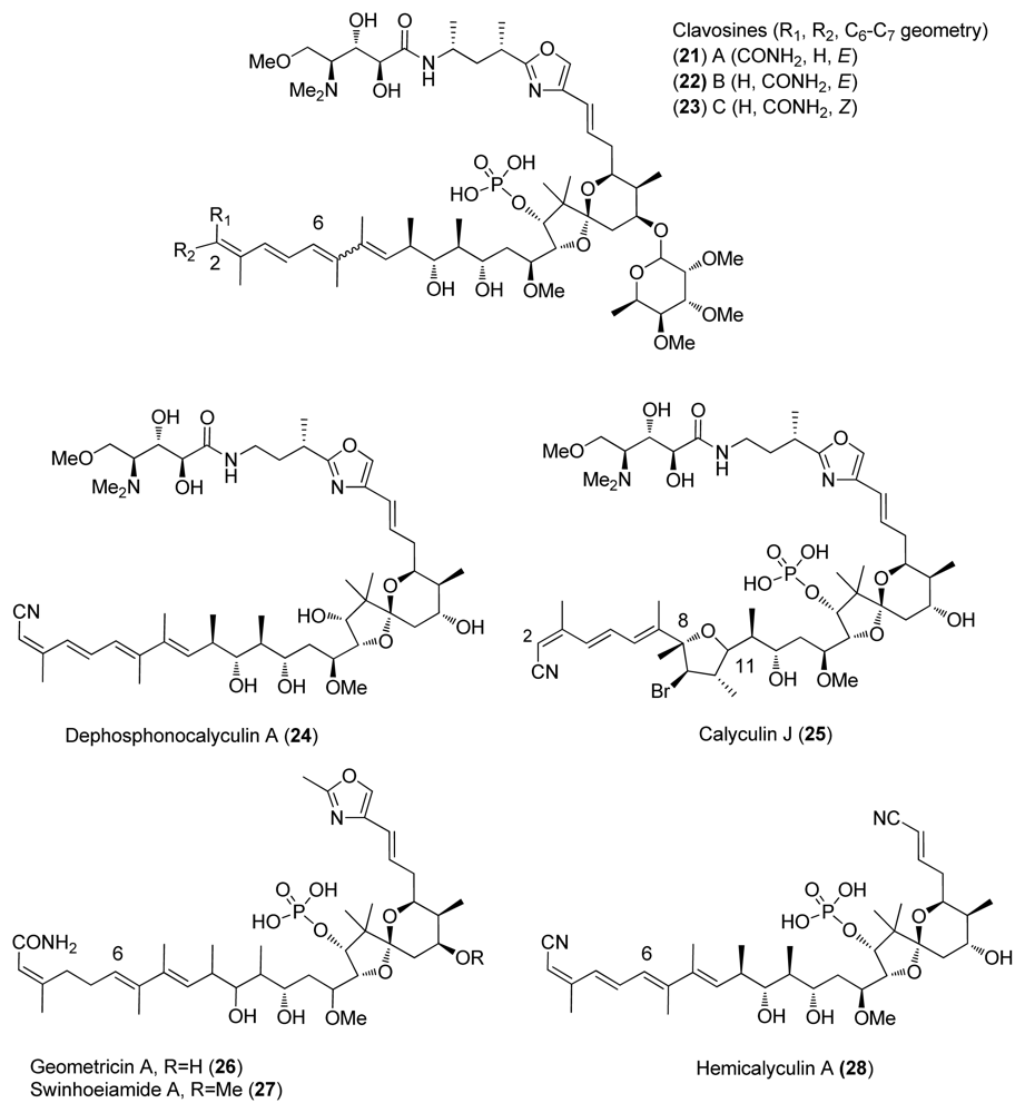
Calyculins are a class of highly cytotoxic metabolites originally isolated from the marine sponge Discodermia calyx by Fusetani and co-workers. The first member, calyculin A, was isolated in 1986 from a sponge collected in the Gulf of Sagami, near Tokyo Bay [815]. The sponge still remains the primary source of the natural product. The structures of different calyculins and structurally-related calyculinamides are shown in Figure 1. The most naturally abundant members of the family are calyculins A and C.
The structure of complex natural products may sometimes lead, even with the help of modern analytical methods, to misassignments of the absolute stereochemistry. In such cases, total synthesis can be the key for proving the absolute stereochemistry of the natural product. Calyculins provide an excellent example as Shioiri and co-workers ascertained the absolute stereochemistry of calyculins by synthesis in 1991 shortly after Fusetani disclosed the absolute configuration of calyculin A [16,17]. In their original article, Fusetani and co-workers presented a structure for calyculin A that appeared to be the enantiomer of the natural product [11]. Although being very clear about the uncertainty of the absolute configuration, the then ongoing synthetic efforts towards the calyculins had been directed to the non-natural enantiomer. As a consequence, three of the six published total syntheses of calyculins have yielded the wrong enantiomer [1823].

2. Importance of Protein Phosphatases

Phosphorylation-dephosphorylation of proteins is one of the most essential mechanisms for the proper functioning of cells. It affects almost all cellular functions such as metabolism, signal transduction, cell division, and memory. Protein kinases have long been known for the regulatory properties of phosphorylation and dephosphorylation. Although it has been recognised only later, protein phosphatases (PP) have also a great influence for these regulation processes. Phosphatases that catalyze dephosphorylation of serine and threonine residues are encoded by the phospho protein phosphatase (PPP) and protein phosphatase magnesium-dependent (PPM) gene families, whereas the protein tyrosine phosphatases (PTPs) dephosphorylate phosphotyrosine amino acids [2,3]. PP enzymes play a very dynamic role in cellular signalling, particularly because they can be turned on and off through very tight regulation of their subunit composition and selective targeting. These functions are regulated by allosteric modification using second messengers and reversible protein phosphorylation to create specific subcellular multi-protein signalling modules [2,6,7].
The total number of phosphatases discovered is over 100 but it has been estimated that the total number could be as many as 1,000 [6]. PP1, PP2A, PP2B, and PP2C are the most widely studied phosphatases and also account for the majority of the protein serine/threonine activity in vivo. PP1, PP2A, and PP2B belong to the family of PPPs and their enzymatic activity is dependent upon Ca2+/Calmodulin, whereas PP2C of the PPM family is Mg2+ dependent [2,3].
Extracellular signals, such as hormones and growth factors, affect the regulatory subunits and thereby modify the substrate specificity of PP1, which is involved in glycogen metabolism, muscle contraction, cell cycle progression, neuronal activities, and splicing of RNA. Recently, PP2A has been the focus of important interest since it accounts for 1% of total cellular proteins, and for the major portion of serine and threonine phosphatase activity in most tissues and cells. Although PP2A is involved in a great variety of cellular processes, including cell metabolism, signalling, and cell cycle control as well as the control of telomerase activity, its specific role is less delineated [2,3].
The holoenzyme of PP2A consists of three subunits, named A, B, and C. The catalytic subunit C is always associated with the scaffolding subunit A, which modulates its enzymatic properties by coordinating the protein-protein targeting to protein kinases and cytoskeletal proteins [7]. The holoenzyme of PP1 contains also a catalytic subunit C. PP1’s and PP2A’s C subunits are structurally related, and share 50% amino acid identity [6]. The regulative subunit B, subdivided into B, B′, B″, is encoded by separate genes, and can bind to AC with wide variety of heteromeric complexes. It is believed that individual subunits cannot exist individually in vivo; however, AC dimers are abundant in tissues. To date, two isoforms (α, β) of subunits A and C have been described, and there is an ever-growing number of B-type isoforms. The homologues of mammalian PP2A subunits have been identified from diverse origins such as algae, higher plants, and yeast. Moreover, although PP2A is primarily a serine and threonine phosphatase, it can, in specific circumstances, display an independent phosphotyrosyl phosphatase (PTP) activity. The diversity and selectivity of PP2A has been linked to the coverable three dimensional holoenzyme [7].
Protein phosphatase signalling plays an important role in many human diseases [35]. Unfortunately, studies towards determining the signalling mechanism have been slowed down by the absence of a PP2A crystal structure [4]. Many observations support the role of PP2A in tumorogenesis although PP2A inhibitors can also display anti-tumour activity [35]. The mutations in the gene encoding the subunit A in human breast, lung, and colorectal carcinomas, as well as in melanomas strengthen the notion of tumorigenesis activity [4,7]. However, it has not been unequivocally established so far whether such mutations, examples of which have been found in human cancer cells, result in the activation of an oncogenic function or rather in the inactivation of the presumed tumour suppressive role of PP2A. The exact effect of PP2As has been found complicated since it can exert inhibiting as well as stimulating control on cell proliferation. This might indicate activity of several different PP2A complexes during these processes [5].
The major members of PPP family are highly concentrated in the brain, and are fundamental elements of complex signalling system controlling neuronal function. PP1 is widely distributed in neurons and has multiple functions. Targeted inhibition of PP1 is a potential strategy for minimizing the symptoms associated with Parkinson’s disease [4]. PP2A activity also affects human neurodegenerative diseases. In Alzheimer’s disease, the activity levels of PP2A are significantly decreased. Altogether, PP2A-dependent PI 3-kinase signalling plays a crucial role in neuronal survival [4,7].
Both PP1 and PP2A are involved in the mediation of insulin action on carbohydrate and lipid metabolism. More specifically, activation of PP1 and inactivation of PP2A can affect insulin stimulation. Type 2 diabetes mellitus is characterized by variation of insulin resistance. Therefore, molecules involved in the insulin signalling cascade are potential targets for therapeutic drug design; both PP1 and PP2A have been involved in these studies.
PP2A signalling also regulates the transcription factors Sp1 and NK-κB which are essential modulators of cellular gene expression and viral transcription of many human viruses, such as HIV-1, cytomegalovirus, hepatitis B, herpes simplex type 1, Epstein-Bass virus, and papillomavirus. Recent studies also suggest that PP2A signalling participates in parasite-transmitted human diseases such as malaria [7].

3. Inhibition of Protein Phosphatases PP1 and PP2A by Naturally Occurring Toxins

In contrast to many enzymes, protein phosphatases, especially PP1 and PP2A, exhibit broad and overlapping substrate specificity, with no apparent substrate consensus sequence. Because the protein phosphatases affect other proteins and have literally hundred of substrates, it has been challenging to describe the mode of action of these biological catalysts and their regulation. For that reason, much of the information gathered from the functioning of protein phosphatases is based on inhibition studies [6].
Protein inhibitors have been used to study the mechanism of protein phosphatase inhibition. However, they suffer from some shortages: proteolytic degradation, poor membrane permeability, high molecular weight, potential instability, and often unavailability in sufficient quantity. To avoid these problems, small molecule inhibitors are often used. Many naturally occurring molecules, with wide structurally diversity, have been identified to either selectively or specifically inhibit the phosphatases. Alkaloids, terpenes, oligosaccharides, and polyketides have evolved to imitate and/or complement small areas of molecular surfaces of protein-peptides [6].
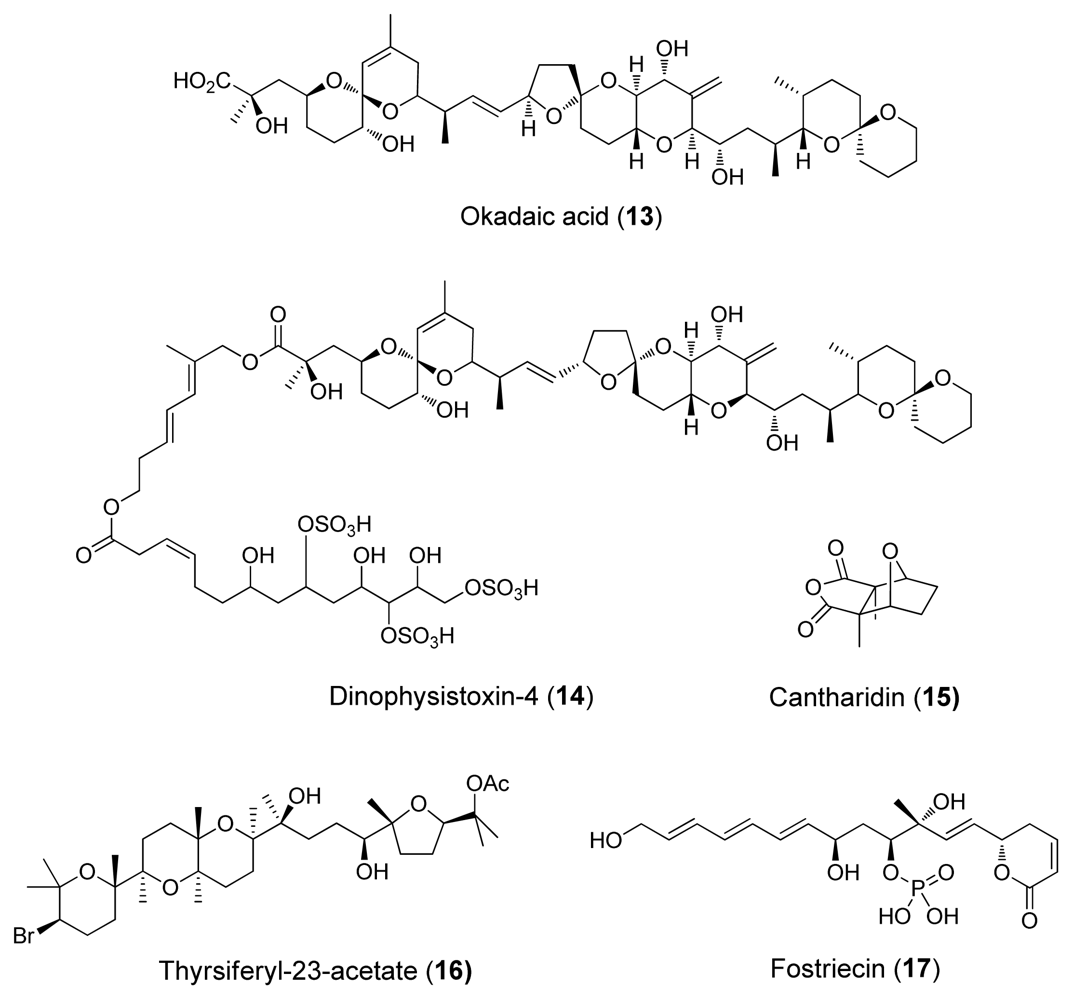
Several natural products from different structural groups have been identified to inhibit serine/threonine-specific protein phosphatases. The natural toxin inhibitors are also known as the okadaic acid class inhibitors (Figures 14). Okadaic acid, the causative agent of diarrhetic seafood poisoning [4], was the first of these inhibitors discovered in 1981. It is a marine polyketide initially found from marine sponges Halicondria okadai and Halicondria melanodocia.
Cyclic peptides such as microcystins (e.g., microcystin–LR (18)) and nodularins were initially isolated from blue green algae and are potent inhibitors of PP1 and PP2A, but poor PP2B and PP2C inhibitors. Another cyclic peptide motuporin (19), also known as nodularin-V, was isolated from the marine sponge Theonella swinhoei gray (Figure 3) [4]. Both microcystins and motuporin share the rare aminoacid ADDA, which interacts with the hydrophobic groove of PP1.
As mentioned earlier several different structural groups can bind PP1 and PP2A, which makes the classification of these inhibitors challenging. The inhibitors can be classified by structure but a valid choice is classification based on PP1/PP2A selectivity. Based on different inhibition studies and screening, a large number of structurally interesting natural products has been identified to bind PP1 and PP2A more or less selectively. The IC50 values of selected molecules are collected in Table 1, as well as the origin and structural class.
From the biological activity data (Table 1) it can be observed that okadaic acid (13), dinophysistoxin (14), cantharidin (15) and its derivative, thyrsiferyl-23-acetate (16), as well as phosphate-bearing inhibitor fostriecin (17) (Figure 2) are selective PP2A inhibitors. Although some common structures such as spiroketal moieties can be identified, the observed PP1 and PP2A differences cannot be adequately explained with the current structure-activity relationship data [6]. However, the binding data of Table 1 indicates that tautomycin (20) (Figure 4) and calyculins show slight PP1 selectivity. Tautomycin was first isolated from Streptomyces spiroverticillatus and is the first inhibitor to display preferential inhibition of PP1 (Figure 4) [4].
Mutagenesis and natural products studies indicate that acidic groove residues are a key feature in the active site of PP1 [4]. This could mean that the binding region in PP2A is more hydrophobic than the one in PP1, and therefore more accessible to hydrophobic inhibitors such as thyrsiferyl-23-acetate (16) (Figure 2).

4. Calyculins and Related Structures

4.1. Origin

The first isolated calyculin was calyculin A (1) in 1986, followed by calyculins B-D (24) in 1988, calyculins E-H (58) in 1990 and calyculin J (25) in 1997 [915]. D. calyx belongs to the order of lithistid sponges, which are an artificial assemblage of species of diverse origin known from their ability to produce diverse array of biologically active metabolites such as polyketides, cyclic peptides, alkaloids, pigments, and novel sterols [9].
Calyculin related structures have also been found from other marine sponges, such as Lamellomorpha strongylata which was collected at the Chantam Rise off the East Coast of South Island of New Zealand in 1995 and whose extraction afforded calyculinamides A (10) and B (11) [10]. Calyculinamide A (10), calyculinamide F (12), des-N-methyl calyculin A (9), and dephosphocalyculin A (24) were isolated in 1997 from D. calyx [13]. Further, calyculin derivatives clavosines A-C (2123) were isolated in 1998 from the marine sponge Myriastra clavosa (Figure 5) [25]. In 2001, Epipolasid sponge Lufariella geometrica was collected at Heron Island’s Wistari Reef, Australia, and allowed the isolation of another novel calyculin derivative, geometricin A (26) [26]. The latest isolated calyculin derivative is swinhoeiamide A (27) from the lithistid sponge Theonella svinhoei [27].
To this day, totally eighteen calyculins and calyculin related structures have been isolated. Calyculin A is formed from four different structural regions: C1–C8 tetraene, C9–C25 dipropionate spiroketal, C26–C32 oxazole and C33–C37 amino acid, these subunits are represented in Figure 1. Calyculins differ from each other by the methyl group at C32 and the geometry of C2,3 and C6,7 olefins [915]. The geometry of C2,3 and C6,7 olefins are also the critical sites of the structural differences in calyculinamides as well as in clavosines. In the latter, the C21 hydroxyl group is also in the S configuration, and is glycosylated by a trimethoxyrhamnose [10,13,25]. Calyculin J (25) is a C9 brominated derivative of 1 where C8–11 and C11 oxygen form a tetrahydrofuran ring. Geometricin A (26), swinhoeiamide A (27), and hemicalyculin A (28) could be described as rump calyculin derivatives since the most significant difference comparing to calyculins is the lack of the polar region [2628].

4.2. Crystal structures of calyculins and their binding to protein phosphatases

Several research groups have studied the structure-activity relationships (SARs) of naturally occurring toxins to PP1 and PP2A. Quinn et. al. developed a pharmacophore model for the binding of okadaic acid (13), calyculin A (1) and microcystin LR (18) to PP1 [29]. Competitive binding assays with 13, 1, 18 and tautomycin (20) suggested that at least these toxins share a common binding site [30]. The absolute stereochemistry of the calyculins was first published in 1991 by Shioiri et. al. [16]. The first publication of X-ray structure of PP1 in 1995 [31] soon inspired four docking studies in 1997 [24,3234]. However, the first two groups, Armstrong and Holmes, used the incorrect enantiomer of crystal structure. Their initial idea was that the binding of the toxins would not change the structure significantly [8]. This seems to be possible for cyclic microcystines and nodularins; however, with open chain molecules such as calyculins, this approach is unlikely [34].
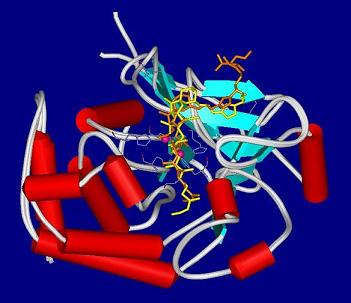
Calyculins as well as other inhibitors have been targets of continuous study and several binding models have been proposed [28,3537]. The binding mode of calyculin A (1) to the active site of PP1 is shown in Figure 6: 1 is represented according to its crystal structure in orange [35], in yellow is the calyculin model built by Koskinen [34].
The SARs of calyculins indicate that the phosphate, the hydroxyl C13, and the hydrophobic polyketide tail are essential for their inhibitory action. The dipeptide portion was less important in the interaction with enzymes, but essential for cytotoxicity [28,35]. However, compared to 1, dephosphonocalyculin A (24) was inactive, which was already examined [15]. This could indicate that phosphate group is less important for the binding.
It should be noted that the published models are still speculative. The prediction of enzyme-inhibitor interaction is challenging because there are so many parameters affecting the system. Site-directed mutagenesis studies and SAR data for serine/threonine protein phosphatases are useful, but the interpretation of the results can be difficult. Even the models of most simple enzymes contain so wide range of contacts that the interpretation is difficult. Calyculin fragments would give a useful addition to PP1, PP2A binding and SAR studies. In future, design of simpler and more selective inhibitors would also be possible.

5. Synthetic Approaches towards Calyculins

The fascinating structures of calyculins have drawn a great amount of attention and resources. The first total synthesis of ent-calyculin A (1) was published by Evans et al. in 1992 [18]. Two years later, Masamune et al. published the first total synthesis of the natural enantiomer of 1 [19]. In 1996, the Shioiri group published a formal total synthesis of 1 [20]. Total synthesis of ent-calyculin A and B by Smith et al. [22] and calyculin C by the Armstrong group were published in 1998 [21]. The latest total synthesis of ent-calyculin A was published by Barrett et al. in 2001 [23]. The Koskinen group has been involved in the preparation of individual fragments [3846]. Only the studies of these seven groups will be examined in detail in this review.
The total synthesis of calyculins has been reviewed by Jacobs and Itching in 1998 [47], and by Pihko and Koskinen in 1999 [48]. The retrosynthetic analyses, as well as the preparation of individual fragments, and the final assembly of the fragments are presented in the following section. In order to compare the different methods, we will first present the preparation of the different fragments separately. Then, the assembly of these fragments to reach the calyculins will be discussed. The total syntheses of calyculins will be presented in chronological order and, for clarity, the different fragments will be described in the same order of publishing year of the total syntheses.

5.1. Retrosynthetic analysis

The retrosynthetic analysis of the calyculin skeleton divides it into three fragments: the C1–C8 tetraene subunit, the C9–C25 dipropionate spiroketal subunit, and the C26–C37 amino acid oxazole subunit (Scheme 1).
Further, the C26–C37 amino acid oxazole subunit is divided into two subunits: the amino acid C33–C37 subunit and the oxazole C26–C32 subunit (Scheme 2).
The retrosynthetic analysis and the executions of the different fragments are described in the following sections.

5.2. C1–C8 tetraene fragment

For the synthesis of this fragment, a number of renowned reactions can be highlighted: Horner-Wadsworth- Emmons (HWE), Peterson olefination, Stille coupling, and related Negishi and Suzuki couplings. Evans and co-workers [18] were the first to report that the HWE reaction to couple the entire tetraene is not possible due to the unstable cyano group and tetraenes tendency to isomerisation. The uses of sp2-sp2 couplings methods such as the Stille coupling were tempting since the double bond geometries of the starting materials are completely retained in the reaction.
Based on the disconnections of the building blocks, the retrosynthetic analysis of tetraene can be divided into three groups (Scheme 3). Smith’s group choose to disconnect in position a and d which gives nitrile 29 and phosphate 30 [22], whereas Evans’ and Armstrong’s target was to create vinyl iodide 31 and phosphate 32, via b and d disconnections [18,21]. Finally, Masamune and Barrett chose c and d disconnections involving intermediates 33 and 34 [19,23].
In addition, a few other approaches and ideas for the synthesis of this fragment have been published. For example, Negishi et al. recently proposed that propyne bromoboration and tandem Pd-catalyzed cross coupling could be used in the synthesis of C1–C8 fragment [49].

5.2.1. Evans [18]

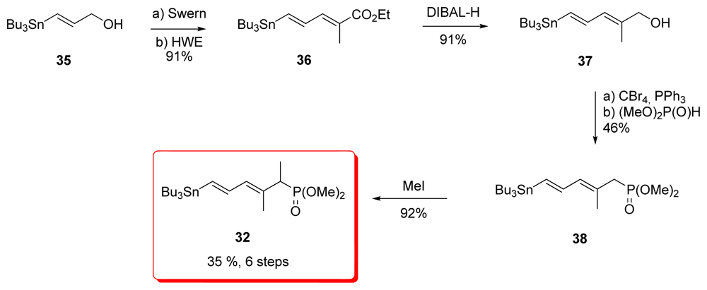
Evans’ synthesis of phosphonate diene 32 began with vinyl stannane 35 which was prepared by reduction of methyl (E)-3-(tributylstannyl)-2-propionate (Scheme 4). Swern oxidation followed by HWE reaction gave diene ester 36 as a 19:1 E:Z mixture. Reduction of the ester furnished alcohol 37, which was converted to phosphonate 38 by the Michaelis-Becker method. Final methylation of 38 completed the synthesis of targeted phosphonate 32.

5.2.2. Masamune [19]

The fact that tributylstannyl moieties can be easily converted to the corresponding iodide was utilized in the synthesis of the tetraene fragment by Masamune via fragments 33 and 34. However, the synthesis of 33 has not been published, and the preparation described here has been found in the PhD thesis of S. A. Filla [50]. Addition of Lipschutz higher cuprate Bu3SN(Bu)Cu(CN)Li2 to ethyl butynoate 39 occurred regioselectively to furnish the (Z)-enoate 40 in good yield (Scheme 5). Treatment of 40 with the Weinreb reagent produced nitrile 41. Tin-iodide exchange gave vinyl iodide 42 whose Stille coupling with trans-1,2-bis(tri-n-butylstannyl)ethylene (43) produced the expected stannane 33 in a modest 40% yield.
The protected 3-butyl-2-ol 44 was reacted with methylcopper (I) reagent followed by iodination to give vinyl iodide 45. (Scheme 10) This was converted to phosphonate 46 by an Arbuzov reaction and subsequent tin-iodine exchange with CuSnBu3 yielded 34. This last step appeared to be the weakest link in this part, lowering the global yield.

5.2.3. Shioiri [20]

Shioiri group’s strategy was to introduce the whole tetraene, actually the C1–C12, as a single moiety to the rest of the target molecule. Stannyl cupration of alcohol 47 was the key step, affording the stannyl derivative 48 in 87% yield. Further conversion of the alcohol to the corresponding nitrile generated 33 (Scheme 7).
The synthesis of the other coupling partner began with alcohol 49 which was readily available from l-(+)-tartrate (Scheme 8). Parikh-Doering oxidation and subsequent HWE reaction were followed by conversion of the acetonide to the bis-TBS ether derivative 50. Weinreb amide formation and reaction with methyl magnesium bromide furnished methyl ketone 51, which was converted to (E)-vinyl iodine 52 by use of the Takai and Utimoto’s chromium reagent. Selective deprotection of the primary TBS group was followed by conversion of the resulting alcohol to the corresponding methyl ketone 53. Final Stille coupling of 53 with stannane 33 produced 54. Shioiri’s group has also published another strategy for the synthesis of this subunit; however, this method has not been used in the formal total synthesis [49].

5.2.4. Smith [22,51]

Smith et al. began by synthesizing the iodo phosphonate 56 in two steps from allylic alcohol 55 (Scheme 9). This was followed by a two stage one-pot coupling of the three components. The first part, the Negishi coupling of the organozinc compound 57 with bromoboronate 58 was followed by a Suzuki coupling with iodide 55 affording phosphonate 59 in 64% yield. Final methylation of 59 furnished 30.

5.2.5. Armstrong [21,52]

Armstrong’s synthesis of the tetraene fragment also began with the iodo alcohol 55 and proceeded successfully using classical transformations to generate phosphonate 61, which unfortunately appeared to be too unreactive toward hindered aldehydes (Scheme 10). For this reason, the authors decided to convert 61 to diene 32. Unfortunately, this transformation proceeded with a low yield of 33% over two steps, lowering the overall yield.

5.2.6. Barrett [23,53]

For the preparation of stannane 33, Barrett et al. used a strategy similar in every aspect to the one presented earlier by Masamune; however, as mentioned before, Masamune’s results were not published. Conjugate addition of tributylstannyl cuprate to ethyl butynoate 39 gave (Z)-enoate 40. Conversion of ethyl ester of 40 to the corresponding nitrile 41 was achieved via the amide in 2 steps. Metal-halogen exchange furnished vinyl iodide 42 and Stille coupling with 43 were the lasts steps for the preparation of 33.
The key idea of the synthesis of the C6-C14 fragment was to construct the vinyl iodide 67 via methyl zirconation–iodonolysis of alkyne 66 using Negishi’s procedure (Scheme 12). The synthesis started with commercially available methyl (S)-3-hydroxy-2-methylpropanoate (62) which was efficiently converted to homoallylic alcohol 64 by Brown’s homologation with (−)-63 [54,55], setting the stereochemistry at C11 and C12 with excellent diastereomeric excess (>96%). Further standard steps led to ester 65, which was then reduced to aldehyde and homologated to the corresponding alkyne 66 using the Corey-Fuchs protocol. Methylzirconation-iodinolysis of 66 furnishing the corresponding vinyl iodide and final formation of the methyl ketone at C14 were the final steps for the preparation of 67.

5.2.7. Koskinen [40]

Koskinen et al. reported a short and efficient synthesis of alcohol 37, which was used by Evans for the preparation of phosphonate 32 (Scheme 13). Sequential treatment of distannyl compound 43 with n-BuLi and ZnCl2, followed by Pd-catalyzed Negishi coupling with bromo-ester 68, furnished diene 69 in 95% yield. Reduction with DIBAL-H afforded the alcohol 37. Introduction of the phosphonate moiety included bromination, Michaelis-Becker reaction and final methylation, following the procedure published by Evans et al. [18] gave 32.

5.3. Synthesis of the C9–C25 dipropionate-spiroketal subunit

The C9–C25 spiroketal-propionate subunit forms the core of the calyculins. With eleven stereocenters, a phosphate-bearing spiroketal, and anti, anti, anti dipropionate segment, the synthesis of this fragment is most challenging.
The construction of the C9–C25 spiroketal-propionate subunit can be divided into four groups (Scheme 14). Armstrong’s [21] and Koskinen’s group [4446] chose to introduce the dipropionate in two parts and to use Brown’s asymmetric crotylborane chemistry, while Masamune et al. selected the asymmetric aldol strategy [19]. In contrast, Evans et al. used a chiral β-ketoamide aldol methodology [18]. Shioiri et al. used the Ziegler-Brückner aldol-oxidative degradation method [20] and Smith’s group chose to use vinyl cuprate–epoxide coupling [22].
For the disconnection on the spiroketal, Smith and co-workers strategy was to create the C19-C20 bond by addition of a dithiane to an epoxide (via disconnection d). All the other groups decided to use more or less similar aldol-type strategy to create the C20–C21 bond (disconnection e).
Trost and co-workers have also published a synthesis of the C15–C24 spiroketal core; the synthesis being based on their methodology on ruthenium-catalyzed cyclization and allyl alcohol addition process [56].

5.3.1. Evans [18]

The authors exploited their studies on the oxazolidone chiral auxiliaries in this synthesis, creating six of the eleven stereocentres with this method. The synthesis of the spiroketal fragment began with the known acid 70, which could be prepared from diethyl isopropylidenemalonate in three steps (the yields of these early transformations were not reported in the original procedure) [57]. Compound 70 was converted to the (S)-phenylalanine-derived oxazolidone, followed by auxiliary-based asymmetric hydroxylation at C17 which afforded 71 as a single diastereomer. Removal of the chiral auxiliary and PMB protection at C17 were followed by chelation controlled addition of methoxyallylstannane 73 to aldehyde 72, affording alcohol 74 as a 7.5:1 mixture of diastereomers. Silylation and regioselective Rh-catalysed hydroboration gave alcohol 75. Finally, pivaloyl protection and oxidative cleavage of the double bond afforded ketone 76 (Scheme 15).
Mukayama aldol coupling of between 76 and 77, prepared in 3 steps via classical methods, afforded 78 as a single diastereomer in 80% yield. Spiroketal formation was effected with acid catalysis, furnishing 79 in a 5:1 ratio of diastereomers (Scheme 16).
Spiroketal 79 was then hydroborated and TBS protected at C25, followed by pivalate removal and Swern oxidation at C13, resulting in the formation of aldehyde 80 (Scheme 17). Addition of 80 to the titanium enolate derived from β-ketoamide 81 provided the syn, syn adduct 82 exclusively. Anti selective reduction at C11 was then performed, yielding diol 83. The configuration of the C13 alcohol had then to be inverted via a Mitsunobu reaction to give 84. Finally, standard transformations provided aldehyde 85, which represents the C9–C25 subunit of calyculins.

5.3.2. Masamune [19]

Masamune et al. chose to create the C10–C13 dipropionate segment before constructing the spiroketal (Scheme 18). The synthesis began with d-threonine derivative 86. Claisen condensation with methyl isobutyrate, lactonisation, and TBS protection gave 87. Reduction at C17 with KHBEt3, with >10:1 diastereselectivity, PMB protection, and conversion to silyl enol ether provided 88. Then, the titanium-chelated Mukaiyama aldol addition of 88 to aldehyde 89 provided 90 with a 10:1 diastereoselectivity. Reduction at C15 with Me4NHB(OAc)3, diol protection, and a debenzylation-oxidation sequence provided aldehyde 91.
The authors exploited their studies with chiral borolanyl triflates in asymmetric aldol reactions. The advantage of these reagents is that the intrinsic Felkin bias can be overridden in aldol reactions with chiral aldehydes. This was applied to the construction of the C10–C13 anti, anti, anti dipropionate. Reacting aldehyde 91 with enolate 92 produced 93 with an excellent 12:1 diastereoselectivity. Acetonide migrations to the more stable syn adduct, reduction of the thioester, protection of the alcohol, and C15 methylation provided compound 94. The standard final four steps completed the preparation of the C9-C20 methyl ketone 95.
The key aldol reaction between 95 and aldehyde 96 was performed in the presence of bulky (cHex)2BCl, leading to the exclusive formation of 97, without any trace of its C21 epimer (Scheme 19). Desilylation and treatment with formic acid provided spiroketal 98 as a single diastereomer. TBS protection and removal of PMB constituted the last steps of the C9–C25 fragment 99.

5.3.3. Shioiri [20,58]

Shioiri’s group formal total synthesis of calyculin A consisted of a variety of studies and different strategies. The synthesis of C14–C20 ketone 105 began from diethyl l-tartrate 100 which was converted in five steps to the key intermediate 101. Aldol reaction with ketene acetal 102, in the presence of chiral borane reagent 103, stereoselectively produced 104, which was then easily converted to the corresponding methyl ketone 105.
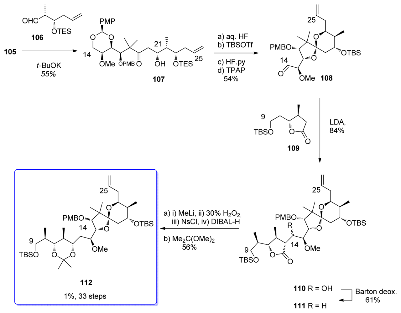
The aldol reaction between 105 and aldehyde 106, easily prepared from dimethyl l-malate, proceeded with an excellent diastereoselectivity to give 107 in a 18:1 ratio (Scheme 21). Interestingly, only the potassium enolate of 105 gave satisfactory results in the formation of the syn aldol adduct, the lithium and sodium enolates giving only poor diastereoselectivity. Spiroketalization was then performed in aqueous HF. Further protection-deprotection sequence followed by TPAP oxidation gave aldehyde 108. Coupling of 108 with the enolate of C9–C13 lactone 109 furnished 110 as a mixture of diastereomers. Barton deoxygenation of the hydroxyl group at C14 furnished 111, together with its C13 epimer in a 4:1 ratio (the latter being further epimerized with MeLi to give the desired 111). Degradation of the γ–lactone to the 1,3-acetonide was performed using Ziegler-Brückner conditions and further protection of the diol to gave 112.

5.3.4. Smith [22,59,60]

Smith published the synthesis of the spiroketal core of ent-calyculin A in 1991, using a novel dithiane-epoxide coupling strategy. Brown’s crotylation on 3-benzyloxypropanal, using (Z)-crotylboron reagent (+)-113, furnished alcohol 114 in 99% enantiopurity (Scheme 22). Boc-protection and electrophilic cyclization in the presence of IBr afforded the syn, syn carbonate 115, with good α/β selectivity of 13.9/1. The synthesis of C20–C25 coupling moiety was finished with cleavage of the carbonate group, and TBS protection to give epoxide 116.
The C16-C19 dithiane moiety 118 was easily prepared from alcohol 117, via consecutive Swern oxidation, dithiane formation and diol protection (Scheme 23). Key coupling was performed by metalation of 118 with n-BuLi followed by addition of DMPU and epoxide 116, to furnish alcohol 119. Silyl protection, acetal reduction, and Parikh-Doering oxidation then afforded aldehyde 120. The stereochemistry of C16 was created by chelation-controlled addition of vinylmagnesium bromide to 120 to give alcohol 121 with a >20:1 diastereoselectivity. Two different conditions for the spirocyclisation were employed. The first, using aqueous HF, produced 122a as a single diastereomer in 88% yield but resulted in the loss of PMB group at C17. To retain this useful protecting group, an alternative sequence was applied. Sequential treatment of 121 with TBAF and HgCl2/CaCO3 afforded a mixture of 122b and its C19 epimer. Fortunately, the latter could be quantitatively converted to 122b upon exposure to p-TsOH, with a global yield of 76%.
After extensive experimentation, the authors found out that Payne epoxidation of 122b occurred in good syn diastereoselectivity and yield (9.5:1, 89%) to give 123. After TBS-protection, the epoxide was coupled with the vinyl cuprate derived from 124 giving access to 125 as a single diastereomer in 83% yield. Methylation of the C15 hydroxyl group of 125, and oxidation of the alkene were followed by DIBAL-H reduction to provide 126 with >12:1 diastereoselectivity at C13 (probably via internal hydride delivery by prior coordination at the C15 methoxy group). Finally, protecting group manipulations provided compound 127.

5.3.5. Armstrong [21,52]

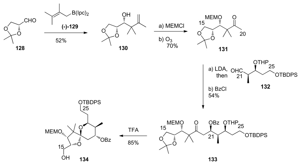
Armstrong’s group used Brown’s chiral allylborane reagents in their synthesis of C9–C25 fragment. The synthesis of spiroketal core starts with the reaction of d-glyceraldehyde 128 with the enantiopure allylboron reagent (−)-129 (Scheme 25). This addition occurred in good yield and excellent stereoselectivity to provide 130. After MEM-protection and ozonolysis, ketone 131 was obtained. Aldol reaction between 131 and aldehyde 132 and further benzoyl protection furnished compound 133. Upon exposure to TFA, 133 underwent the expected tandem deprotection-spirocyclisation to give the desired spiroketal 134 as a single enantiomer.
Swern oxidation of alcohol 134 was followed by reaction with allylmagnesium bromide in the presence of ZnCl2 to give homoallylic alcohol 135 (Scheme 26). Unfortunately, the Felkin-Ahn mode was followed for this addition, resulting in the formation of the stereochemistry at C15 opposite to that present in the natural product. Inversion at C15 was performed by consecutive oxidation-reduction. Finally, the new C15 hydroxyl was methylated to give 136. Ozonolysis of 136 afforded the corresponding aldehyde that, upon submission to an asymmetric Brown’s crotylboration with (+)-63 and benzoyl protection, gave olefin 137 as a single diastereomer. Second ozonolysis gave the corresponding aldehyde which was the substrate for a further Brown’s crotylation. Unfortunately, this step proceeded with very low diastereoselectivity; the expected anti, anti, anti adduct 138 being isolated in only 40% yield; the anti, syn, anti isomer being isolated in 30%. This disappointing result led the authors to study the influence of the different protecting groups at C25, C21 and C13 on the crotylation process. Unfortunately, all the other protecting group combination gave lower yield of the expected 138, the undesired isomer always being the main product. The tetra-TBS protected compound 139 was then prepared by simple protection group conversion of 138, followed by MEM-deprotection.

5.3.6. Barrett [23,61]

Barrett et al. also used the Brown’s crotyl boration reagents for the synthesis of the spiroketal moiety of calyculins. The synthesis began with ester 140 which was converted to allylic alcohol 141 (Scheme 27). Sharpless asymmetric epoxidation, ring opening and protection of the resulting diol furnished acetonide 142. Deprotection of the benzyloxymethyl ether, and subsequent Swern oxidation completed the synthesis of aldehyde 143.
Addition of (Z)-borane (−)-145 to aldehyde 144 proceeded effectively to yield the syn adduct 146 with >95% stereocontrol (Scheme 28). Transformation of 146 to the corresponding methyl ketone 147 was then carried out using standard transformations. Key aldol reaction between 147 and aldehyde 143 yielded, after acidic treatment, to the spiroketal 148, as a 2:1 mixture of C21 epimers. Swern oxidation followed by K-selectride reduction furnished diol 149 as a single diastereomer. Final protection-oxidation steps led to compound 150.

5.3.7. Koskinen [4246]

The Koskinen group has presented separate studies for the preparation of the dipropionate and the spiroketal moieties. The key lactone 153 was prepared in five steps from ketoester 151 and aldehyde 152 (Scheme 29). MEM-protection, benzyl deprotection and further oxidation provided aldehyde 154. This aldehyde was subjected to a Brown’s crotylation, affording the expected homoallylic alcohol in a 6:1 separable mixture of diastereomers, in favor of the expected product. Further ozonolysis furnished aldehyde 155, which was, in turn, subjected to a crotylation reaction. Based on previous studies by Armstrong [21,52] showing that a second Brown’s crotylation on similar substrates gave only poor selectivity, the authors decided to use the Roush (Z)-crotyl trifluorosilane 156 for this reaction [62]. This transformation pleasingly afforded a single isomer and further acetonide protection furnished 157, whose analysis proved the anti, anti, anti relationships in the stereotetrad. This strategy proved to be efficient in terms of selectivity, but suffers from poor yields. Current studies in our lab recently showed that this methodology could be improved and much better yields were obtained on new substrates used in the course of the synthesis of calyculins (unpublished results).
Koskinen also recently published a preparation of the C13–C25 spiroketal core of calyculins (Scheme 30) [46]. Lactone 153 was allyl-protected, reduced with LiAlH4, and the diol protected as a TES ether. Further selective deprotection-oxidation of the primary alcohol, and homologation of the aldehyde to the corresponding alkyne using the Ohira-Bestmann protocol produced 158 in good yield. Key coupling between acetylene 158 and thioester 159 furnished ynone 160. This was subjected to acidic treatment, leading to TES-deprotection followed by a double intramolecular hetero-Michael addition (DIHMA) to yield 161 as a single enantiomer. The DIHMA spiroketalisation differs from all the other methods described for this fragment.

5.4. Syntheses of the C26-C32 oxazole fragment

Compared to the synthesis of C9-C25 spiroketal-propionate subunit, the C26–C32 oxazole fragment seems to be less difficult; however, formation of the C30 stereocenter creates challenges. Construction of the suitably substituted oxazole in good yield without epimerization proved challenging.
Retrosynthetically, the C26-C32 can be simplified to a chiral aminoacid (Scheme 31). The Evans’ oxazolidone was used as source of chirality by Evans [18], Smith [22], and Barrett [23] whereas Masamune used the Sharpless epoxidation [19]. Shioiri decided to use a hydroxy acid as the starting material [20]. Finally, Armstrong started from l-pyroglutamic acid [21], while Koskinen used d-alaninal [39].

5.4.1. Evans [18]

The key step of the oxazole synthesis was the diastereoselective Michael addition of N-propionyloxazolidone 162 to tert-butyl acrylate, setting up the correct stereochemistry for 163 at C30 in 88% yield with >95:5 diastereoselectivity (Scheme 32). Cleavage of the tert-butyl ester followed by Curtius rearrangement afforded amino acid 164, which was coupled with l-serine methyl ester to give 165. Cyclisation with thionyl chloride afforded the corresponding oxazoline 166. Finally, oxidation by trapping the enolate of 166 with PhSeCl followed by oxidative elimination afforded oxazole 167.

5.4.2. Masamune [19,63]

The starting point for the oxazole fragment 172 was the epoxy alcohol 168, obtained by Sharpless asymmetric epoxidation of the corresponding allylic alcohol; unfortunately, no details about the yield or selectivity were reported (Scheme 33). Regioselective ring-opening of 168 with Me3Al provided the diol 169, with the requisite stereochemistry at C30. Oxidative cleavage of 169, further oxidation to acid and conversion to amide furnished 170. Treatment of 170 with ethyl bromopyruvate furnished, after dehydration, oxazole 171. To avoid epimerization, this reaction had to be carried out in the presence of 3, 4-epoxycyclopentene. Final preparation of oxazole 172 was achieved with standard transformations.

5.4.3. Shioiri [20,64]

For the synthesis of oxazole fragment of calyculin A, Shioiri et al. used methyl (S)-3-hydroxy-2-methylpropionate [(S)-174]. TBS protection, DIBAL-H reduction to the corresponding aldehyde, and Wittig olefination gave the unsaturated ester 175 (Scheme 34). This was followed by hydrogenation of the double bond, and cleavage of the TBS-deprotection to yield alcohol 176. Oxidation of 176 to the corresponding acid and coupling with l-serine methyl ester gave 177. For the conversion to the oxazoline 178, the authors applied their own method which uses triflic anhydride, diphenyl sulfoxide and potassium phosphate. Oxazoline 178 was obtained in 66% yield, without any epimerization in the process. Oxidation with NiO2 provided the corresponding oxazole. Finally, after removal of the tert-butyl ester, Curtius rearrangement gave the amino ester 179. This synthesis afforded the enantiomer of the fragment present in the natural product. However, the enantiomer of 179 can easily be obtained by using (R)-3-hydroxy-2-methylpropionate [(R)-174] as starting material.
The authors presented also a method for the preparation of the C26–C32 oxazole part of calyculin C, which adds an extra methyl group at C32 [57]. This method relied on the asymmetric ring opening of a prochiral cyclic anhydride. However, this strategy has not been used in any total synthesis and therefore will not be described here in detail.

5.4.4. Smith [22,51]

The synthesis started with 4-chlorobutyl chloride (180) which was converted to oxazolidinone 181 (Scheme 35). Methylation (87% yield, 95% diastereoselectivity) followed by removal of the chiral auxiliary furnished acid 182. Condensation with l-serine methyl ester in the presence of diethylcyanophosphonate (DECP) as the coupling agent yielded amide 183. Exposure of 183 to Burgess reagent followed by Narrish-Singh oxidation produced the desired oxazole ring. Finally, azide reduction furnished 184 in good yield and without epimerisation.

5.4.5. Armstrong [21,65]

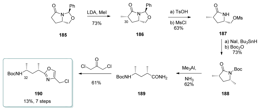
Comparing to other syntheses of this subunit, Armstrong targeted calyculin C, which contains one additional methyl at C32. Bicyclic N,O-acetal 185, which was prepared from (S)-pyroglutamic acid, was methylated at C30 to give 186 in a 80:20 ratio of diastereomers (Scheme 36). Acetal hydrolysis followed by mesylation afforded lactam 187. Radical deoxygenation of the in situ formed iodide and Boc-protection yielded 188. Ring opening by Me3Al in the presence of ammonia furnished the open-chain amide 189. Finally, oxazole 190 was obtained by reaction of 189 with 1,3-dichloroacetone.

5.4.6. Barrett [23,66]

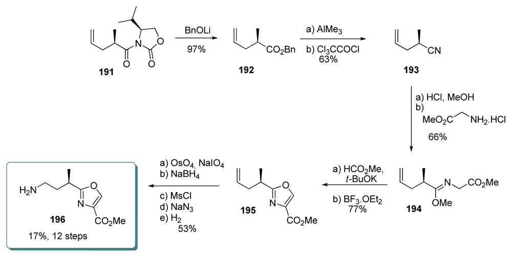
Barrett et al. synthesized the oxazole unit by using a modified Cornforth-Meyers approach (Scheme 37). The synthesis started with oxazolidinone 191, which was reacted with lithium benzyloxide to give ester 192.
Nitrile 193 was obtained by submitting 192 to Me3Al and dehydration. Addition of MeOH and HCl to 193 produced an intermediate imidate ion, which reacted with glycine methyl ester to produce 194. The Cornforth-Meyers procedure, using methyl formate in the presence of t-BuOK and BF3.OEt2, provided oxazole 195. Final stages transformed the terminal alkene of 195 to the corresponding primary amine 196 in 5 steps. No racemization was observed during the entire process.

5.4.7. Koskinen [39]

Koskinen group’s strategy towards calyculin C was to use cyclic stereocontrol to create the syn-isomer of C26–C32 (Scheme 38). d-Alaninal derivative 197 was subjected to Still-Gennari modification of HWE olefination to give the corresponding (Z)-enoate, whose cyclization under Ragnarsson-Grehn conditions (Boc2O, cat. DMAP) and hydrogenation furnished lactam 198, together with 9% of its anti diastereomer. Hydrolysis of 198 followed by coupling with l-serine methyl ester afforded amides 199 (at this stage, the anti diastereomer could be separated by chromatography). Conversion to oxazole 200 was achieved by treatment with Burgess reagent to give the oxazoline, followed by oxidation. This oxidation proved to be difficult, and the best results were obtained by using either CuBr2/HMTA/DBU as discussed earlier [22,51] or by temporary TMS protection of the carbamate hydrogen, deprotonation of the oxazoline and oxidation of the enolate with I2. Those two strategies yielded 42% of oxazole 200.

5.5. Syntheses of the C33–C37 amino acid fragment

Because of the three chiral centres and possibly reactive amine, the synthesis of C33–C37 is also a challenging target. For the synthesis of this fragment, most groups used carbohydrates. It can be also noticed that the left half of the fragment resembles serine. This was exploited by the Shioiri, Barrett, and Koskinen groups (Scheme 39) [20,38,65,66].

5.5.1. Evans [18]

Sarcosine derived oxazolidinone was prepared from 201 and further alkylated with dimethoxyethane in good yield (80%) and diastereoselectivity (98:2) to produce 202 (Scheme 40). Displacement of the chiral auxiliary with LiBH4 followed by Swern oxidation, where Hunig’s base was employed to prevent racemisation, cleanly furnished aldehyde 203. Enolization of 204 in the presence of Sn(OTf)2 and TMEDA followed by addition of aldehyde 203 afforded the expected anti aldol 205 in 60% yield. Unfortunately significant amounts of other diastereomers were also formed in the reaction.

5.5.2. Masamune [19,63]

The synthesis of the aminoacid derivative 209 began with the reaction of lactone 206, easily prepared from gulonolactone, with the Weinreb’s reagent of methylamine (Scheme 41). The crude hydroxyamine obtained was then mesylated and treated with t-BuOK to provide lactam 207, with reverse stereochemistry at C35 to that present in 206. Cleavage of acetonide liberated the corresponding diol which was protected as the dibenzyl ether 208. Treatment of 208 with Meerwein’s reagent provided the corresponding imidate, which after hydrolysis, reaction with formaldehyde and reduction with cyanoborohydride, furnished aminoester 209, the enantiomer of the C33–C37 fragment present in calyculins. However, in the course of the total synthesis of natural calyculin A, the authors briefly state that the synthesis of the fragment with the correct stereochemistry had been performed with significant improvements compared to 209.

5.5.3. Shioiri [20,67]

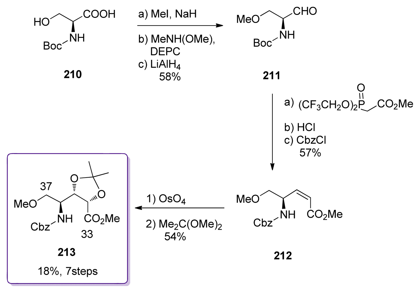
Shioiri et al. also published a synthesis of the C33–C37 fragment. Their strategy was based on OsO4 dihydroxylation of the l-serine derived Z-alkene 212 (Scheme 42). Boc-l-serine 210 was O-methylated and converted to aldehyde 211. Reaction with Still-Gennari’s phosphonate and Boc replacement by Cbz group furnished the Z-enoate 212. Dihydroxylation in the presence of dihydroquinine p-chlorobenzoate produced the corresponding diol (80% total yield, 80:20 diastereoselectivity), which was then protected as its acetonide 213.
Shioiri’s group has also earlier published a synthesis of enantiomer of the C33–C37 fragment. Although it was not utilized in the total synthesis, it had a great effect in determining the absolute stereochemistry of the calyculins.

5.5.4. Smith [22,51]

The starting point of the preparation of the amino acid subunit was commercially available iso-propylidene d-erythronolactone (214, Scheme 43). Treatment of 214 with PMBNH2 in the presence of Me3Al gave the ring opened hydroxyl amide. Parikh-Doering oxidation of the primary alcohol and dehydration led to the formation of 215, in a 7:1 anomeric mixture in favor of the β–anomer. In the presence of the TMS-enol ether of pinacolone and BF3.OEt2, 215 elegantly led to the formation of ketone 216 in 81% yield and as a single diastereomer. Formation of the TMS-silyl ether of 216 followed by reduction provided alcohol 217. Methylation of the free hydroxyl group of 217, PMB-Boc protecting group exchange and basic hydrolysis were the last steps for the preparation of 218.

5.5.5. Armstrong [21,65]

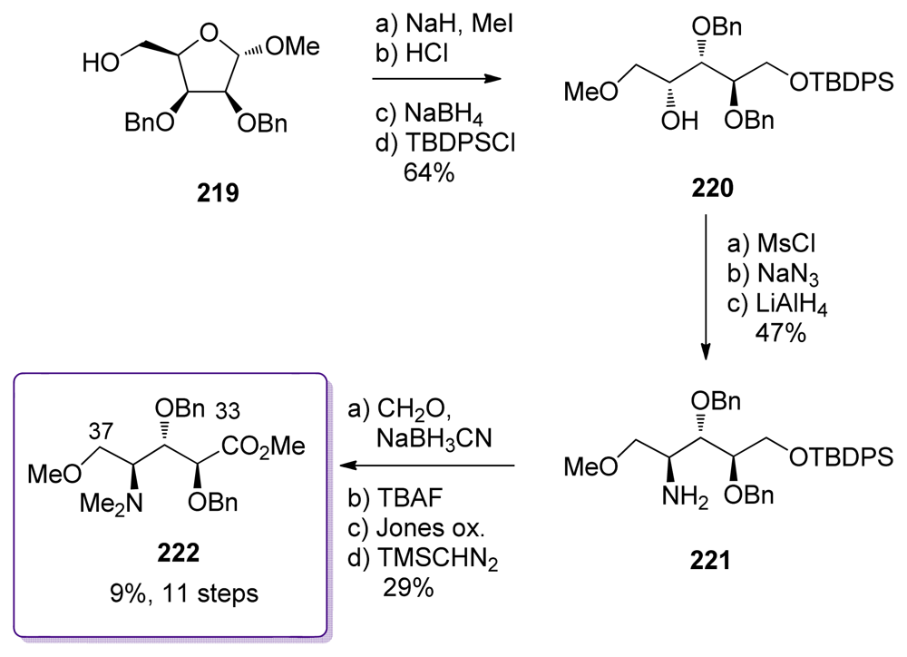
The synthesis started from alcohol 219, available in four steps from d-lyxose (Scheme 44). O-Methylation, acidic hydrolysis followed by mild reduction yielded the corresponding diol which was selectively silyl-protected to give 220. The C36 stereochemistry of 220 is opposite to that found in the natural product. Inversion was achieved via mesylation, azidation and reduction to furnish amine 221. Classical steps of amine-methylation, silyl deprotection and oxidation of the primary alcohol to the corresponding methyl ester completed the preparation of 222. The overall yield for this fragment remains low mainly because of the difficulties encountered in the Jones oxidation.

5.5.6. Barrett [23,66]

Like the other fragments in the Barrett’s total synthesis of ent-calyculin A, the aminoacid part was also prepared using an allylboration strategy (Scheme 45) [54,55]. Key reaction of Garner’s aldehyde 223 with the silylated allylborane derivative 224 followed by oxidative cleavage of the C-Si bond produced stereospecifically diol 225, which was then protected as a di-PMB ether. Selective hydrolysis of the isopropylidene ketal gave alcohol 226. O- and N-methylation preceeded the oxidative cleavage of the double bond, which was further oxidized to acid to furnish amino acid 227.

5.5.7. Koskinen [38]

The synthesis of the aminoacid fragment C33-C38 by Koskinen et al. began with the l-serine derived aldehyde 228 which gave, after treatment with the Still-Gennari phosphonate, the Z-enoate 229 (Scheme 46). The key-step for this sequence was the stereospecific dihydroxylation. Taking advantage of the allylic strain of Z-olefins enhanced by the presence of the cyclic protecting group pattern, treatment of 229 with OsO4 led to the formation of a single diol 230, whose analysis proved >99% optical purity. After acetate protection, the acetonide was cleaved to amino alcohol 231. O-Methylation, Boc-deprotection and N-dimethylation finished this synthesis to give 232.

5.6. Finishing the total synthesis: introduction of phosphonate and assembly of fragments

The syntheses of the four fragments of calyculins have without a doubt created a great challenge to all chemists involved in the synthesis work. However, before the final assembly of the fragments, representing the final judgement of the efficiency of the total synthesis, there was still one problem to solve: the introduction of the C17 phosphate group. The C17 is placed in very shielded and hindered position in the spiroketal core. It therefore requires a reactive electrophile for the activation, but at the same time it has to be mild enough not to react with other parts of the molecule. It is interesting that even if phosphate groups are common in natural products, techniques for their introduction are still limited.
Because the Evans and Masamune groups were the first ones involved in the total synthesis of calyculins, they performed the pioneering work in that field, by studying the different possibilities protecting phosphate groups [18,19,68]. They both ended up using phosphorous (III) compounds as reactive electrophiles; this technique being used later by all the other groups.
The efforts towards the total synthesis of any natural product are truly tested in the coupling of fragments. Without a good method for that, even the greatest synthesis does not complete its final goal. The completion of the six published total synthesis of calyculins are discussed next.

5.6.1. Total synthesis of ent-calyculin A by Evans [18]

The total synthesis published by Evans and co-workers was the first completed synthesis of a member of the calyculin family; however, the molecule obtained appeared to be the enantiomer of calyculin A. Thanks to this work, the absolute configuration of calyculins was finally determined.
The assembly of the fragments is shown in Scheme 47 and Scheme 48. It should be noted that the protection of hydroxyl groups was planned as a cumulative silicon strategy. If a protection needed to persist until the end of the synthesis, silicon-based protecting groups were used while more temporary protecting groups were non-silicon based.
To produce the C26–C37 subunit, the oxazole 167 was first Boc-deprotected and coupled with oxazolidone 205 in the presence of Me3Al, pleasingly leading to the formation of the amide bond and PMB-deprotection in the same operation (Scheme 47). Diol 233 was thus obtained and was further TES-protected, which was followed by CBz-removal, reductive methylation of the amine, and reduction of the ester group to furnish 234. Conversion of the alcohol to the corresponding tributylphosphonium salt, precursor for the Wittig reaction, terminated the preparation of 235.
The preparation of the C1–C25 fragment was finished with the HWE reaction between aldehyde 85 and stannyl phosphonate 32 which afforded the corresponding stannyl triene as a 7:1 E:Z mixture (Scheme 48). Stille coupling of the triene with vinyl iodide 31 gave the tetraene 236. The phosphonate at C17 was then introduced, by treating 236 with PCl3, followed by PMBOH and in situ oxidation of the intermediate phosphite with H2O2. Selective removal of the primary TBS at C25 and subsequent oxidation produced aldehyde 237. Wittig reaction between 237 and the phosphonium salt 235 afforded the protected product 238. Treatment of 238 by HF finished the first total synthesis of ent-calyculin A.

5.6.2. Total synthesis of calyculin A by Masamune [19,63]

Masamune et al. published in 1994 the first total synthesis of naturally occurring calyculin A. The assembly of C26–C37 fragment is described in Scheme 49. Ester ent-209 was saponified and then coupled with amine ent-172 which gave amide 239. Selective deprotection of the C34 and C35 benzyl moieties in the presence of C26 PMB group was achieved by performing the hydrogenation in a HCO2H/MeOH solvent mixture. Further TES-protection of the resulting diol, PMB removal and oxidation to aldehyde produced 240.
Then, the phosphate introduction at the C17 alcohol of spiroketal 99 was carried out (Scheme 50). The authors chose trimethylsilylethyl phosphate ester, a protecting group they had previously studied [68], which was introduced by successively treating 99 with TMSCH2CH2OPCl2, TMSCH2CH2OH and H2O2, to yield 241. To create the C25–C26 double bond, the Masamune group decided to use Julia-Lythgoe conditions. In this purpose, the benzyl group at C25 was removed and conversion of the alcohol to the corresponding sulfone was carried out. Additional protecting group exchange at C9 was also performed to give sulfone 242. Key Julia-Lythgoe olefination between 242 and 240 was then performed, providing 243. The geometry of the newly formed C25-C26 double bond was described as being predominantly E, however no E:Z ratio was reported. Oxidation of the primary alcohol of 243 was followed by reaction with phosphonate 34 and tin/iodide exchange. Stille coupling with stannyl 33 furnished compound 244. Final HF treatment completed the first total synthesis of natural calyculin A.

5.6.3. Formal total synthesis of calyculin A by Shioiri [20]

The formal total synthesis of ent-calyculin A by Shioiri and co-workers converges on the C9–C37 intermediate 243 in the Masamune’s synthesis. This synthesis allows a comparable overall yield with considerably shorter synthesis.
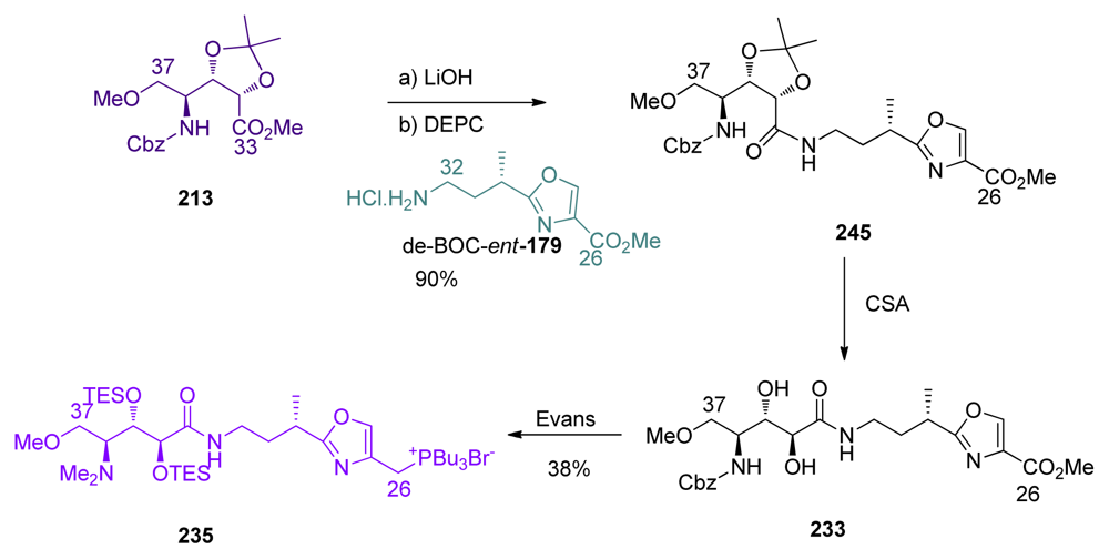
The C26–C37 fragment was synthesized efficiently from 213 and de-BOC-ent-179 (Scheme 51). The coupling of the fragments C26–C32 and C33–C37 in the presence of DEPC furnished amide 245 in 90% yield. Acetonide deprotection furnished diol 233, which was converted to phosphonium 235 following Evans procedure [18].
PMB-removal at C17 on 112 was followed by phosphorylation, C9 protecting group exchange and ozonolysis at C25 to give aldehyde 246 (Scheme 52). Wittig reaction between 246 and phosphonium salt 235, followed by TMS-deprotection at C9 furnished compound 243, the intermediate previously reported by Masamune.

5.6.4. The total synthesis of ent-calyculin A and B by Smith [22,51,69]

Smith and co-workers tenacious work afforded the total syntheses of ent-calyculin A and B in 1999. The coupling of C33–C37 oxazole 184 and C26–C32 acid 218 was carried out in the presence of DEPC, as described by Shioiri, to give 247 (Scheme 53). Boc-deprotection was followed by methylation of the amine, and acetonide deprotection liberated diol which was protected with DEIPS group, to yield 248. Finally, conversion of the methyl ester at C26 to the phosphonium salt 249 was carried out via the corresponding chloride.
Phosphate introduction at C17 of 127 was followed by benzyl deprotection and oxidation to furnish aldehyde 250 (Scheme 54). Wittig olefination between 250 and phosphonium chloride 249 provided 251 as a 9:1 E:Z mixture in 84% yield. Pivaloyl-removal at C9 and subsequent oxidation of 251 furnished the corresponding aldehyde, which was olefinated via HWE reaction with phosphonate 30 to give methyl ketone 252, after acidic treatment, in excellent 92% yield and 15:1 E:Z ratio. Peterson olefination of 252 furnished a 1.7:1 mixture of the E and Z isomers, which respectively corresponds to protected ent-calyculin A and ent-calyculin B. After chromatographic separation, final HF treatment produced ent-calyculin A in 69% yield and ent-calyculin B in 84% yield.

5.6.5. Total synthesis of calyculin C by Armstrong [21,65]

The total synthesis of calyculin C by Armstrong was also a result of perseverant work. Armstrong and co-workers were also the first to prepare calyculin C and prove its stereochemistry. Boc-deprotection of 190 was followed by coupling with ester 222 (Scheme 55). Unfortunately, this reaction produced a 2.7:1 mixture of C34 epimers. Further studies proved that the main product was the undesired isomer; the expected diastereomer 253 being isolated in only 17%. Conversion of the chloride to the phosphonium salt and benzyl-deprotection then furnished 254.
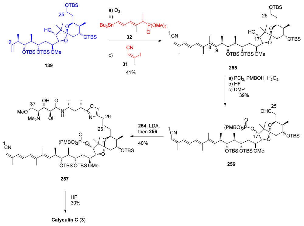
The final stages of the synthesis followed Evans’ example (Scheme 56). Fragment 139 was converted to the tetraene 255. C17-phosphonate protection, C25-deprotection and subsequent oxidation furnished aldehyde 256. Olefination between 256 and 254 furnished compound 257 in modest yield. Final HF treatment completed the total synthesis of calyculin C 3.

5.6.6. Formal total synthesis of ent-calyculin A by Barrett [23]

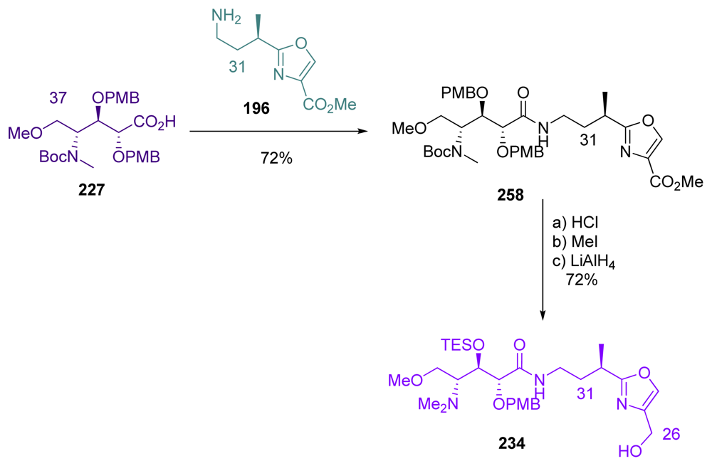
Barrett et al. last steps for completing the total synthesis of ent-1 began with the coupling of aminoacid 227 and oxazole 196 fragments, to give amide 258 (Scheme 57). The latter was then converted to Evans’s intermediate 234, by a three-step/one-pot sequence involving Boc-deprotection, N-methylation and reduction of the ester at C26.
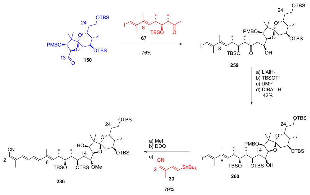
Aldol reaction between methyl ketone 67 and aldehyde 150 gave in a diastereoselective manner β-hydroxyketone 259, unfortunately carrying the wrong stereochemistry at C15 (Scheme 58). LiAlH4 reduction at C13 furnished the corresponding diol in 73%, together with its C13-epimer in 15% yield. Selective monosilylation at C13 was followed by inversion of stereochemistry at C15 by successive Dess-Martin oxidation and DIBAL-H reduction to produce 260. O-Methylation at C15, PMB-removal at C17 and final Stille coupling with stannane 33 finished the synthesis of Evans’ intermediate 236.

6. Conclusions

Altogether six total or formal synthesis of calyculins have been published. These syntheses can be divided into three groups:
  • Masamune and Armstrong have described the total synthesis of natural calyculins A and C, respectively
  • Evans and Smith have completed the total synthesis of the enantiomer of naturally occurring calyculin A (and B for Smith)
  • Shioiri and Barrett have published highly advanced intermediates, previously prepared by Masamune and Evans respectively and therefore accomplished formal synthesis of natural and non natural calyculin A.
Even if the basic retrosynthetic analysis appeared to be quite similar in the different synthesis, the different points of views and methods makes the comparison challenging. The overall yields and number of steps of the different syntheses are compiled in Table 2.
Each of these syntheses resulted from extensive efforts and has to be considered as high level work. As discussed earlier, the structure of calyculins makes the total synthesis very demanding in every aspect; every fragment had its own features and presented challenges. Of course, theses syntheses are not perfect and some drawbacks could be noted, like a C13-stereochemistry inversion for Evans, a low-yielding iodide-tin exchange for Masamune, protecting groups exchange for Shioiri, low selectivity in the final Peterson olefination for Smith (which fortunately led to the formation of ent-calyculin B), stereochemistry inversions and poor selectivity in the second Brown crotylation for Armstrong or a C15-stereochemistry inversion for Barrett. However, all these syntheses were performed before 2000 and all the new tools that have appeared in the last decade for stereoselective transformations were not available. For all these reasons, these total syntheses of the structurally much elaborated calyculins deserve the biggest respect from the synthetic community.
Another goal should still lie in a better understanding of the PP inhibition-activity of calyculins and related toxins. Improved design and synthetic methods should also lead to the design of simpler synthetic inhibitors that could compete with the activity of natural toxins.

Acknowledgements

This research has been financially supported by the Academy of Finland (Grant No 123485).
  • Samples Availability: Available from the authors.

References

  1. Nakao, Y; Fusetani, N. Enzyme inhibitors from marine invertebrates. J Nat Prod 2007, 70, 689–710. [Google Scholar]
  2. Barford, D; Das, AK; Egloff, M-P. The strucrure and mechanism of protein phosphatases: Insights into catalysis and regulation. Annu Rev Biophys Biomol Struct 1998, 27, 133–164. [Google Scholar]
  3. Cohen, PTW. Novel protein serine/threonine phosphatases: Variety is the spice of life. Trends Biochem Sci 1997, 22, 245–251. [Google Scholar]
  4. McCluskey, A; Sim, ATR; Sakoff, JA. Serine-Threonine protein phosphatase inhibitors: Development of potential therapeutic strategies. J Med Chem 2002, 45, 1151–1175. [Google Scholar]
  5. Schönthal, AH. Role of serine/threonine protein phosphatase 2A in cancer. Cancer Lett 2001, 170, 1–13. [Google Scholar]
  6. Sheppeck, JE; Gauss, C-M; Chamberlin, AR. Inhibition of the ser-thr phosphatases PP1 and PP2A by naturally occurring toxins. Bioorg Med Chem 1997, 5, 1739–1750. [Google Scholar]
  7. Sontag, E. Protein phosphatase 2A: The trojan horse of cellular signaling. Cell Signaling 2001, 13, 7–16. [Google Scholar]
  8. Kato, Y; Fusetani, N; Matsunaga, S; Hashimoto, K; Koseki, K. Bioactive marine metabolites. 24. Isolation and structure elucidation of calyculins B, C, and D, novel antitumor metabolites, from the marine sponge Discodermia calyx. J Org Chem 1988, 53, 3930–3932. [Google Scholar]
  9. Bewley, CA; Faulkner, DJ. Lithistid sponges: Star performers or hostes to the stars. Angew Chem Int Ed 1998, 37, 2162–2178. [Google Scholar]
  10. Dumdei, EJ; Blunt, JW; Munro, MHG; Pannell, LK. Isolation of calyculins, calyculinamides, and swinholide H from the New Zealand deep-water marine sponge lamellomorpha strongylata. J Org Chem 1997, 62, 2636–2639. [Google Scholar]
  11. Kato, Y; Fusetani, N; Matsunaga, S; Hashimoto, K; Fujita, S; Furuya, T. Bioactive marine metabolites. Part 16. Calyculin A. A novel antitumor metabolite from the marine sponge Discodermia calyx. J Am Chem Soc 1986, 108, 2780–2781. [Google Scholar]
  12. Kato, Y; Fusetani, N; Matsunaga, S; Hashimoto, K; Koseki, K. Bioactive marine metabolites. 24. Isolation and structure elucidation of calyculins B, C, and D, novel antitumor metabolites, from the marine sponge Discodermia calyx. J Org Chem 2002, 53, 3930–3932. [Google Scholar]
  13. Matsunaga, S; Fujiki, H; Sakata, D; Fusetani, N. Calyculins E, F, G, and H, additional inhibitors of protein phosphatases 1 and 2A, from the marine sponge discodermia calyx. Tetrahedron 1991, 47, 2999–3006. [Google Scholar]
  14. Matsunaga, S; Wakimoto, T; Fusetani, N. Isolation of four new calyculins from the marine sponge Discodermia calyx. J Org Chem 1997, 62, 2640–2642. [Google Scholar]
  15. Matsunaga, S; Wakimoto, T; Fusetani, N; Suganuma, M. Isolation of dephosphonocalyculin A from the marine sponge, Discodermia calyx. Tetrahedron Lett 1997, 38, 3763–3764. [Google Scholar]
  16. Hamada, Y; Tanada, Y; Yokokawa, F; Shioiri, T. Stereoselective synthesis of 2,3-dihydroxy-4-dimethylamino-5-methoxypentanoic acid, a fragment of calyculins -Determination of the absolute configuration of calyculins. Tetrahedron Lett 1991, 32, 5983–5986. [Google Scholar]
  17. Matsunaga, S; Fusetani, N. Absolute stereochemistry of the calyculins, potent inhibitors of potent phosphatases 1 and 2A. Tetrahedron Lett 1991, 32, 5605–5606. [Google Scholar]
  18. Evans, DA; Gage, JR; Leighton, JL. Total synthesis of (+)-calyculin A. J Am Chem Soc 1992, 114, 9434–9453. [Google Scholar]
  19. Tanimoto, N; Gerritz, SW; Sawabe, A; Noda, T; Filla, SA; Masamune, S. The synthesis of naturally occurring (−)-calyculin A. Angew Chem Int Ed 1994, 33, 673–675. [Google Scholar]
  20. Yokokawa, F; Hamada, Y; Shioiri, T. Total synthesis of calyculin A—Construction of the C(9)–C(37) fragment. Chem Commun 1996, 7, 872. [Google Scholar]
  21. Ogawa, AK; Armstrong, RW. Total synthesis of calyculin C. J Am Chem Soc 1998, 120, 12435–12442. [Google Scholar]
  22. Smith, AB; Friestad, GK; Duan, JJW; Barbosa, J; Hull, KG; Iwashima, M; Qiu, Y; Spoors, PG; Bertounesque, E; Salvatore, BA. Total synthesis of (+)-calyculin A and (−)-calyculin B. J Org Chem 1998, 63, 7596–7597. [Google Scholar]
  23. Anderson, OP; Barrett, AGM; Edmunds, JJ; Hachiya, S-I; Hendrix, JA; Horita, K; Malecha, JW; Parkinson, CJ; VanSickle, A. Applications of crotyldiisopino campheylboranes in synthesis: A formal total synthesis of (+)-calyculin A. Can J Chem 2001, 79, 1562–1592. [Google Scholar]
  24. Gupta, V; Ogawa, AK; Du, X; Houk, KN; Armstrong, RW. A model for binding of structurally diverse natural product inhibitors of protein phosphatases PP1 and PP2A. J Med Chem 1997, 40, 3199–3206. [Google Scholar]
  25. Fu, X; Schmitz, FJ; Kelly-Borges, M; McCready, TL; Holmes, CFB. Clavosines A-C from the marine sponge myriastra clavosa: Potent cytotoxins and inhibitors of protein phosphatases 1 and 2A. J Org Chem 1998, 63, 7957–7963. [Google Scholar]
  26. Kehraus, S; Konig, GM; Wright, AD. A new cytotoxic calyculinamide derivative, geometricin A, from the australian sponge luffariella geometrica. J Nat Prod 2002, 65, 1056–1058. [Google Scholar]
  27. Edrada, RA; Ebel, R; Supriyono, A; Wray, V; Schupp, P; Steube, K; van Soest, R; Proksch, P. Swinhoeiamide A, a new highly active calyculin derivative from the marine sponge Theonella swinhoei. J Nat Prod 2002, 65, 1168–1172. [Google Scholar]
  28. Wakimoto, T; Matsunaga, S; Takai, A; Fusetani, N. Insight into binding of calyculin A to protein phosphatase 1: Isolation of hemicalyculin A and chemical transformation of calyculin A. Chem Biol 2002, 9, 309–319. [Google Scholar]
  29. Quinn, RJ; Taylor, C; Suganuma, M; Fujiki, H. The conserved acid binding domain model of inhibitors of protein phosphatases 1 and 2A: Molecular modelling aspects. Bioorg Med Chem Lett 1993, 3, 1029–1034. [Google Scholar]
  30. Takai, A; Sasaki, K; Nagai, H; Mieskes, G; Isobe, M; Isono, K; Yasumoto, T. Inhibition of spesific binding of okadaic acid to protein phosphatase 2A by microcystin-LR, calyculin-A and tautomycin: Method of analysis of interactions of tight-binding with target protein. Biochem J 1995, 306, 657–665. [Google Scholar]
  31. Goldberg, J; Huang, H-b; Kwon, Y-g; Greengard, P; Nairn, AC; Kuriyan, J. Three dimensional structure of the catalytic subunit of protein serine/threonine phosphatase-1. Nature 1995, 376, 745–753. [Google Scholar]
  32. Bagu, JR; Sykes, BD; Craig, MM; Holmes, CFB. A molecular basics for different interactions of marine toxins with protein phosphatase-1. J Biol Chem 1997, 272, 5087–5097. [Google Scholar]
  33. Gauss, C-M; Sheppeck, JE; Nairn, AC; Chamberlin, R. A molecular modeling analysis of the binding interactions between the okadaic acid class of natural product inhibitors and the ser-thr phosphatases, PP1 and PP2A. Bioorg Med Chem 1997, 5, 1751–1773. [Google Scholar]
  34. Lindvall, MK; Pihko, PM; Koskinen, AMP. The binding model of calyculin A to protein phosphatase.1: A novel spiroketal vector model. J Biol Chem 1997, 272, 23312–23316. [Google Scholar]
  35. Kita, A; Matsunaga, S; Takai, A; Kataiwa, H; Wakimoto, T; Fusetani, N; Isobe, M; Miki, K. Crystal structure of the complex between calyculin A and the catalytic subunit of protein phosphatase 1. Structure 2002, 10, 715–724. [Google Scholar]
  36. McCready, TL; Islam, BF; Schmitz, FJ; Luu, HA; Dawson, JF; Holmes, CFB. Inhibition of protein phosphatase-1 by clavosines A and B- novel members of the calyculun family of toxins. J Biol Chem 2002, 9, 4192–4198. [Google Scholar]
  37. Volter, KE; Embrey, KJ; Pierens, GK; Quinn, RJ. A study of the binding requirements of calyculin A and dephosphonocalyculin A with PP1, development of a molecular recognition model for the binding interactions of the okadaic acid class of compounds with PP1. Eur J Pharm Sci 2001, 12, 181–194. [Google Scholar]
  38. Koskinen, AMP; Chen, J. Enantiospecific synthesis of polyhydroxy amino acids. Synthesis of the C33-C38 portion of calyculins. Tetrahedron Lett 1991, 32, 6977–6980. [Google Scholar]
  39. Pihko, PM; Koskinen, AMP. Synthesis of the C26–32 oxazole fragment of calyculin C: A test case for oxazole syntheses. J Org Chem 1998, 63, 92–98. [Google Scholar]
  40. Pihko, PM; Koskinen, AMP. The triumph of zinc: A short and highly efficient synthesis of the calyculin C1-C9 tetraene building block by Negishi Coupling. Synlett 1999, 12, 1966–1968. [Google Scholar]
  41. Pihko, PM; Koskinen, AMP; Nissinen, MJ; Rissanen, K. Formation of stable spiro[4.4] ortho ester aminals during the synthesis of the C26-C32 oxazole fragment of calyculin C. J Org Chem 1999, 64, 652–654. [Google Scholar]
  42. Karisalmi, K; Koskinen, AMP. Studies towards the synthesis of the C(9)-C(20) lactone-dipropionate fragment of calyculin C. Synthesis 2004, 9, 1331–1342. [Google Scholar]
  43. Karisalmi, K; Koskinen, AMP. Stereoselective synthesis of the C9-C19 lactone-dipropionate fragment of calyculin C. Tetrahedron Lett 2004, 45, 8245–8248. [Google Scholar]
  44. Rauhala, V; Nevalainen, M; Koskinen, AMP. An expedient synthesis of spiroketals: Model studies for the calyculin C16-C25 fragment. Tetrahedron 2004, 60, 9199–9204. [Google Scholar]
  45. Rauhala, V; Nättinen, K; Rissanen, K; Koskinen, AMP. The reaction mechanism of spirocylization and stereo selectivity studies for the calyculin C16-C25 fragment. Eur J Org Chem 2005, 4119–4126. [Google Scholar]
  46. Habrant, D; Stewart, AJW; Koskinen, AMP. Towards the total synthesis of calyculin C: Preparation of the C13-C25 spirocyclic core. Tetrahedron 2009, 65, 7927–7934. [Google Scholar]
  47. Jacobs, MF; Kitching, W. Spiroacetals of marine origin. Curr Org Chem 1998, 2, 395–436. [Google Scholar]
  48. Pihko, PM. Chemistry and biology of the calyculins. In Acta Univ Oul, Ser A (Sci Rer Nat); Volume 325, PhD Thesis; Dept Chemistry, University of Oulu: Finland, 1999. [Google Scholar]
  49. Wang, C; Tobrman, T; Xu, Z; Negishi, E-i. Highly regio- and stereoselective synthesis of (Z)-trisubstituted alkenes via propyne bromoboration and tandem Pd-catalyzed cross-coupling. Org Lett 2009, 11, 4092–4095. [Google Scholar]
  50. Filla, SA. Discodermolide synthesis: Fragments A and BCD; PhD Thesis; Dept Chemistry, Massachusetts Institute of Technology: Cambridge, MA, USA, 1994. [Google Scholar]
  51. Smith, AB; Friestad, GK; Duan, JJW; Barbosa, J; Hull, KG; Iwashima, M; Qiu, Y; Spoors, PG; Bertounesque, E; Salvatore, BA. Total synthesis of (+)-calyculin A and (−)-calyculin B: Cyanotetraene construction, asymmetric synthesis of the C(26–37) oxazole, fragment assembly, and final elaboration. J Am Chem Soc 1999, 121, 10478–10486. [Google Scholar]
  52. Scarlato, GR; DeMattei, JA; Chong, LS; Ogawa, AK; Lin, MR; Armstrong, RW. Asymmetric synthesis of calyculin C. 1. Synthesis of the C1–C25 fragment. J Org Chem 1996, 61, 6139–6152. [Google Scholar]
  53. Barrett, AGM; Edmunds, JJ; Hendrix, JA; Horita, K; Parkinson, CJ. Stereocontrolled synthesis of calyculin A: Construction of the C(1)–C(14) tetraene nitrile unit. Chem Commun 1992, 1238–1240. [Google Scholar]
  54. Brown, HC; Bhat, KS. Enantiomeric (Z)- and (E)-crotyldiisopinocampheylboranes. Synthesis in high optical purity of all four possible stereoisomers of b-methylhomoalyl alcohols. J Am Chem Soc 1986, 108, 293–294. [Google Scholar]
  55. Brown, HC; Jadhav, PK; Bhat, KS. Chiral synthesis via organoboranes. 13 A highly diastereoselective and enentioselective addition of [(Z)-g-alkoxyallyl]diisopinocampheylboranes to aldehydes. J Am Chem Soc 1988, 110, 1535–1538. [Google Scholar]
  56. Trost, BM; Flygare, JA. A synthesis of the spiroketal subunit of (−)-calyculin A. Tetrahedron Lett 1994, 35, 4059–4062. [Google Scholar]
  57. Yokokawa, F; Hamada, Y; Shioiri, T. A synthesis of the oxazole part of calyculins; Part 2. Synlett 1992, 151–152. [Google Scholar]
  58. Takebuchi, K; Hamada, Y; Shioiri, T. Synthesis of the C13–C19 unit in the spiroketal fragment of calyculins. Tetrahedron Lett 1994, 35, 5239–5242. [Google Scholar]
  59. Smith, AB; Duan, JJW; Hull, KG; Salvatore, BA. Calyculin synthetic studies. Stereoselective construction of the C(14)–C(25) spiroketal subunit. Tetrahedron Lett 1991, 32, 4855–4858. [Google Scholar]
  60. Smith, AB; Friestad, GK; Duan, JJW; Barbosa, J; Hull, KG; Iwashima, M; Qiu, Y; Spoors, PG; Bertounesque, E; Salvatore, BA. Total synthesis of (+)-calyculin A and (−)-calyculin B: Asymmetric synthesis of the C(9–25) spiroketal dipropionate subunit. J Am Chem Soc 1999, 121, 10468–10477. [Google Scholar]
  61. Barrett, AGM; Edmunds, JJ; Horita, K; Parkinson, CJ. Stereocontrolled synthesis of calyculin A: Construction of the C(15)–C(25) spiroketal unit. Chem Commun 1992, 1236–1238. [Google Scholar]
  62. Chemler, SR; Roush, WR. Stereochemistry of the allylation and crotylation reactions of a-methyl-b-hydroxy aldehydes with allyl- andcrotyltrifluorosilanes. Synthesis of anti-anti-dipropionate stereotriads and stereodivergent pathways for the reactions with 2,3-anti and 2,3-syn-a-methyl-b-hydroxy aldehydes. J Org Chem 2003, 68, 1319–1333. [Google Scholar]
  63. Vaccaro, HA; Levy, DE; Sawabe, A; Jaetsch, T; Masamune, S. Towards the synthesis of calyculin: A synthetic intermediate corresponding to the C(26)–C(37) fragment. Tetrahedron Lett 1992, 33, 1937–1940. [Google Scholar]
  64. Yokokawa, F; Hamada, Y; Shioiri, T. A synthesis of the oxazole part of calyculins; Part 1. Synlett 1992, 149–150. [Google Scholar]
  65. Ogawa, AK; DeMattei, JA; Scarlato, GR; Tellew, JE; Chong, LS; Armstrong, RW. Asymmetric synthesis of calyculin C. 2. synthesis of the C26–C37 fragment and model Wittig couplings. J Org Chem 1996, 61, 6153–6161. [Google Scholar]
  66. Barrett, AGM; Edmunds, JJ; Malecha, JW; Parkinson, CJ. Stereocontrolled synthesis of calyculin A: Construction of the C(26)–C(37) amide-oxazole unit. Chem Commun 1992, 1240–1242. [Google Scholar]
  67. Yokokawa, F; Hamada, Y; Shioiri, T. Synthesis of the C26–C37 fragment of calyculin A having natural configuration. Synlett 1992, 703–705. [Google Scholar]
  68. Sawabe, A; Filla, SA; Masamune, S. Use of 2-trimethylsilylethyl as a protecting group in phosphate monoester synthesis. Tetrahedron Lett 1992, 33, 7685–7686. [Google Scholar]
  69. Smith, AB; Salvatore, BA; Hull, KG; Duan, JJW. Calyculin synthetic studies. 2. Stereocontrolled assembly of the C(9)-C(13) dithiane and C(26)-C(37) oxazole intermediates. Tetrahedron Lett 1991, 32, 4859–4862. [Google Scholar]
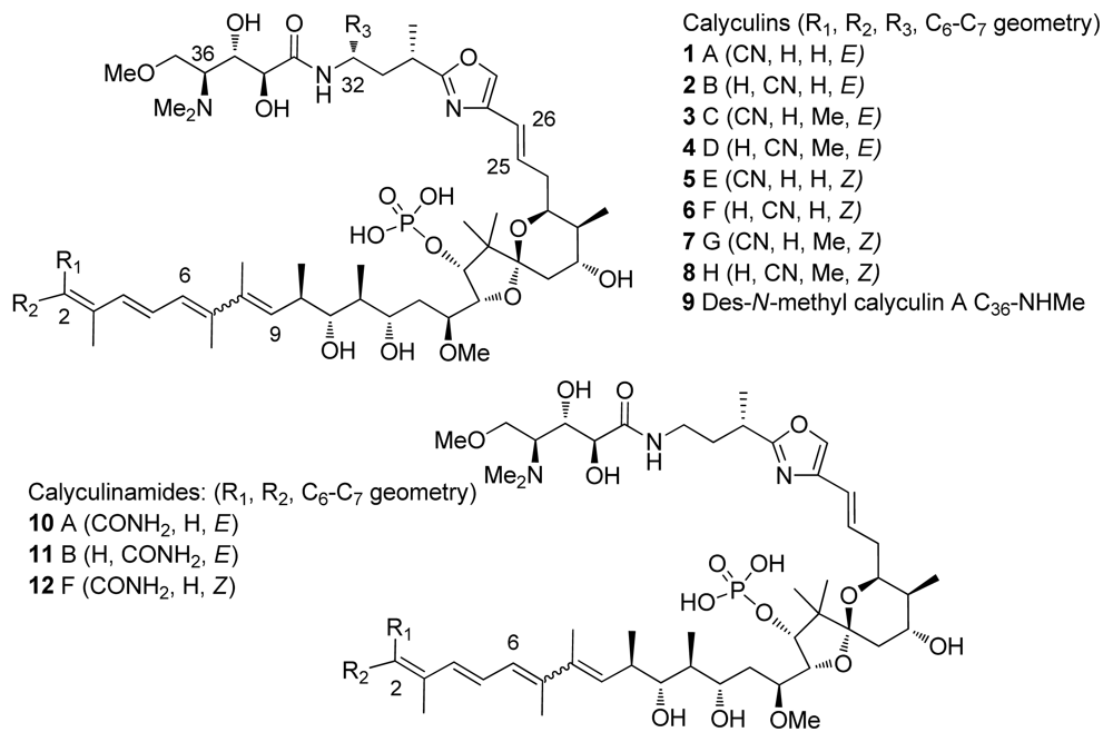
Figure 1. Calyculins and calyculinamides.
Marinedrugs 08 00122f1
Figure 2. PP2A-selective inhibitors.
Marinedrugs 08 00122f2
Figure 3. Right: microcystin-LR (18); left: motuporin (19).
Marinedrugs 08 00122f3
Figure 4. Tautomycin 20.
Marinedrugs 08 00122f4
Figure 5. Calyculin related structures.
Marinedrugs 08 00122f5
Figure 6. Binding models of calyculin A to PP1.
Marinedrugs 08 00122f6
Scheme 1. Retrosynthetic analysis of the calyculin skeleton (X, Y and Z denote the functional groups suitable for coupling).
Marinedrugs 08 00122f7
Scheme 2. Retrosynthetic analysis of the C26–C37 fragment (X denotes the functional group suitable for coupling with the fragment C9–C25).
Marinedrugs 08 00122f8
Scheme 3. Retrosynthetic analysis of C1–C9 fragment (X denotes the functional groups suitable for coupling).
Marinedrugs 08 00122f9
Scheme 4. Preparation of phosphonate 32.
Marinedrugs 08 00122f10
Scheme 5. Preparation of stannane 33 by Masamune.
Marinedrugs 08 00122f11
Scheme 6. Preparation of phosphonate 34.
Marinedrugs 08 00122f12
Scheme 7. Preparation of stannane 33 by Shioiri.
Marinedrugs 08 00122f13
Scheme 8. Preparation of tetraene 54.
Marinedrugs 08 00122f14
Scheme 9. Preparation of phosphonate 30.
Marinedrugs 08 00122f15
Scheme 10. Preparation of phosphonate 32.
Marinedrugs 08 00122f16
Scheme 11. Preparation of stannane 33 by Barrett.
Marinedrugs 08 00122f17
Scheme 12. Synthesis of vinyl iodide 67.
Marinedrugs 08 00122f18
Scheme 13. Preparation of 32 by Koskinen.
Marinedrugs 08 00122f19
Scheme 14. Retrosynthetic analysis of C9-C25 fragment. (Y denotes the functional groups suitable for coupling and P protective groups).
Marinedrugs 08 00122f20
Scheme 15. Preparation of ketone 76.
Marinedrugs 08 00122f21
Scheme 16. Formation of spiroketal 79.
Marinedrugs 08 00122f22
Scheme 17. Preparation of 85.
Marinedrugs 08 00122f23
Scheme 18. Preparation of 95.
Marinedrugs 08 00122f24
Scheme 19. Preparation of spiroketal 99.
Marinedrugs 08 00122f25
Scheme 20. Preparation of ketone 105.
Marinedrugs 08 00122f26
Scheme 21. Synthesis of spiroketal 112.
Marinedrugs 08 00122f27
Scheme 22. Synthesis of epoxides 116.
Marinedrugs 08 00122f28
Scheme 23. Preparation of spiroketal 122.
Marinedrugs 08 00122f29
Scheme 24. Preparation of fragment 127.
Marinedrugs 08 00122f30
Scheme 25. Preparation of spiroketal 134.
Marinedrugs 08 00122f31
Scheme 26. Preparation of 139.
Marinedrugs 08 00122f32
Scheme 27. Preparation of aldehyde 143.
Marinedrugs 08 00122f33
Scheme 28. Preparation of spiroketal 150.
Marinedrugs 08 00122f34
Scheme 29. Preparation of 157.
Marinedrugs 08 00122f35
Scheme 30. Preparation of spiroketal 161.
Marinedrugs 08 00122f36
Scheme 31. Retrosynthetic analysis of C26–C32 fragment (X denotes the functional groups suitable for coupling and P the protective group).
Marinedrugs 08 00122f37
Scheme 32. Preparation of oxazole 167.
Marinedrugs 08 00122f38
Scheme 33. Preparation of oxazole 172.
Marinedrugs 08 00122f39
Scheme 34. Preparation of oxazole 179.
Marinedrugs 08 00122f40
Scheme 35. Preparation of oxazole 184.
Marinedrugs 08 00122f41
Scheme 36. Preparation of oxazole 190.
Marinedrugs 08 00122f42
Scheme 37. Preparation of oxazole 196.
Marinedrugs 08 00122f43
Scheme 38. Preparation of oxazole 200.
Marinedrugs 08 00122f44
Scheme 39. Retrosynthetic analysis of C33–C37 fragment.
Marinedrugs 08 00122f45
Scheme 40. Preparation of 205.
Marinedrugs 08 00122f46
Scheme 41. Preparation of 209.
Marinedrugs 08 00122f47
Scheme 42. Preparation of ester 213.
Marinedrugs 08 00122f48
Scheme 43. Preparation of 218.
Marinedrugs 08 00122f49
Scheme 44. Preparation of 222.
Marinedrugs 08 00122f50
Scheme 45. Preparation of 227.
Marinedrugs 08 00122f51
Scheme 46. Preparation of 232.
Marinedrugs 08 00122f52
Scheme 47. Preparation of the C26–C37 fragment 235.
Marinedrugs 08 00122f53
Scheme 48. Final steps for the synthesis of ent-calyculin A.
Marinedrugs 08 00122f54
Scheme 49. Preparation of C26–C37 fragment 240.
Marinedrugs 08 00122f55
Scheme 50. Final steps to calyculin A.
Marinedrugs 08 00122f56
Scheme 51. Preparation of C26–C37 235 by Shioiri.
Marinedrugs 08 00122f57
Scheme 52. Preparation of 243.
Marinedrugs 08 00122f58
Scheme 53. Preparation of C26–C37 fragment 249.
Marinedrugs 08 00122f59
Scheme 54. Final steps to ent-calyculin A and B.
Marinedrugs 08 00122f60
Scheme 55. Preparation of C26–C37 fragment 254.
Marinedrugs 08 00122f61
Scheme 56. Final steps to calyculin C.
Marinedrugs 08 00122f62
Scheme 57. Preparation of the C26–C37 fragment 234 by Barrett.
Marinedrugs 08 00122f63
Scheme 58. Preparation of the C1–C25 fragment 236 by Barrett.
Marinedrugs 08 00122f64
Table 1. PP1 and PP2A inhibitors.
Name of the inhibitorIsolation originStructural ScaffoldIC50 nM a
PropertiesRef.
PP1PP2A
Microcystin-LR (18)Blue green algaeCyclic peptide0.3–0.60.04–2.0Liver toxin[24]
Nodularin-V (19)Blue green algaeCyclic peptide0.5–30.03–1.0Liver toxin[24]
Cantharidin (15)Blister beetlesTerpenoid0.5–2.00.2Natural defensive toxicant[6]
Thyrsiferyl- 23-acetate (16)Red algae, L. ObtusaTerpenoid>1(4–16)·10−3[6]
Okadaic acid (13)DinoflagellatesPolyketide10–13000.02–1.0Tumour promoter[24]
Dinophysistoxin-4 (14)DinoflagellatesPolyketide~200~2[4]
Calyculin A (1)Marine sponge D. calyx.Polyketide0.4–2.00.25–3Tumour promoter[24]
Calyculin C (3)Marine sponge D. calyx.Polyketide0.62.8Tumour promoter[24]
Tautomycin (20)Bacterium, Streptomyces verticillatusPolyketide1.1–7.5110–23.1Antibiotic[24]
Fostriecins (17)Bacterium, Streptomyces pulveraceusPolyketide0.1313.4·10−6Antitumoric activity[4]
aThe determined IC50 values are not always directly comparable from source to source. They may vary depending on the substrate, and on the purity, concentration and origin of the purified protein.
Table 2. Overview of the total synthesis.
GroupTargetPub. yearNumber of steps aOverall yield (%) aNumber of steps bOverall yield (%) b
Evansent-Calyculin A1992330.5436-
MasamuneCalyculin A1994430.31450.18
ShioiriCalyculin A1996320.092320.092
Smithent-Calyculin A1998350.89370.79
ArmstrongCalyculin C1998300.018300.018
Barrettent-Calyculin A2001340.9340.9
aLongest linear sequence based on the reported starting materials.
bLongest linear sequence based on commercially available starting materials.