Article

PA from a Recent H9N2 (G1-Like) Avian Influenza A Virus (AIV) Strain Carrying Lysine 367 Confers Altered Replication Efficiency and Pathogenicity to Contemporaneous H5N1 in Mammalian Systems

1
Center of Scientific Excellence for Influenza Viruses, National Research Centre (NRC), 12622 Giza, Egypt
2
Institute of Medical Virology, Justus Liebig University Giessen, Schubertstrasse 81, 35390 Giessen, Germany
3
St. Jude Children’s Research Hospital, 262 Danny Thomas Place, Memphis, TN 38105, USA
4
Anatomy and Embryology Department, Faculty of Medicine, Jouf University, Sakaka 2014, Saudi Arabia
5
Department of Epidemiology, Human Genetics, and Environmental Sciences, University of Texas, Houston, TX 77030, USA
6
Human Link, Baabda 1109, Lebanon
*
Authors to whom correspondence should be addressed.
These authors contributed equally to this work.
Viruses 2020, 12(9), 1046; https://doi.org/10.3390/v12091046
Submission received: 26 August 2020 / Revised: 15 September 2020 / Accepted: 18 September 2020 / Published: 20 September 2020
(This article belongs to the Special Issue Evolution and Pathogenesis of Avian and Animal Influenza Viruses)

Abstract

:
Egypt is a hotspot for H5- and H9-subtype avian influenza A virus (AIV) infections and co-infections in poultry by both subtypes have been frequently reported. However, natural genetic reassortment of these subtypes has not been reported yet. Here, we evaluated the genetic compatibility and replication efficiency of reassortants between recent isolates of an Egyptian H5N1 and a H9N2 AIV (H5N1EGY and H9N2EGY). All internal viral proteins-encoding segments of the contemporaneous G1-like H9N2EGY, expressed individually and in combination in the genetic background of H5N1EGY, were genetically compatible with the other H5N1EGY segments. At 37 °C the replication efficiencies of H5N1EGY reassortants expressing the H9N2EGY polymerase subunits PB2 and PA (H5N1PB2-H9N2EGY, H5N1PA-H9N2EGY) were higher than the wild-type H5N1EGY in Madin-Darby canine kidney (MDCK-II) cells. This could not be correlated to viral polymerase activity as this was found to be improved for H5N1PB2-H9N2EGY, but reduced for H5N1PA-H9N2EGY. At 33 °C and 39 °C, H5N1PB2-H9N2EGY and H5N1PA-H9N2EGY replicated to higher levels than the wild-type H5N1EGY in human Calu-3 and A549 cell lines. Nevertheless, in BALB/c mice both reassortants caused reduced mortality compared to the wild-type H5N1EGY. Genetic analysis of the polymerase-encoding segments revealed that the PAH9N2EGY and PB2H9N2EGY encode for a distinct uncharacterized mammalian-like variation (367K) and a well-known mammalian signature (591K), respectively. Introducing the single substitution 367K into the PA of H5N1EGY enabled the mutant virus H5N1PA-R367K to replicate more efficiently at 37 °C in primary human bronchial epithelial (NHBE) cells and also in A549 and Calu-3 cells at 33 °C and 39 °C. Furthermore, H5N1PA-R367K caused higher mortality in BALB/c mice. These findings demonstrate that H5N1 (Clade 2.2.1.2) reassortants carrying internal proteins-encoding segments of G1-like H9N2 viruses can emerge and may gain improved replication fitness. Thereby such H5N1/H9N2 reassortants could augment the zoonotic potential of H5N1 viruses, especially by acquiring unique mammalian-like aa signatures.

1. Introduction

Highly pathogenic avian influenza A viruses (HPAIVs) of the H5N1 subtype have crossed the animal/human barrier in 1997 causing lethal zoonotic infections in humans [1]. During the second wave of human HPAIV H5N1 infections—specifically in December 2005, HPAIV H5N1 (clade 2.2.1) have been reported in Egypt in migratory birds [2]. Subsequently in mid-February 2006, HPAIV H5N1 were transmitted to farmed and backyard poultry in Egypt [3,4] where they kept circulating. They were therefore officially declared “endemic” in the Egyptian poultry sector since 2008 [5]. In parallel, human infections with HPAIV H5N1 are annually reported since 2006, classifying Egypt as the country with the highest cumulative number of human cases in the world [1].
Concomitantly, low pathogenic avian influenza A virus (LPAIV) of the H9N2 subtype are circulating extensively worldwide in poultry for several decades, resulting in a high genetic diversity [1,6]. In 2010, LPAIV H9N2 of a G1-like lineage were detected in different avian species in Egypt [7,8,9]. Currently, the virus appears to be endemic in Egyptian poultry [6]. Since 2011, these viruses crossed the species barrier to humans due to their mammalian-like characteristics, causing mild to moderate human infections in different countries of the Middle East including Egypt [1,6]. The global concern about these H9N2 viruses is mainly associated with their ability to donate their genes to other avian influenza viruses (AIV) giving rise to high and low pathogenic AIVs that might thereby achieve the capability to efficiently cross the specie barrier and infect humans such as the LPAIV H7N9, reported in China since 2013 [1,10].
As both LPAIV H9N2 and HPAIV H5N1 subtypes are co-circulating in birds in several regions of the world co-infections with both subtypes have been frequently reported in individual avian hosts [11]. Consequently, this scenario increases the probability of genetic reassortment between both strains and could result in reassortants with enhanced zoonotic potential [10]. The limited detection of such reassortants in nature is probably due to insufficient surveillance, genetic incompatibility of possible H5N1/H9N2 segment combinations/constellations and/or negative selective pressure against such H5N1/H9N2 reassortants with new genetic constellations that render the virus replication incompetent, compared to the parental strains.
To better validate this potential threat, the present study set out to investigate the genetic compatibility and replication efficiency of reassortants, derived from recent avian influenza A/H5N1 and A/H9N2 viruses in vitro and pathogenicity in BALB/c mice. Moreover, a novel mammalian-like marker for avian influenza A viruses (IAVs) in the polymerase acidic (PA) protein was also described and characterized.

2. Materials and Methods

2.1. Cells, Viruses and Plasmids

Madin-Darby canine kidney (MDCK-II) cells, human lung adenocarcinoma epithelial cells (A549), human embryonic kidney cells (293T) and cultured human airway epithelial cells (Calu-3) were maintained in Dulbecco’s modified Eagle medium (DMEM) (Invitrogen, Carlsbad, CA, USA) supplemented with 100 I.U./mL penicillin, 100 μg/mL streptomycin (P/S) and 10% fetal bovine serum (FBS). All cell monolayers were incubated at 37 °C in a humidified CO2 incubator.
The highly pathogenic avian influenza virus A/chicken/Egypt/N12640A/2016(H5N1), designated hereafter as H5N1EGY, and the low pathogenic virus A/chicken/Egypt/S12568C/2016(H9N2), designated hereafter as H9N2EGY, were isolated from infected chicken in Egypt in 2016 by the Center of Scientific Excellence for Influenza Viruses (CSEIV), National Research Centre, Egypt. Both H5N1EGY and H9N2EGY were plaque-purified and propagated in the allantoic fluids of 10-day-old specific-pathogen-free (SPF) embryonated eggs. Inoculated eggs were incubated for 48 h at 37 °C and then chilled at 4 °C for 4 h before harvesting. Harvested stocks were titred by focus forming assay as previously described [12] and stored at −80 °C.
Plasmids expressing the eight viral segments of human influenza virus A/Anhui/1/2013(H7N9) designated H7N9Anhui [13], pMP-PB2-anhui, pMP-PB1-anhui, pMP-PA-anhui, pMP-HA-anhui, pMP-NP-anhui, pMP-NA-anhui, pMP-M-anhui and pMP-NS-anhui, were obtained from the plasmid stocks at the Institute of Medical Virology, Justus Liebig University Giessen, Germany.
Complete sets of pMPccdB plasmids encoding the eight viral segments of H5N1EGY and H9N2EGY were constructed as previously described [14,15,16]. Briefly, the full length sequences of the 8 viral segments were amplified by RT-PCR using universal set of primers [14,16] and cloned into pMPccdB as previously described [14].

2.2. Reverse Genetics (Rg) Systems for H5N1EGY and H9N2EGY and Generation of Reassortant, Mutant and Wild-Type Strains

To generate wild-type strains and H5N1-subtype reassortants with specific genetic constellations (Figure 1), 8 μg of plasmid DNA (1 μg of each plasmid) encoding different combinations of the eight viral segments were transfected into a co-culture of 293T/MDCK-II cells (ratio 3:1) as previously described [14,17,18]. The transfection mixture was harvested 72 h post-transfection (p.t.). For LPAIV H9N2EGY, 1 µg/mL of L-(tosylamido-2-phenyl) ethyl chloromethyl ketone (TPCK)-treated trypsin was added to the transfection mixture 24 h p.t. and the transfection mixture was then harvested 48 h after addition of TPCK-treated trypsin. An aliquot of 100 µL of each supernatant was used to inoculate SPF embryonated eggs for another 48 h. The harvested viruses of compatible genetic constellations (Figure 1), were titrated using focus forming assay, titred, aliquoted and stored at −80 °C for further use.
Mutations of interest were introduced into the corresponding PA genes by using QuikChange site-directed mutagenesis (Agilent, Santa Clara, CA, USA) and specific mutagenesis primers (Table 1) according to manufacturer instructions. Sanger sequencing was performed to confirm the introduction of these mutants. Variant viruses H5N1EGY_R367K and H5N1PA-H9N2Egy_K367R carrying polymerase acidic (PA)-encoding segments with specific mutations were rescued individually by transfecting a co-culture of 293T/MDCK cells with eight pMPccdB plasmids corresponding to the unchanged seven segments of the virus and the mutated one (7 WT viral segments + 1 mutant PA viral segment), using Trans-IT2020 (Mirus Bio, Madison, WI, USA) as described previously [14,19]. Recombinant viruses were rescued as described above and were subsequently sequenced to confirm the correct variant PB2 and PA segment in the genetic backbone of the other seven segments. Stocks of viruses harvested from infected MDCK-II cells were titrated by focus forming assay and kept at −80 °C until further use.

2.3. In Vitro Replication Efficiency of Reassortant and Wild-Type Strains

To determine the monocycle and multicycle replication efficiency of reassortant and wild-type strains, confluent monolayers of MDCK-II cells were infected in triplicate with reassortant and wild-type strains in PBS/BA (phosphate-buffered saline (PBS) containing 0.2% bovine albumin (BA), 1 mM MgCl2, 0.9 mM CaCl2, P/S) at multiplicity of infection (MOI) of 0.01 in a humidified CO2 incubator at 37 °C. The reassortant H5N16H7N9Anhui, carrying the mammalian adapted H9N2-derived internal proteins-encoding segments (PB2, PB1, PA, NP, M, NS) of human influenza H7N9Anhui virus and the HA and NA of H5N1EGY, was included as a positive control. After 1 h of virus-cell incubation at room temperature (RT), the inoculum was discarded and cell monolayers were washed twice with PBS++ (PBS containing 100 mg/l CaCl2 and 100 mg/l MgCl2) and replaced with infection medium DMEM/BA (DMEM, supplemented with 0.2% BA, 1% P/S and 1 μg/mL TPCK-treated trypsin). Supernatants were collected at 8 (monocycle) and 24 (multicycle) h post-infection (p.i.) and stored at − 80 °C.

2.4. Flow Cytometry Analysis of Viral Polymerase Activity

To analyze the effect of H9N2EGY polymerase subunits on the overall H5N1EGY viral polymerase expression activity, the pPOLI-GFP-RT plasmid was used as a reporter gene. The pPOLI-GFP-RT plasmid expresses a viral RNA (vRNA)-like POLI-transcript encoding the green fluorescent protein plasmid [20]. The viral ribonucloprotein complex (vRNP), composed of the different combinations of the co-expressed polymerase subunits and nucleoprotein, replicate and transcribe the (vRNA)-like POLI-transcript expressed by pPOLI-GFP-RT into GFP mRNA. The expressed GFP activity in the transfected cells is corresponding to the replication/transcription efficiency of the viral polymerase subunits. Briefly, 70–80% confluent monolayers of 293T cells were transfected with pPOLI-GFP-RT (2 µg) and four pMPccdB expression plasmids encoding the PB1 (1 µg), PB2 (1 µg), PA (1 µg), and NP (2 µg) proteins from either H5N1EGY, H7N9Anhui or H9N2EGY and combinations thereof. Further, 293T cells, transfected only with pPOLI-GFP-RT (2 µg) were used as a control. The transfection mixtures were incubated at 37 °C and 5% CO2 for 8 h. Afterwards, the transfection mixtures were replaced with 2 mL Opti-MEM containing 0.2% BA and 1% P/S. GFP expression was monitored daily using fluorescent microscope (Carl Zeiss, Oberkochen, Germany). At 48 h p.t., the control and transfected cells were washed and harvested individually in PBS. A volume of 200 μL of each cell sample was mildly centrifuged and the supernatants were discarded. The cell pellets were re-suspended in 300 μL of FACS buffer (PBS with 0.5–1% BSA or 5–10% FBS, and 0.1% NaN3 at 1:10 ratio) and analyzed for percentage of GFP positive cells with fluorescein isothiocyanate (FITC)-channel using BD LSRFortessa cell analyzer (BD Biosciences, San Jose, CA, USA). The control 293T cells were used as negative control to eliminate any auto-fluorescence.

2.5. Temperature-Dependent Replication Kinetics of Reassortant and Wild-Type Strains

In order to determine the effect of temperature on the replication ability of the reassortant viruses, low MOI growth curves were carried out in triplicate by infecting A549 and Calu-3 cell cultures in 6-well plates at an MOI of 0.001. The reassortant H5N16H7N9Anhui was included as a positive control. In parallel, cryopreserved normal human bronchial epithelial (NHBE) cells were seeded and differentiated as described previously [21]. Briefly, undifferentiated cells were seeded on transwell plates (Corning, New York, NY USA) and grown in a mixture of DMEM (Invitrogen) and Bronchial Epithelial Cell Growth Basal Medium (BEGM) (Lonza, Köln, Germany) supplemented with retinoic acid (75 nM). Confluent cell layers were further differentiated under air-liquid interface conditions for at least 4 additional weeks until they resembled a pseudostratified human airway epithelia. Cells were then infected with an MOI of 1. The infected cells were incubated with virus diluted in PBS++ for 1 h at RT. The infection inocula were then aspirated, the cells washed twice with PBS++ and supplemented with 2 mL of infection (DMEM/BA) medium for 36 h at 37 °C (NHBE) 33 °C and 39 °C (A549 and Calu-3). Supernatants were collected at 6, 12, 24 and 36 h p.i. and stored for further use at −80 °C.

2.6. Pathogenicity of Reassortant Strains versus Wild-Type H5N1EGY in BALB/c Mice

To determine body weight and survival rate in vivo, female BALB/c mice (6–8 week-old) were divided into four groups (10 mice/group). Three groups of mice were anesthetized with isoflurane and intranasally inoculated with a challenge dose (105 FFU in 100 µL PBS) of each virus “H5N1EGY, H9N2EGY, H5N1PB2-H9N2EGY, or H5N1PA-H9N2EGY” and an uninfected control group was anesthetized and intranasally inoculated with 100 µL of 1X PBS. Infected and control mice were then monitored for 14 days post-infection (dpi) for body weight loss and survival rate. Mortality was recorded either as an actual death or loss of ≥25% of initial body weight, which is the threshold at which the animals were humanely euthanized.

2.7. Luciferase Reporter Assay

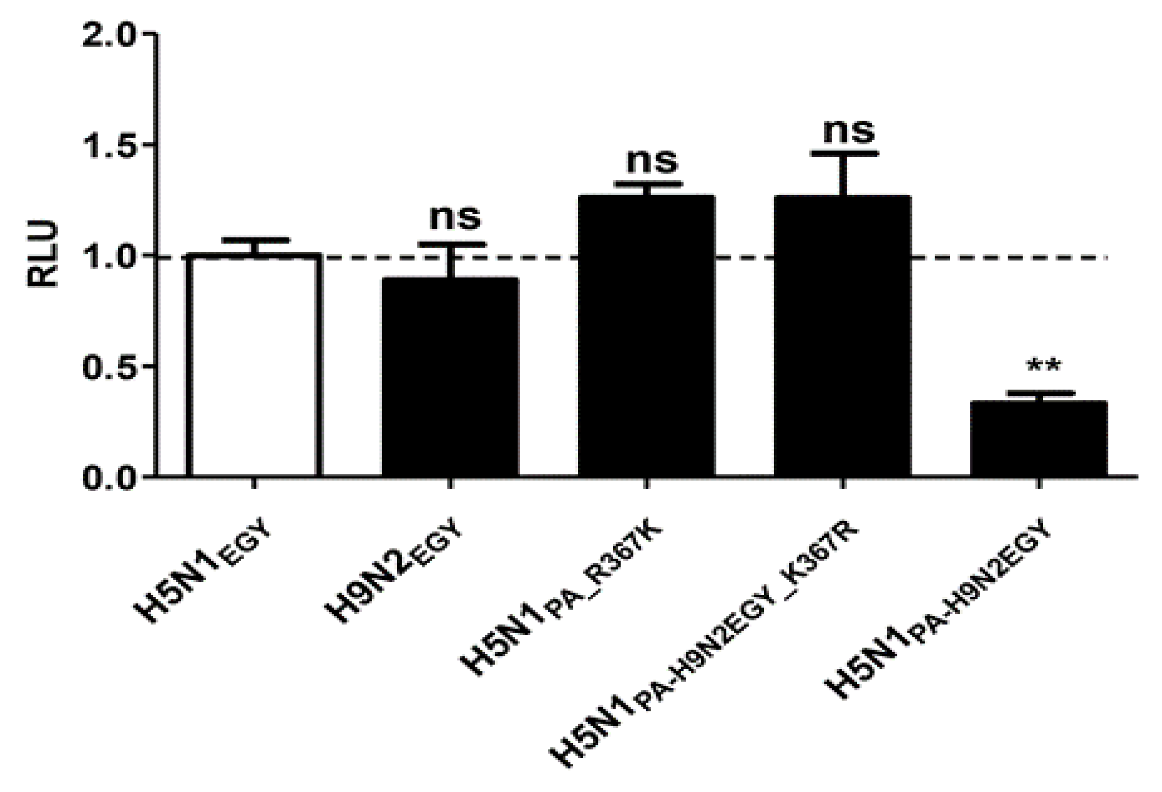
The Luciferase reporter gene assay was performed as previously described [13] with minor modifications. Briefly, 293T (2 × 10⁶ cells/well) were seeded in 6-well plates and co-transfected with 1 µg of each pMPccdB expression plasmid encoding the individual viral ribonucleoprotein complex (vRNP; PB2, PA, PB1 and NP) components of H5N1EGY, or H9N2EGY, or H7N9Anhui, or vRNP components of H5N1EGY expressing individually the mutated PA_R367K of H5N1EGY or PA_K367R of H9N2EGY. The four expression plasmids were mixed with 40 ng pRL-SV40 (Renilla luciferase expression plasmid for normalization), and 200 ng of a firefly luciferase reporter plasmid pHW72-Luc expressing negative-sense firefly luciferase flanked by noncoding sequences of NS segment under the control of a species-specific polymerase I promoter. Transfection was performed using Trans-IT2020 as previously described [14] for 8 h. After 24 h of transfection, the cells were harvested, washed one time with 1x PBS, and lysed with 200 μL of “1x passive lysis buffer” (Promega, Madison, WI, USA). The Firefly/Renilla luciferases were quantified using a Dual-Luciferase Reporter Assay System (Promega) and measured using a Spark 10M multimode microplate reader (TECAN). Relative luminometer units (RLU), normalized to Renilla luciferase, refer to fold induction of polymerase activity.

2.8. Pathogenicity of Mutated Strains versus Wild-Type H5N1EGY in BALB/c Mice

To investigate the role of the 367K in the pathogenesis of H5N1EGY infection in vivo, female BALB/c mice (6–8 week-old) were divided into six groups (10 mice/group). Five groups of mice were anesthetized with isoflurane and intranasally inoculated with a challenge dose (103 FFU in 30 µL PBS) of each mutant versus the corresponding reassortant and wild-type H5N1EGY and H9N2EGY viruses. An uninfected control group was anesthetized and intranasally inoculated with 30 µL of 1X PBS. Infected and control mice were then monitored for 14 dpi for body weight loss and survival rate. Mortality was recorded either as an actual death or loss of ≥25% of initial body weight, which is the threshold at which animals were humanely euthanized.

2.9. Ethical Statement and Biosafety

All animal experiments and procedures were conducted in accordance with the guidelines and regulations of approved by the Medical Research Ethics Committee (MREC) of the National Research Centre (NRC), Egypt (permission code: 16–247; permission date: 1 August, 2016). All experiments using infectious virus were performed in accordance with the Egyptian and German biosafety regulations pertaining to the propagation of influenza viruses. All experiments involving IAVs were performed using Biosafety Level 3 containment laboratories approved for such use by the local authorities (RP, Giessen, Germany).

2.10. Statistical Analysis

Statistical analysis and graphical data presentations were performed using GraphPad Prism 5.0 software (GraphPad Software, San Diego, CA, USA). Significances were determined either with repeated-measures ANOVA with Bonferroni’s post-hoc test, one-way analysis of variance ANOVA followed by Dunnett’s multiple comparison post hoc Test, one-way ANOVA followed by Tukey’s post-hoc test or a two-tailed unpaired t-test with Welch’s correction as indicated in each relevant figure legend. All data are presented as mean ± SD.

3. Results

3.1. Gene segments of H9N2EGY Show High Genetic Compatibility in the Genetic Background of H5N1EGY

HPAIV H5N1EGY and LPAIV H9N2EGY were isolated from infected chicken in Egypt in 2016. Both strains have no history of previous adaptation to mammalian cell culture systems; therefore, they were selected to study the effect of H9N2EGY gene segments in genetic background of H5N1EGY on the replication ability, polymerase activity in vitro and virulence in mammalian systems in vivo.
To investigate these aspects we successfully established complete reverse genetic (Rg) systems for both wild-type strains and specific H5N1EGY reassortants, expressing the internal proteins-encoding segments of H9N2EGY individually or in combination (Figure 1), were generated. Additionally, a recombinant H5N1 reassortant expressing mammalian-adapted internal protein-encoding segments of the human influenza virus isolate A/Anhui/1/2013 (H7N9Anhui) and the HA and NA of H5N1EGY was generated as a control. This control represents a reassortant H5N1 virus carrying internal protein-encoding segments derived from a BJ16-like H9N2-subtype AIV [22] as most of the human H9N2 isolates were genetically classified either as G1- or as BJ16-like H9N2 [6].
Interestingly, all reassortants were rescued indicating that all genetic combinations were compatible in the genetic background of H5N1EGY indicating the high genetic compatibility between H9N2EGY and H5N1EGY.

3.2. Improved Replication Efficiency of H5N1EGY Reassortants Expressing PB2 and PA of H9N2EGY in MDCK-II Cells

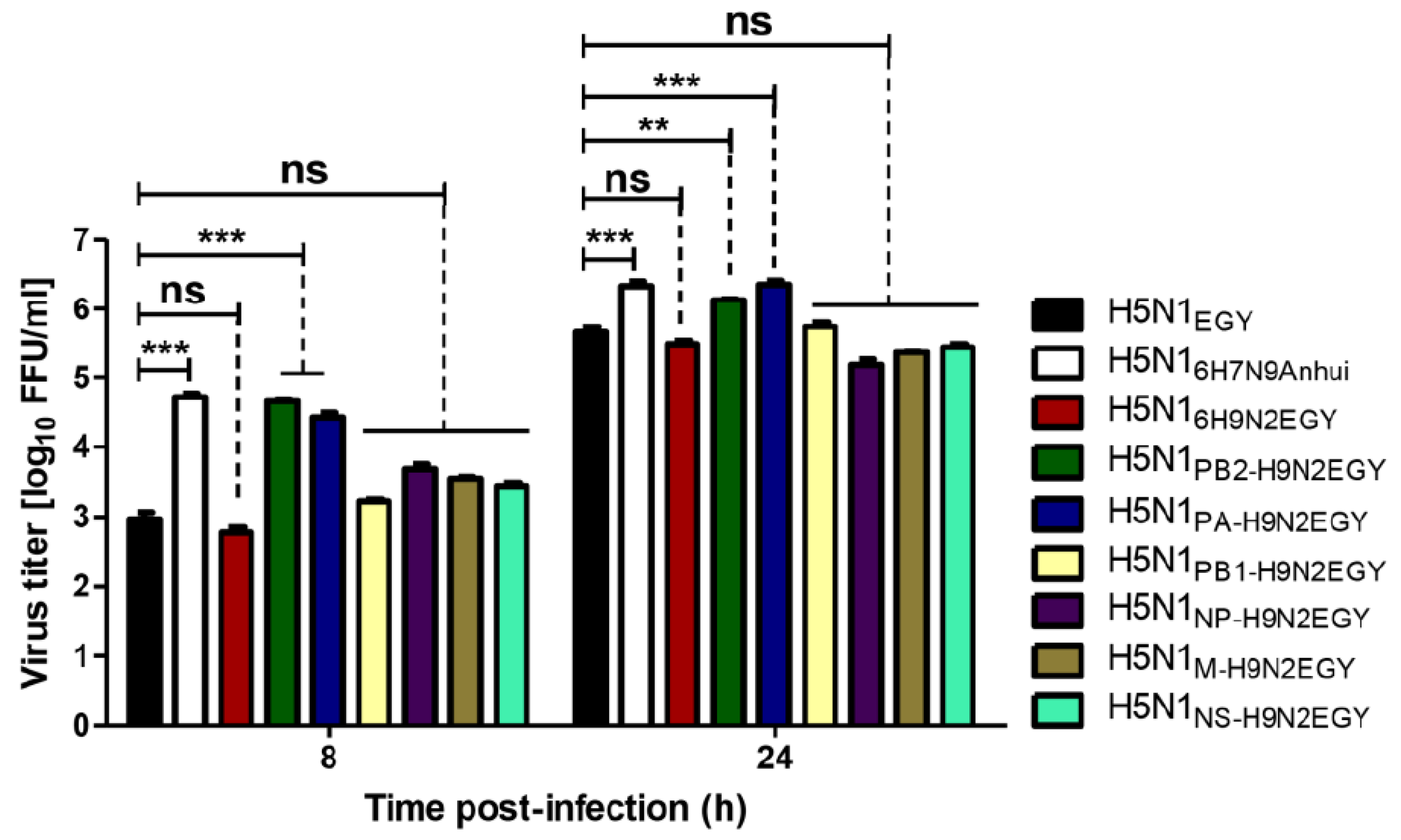
The replication efficiencies of H5N1EGY reassortants were characterized by infection of mammalian MCDK-II cells at a low multiplicity of infection (MOI) = 0.01 at 37 °C for 8 h (single replication cycle) and 24 h (multi-replication cycles). Out of the 7 reassortants, only two H5N1 reassortants, which express the PB2 and PA polymerase subunits of H9N2EGY (H5N1PB2-H9N2EGY and H5N1PA-H9N2EGY) replicated significantly higher than the parent H5N1EGY strain (Figure 2). The replication efficiencies of the other H5N1EGY reassortants carrying gene segments of H9N2EGY were comparable or lower than those of the H5N1EGY and the control H5N16H7N9Anhui.

3.3. Impact of the Polymerase Subunits of H9N2EGY on the Polymerase Activity of Reassortant H5N1EGY

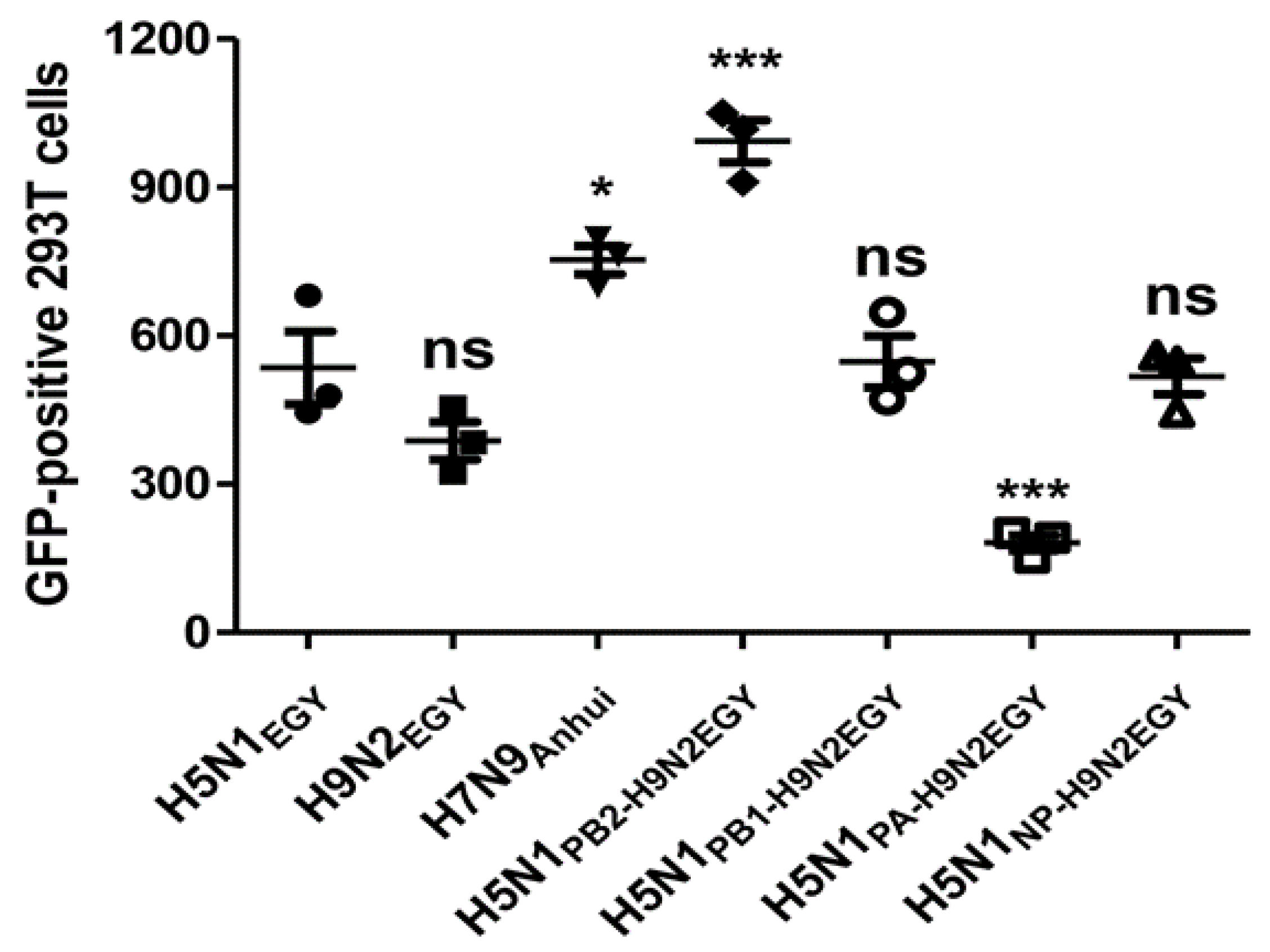
To investigate whether the enhanced in vitro replication efficiency for H5N1PB2-H9N2EGY and H5N1PA-H9N2EGY was accompanied with an improved polymerase activity of the reassortants, a mini-genome assay using combinations of PB2, PB1, PA, and NP of the parent strains, H5N1EGY and H9N2EGY, together with a vRNA-like transcript expressing the GFP-reporter gene followed by flow cytometry analysis was applied. Interestingly, the polymerase comprising PB1, PA and NP of H5N1EGY and the PB2 of H9N2EGY showed a significant activity increase compared to the activities of either the parental vRNP-H5N1EGY or vRNP-H9N2EGY polymerases (Figure 3). Yet, the polymerase activity constituted by the PB2, PB1 and NP of H5N1EGY together with the PA of H9N2EGY, was significantly lower than the polymerase activities of the parental polymerases of either H5N1EGY or vRNP-H9N2EGY. This indicates that the PA protein of H9N2EGY improves H5N1EGY fitness by other mechanisms, rather than improving the polymerase activity of the H5N1PA-H9N2EGY reassortant strain.

3.4. Replication Efficiency of Reassortant and Wild-Type H5N1EGY in Human Lung Cells Increases at Elevated Temperature Resembling Lung Ambient Temperature in Humans

In an attempt to better estimate the outcome of human infections with reassortants between H5N1EGY and H9N2EGY, we set out to examine the influence of site-specific mammalian body temperatures on viral replication efficiency. Therefore, we investigated whether different temperatures resembling either the initial site of IAV replication in the upper respiratory tract (33 °C) or the later replication site of the lower respiratory tract (39 °C) in case of viral pneumonia [23], would affect the replication efficiency of the H5N1PB2-H9N2EGY and H5N1PA-H9N2EGY versus wild-type H5N1EGY and the control H5N16H7N9Anhui. To this point, mammalian cell lines Calu-3 as an in vitro model for bronchial epithelial cells of the upper respiratory tract (URT) [24] and A549 as a type-II alveolar epithelial cell model of the lower respiratory tract (LRT) [25] were infected in triplicates with the indicated viruses at low MOI of 0.001.
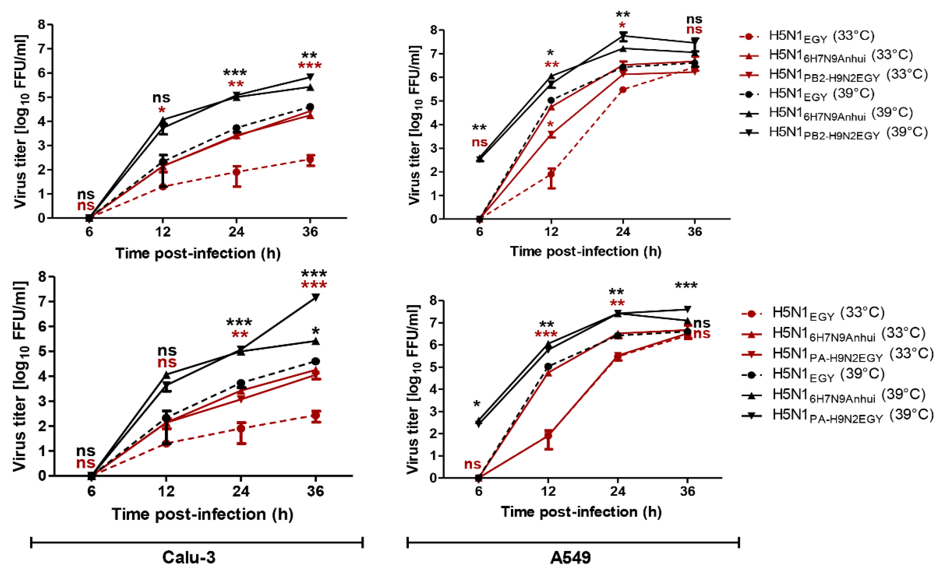
The obtained results (Figure 4) demonstrated that in Calu-3 cells both reassortants H5N1PB2-H9N2EGY and H5N1PA-H9N2EGY replicate to higher levels than the parental H5N1EGY, but comparable to the control H5N16H7N9Anhui at both temperature settings. In A549 cells, the H5N1PB2-H9N2EGY replicated significantly higher than the wild-type H5N1EGY, but comparable to control H5N16H7N9Anhui at 33 °C and 39 °C. However, the replication of H5N1PA-H9N2EGY in A549 cells was only higher than the wild-type H5N1EGY at 39 °C. These data demonstrate the ability of both reassortant viruses to replicate efficiently at different temperature conditions, mimicking the temperatures of the different lung compartments during AIV invasion at the initial site of infection and subsequently during alveolar infection.

3.5. Reassortants H5N1PB2-H9N2EGY and H5N1PA-H9N2EGY Showed Similar to Lower Virulence When Compared to the Wild-Type H5N1EGY in Mice

To analyze the characteristics of H5N1PB2-H9N2EGY and H5N1PA-H9N2EGY in vivo we inoculated female BALB/c mice with the reassortant viruses and the wild-type H5N1EGY. After inoculation (105 PFU/100 µL) with H5N1PB2-H9N2EGY and H5N1EGY all mice showed comparable reduction in body weight at 1 dpi compared to the relative body weight immediately before infection and compared to the PBS control group (Figure 5a). Unlike H5N1EGY-infected mice, which started to regain body weight at 11 dpi (2 mice only), the body weight of H5N1PB2-H9N2EGY inoculated mice started to increase faster at 5 dpi to the end of the experiment. However, the H5N1PA-H9N2EGY inoculated mice exhibited transient reduction in body weight at 5 dpi and the body weight started to increase at 9 dpi to the end of the experiment, although at significantly lower levels than the control group (Figure 5a). Interestingly, the survival rates between the reassortants and the wild-type differed strongly (Figure 5b). Here, 60% of the mice survived the infection with the reassortants, while only 20% of the mice survived the infection with the wild-type virus.
These data suggest that the reduced pathogenicity of the reassortant H5N1PB2-H9N2EGY and H5N1PA-H9N2EGY viruses may be attributed to specific genetic difference between the PA and PB2 segments of H9N2EGY and H5N1EGY.

3.6. The Genetic Analysis of the Polymerase-Encoding Segments from H9N2EGY Revealed that the PAH9N2EGY and PB2H9N2EGY Encode for Distinct Mammalian-Like Variations

To investigate the molecular basis behind the enhanced replication efficiency of H5N1PB2-H9N2EGY and H5N1PA-H9N2EGY in vitro, we compared the genetic differences between the PB2 and PA segments from H5N1EGY with their corresponding segments in H9N2EGY. Genetic analysis revealed that the PB2 and PA of H5N1EGY and H9N2EGY, namely PB2H9N2EGY and PAH9N2EGY, shared 86% and 89.9% nucleotides and 96.4% and 95.4% amino acids (aa) identity, respectively. The PB2 and PA of H5N1EGY and H9N2EGY differ in 26 and 33 aa, respectively (Table 2).
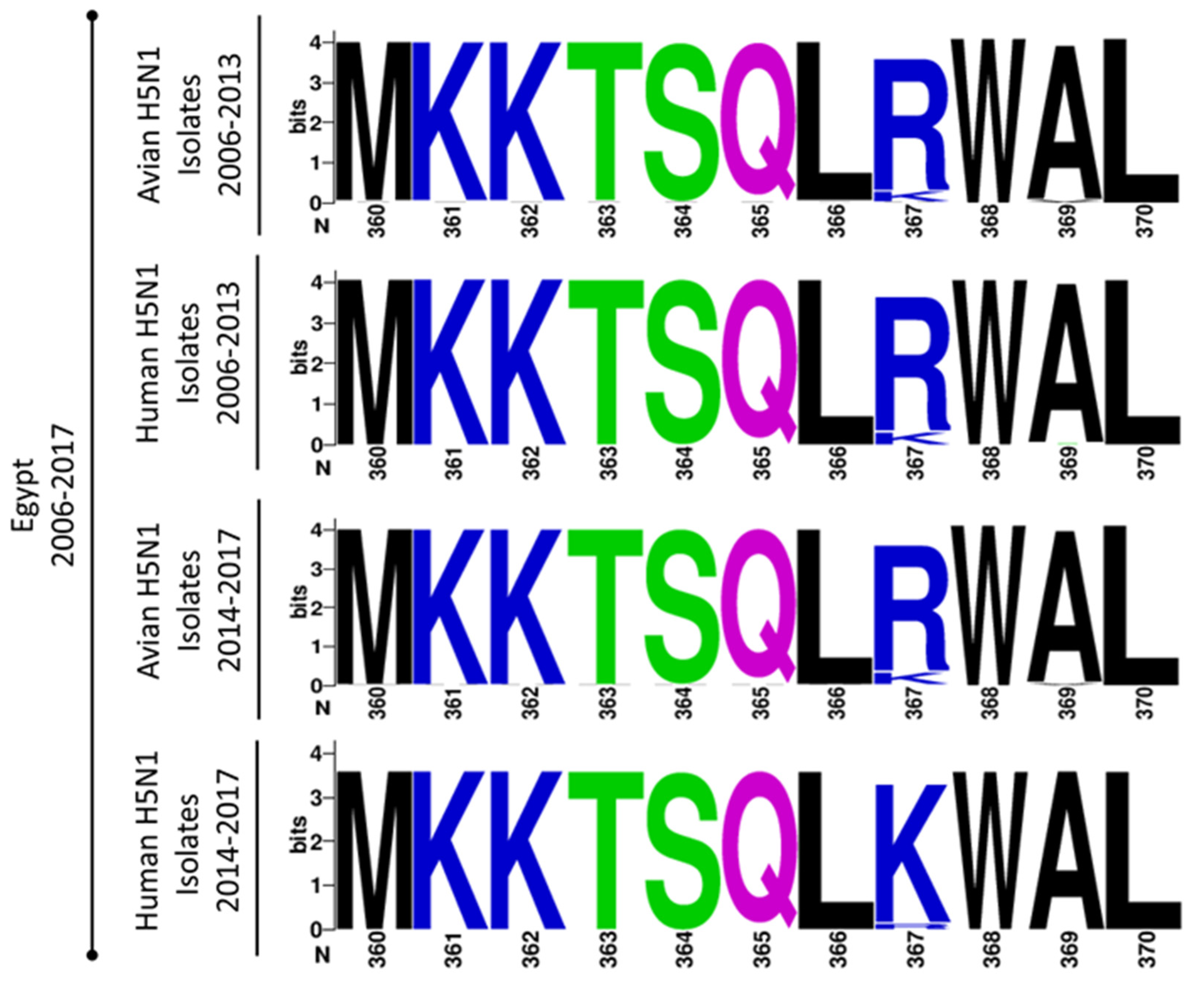
Except for the well-known mammalian signature 591K [26,27,28], and the yet un-described 367K (Figure 6) in the PB2H9N2EGY and PAH9N2EGY, respectively, all other variations in the PB2H9N2EGY and PAH9N2EGY were predominant in the corresponding polymerase protein-encoding segments of circulating H9N2 and H5N1 AIV isolates, but were less frequently observed in human isolates of the same subtypes. Unlike Q591K [28], the impact of the R367K substitution in the PA on the replication efficiency, polymerase activity and virulence of H5N1 strains has not been characterized yet.

3.7. The H5N1EGY Mutant Expressing PAR367K Replicates Efficiently in Primary Human Bronchial Epithelial Cells and Continuous Human Cell Culture Models in a Temperature-Dependent Manner

To better understand the impact of lysine at position 367 in the PA of H9N2EGY, primary human bronchial epithelial (NHBE) cells were infected (MOI = 1) with wild-type H5N1EGY, H5N1PA_R367K and H5N1PA-H9N2EGY_K367R mutants and reassortant H5N1PA-H9N2EGY. At 37 °C, the reassortant H5N1PA-H9N2EGY and the mutant H5N1PA_R367K showed significant higher virus replication than the wild-type virus at 12–48 h p.t. (Figure 7a). The reassortant H5N1PA-H9N2EGY_K367R demonstrated lower replication efficiency than the H5N1PA-H9N2EGY and H5N1PA_R367K viruses, but still higher than the H5N1EGY (Figure 7a).
Based on the fact that the initial infection and onward transmission of an influenza virus depends on efficient virus replication in the human respiratory tract, we tested the replication of the mutant H5N1PA_R367K versus the wild-type H5N1EGY in A549 (Figure 7b) and Calu-3 (Figure 7c) at two different temperature settings, 33 °C and 39 °C, representing the initial site of infection and the subsequent alveolar infection, respectively. Interestingly, H5N1PA_R367K showed significant higher replication than the wild-type H5N1EGY in A549 cells at 12–24 h p.i. at 39 °C and 24–36 h p.i. at 33 °C (Figure 7b). In Calu-3 cells, comparable replication kinetics were observed for H5N1PA_R367K and H5N1EGY at 39 °C. However, H5N1PA_R367K showed significant higher replication than the wild-type at 36 h p.i. at 33 °C (Figure 7c). This indicates a possible improvement of the viral fitness of H5N1PA_R367K at the initial site of infection.

3.8. Replication Efficiency of H5N1PA_R367K Is Not Associated with an Enhanced Polymerase Activity

To investigate whether the improved replication efficiency of H5N1PA_R367K in mammalian cell culture models is linked to an improved polymerase activity, we further examined and compared the effect of the PA mutations in a mini-genome reporter assay using plasmids expressing the three subunits (PB2, PB1, PA) of the viral RNA-dependent RNA polymerase (RdRp) and the viral nucleoprotein (NP) of H5N1EGY or H9N2EGY (controls) or combinations of H5N1EGY PB2, PB1 and NP with mutated PA of H5N1EGY (PA_R367K) or of H9N2EGY (PA-H9N2EGY_K367R), as well as wild-type PA of H9N2EGY (PA-H9N2EGY). Along with the three RdRp subunits and NP expressing plasmids, a Renilla luciferase expression plasmid (transfection control) and a vector expressing a vRNA-like Pol-1 transcript encoding the firefly luciferase was co-transfected (Figure 8). The results show that both the H5N1EGY PA_R367K and the H5N1EGY PA-H9N2EGY_K367R slightly improved the polymerase activity of the Renilla luciferase expression by the H5N1EGY vRNP with PA alterations, but not significantly when compared to the wild type H5N1EGY vRNP. The fact that the H9N2EGY PA-H9N2EGY reduced the H5N1EGY vRNP Renilla luciferase expression activity correlates well with the result of the mini-genome analysis with the GFP-expressing reporter gene (Figure 3). In the light of these data, we conclude that the improved replication efficiency of H5N1PA_R367K is not directly related to an enhanced polymerase activity.

3.9. The PA_R367K Contributes to the Pathogenicity of H5N1EGY in Mice

To examine the effect of R367K mutation in the H9N2EGY to the pathogenicity in mice, we intranasally infected 6 to 8-week-old healthy female BALB/c mice (10 mice in each group) with a lower infectious dose (103 PFU) in 30 µL PBS of the recombinant wild-type strains (H5N1EGY and H9N2EGY), the reassortant (H5N1PA-H9N2EGY), and the H5N1 expressing PAH5N1EGY_R367K (H5N1PA_R367K) or the mutated PAH9N2EGY_K367R (H5N1PA-H9N2EGY_K367R). Mice inoculated with these viruses were monitored for 14 days for weight loss and mortality (Figure 9a). The mice infected with H9N2EGY, H5N1PA-H9N2EGY and H5N1PA-H9N2EGY_K367R virus showed less than 5% of weight loss in the next 14 days p.i. The mice demonstrated around 10–20% of weight loss after infection with the wild-type H5N1EGY and the corresponding H5N1PA_R367K mutant. Even though the pattern of body weight reduction was comparable between the H5N1EGY and H5N1PA_R367K, the H5N1EGY-infected mice started to gain weight at day 9 p.i.
In contrast the survival rates of the mice infected with the different viruses varied strongly (Figure 9b). Here it showed that while all mice infected with H9N2EGY and H5N1PA-H9N2EGY_K367R survived, only 80% of H5N1PA-H9N2EGY-infected mice and 60% of H5N1EGY-infected mice survived. Most strikingly, all mice infected with H5N1PA_R367K died without exceeding the 25% body weight reduction limit. These data suggest that the R367K substitution alone in the PA of H5N1EGY could contribute to the enhanced mortality of H5N1PA_R367K in mice.

4. Discussion

The genome of influenza viruses (IVs) shows a high genetic plasticity based on nucleotide exchange due to the error prone polymerase, as well as gene segment reassortment between different viruses upon co-infections with different virus strains [1]. This allows these viruses to escape selective pressures, such as antibodies, and to quickly adapt to new host species. For AIV these genome modifications can lead to a dynamic interspecies transmission resulting in the continuous emergence of unexpected AIV variants with a varying potential for a bird-to-human transmission [30]. Historically, influenza A virus (IAV) reassortants represent potential threats to the public health in the form of global pandemic influenza outbreaks [1,31]. Along this line, IAV segments encoding the viral internal proteins of H9N2-subtype LPAIV were recently found to augment the zoonotic potential of different AIV subtypes [1,10]. By successful employment of reverse genetics systems, we were able to demonstrate in this study that the gene segments derived from a contemporary G1-like H9N2-subtype AIV, isolated from chicken in Egypt in 2016, show a high genetic compatibility in the genetic background of a H5N1-subtype HPAIV, also isolated from chicken in Egypt in 2016. All seven possible H5N1 reassortants carrying a single H9N2-subtype internal gene segment individually (H5N1PB2-H9N2EGY, H5N1PB1-H9N2EGY, H5N1PA-H9N2EGY, H5N1NP-H9N2EGY, H5N1M-H9N2EGY and H5N1NS-H9N2EGY) or in combination (H5N16H9N2EGY) were compatible to the remaining H5N1EGY segments and could be rescued. These results are in agreement with a recent study showing remarkably high genetic compatibility between a HPAIV H5N1-subtype (clade 2.2.1.2) and a LPAIV H9N2-subtype (G1-like) strain, both isolated in 2013 [32]. Nevertheless, another study, analyzing natural reassortment between A/chicken/Egypt/AR236/2015 (H5N1) and A/chicken/Egypt/AR755/2013 (H9N2) in co-infected embryonated chicken eggs reported the recovery of only five H5N1-subtype reassortants in a total of 100 plaque-picked viruses [33]. Yet, the authors did not exclude the possibility that more reassortants with different genetic constellations might have been present in the allantoic fluid of the mixing vessel [33]. Importantly, our study provides an additional evidence that, similar to H7N9-, H10N8- and H5N6-subtype AIVs [1,10], the H9N2-subtype viruses are able, to donate their internal-proteins encoding segments via a unidirectional genetic transfer to H5N1-subtype strains [33]. In complement to this study, we previously tested the genetic compatibility of viral segments derived from HPAI A/duck/Egypt/Q4596D/2012 (H5N1) (H5N1/Q4596D) in the genetic backbone of LPAI A/chicken/Egypt/S4456B/2011 (H9N2) (H9N2/S4456B) using Rg approach of influenza viruses [34]. Except for NP segment from H5N1/Q4596D, all internal protein-encoding segments were compatible with the complementary seven segments from H9N2/S4456B and viruses could be rescued [34]. Although the compatibility results is very likely a strain specific characteristic and cannot be expanded to the subtype as a general rule, the data provided in our work further indicate a high genetic compatibility between both investigated strains and support the assumption that H5N1/H9N2 reassortants have no genetic restrictions to emerge naturally in the populations.
In MDCK–II cells, only H5N1PB2-H9N2EGY and H5N1PA-H9N2EGY replicated significantly higher than the parental H5N1EGY, but comparable to the replication efficiency of H5N16H7N9Anhui. The reassortant H5N16H7N9Anhui virus comprises the internal protein-encoding segments of the human AIV-isolate A/Anhui/1/2013(H7N9) strain (H7N9Anhui). Genetic analysis of the human 2013 H7N9 strains in China indicated that the internal-protein encoding segments were likely derived from a BJ16-like H9N2-subtype AIV [22]. Notably, most of the human H9N2 isolates were genetically classified as G1- or BJ16-like H9N2 [6]. This might explain the ability of the PB2 or PA segment from G1-like H9N2EGY to improve the replication fitness of H5N1EGY to similar levels as the H5N1EGY expressing the internal-protein encoding segments of the human adapted BJ16-like H9N2 of H7N9Anhui.
Our data are partially consistent with those of Arai et al. showing that the replication of a reassortant H5N1-subtype HPAIV containing the NS and/or the PB2 or PA gene segment of a H9N2-subtype LPAIV tended to be higher in mammalian cells at 37 °C and 33 °C than that of the wild-type H5N1 strain [32]. Contrary to Arai et al., we showed that the H5N1EGY reassortant expressing the NS proteins of H9N2EGY propagated to comparable levels like the wild-type H5N1EGY at least at 37 °C. By comparing the NS1 and NS2 of 2016 H9N2EGY and A/chicken/Egypt/CL42/2013 (CL42) (H9N2) used by Arai et al., we found that they share 91.7% and 95% aa similarly, respectively. The NS1 proteins of our H9N2EGY and CL42 strains belong to allele “A” and share the same pattern of mammalian-like markers including P42S and mammalian like PDZ motifs “ESKV for H5N1EGY and KSEV for CL42”. Similar to our finding, Arai and his colleagues showed that the 6 internal-protein encoding segments or the NS segment alone of the H9N2-subtype LPAIV could also enhance the replication of the corresponding H5N1 reassortants to higher levels than the wild-type H5N1 strain, indicating that the in vitro replication was related to the internal gene cassette rather than the surface proteins-encoding segments [32]. Contrary to our data and the data shown by Arai et al., the natural reassortments studied by Naguib et al. have shown that the replication efficiency of natural H5N1 reassortants expressing the PB2 or the six internal-protein encoding segments of H9N2 (clone C46) were either retarded or unaltered, compared to the wild-type H5N1 strain [33]. This might indicate that only specific aa variations in the studied H5N1 and H9N2 strains in each study, collected on 2013, 2015 and 2016, might affect the impact of the reassortment on the replication efficiency of the corresponding H5N1-subtype reassortants.
To study the mechanism(s) underlying the replication differences determined for the two polymerase reassortants (PB2 and PA), the expression activity of different combinations of polymerase subunits and nucleoprotein genes (PB2, PB1, PA, and NP) from H9N2EGY in the genetic background of H5N1EGY was determined in vitro by minigenome assays in human 293T cells. Despite the fact that both the PB2 and the PA of H9N2EGY enhance the replication of H5N1EGY virus, only the PB2 polymerase subunit of H9N2EGY combined with the other H5N1EGY components could augment the overall in vitro polymerase activity to levels higher than those activities of the wild-type H5N1EGY and H7N9Anhui. In the light of these results, the enhanced propagation efficiency of the H5N1PA-H9N2EGY virus must be due to other mechanisms, e.g., improved functional protein interaction of the PAH9N2EGY with cellular- and/or other viral proteins rather than improving the overall polymerase activity. Yet, another study showed that both PB2 and PA of a H9N2 strain (isolated from chicken in Egypt in 2013) could slightly improve the polymerase activity of another H5N1 virus (isolated from chicken in Egypt in 2013), even though not significantly [32]. As this demonstrates that the potential for H9N2 AIV genes to enhance replication of H5N1 AIV depends on specific, not-well understood mechanisms and factors, we aimed to provide a more detailed insight.
The initial site of IAV replication in the upper respiratory tract (33 °C). Normally, the internal body temperature ranges from 37 °C to 38 °C. Mild fever is occurring when body temperature ranges from 38.1–39 °C. Moderate and high grade fever is 39.1–40 °C and high-grade fever is 40.1–41.1 °C, respectively [35,36]. Drastic and irreparable changes in organ structure and function can occur when body temperature rises above 41.1 °C [36]. The temperature 39 °C was chosen to assess the viral replication of the investigated viruses as an intermediate temperature representing low-to-moderate fever during viral pneumonia. The replication efficiencies of H5N1PB2-H9N2EGY and H5N1PA-H9N2EGY reassortants in human cells was higher than that of the parental H5N1EGY virus at 33 °C comprising the temperature of the upper respiratory tract at the initial site of infection, and at 39 °C representing the average temperature of lower respiratory tract during infection (Figure 4). This is in agreement with the work of Arai et al. [32] and strongly supports the notion that genetic recombination between H5N1- and H9N2-subtype viruses (in birds) may well yield hybrid viruses with an improved replication competence in mammalian species.
To ensure the infection of the URT and LRT of mice with the wild-type and reassortant strains, a high infectious dose “105 FFU” of each virus in a relatively large volume “100 µL” was applied. We demonstrated that both reassortants H5N1PB2-H9N2EGY and H5N1PA-H9N2EGY did not potentiate the virulence of the wild-type H5N1EGY. Our data are consistent with previous studies showing that H5N1 reassortants expressing the polymerase subunits of H9N2 were less virulent in mice [32] and ferret models [33] than the respective wild-type H5N1EGY.
Zoonotic potential is an important characteristic that needs to be assessed for emerging animal IAV, and the reliable prediction of zoonotic potential based on nucleotide sequence data would significantly help to select for specific animal infection studies. To this point, we assessed the genetic difference between PB2H5N1EGY and PB2H9N2EGY and between PA H5N1EGY and PAH9N1EGY. Two interesting variations were deduced from the sequence analyses; 591K in PB2H9N2EGY and 367K in PAH9N1EGY (Table 3).
The impact of these aa in H9N2, H5N1 and H7N9 strains in comparison to the fundamental 627K adaptation marker in PB2 polymerase subunit has been previously studied [28]. For this reason, we focused on the characterization of the impact of 367K in PAH9N1EGY on viral replication, polymerase activity and pathogenicity of H5N1EGY. Notably, we found a remarkable prevalence of the lysine “K” instead of argenine “R” at aa residue 367 in human isolates of H5N1 AIV since 2014 [37]. The emergence of Q367K substitution in the field was coincident with a substantial increase in the number of human cases of H5N1 infection during the 2014–2015 flu season in Egypt [37], which may reflect its possible contribution to the overall high morbidity of avian influenza H5N1 viruses among Egyptian human population in 2014–2015. We demonstrated here for the first time that the PA_367K could improve the replication kinetics of H5N1EGY in different animal cell culture models in a temperature dependent manner, especially at human URT temperature of 33 °C, regardless to the overall activity of the viral polymerase. To study the impact of the PA_367K mutation in vivo, we intentionally minimized the infectious dose of the studied viruses and the applied volume (30 µL) to ensure that the viruses were restricted to the URT of the infected mice. Interestingly PA_367K demonstrated the ability to potentiate the mortality caused by H5N1EGY as demonstrated by survival rates of mice infected with mutant and wild-type viruses.
Taken together, our results demonstrate that the co-circulation of H5N1-subtype HPAIV and H9N2-subtype LPAIV in Egyptian poultry may result in co-infection and viral reassortment, thus H5N1 reassortants with an increased zoonotic potential may emerge and pose a public health risk. Specifically the lysine at position 367 of the PA seems to have a strong impact. The data presented here question the role of genetic compatibility or viral replication fitness of H5N1/H9N2 reassortants to the low or rare detection of natural genetic reassortment events among HPAI H5N1 and LPAI H9N2 viruses in Egypt. Additionally, the ability of H9N2 strains to potentiate other AIVs may be attributed to the fact that H9N2 viruses accommodate for several human adaptation markers in their genome, which may enable the reassortant AIVs to cross the species barrier.

Author Contributions

Conceptualization, A.M., M.A.A. and S.P.; methodology, A.M., S.H.M., C.M., H.Z.N., R.E.-S., and M.S.; formal analysis, A.M., S.H.M., M.S., C.M., H.Z.N., R.E.-S., M.A.A. and S.P.; investigation, A.M., R.E.-S., M.S., A.K., G.K., M.A.A. and S.P.; data curation, A.M., G.K. and S.P.; writing—original draft preparation, A.M.; S.P.; writing—review and editing, A.M., G.K., M.A.A. and S.P. All authors have read and agree to the published version of the manuscript.

Funding

This research was partially funded by the Science and Technology Development Fund (STDF) in Egypt, under contract number (5175 to MAA) and the National Institute of Allergy and Infectious Diseases, National Institutes of Health, US Department of Health and Human Services (under contract HHSN272201400006C). This work was also supported by Egyptian National Research Centre (NRC)-funded projects (TT110801 and 12010126 to AM). The work of SP was supported in parts by the DFG-funded Collaborative Research Centre 1021 (RNA viruses: RNA metabolism, host response and pathogenesis), the German Centre for Infection Research, partner site Giessen (DZIF, TTU 01.806). S.P. is a member of the German FluResearchNet, a nationwide research network on zoonotic influenza. The funders had no role in study design, data collection and analysis, decision to publish, or preparation of the manuscript.

Acknowledgments

We thank Erich Hoffmann (Memphis, TN) for the pHW72-luciferase plasmid and Rebecca Badra (Human Link, Lebanon) for her assistance in reviewing the final draft of the manuscript.

Conflicts of Interest

The authors declare no conflict of interest.

References

  1. Mostafa, A.; Abdelwhab, E.M.; Mettenleiter, T.C.; Pleschka, S. Zoonotic potential of influenza a viruses: A comprehensive overview. Viruses 2018, 10, 497. [Google Scholar] [CrossRef] [PubMed] [Green Version]
  2. Saad, M.D.; Ahmed, L.S.; Gamal-Eldein, M.A.; Fouda, M.K.; Khalil, F.; Yingst, S.L.; Parker, M.A.; Montevillel, M.R. Possible avian influenza (h5n1) from migratory bird, egypt. Emerg. Infect. Dis. 2007, 13, 1120–1121. [Google Scholar] [CrossRef] [PubMed]
  3. Bahgat, M.M.; Kutkat, M.A.; Nasraa, M.H.; Mostafa, A.; Webby, R.; Bahgat, I.M.; Ali, M.A. Characterization of an avian influenza virus h5n1 egyptian isolate. J. Virol. Methods 2009, 159, 244–250. [Google Scholar] [CrossRef] [PubMed]
  4. Naguib, M.M.; Verhagen, J.H.; Samy, A.; Eriksson, P.; Fife, M.; Lundkvist, Å.; Ellström, P.; Järhult, J.D. Avian influenza viruses at the wild–domestic bird interface in egypt. Infect. Ecol. Epidemiol. 2019, 9, 1575687. [Google Scholar] [CrossRef] [PubMed] [Green Version]
  5. Abdelwhab, E.M.; Hassan, M.K.; Abdel-Moneim, A.S.; Naguib, M.M.; Mostafa, A.; Hussein, I.T.M.; Arafa, A.; Erfan, A.M.; Kilany, W.H.; Agour, M.G.; et al. Introduction and enzootic of a/h5n1 in egypt: Virus evolution, pathogenicity and vaccine efficacy ten years on. Infect Genet Evol. 2016, 40, 80–90. [Google Scholar] [CrossRef] [PubMed]
  6. Peacock, T.H.P.; James, J.; Sealy, J.E.; Iqbal, M. A global perspective on h9n2 avian influenza virus. Viruses 2019, 11, 620. [Google Scholar] [CrossRef] [Green Version]
  7. Kandeil, A.; El-Shesheny, R.; Maatouq, A.M.; Moatasim, Y.; Shehata, M.M.; Bagato, O.; Rubrum, A.; Shanmuganatham, K.; Webby, R.J.; Ali, M.A.; et al. Genetic and antigenic evolution of h9n2 avian influenza viruses circulating in egypt between 2011 and 2013. Arch. Virol. 2014, 159, 2861–2876. [Google Scholar] [CrossRef]
  8. Arafa, A.-S.; Hagag, N.; Erfan, A.; Mady, W.; El-Husseiny, M.; Adel, A.; Nasef, S. Complete genome characterization of avian influenza virus subtype h9n2 from a commercial quail flock in egypt. Virus Genes 2012, 45, 283–294. [Google Scholar] [CrossRef]
  9. El-Zoghby, E.F.; Arafa, A.S.; Hassan, M.K.; Aly, M.M.; Selim, A.; Kilany, W.H.; Selim, U.; Nasef, S.; Aggor, M.G.; Abdelwhab, E.M.; et al. Isolation of h9n2 avian influenza virus from bobwhite quail (colinus virginianus) in egypt. Arch. Virol. 2012, 157, 1167–1172. [Google Scholar] [CrossRef]
  10. Chang, H.-P.; Peng, L.; Chen, L.; Jiang, L.-F.; Zhang, Z.-J.; Xiong, C.-L.; Zhao, G.-M.; Chen, Y.; Jiang, Q.-W. Avian influenza viruses (aivs) h9n2 are in the course of reassorting into novel aivs. J Zhejiang Univ Sci B 2018, 19, 409–414. [Google Scholar] [CrossRef]
  11. Kayali, G.; Kandeil, A.; El-Shesheny, R.; Kayed, A.S.; Maatouq, A.M.; Cai, Z.; McKenzie, P.P.; Webby, R.J.; El Refaey, S.; Kandeel, A.; et al. Avian influenza a(h5n1) virus in egypt. Emerg. Infect. Dis. 2016, 22, 379–388. [Google Scholar] [CrossRef] [PubMed]
  12. Ma, W.; Brenner, D.; Wang, Z.; Dauber, B.; Ehrhardt, C.; Hogner, K.; Herold, S.; Ludwig, S.; Wolff, T.; Yu, K.; et al. The ns segment of an h5n1 highly pathogenic avian influenza virus (hpaiv) is sufficient to alter replication efficiency, cell tropism, and host range of an h7n1 hpaiv. J. Virol. 2010, 84, 2122–2133. [Google Scholar] [CrossRef] [PubMed] [Green Version]
  13. Petersen, H.; Mostafa, A.; Tantawy, M.A.; Iqbal, A.A.; Hoffmann, D.; Tallam, A.; Selvakumar, B.; Pessler, F.; Beer, M.; Rautenschlein, S.; et al. Ns segment of a 1918 influenza a virus-descendent enhances replication of h1n1pdm09 and virus-induced cellular immune response in mammalian and avian systems. Front. Microbiol. 2018, 9. [Google Scholar] [CrossRef] [PubMed]
  14. Mostafa, A.; Kanrai, P.; Ziebuhr, J.; Pleschka, S. Improved dual promotor-driven reverse genetics system for influenza viruses. J Virol Methods 2013, 193, 603–610. [Google Scholar] [CrossRef] [PubMed]
  15. Mostafa, A.; Kanrai, P.; Petersen, H.; Ibrahim, S.; Rautenschlein, S.; Pleschka, S. Efficient generation of recombinant influenza a viruses employing a new approach to overcome the genetic instability of ha segments. Plos One 2015, 10, e0116917. [Google Scholar] [CrossRef] [Green Version]
  16. Hoffmann, E.; Stech, J.; Guan, Y.; Webster, R.G.; Perez, D.R. Universal primer set for the full-length amplification of all influenza a viruses. Arch. Virol. 2001, 146, 2275–2289. [Google Scholar] [CrossRef]
  17. Neumann, G.; Watanabe, T.; Ito, H.; Watanabe, S.; Goto, H.; Gao, P.; Hughes, M.; Perez, D.R.; Donis, R.; Hoffmann, E.; et al. Generation of influenza a viruses entirely from cloned cdnas. Proc. Natl. Acad. Sci. USA 1999, 96, 9345–9350. [Google Scholar] [CrossRef] [Green Version]
  18. Hoffmann, E.; Neumann, G.; Kawaoka, Y.; Hobom, G.; Webster, R.G. A DNA transfection system for generation of influenza a virus from eight plasmids. Proc. Natl. Acad. Sci. USA 2000, 97, 6108–6113. [Google Scholar] [CrossRef] [Green Version]
  19. Mostafa, A.; Kanrai, P.; Ziebuhr, J.; Pleschka, S. The pb1 segment of an influenza a virus h1n1 2009pdm isolate enhances the replication efficiency of specific influenza vaccine strains in cell culture and embryonated eggs. J. Gen. Virol. 2016, 97, 620–631. [Google Scholar] [CrossRef]
  20. Pleschka, S.; Jaskunas, R.; Engelhardt, O.G.; Zurcher, T.; Palese, P.; Garcia-Sastre, A. A plasmid-based reverse genetics system for influenza a virus. J. Virol. 1996, 70, 4188–4192. [Google Scholar] [CrossRef] [Green Version]
  21. Müller, C.; Obermann, W.; Schulte, F.W.; Lange-Grünweller, K.; Oestereich, L.; Elgner, F.; Glitscher, M.; Hildt, E.; Singh, K.; Wendel, H.-G.; et al. Comparison of broad-spectrum antiviral activities of the synthetic rocaglate cr-31-b (−) and the eif4a-inhibitor silvestrol. Antivir. Res. 2020, 175, 104706. [Google Scholar]
  22. Zhang, Y. Isolation and characterization of h7n9 avian influenza a virus from humans with respiratory diseases in zhejiang, china. Virus Res. 2014. [Google Scholar] [CrossRef] [PubMed]
  23. Shi, Y.; Lu, P.; Yang, Y.; Hu, C.; Jin, H.; Kong, L. Human infected h7n9 avian influenza. Radiol. Influenza 2020, 77–104. [Google Scholar]
  24. Kreft, M.E.; Jerman, U.D.; Lasič, E.; Hevir-Kene, N.; Rižner, T.L.; Peternel, L.; Kristan, K. The characterization of the human cell line calu-3 under different culture conditions and its use as an optimized in vitro model to investigate bronchial epithelial function. Eur. J. Pharm. Sci. 2015, 69, 1–9. [Google Scholar] [CrossRef] [PubMed]
  25. Foster, K.A.; Oster, C.G.; Mayer, M.M.; Avery, M.L.; Audus, K.L. Characterization of the a549 cell line as a type ii pulmonary epithelial cell model for drug metabolism. Exp. Cell Res. 1998, 243, 359–366. [Google Scholar] [CrossRef] [PubMed]
  26. Mok, C.K.; Lee, H.H.; Lestra, M.; Nicholls, J.M.; Chan, M.C.; Sia, S.F.; Zhu, H.; Poon, L.L.; Guan, Y.; Peiris, J.S. Amino acid substitutions in polymerase basic protein 2 gene contribute to the pathogenicity of the novel a/h7n9 influenza virus in mammalian hosts. J. Virol. 2014, 88, 3568–3576. [Google Scholar] [CrossRef] [PubMed] [Green Version]
  27. Yamada, S.; Hatta, M.; Staker, B.L.; Watanabe, S.; Imai, M.; Shinya, K.; Sakai-Tagawa, Y.; Ito, M.; Ozawa, M.; Watanabe, T.; et al. Biological and structural characterization of a host-adapting amino acid in influenza virus. Plos Pathog. 2010, 6, e1001034. [Google Scholar] [CrossRef] [PubMed] [Green Version]
  28. Wang, C.; Lee, H.H.Y.; Yang, Z.F.; Mok, C.K.P.; Zhang, Z. Pb2-q591k mutation determines the pathogenicity of avian h9n2 influenza viruses for mammalian species. Plos One 2016, 11, e0162163. [Google Scholar] [CrossRef]
  29. Schneider, T.D.; Stephens, R.M. Sequence logos: A new way to display consensus sequences. Nucleic Acids Res. 1990, 18, 6097–6100. [Google Scholar] [CrossRef]
  30. Kanrai, P.; Mostafa, A.; Madhugiri, R.; Lechner, M.; Wilk, E.; Schughart, K.; Ylösmäki, L.; Saksela, K.; Ziebuhr, J.; Pleschka, S. Identification of specific residues in avian influenza a virus ns1 that enhance viral replication and pathogenicity in mammalian systems. J. Gen. Virol. 2016, 97, 2135–2148. [Google Scholar] [CrossRef]
  31. Shao, W.; Li, X.; Goraya, M.U.; Wang, S.; Chen, J.L. Evolution of influenza a virus by mutation and re-assortment. Int J Mol Sci 2017, 18, 1650. [Google Scholar] [CrossRef] [PubMed] [Green Version]
  32. Arai, Y. Genetic compatibility of reassortants between avian h5n1 and h9n2 influenza viruses with higher pathogenicity in mammals. J. Virol. 2019. [Google Scholar] [CrossRef] [PubMed] [Green Version]
  33. Naguib, M.M. Natural reassortants of potentially zoonotic avian influenza viruses h5n1 and h9n2 from egypt display distinct pathogenic phenotypes in experimentally infected chickens and ferrets. J. Virol. 2017. [Google Scholar] [CrossRef] [PubMed] [Green Version]
  34. Moatasim, Y.; Kandeil, A.; Mostafa, A.; Elghaffar, S.K.A.; El Shesheny, R.; Elwahy, A.H.M.; Ali, M.A. Single gene reassortment of highly pathogenic avian influenza a h5n1 in the low pathogenic h9n2 backbone and its impact on pathogenicity and infectivity of novel reassortant viruses. Arch. Virol. 2017, 162, 2959–2969. [Google Scholar] [CrossRef]
  35. Ogoina, D. Fever, fever patterns and diseases called ‘fever’ – A review. J. Infect. Public Health 2011, 4, 108–124. [Google Scholar] [CrossRef] [PubMed] [Green Version]
  36. Del Bene, V.E. Temperature. In Clinical Methods: The History, Physical, and Laboratory Examinations; Walker, H.K., Hall, W.D., Hurst, J.W., Eds.; Butterworths; Boston, MA, USA, 1990. [Google Scholar]
  37. El-Shesheny, R.; Mostafa, A.; Kandeil, A.; Mahmoud, S.H.; Bagato, O.; Naguib, A.; Refaey, S.E.; Webby, R.J.; Ali, M.A.; Kayali, G. Biological characterization of highly pathogenic avian influenza h5n1 viruses that infected humans in egypt in 2014-2015. Arch. Virol. 2017, 162, 687–700. [Google Scholar] [CrossRef]
  38. Arai, Y.; Kawashita, N.; Ibrahim, M.S.; Elgendy, E.M.; Daidoji, T.; Ono, T.; Takagi, T.; Nakaya, T.; Matsumoto, K.; Watanabe, Y. Pb2 mutations arising during h9n2 influenza evolution in the middle east confer enhanced replication and growth in mammals. Plos Pathog. 2019, 15, e1007919. [Google Scholar] [CrossRef]
Figure 1. Genetic compatibility between Egyptian H5N1- and H9N2-viruses. The six internal-proteins encoding segments of LPAIV H9N2EGY, were placed individually and in combination, into the genetic background of HPAIV H5N1EGY. The gene segments from H5N1EGY and H9N2EGY are colored in grey and red, respectively. The six internal-proteins encoding segments from H7N9Anhui (control) are colored in green. All genetic constellations used in this study were transfected to co-culture of 293T/MDCK-II cells and the rescued wild-type, reassortant and mutant viruses were propagated on embryonated SPF eggs. All genetic combinations were compatible showing variable hemagglutination unit (HAU) and focus forming unit (FFU) titers.
Viruses 12 01046 g001
Figure 2. Replication efficiency of the H5N1EGY reassortants. Mammalian Madin-Darby canine kidney (MDCK-II) cells were infected (in triplicate) with H5N1EGY reassortants expressing internal proteins-encoding H9N2EGY genes or with the wild-type H5N1EGY or the control H5N16H7N9Anhui virus at MOI of 0.01, cultured at 37 °C for single replication cycle (8 h) and multiple replication cycles (24 h) p.i. Subsequently the virus titers were determined. Error bars reflect standard deviation (SD) of three independent experiments. Statistical analysis was performed using repeated measures ANOVA, followed by Bonferroni post hoc test. The significant differences are indicated (** = p < 0.01, *** = p < 0.001 and non-significant = ns).
Viruses 12 01046 g002
Figure 3. In vitro polymerase activity in human 293T cells. 293T cells were transfected with plasmids expressing the three subunits (PB2, PB1, and PA) of the viral RNA-dependent RNA polymerase (RdRp) and the viral nucleoprotein (NP) of H5N1EGY, H9N2EGY, H7N9Anhui or combinations of H5N1EGY RdRp subunits with single RdRp subunits of H9N2EGY. Along with the three RdRp subunits and NP expressing plasmids, a vector expressing a vRNA-like Pol-1 transcript encoding the reporter GFP gene was co-transfected. At 48 h p.t., the control and the transfected cells were analyzed for percentage of GFP positive cells. The significance was tested using one-way ANOVA, followed by Dunnett’s multiple comparison post hoc test and the significant differences are indicated (* = p < 0.05, *** = p < 0.001 and non-significant = ns).
Viruses 12 01046 g003
Figure 4. Replication kinetics of H5N1EGY reassortants in mammalian cell culture models. Calu-3 and A549 cells were infected (in triplicates) with the H5N1EGY reassortants or wild-type H5N1EGY and control H5N16H7N9Anhui at multiplicities of infection (MOIs) of 0.001 and incubated at 33 °C or 39 °C. At 6, 12, 24, 36 h p.i., the cell culture supernatants were collected and the virus titre was determined. Statistical analysis was performed using two-way ANOVA, followed by Bonferroni post hoc test. The significant differences are indicated (* = p < 0.05, ** = p < 0.01, *** = p < 0.001 and non-significant = ns).
Viruses 12 01046 g004
Figure 5. Pathogenicity of H5N1EGY and reassortants H5N1PB2-H9N2EGY and H5N1PA-H9N2EGY in female BALB/c mice. Mice were infected with 105 PFU of each virus in 100 μL phosphate-buffered saline (PBS). The morbidity rate and the mortality rate as demonstrated by weight loss of body (a) and the survival rate (b), respectively, were monitored for 14 dpi. Mice judged moribund (body weight loss >25%) were euthanized.
Viruses 12 01046 g005
Figure 6. Prevalence of lysine and argenine at amino acid (aa) residue 367 among Egyptian human and avian H5N1 isolates (2006 to 2017). The graphic was created via Web-based WebLogo application (http://weblogo.threeplusone.com/create.cgi) [29]. The aa color is given according to their chemical properties. Polar aa “T and S”: green; neutral aa “Q”: purple; basic aa “K and R”: blue; hydrophobic aa “M, L, W, A and L”: black.
Viruses 12 01046 g006
Figure 7. Impact of PA-367K on the replication efficiency of H5N1EGY in mammalian cell culture models. (a) mammalian differentiated primary bronchial epithelial cells (NHBE) cells were infected (in triplicates) with wild-type H5N1EGY, reassortant H5N1PA-H9N2EGY, as well as mutant H5N1PA_R367K and H5N1PA-H9N2EGY_K367R at MOI of 1, cultured at 37 °C for 6–36 h p.i. (b) A549 and (c) Calu-3 were infected with wild-type H5N1EGY and mutant H5N1PA_R367K (MOI = 0.01) and cultured at 33 °C and 39 °C for 6–36 h p.i. Subsequently the virus titers were determined. Error bars reflect standard deviation (SD) of three independent experiments. Statistical analysis was performed using repeated measures ANOVA, followed by Bonferroni post hoc test. The significant differences are indicated (* = p < 0.05, ** = p < 0.01, *** = p < 0.001 and non-significant = ns).
Viruses 12 01046 g007
Figure 8. PA-367K does not significantly alter the in vitro polymerase activity of H5N1EGY in human 293T cells. The 293T cells were transfected with plasmids expressing the three subunits (PB2, PB1, PA) of the viral RNA-dependent RNA polymerase (RdRp) and the viral nucleoprotein (NP) of H5N1EGY or H9N2EGY (controls) or combinations of H5N1EGY PB2, PB1 and NP with mutated PA of H5N1EGY (PA_R367K) or of H9N2EGY (PA-H9N2EGY_K367R), as well as wild-type PA of H9N2EGY (PA-H9N2EGY). Along with the three RdRp subunits and NP expressing plasmids, a Renilla luciferase expression plasmid (transfection control) and a vector expressing a vRNA-like Pol-1 transcript encoding the firefly luciferase was co-transfected. At 48 h p.t., the control and transfected cells were analyzed for Renilla/luciferase expression levels. The significance was tested using one-way analysis of variance ANOVA, followed by Dunnett’s multiple comparison post hoc test and the significant differences are indicated (** = p < 0.01 and non-significant = ns).
Viruses 12 01046 g008
Figure 9. Pathogenicity of wild-type H5N1EGY, H9N2EGY, reassortants H5N1PB2-H9N2EGY and H5N1PA-H9N2EGY, mutants H5N1PA_R367K and H5N1PA-H9N2EGY_K367R in female BALB/c mice. Mice were infected with 103 PFU of each virus in 30 μL PBS. (a) The body-weight reduction rate and the mortality rate as demonstrated by weight loss (a) and the survival rate (b), respectively, were monitored for 14 dpi. Mice judged moribund (body weight loss > 25%) were euthanized.
Viruses 12 01046 g009
Table 1. Site directed mutagenesis primers.
Mutant VirusMutationPrimer NameMutagenesis Primer Sequence
H5N1PA_R367KR367KPA-367F5’-GAAAAAAACGAGCCAGTTAAAGTGGGCACTCGGTGAGAACATG-3’
PA-367R5’-CATGTTCTCACCGAGTGCCCACTTTAACTGGCTCGTTTTTTTC-3’
H5N1PA-H9N2EGY_K367RK367RPA-K367R-H9EGY-F5’-GAAGAAAACAAGCCAATTAAGATGGGCACTCGGTGAGAATATG-3’
PA-K367R-H9EGY-R5’-CATATTCTCACCGAGTGCCCATCTTAATTGGCTTGTTTTCTTC-3’
Table 2. Amino acid differences between PB2 and PA proteins from H5N1EGY and H9N2EGY.
Amino Acid (aa) ResiduePB2Amino Acid (aa) ResiduePA
H5N1EGYH9N2EGYH5N1EGYH9N2EGY
6EG38IV
64IM58SG
66IM94VI
80RK101ED
106AT129TI
129NT184AV
147TI204KR
197RK212LR
249KE269KR
292MI287SA
315IM321GN
339TK323VA
368QR337TA
369KR342ML
377SA351DE
390ND367RK
393TS382ED
451TV388RS
498HQ391KR
521TA396DG
529VI400TS
570IM437HY
591QK448EA
627KE450AV
649IV539KR
661TA554VI
615RK
626RK
653SP
669VI
706LF
712AI
716NK
Table 3. Predominance of the distinct aa residues [28,37,38] in the studied polymerase acidic (PA) and polymerase basic 2 (PB2) proteins in Egyptian avian versus Egyptian human H5N1 isolates.
Distinct Amino Acid (aa) Residues
PA Segment321342351367705707
PAH9N2EGYNLEKSF
PAH5N1EGYGMDRSL
PAH7N9Anhui (Human isolate)NLEKSF
PAH5N1EGY (Human isolates 2006–2013)SLERSF
PAH5N1EGY (Avian isolates 2006–2013)NLERSF
PAH5N1EGY (Human isolates 2014–2017)GMDKSL
PAH5N1EGY (Avian isolates 2014–2017)NLERYF
PB2 Segment591627701
PB2H9N2EGYKED
PB2H5N1EGYQKD
PB2H7N9Anhui (Human isolate)SKR
PB2H5N1EGY (Human isolates 2006–2013)QKD
PB2H5N1EGY (Avian isolates 2006–2013)QKD
PB2H5N1EGY (Human isolates 2014–2017)QKD
PB2H5N1EGY (Avian isolates 2014–2017)QKD