Case Report

The Cross-Talk between Thrombosis and Inflammatory Storm in Acute and Long-COVID-19: Therapeutic Targets and Clinical Cases

by
Domenico Acanfora
1,*,
Chiara Acanfora
1,2,
Marco Matteo Ciccone
3,
Pietro Scicchitano
3,
Alessandro Santo Bortone
4,
Massimo Uguccioni
5 and
Gerardo Casucci
1,*
1
Department of Internal Medicine, San Francesco Hospital, Viale Europa 21, 82037 Telese Terme, Italy
2
Department of Biotechnological and Applied Clinical Sciences, University of L’Aquila, 67100 L’Aquila, Italy
3
Section of Cardiovascular Diseases, Department of Emergency and Organ Transplantation, School of Medicine, University of Bari, 70124 Bari, Italy
4
Division of Cardiac Surgery, Department of Emergency and Organ Transplantation, University of Bari, 70124 Bari, Italy
5
Cardiology Unit, San Camillo Hospital, 00152 Rome, Italy
*
Authors to whom correspondence should be addressed.
Viruses 2021, 13(10), 1904; https://doi.org/10.3390/v13101904
Submission received: 18 August 2021 / Revised: 16 September 2021 / Accepted: 20 September 2021 / Published: 23 September 2021
(This article belongs to the Special Issue Pathogenesis and Potential Antiviral Targets of SARS-CoV-2)

Abstract

:
Severe acute respiratory syndrome coronavirus-2 (SARS-CoV-2) commonly complicates with coagulopathy. A syndrome called Long-COVID-19 is emerging recently in COVID-19 survivors, characterized, in addition to the persistence of symptoms typical of the acute phase, by alterations in inflammatory and coagulation parameters due to endothelial damage. The related disseminated intravascular coagulation (DIC) can be associated with high death rates in COVID-19 patients. It is possible to find a prothrombotic state also in Long-COVID-19. Early administration of anticoagulants in COVID-19 was suggested in order to improve patient outcomes, although exact criteria for their application were not well-established. Low-molecular-weight heparin (LMWH) was commonly adopted for counteracting DIC and venous thromboembolism (VTE), due to its pharmacodynamics and anti-inflammatory properties. However, the efficacy of anticoagulant therapy for COVID-19-associated DIC is still a matter of debate. Thrombin and Factor Xa (FXa) are well-known components of the coagulation cascade. The FXa is known to strongly promote inflammation as the consequence of increased cytokine expression. Endothelial cells and mononuclear leucocytes release cytokines, growth factors, and adhesion molecules due to thrombin activation. On the other hand, cytokines can activate coagulation. The cross-talk between coagulation and inflammation is mediated via protease-activated receptors (PARs). These receptors might become potential targets to be considered for counteracting the clinical expressions of COVID-19. SARS-CoV-2 is effectively able to activate local and circulating coagulation factors, thus inducing the generation of disseminated coagula. LMWH may be considered as the new frontier in the treatment of COVID-19 and Long-COVID-19. Indeed, direct oral anticoagulants (DOACs) may be an alternative option for both early and later treatment of COVID-19 patients due to their ability to inhibit PARs. The aim of this report was to evaluate the role of anticoagulants—and DOACs in particular in COVID-19 and Long-COVID-19 patients. We report the case of a COVID-19 patient who, after administration of enoxaparin developed DIC secondary to virosis and positivity for platelet factor 4 (PF4) and a case of Long-COVID with high residual cardiovascular risk and persistence of blood chemistry of inflammation and procoagulative state.

1. Introduction

The coagulation pathway is a cascade of events that leads to hemostasis in order to heal wounds and prevent spontaneous bleedings. Intrinsic and extrinsic pathways converge to generate and activate fibrin. Severe acute respiratory syndrome (SARS-CoV)-2—responsible for coronavirus disease 2019 (COVID-19) pandemic—seems to evolve with severe coagulopathy [1]. Disseminated intravascular coagulation (DIC) is a common complication in sepsis [2] and can be associated with high death rates in COVID-19 patients [3,4,5,6]. Therefore, DIC and subsequent alterations in coagulation processes in severe COVID-19 may be a hallmark for clinical degeneration in COVID-19 [5]. Sepsis is effectively able to promote the coagulation process: the first step is the generation of the tissue factor (TF), which leads to the activation of the extrinsic pathway of the coagulation process; meanwhile, the increase in plasminogen activator inhibitor-1 (PAI-1) inhibits the fibrinolysis cascade. Tang et al. also reported the down-regulation of Protein C in sepsis: this resulted in the reduction of the anticoagulation properties of Protein C pathway in sepsis [5]. Recent advances in comprehending the pathogenic mechanisms of coagulation and fibrinolysis in sepsis may have therapeutic implications. Recombinant human activated protein C (rhAPC; drotrecogin-alfa activated) was the only pharmacologic therapy that demonstrated reduction in mortality in adults with severe sepsis [7], but no data are available about its administration in patients with COVID-19. Food and Drug Administration has revoked the use of drotrecogin-alfa (activated) due to a significant increase in bleeding and poor efficacy [8]. Systemic corticosteroid administration and long-term bed rest also increase the risk of venous thromboembolism (VTE) in patients with COVID-19. Theoretically, administration of anticoagulants (such as heparin) can be considered as a good therapeutic option in patients with COVID-19, though their efficacy and safety are still a matter of debate in such a context [9]. Thrombin and Factor Xa (FXa) are well-known components of the coagulation cascade. The FXa is known to strongly promote inflammation as a consequence of the increase in cytokine expression. Endothelial cells and mononuclear leucocytes release cytokines, growth factors, and adhesion molecules due to thrombin activation. On the other hand, cytokines can activate coagulation. The cross-talk between coagulation and inflammation is mediated via protease-activated receptors (PARs). These receptors might become potential targets to be considered for counteracting the clinical expressions of COVID-19. COVID-19 is effectively able to activate local and circulating coagulation factors, thus inducing the generation of disseminated coagula [10]. Recently, the International Society of Thrombosis and Hemostasis (ISTH) identified an earlier phase of sepsis-associated DIC, named “sepsis-induced coagulopathy” (SIC), which can benefit from anticoagulants. [11,12] The patients who meet the diagnostic criteria of SIC may benefit from anticoagulant therapy [12]. Given the common use of anticoagulants worldwide, international guidelines about the management of these drugs in patients with COVID-19 are needed. The aim of this report was to evaluate the possible clinical effects of anticoagulants in COVID-19 patients and the role of direct oral anticoagulants (DOACs) as alternative therapeutic options to heparin in relation to their inhibitory effects on PARs. In particular, a dedicated focus was set on the withdrawal of DOACs and substitution with heparins in high-risk patients (i.e., elderly, atrial fibrillation, coronary artery disease, peripheral arterial disease, heart failure, VTE, cancer, etc.) with known or suspected COVID-19.

2. COVID-19 and Older Adults with Comorbidities

Older people are at higher risk for severe and fatal forms of COVID-19 due to their frailty and comorbidities [3,13,14]. Experience from Italy shows a median age at death of 79 years for men and 82 for women [15]. The COVID-19 disease has been defined as a “pandemic” since 11 March 2020 by the World Health Organization (WHO). As of April 2nd, the death rate was double that of Severe Acute Respiratory Syndrome (SARS, 2002–2003) and Middle-East Respiratory Syndrome (MERS, 2013) [16]. This pandemic seems to expand at an exponential rate, doubling the positive cases every 43 h. New COVID-19 populations are generally liable, but elderly people with underlying diseases are more susceptible. Diabetes, hypertension, obesity, cardiovascular disease, and cerebrovascular disease are the most important comorbidities implied in degeneration of clinical conditions of patients with COVID-19 [13,14]. Elderly individuals showed the most severe SARS-CoV-2 phenotypes, were more frequently admitted to the intensive care unit (ICU) and demonstrated higher mortality rates [15,16,17,18]. Yang et al. found that 52% of their COVID-19 population was older than 60 years old with higher prevalence in chronic medical illnesses [19]. Indeed, “Inflammaging”, i.e., a sort of dysregulated immune response with exacerbated inflammatory and depressed immunologic components, is a typical feature of aging and might make the elderly more vulnerable to COVID-19, mainly by promoting the cytokine storm [20]. Interestingly, the resistance of the bat to the toxic effect of COVID-19 is explained by a well-balanced immune response with mild inflammatory component [21]. Furthermore, the proportion of fat mass increases with age [22]. Thus, geriatric patients might be at greater risk of cytokine storm also if their BMI does not fall within the obesity range. Indeed, adipose tissue has a proinflammatory effect [23]. Appropriate medical management of cardiovascular comorbidities, including the correct use of anticoagulants, was supported to possibly promote favorable effects on the prognosis of patients with COVID-19 pneumonia [24].

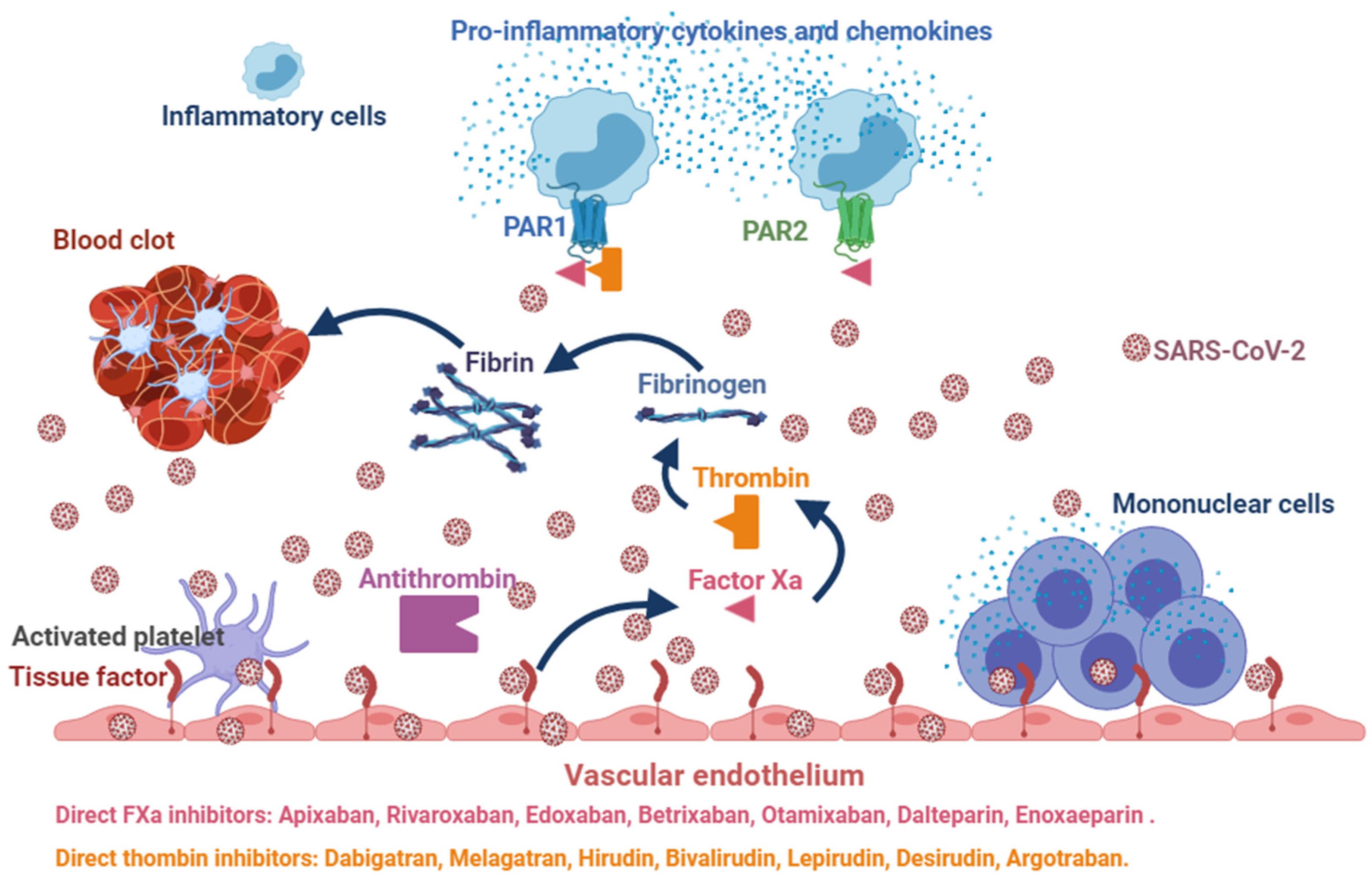
3. Uncertain Effects of Anticoagulants in COVID-19

The virosis induced by SARS-CoV-2 promotes endothelial dysfunction, through direct invasion of endothelial cells [25], which in turn can induce excessive thrombin generation and fibrinolysis shutdown, which are conditions able to promote the activation of coagulation [10,25]. Endothelial dysfunction leads to microvascular dysfunction with consequent vasoconstriction, ischemia, inflammation, edema and a procoagulative state [25]. The reduction in Oxygen (O2) plasma levels in severe COVID-19 may induce a prothrombotic status due to the activation of a hypoxia-inducible transcription factor-dependent signaling pathway [10]. All these conditions may lead to occlusion and micro-thrombosis in pulmonary small vessels of severe COVID-19 patients, as outlined in autoptic reports [4]. Therefore, early administration of anticoagulants in COVID-19 was suggested in order to improve the outcome of patients, although exact criteria for their application were not well-established [26]. Low-molecular-weight heparin (LMWH) was commonly adopted for counteracting DIC and venous thromboembolism (VTE), due to its pharmacodynamics and anti-inflammatory properties [27]. The prophylactic dose of LMWH was used by Tang et al. [28] and such administration was related to low and mild bleeding complication rates. Indeed, DIC may develop due to the occurrence of reduction in platelet count and prolongation of pro-thrombin time (PT). These alterations are related to an increased mortality rate. For this reason, anticoagulation may be challenging. ISTH proposed new SIC criteria to manage anticoagulation in such a context, based upon a dedicated score which had been previously validated [12]. Nevertheless, severe COVID-19 patients poorly met SIC criteria, and therefore should not theoretically undergo anticoagulant treatment. For example, platelet count cannot be considered as a sensitive marker for coagulopathy in severe COVID-19 pneumonia; therefore, one of the SIC criteria—platelets depletion—could not be considered for the final decision making [28]. Conversely, markedly elevated D-dimers—indirect markers for coagulation activation—suggest possible benefits from heparin therapy. Nevertheless, the activation of coagulation may reduce pathogen dissemination and invasion [29]. Therefore, anticoagulation may be dangerous in patients without significant coagulopathy, thus explaining the occurrence of higher mortality rate in heparin users as compared to controls with lower D-dimer plasma levels [28]. LMWH are nowadays considered as the new frontier in counteracting the advancing of COVID-19. Other therapies have not been considered yet. Physicians should consider anticoagulation with heparin or LMWH as not beneficial to unselected patients; only those meeting SIC criteria or with markedly elevated D-dimer may be considered for possible anticoagulation with LMWH [28,30].

4. Potential Benefits of DOACs in COVID-19

The blood coagulation cascade is initiated by tissue factor (TF)/Factor VIIa (TF/FVIIa) complex and conveys to the generation of Xa factor (FXa) first, and then thrombin (II Factor) [31,32,33]. Thrombin is a proteolytic enzyme of coagulation cascade able to cleave fibrinogen into fibrin and activate platelets. Aberrant activation of blood coagulation system can contribute to the degeneration of different pathologies, such as COVID-19 pneumonia (Figure 1).
Vessel thrombosis leads to ischemia and, if the occlusion is prolonged, to the necrosis of tissues and organs, while degradation products may enhance inflammation into vessels mainly due to the activation of specific proinflammatory receptors such as protease-activated receptors (PARs) [34]. Four PARs (PAR1–4) are known, which are ubiquitously expressed [31,34]. PARs typically serve as both receptor and ligand: proteolytic cleavage by an activated coagulation factor leads to exposure of a neoamino terminus which activates the receptor itself, thus leading to widespread intracellular signaling [34]. PAR’s 1, 3 and 4 are usually activated by thrombin, while PAR-2 by the TF/FVIIa complex, factor Xa, and trypsin [31]. Furthermore, PAR-1 acts as the receptor for TF/FVIIa complex and factor Xa. Thrombin activates PAR1, PAR3, and PAR4, whereas FXa primarily activates PAR2 and PAR1 [31,34]. Direct oral anticoagulants (DOACs) specifically inhibit FXa or thrombin: apixaban, edoxaban, and rivaroxaban are inhibitors for FXa, while dabigatran etexilate is a thrombin inhibitor [31]. The role of this compound in sepsis has not been evaluated yet. Posma et al. evaluated the role of each proteases of the coagulation cascade, particularly FXa and thrombin, and PARs in different mouse models of inflammatory diseases, including virosis (H1N1 strain of influenza A virus) [35]. The blood coagulation cascade may be activated during virosis, thus provoking DIC [31]. Inhibition of the TF/FVIIa complex reduced the inflammatory background of the virosis and the mortality rate in primate model infected by Ebola virus [36]. Indeed, the roles of PAR1 and PAR2 in mouse models of viral infections are controversial [37]. PAR1 inhibition protected mice against respiratory syncytial virus and human metapneumovirus infection [38]. In vitro studies with human A549 cells showed that PAR1 inhibition reduced the replication of respiratory syncytial virus and human metapneumovirus infection [35]. Animal studies showed a significant reduction in inflammation in H1N1 infection when rivaroxaban was administered, dabigatran etexilate being unable to reproduce such results [35]. Ellinghaus et al. demonstrated a reduction in thrombin-induced expression of pro-inflammatory genes in human endothelial cells after administration of rivaroxaban and dabigatran. Indeed, dabigatran showed a biphasic reaction compared to rivaroxaban as it transiently increased the expression of pro-inflammatory genes at concentrations below the minimal inhibitory effective concentrations [39]. Nevertheless, poor data are available for translating these results in clinical practice, while no studies have evaluated the effects of DOACs in COVID-19.

5. Clinical Case

A 45-year-old male has been hospitalized for SARS-CoV-2 nasopharyngeal RNA swab positivity for 10 days due to fever, breathlessness, and peripheral oxygen desaturation. He was reported no to have any noteworthy diseases, except for fracture surgery of the right tibia in January 2020. Vital signs on admission were as follows: blood pressure, 150/90 mm Hg; heart rate, 96 beats/min; respiratory rate, 28/min; body temperature, 39.8 °C; oxygen saturation through Venturi mask at 12 L/min 57%; pH 7.49; arterial carbon dioxide partial pressure 36 mmHg; arterial oxygen partial pressure 57 mmHg; PaO2/FiO2 114; Modified Early Warning Score (MEWS) 4 [40]; the severity CT score index was 4 [41] (Figure 2).
The patient was treated with ceftriaxone (2 gr ev od), clarithromycin (500 mg ev bid), methylprednisolone (40 mg ev bid), enoxaparin (4000 UI 0,4 subcutaneous bid), furosemide (20 mg ev od), paracetamol (1 g ev every 4 h). Worsening of patient’s condition was observed on the second day of hospitalization (MEWS 8). High flow non-invasive ventilation was started to FiO2 60%. Laboratory data showed that the disseminated intravascular coagulation (DIC Score = 6) was suggestive of overt DIC [42] (Table 1). Anti-platelet factor 4 (PF4) was present and enoxaparin was stopped. Arterial and venous Doppler ultrasound of the lower limbs and supraortic vessels showed no abnormalities. Abdominal ultrasound, CT scan of the brain and MR angiography of intra- and extra-cranial vessels did not show relevant signs. Based on the European Society of Cardiology Guidance (CV disease/COVID-19 2020) [26], Direct Oral Anticoagulant was started (Rivaroxaban 15 mg bid). On the seventh day, coagulation parameters were normalized (DIC score = 0) and clinical conditions improved with MEWS score 0. The patient was discharged and went home being apyretic and negative SARS-CoV-2 nasopharyngeal RNA swab, with home therapy: Rivaroxaban 15 bid for two weeks followed by Rivaroxaban 20 mg od and Prednisone 25 mg od for seven days. DIC is an acquired syndrome whose main characteristic is an alteration in blood coagulation. Insults or injuries with a high risk of leading to DIC can be both infectious and non-infectious. In the patient observed in our COVID-19 area, we obtained a rapid remission of symptoms and normalization of hematochemical parameters, in particular those relating to DIC, after withdrawal of enoxaparin. The patient experienced exposure to enoxaparin at tibial fracture surgery, and anti-PF4 was also present. The pathophysiology of DIC in our patient is likely to be related to both SARS-CoV-2 infection (Figure 1) and the presence of antiplatelet antibodies (Figure 3).
The choice to treat the patient with rivaroxaban derives primarily from the absolute contraindication to the use of heparins; AVKs have a slow onset of anticoagulant effects and require continuous monitoring of INR; for dabigatran, apixaban, and edoxaban, evidence of efficacy in ill patients is lacking; for rivaroxaban there is evidence of a reduction in fatal and major thromboembolic events in medically ill patients [43]. A timely diagnosis is therefore the best way to reassure patients. Blood levels of fibrinogen, d-dimer, and platelet counts can be useful as more than reliable biological markers of an ongoing DIC or a probable onset of it.

6. Long-COVID-19

Patients discharged from hospital after acute COVID-19 had an increased risk of multiorgan dysfunction, readmission, and mortality [44]. Recent joint guidelines proposed by the National Institute for Health and Care Excellence (NICE), the Scottish Intercollegiate Guidelines Network (SIGN), and the Royal College of General Practitioners (RCGP) have divided COVID-19 infection into 3 phases—‘Acute COVID-19’ (signs and symptoms of COVID-19 infection up to 4 weeks), ‘ongoing symptomatic COVID-19’ (from 4 weeks up to 12 weeks), and ‘post-COVID-19 syndrome’ (when signs and symptoms continue beyond 12 weeks). The term ‘Long-COVID-19’ is given to the signs and symptoms that continue or develop after the ‘acute COVID-19’ phase and include both ‘ongoing symptomatic COVID-19’ and ‘post COVID-19 syndrome’ [45]. In addition to the persistence of symptoms, it is also possible to detect abnormalities of chest radiographs and biomarkers [46]. Interestingly, in Long-COVID-19 patients, it is possible to detect a prolonged elevation of D-dimer, regardless of the inflammatory indices and the severity of the acute phase. In these patients with persistent elevation of the D-dimer, there is an increase in serious thromboembolic complications [47].

7. Materials and Methods and Preliminary Results

To characterize the clinical picture, laboratory findings and prognosis of patients with long-COVID-19 were reported preliminary data of patients belonging to our Institute. We consecutively enrolled 50 patients admitted to our Institute from 1 May 2021 to 30 June 2021 for symptoms characterized by dyspnea, fatigue, cough, headache, loss of appetite, myalgia. Of the patients enrolled, thirty recovered from COVID-19. Table 2 summarizes the clinical characteristics of the study population.
Length of acute COVID-19 was 23.1 ± 8 (range 11–49) days. During the acute phase of COVID-19, according to Severity of COVID-19 WHO Clinical Classification [48], 21 patients were classified as Mild/Moderate and 9 as Severe/Critical. On the Post-COVID-19 Functional Status Scale [49] 7 (23.3%) patients reported No/Negligible functional limitations for, 6 (20%) Slight Functional Limitations and 17 (56.7%) Moderate/Severe Functional Limitations. No differences were found in demographics, medical history and vital signs in patients with Long-COVID-19 compared to no COVID-19 patients. Echocardiographic findings showed that left ventricular ejection fraction was lower in Long-COVID-19 patients. Table 3 summarizes laboratory data of Long-COVID-19 patients and no COVID-19 patients; inflammatory parameters and coagulation pathway were higher in Long-COVID-19 patients.

8. Clinical Case

71-year-old man, diabetic, hypertensive, hypercholesterolemic, chronic ischemic heart disease, previous acute coronary syndrome (ACS) with ST elevation, treated with percutaneous transluminal coronary angioplasty-drug eluting system (PTCA-DES) on the right coronary artery, recurrence of ACS-STEMI August 2019 treated with PTCA-DES of anterior interventricular coronary artery. On March 28, onset of fever (39.8 °C), cough, dyspnea, asthenia; nasopharyngeal swab positive for the presence of SARS-CoV-2 virus RNA. On April 8, worsening of dyspnea and SO2 76%; hospitalization in the COVID-19 area. Non-invasive ventilation in CPAP for 10 days and subsequent high-flow O2-therapy. Discharged April 28, after nasopharyngeal swab was negative for the presence of SARS-CoV-2 virus RNA. On May 26, due to the persistence of asthenia, easy fatigue, dyspnea, cough, polymyalgia and mental confusion, the patient was hospitalized (Functional Status Scale 3). Laboratory parameters showed persistent inflammatory and procoagulative state (Erythrocyte Sedimentation Rate 46 mm; High Sensitivity C Reactive Protein 62.86 mg/L; Interleukin-6 53.45 pg/mL; D-dimer 1199.7 ng/mL; NT-ProBNP 1438.9 pg/mL). Bilateral peripheral areas of consolidation with surrounding ground-glass opacities were observed at CT scan (Figure 4).
The transthoracic echocardiographic study showed left ventricular dilatation, anterior hypokinesia and proximal interventricular septum and a reduction in left ventricular ejection fraction (Left Ventricular (LV) end diastolic dimension, 6.0 cm; LV end diastolic volume, 181 mL; LV end systolic dimension, 5.0 cm; LV end systolic volume, 119 mL; LV ejection fraction, 34%; Left atrial anteroposterior dimension, 4.5 cm; E/A ratio 0.72; PAPs, 19 mmHg). Therapy with ceftriaxone and enoxaparin was initiated and antihypertensive, hypoglycemic and cardioactive drugs were continued. After six days of therapy, the patient’s clinical condition and laboratory parameters improved (Erythrocyte Sedimentation Rate 28 mm; High Sensitivity C Reactive Protein 2.27 mg/L; Interleukin-6 2.45 pg/mL; D-dimer 824.65 ng/mL; NT-ProBNP 436.8 pg/mL). The patient was discharged with the following therapy: Pantoprazole 40 mg/die, Bisoprolol 2.5 mg/die, Rivaroxaban 2.5 mg/bid, acetylsalicylic acid 100 mg/die, Sacubitril/Valsartan 24 mg–26 mg/bid, Repaglinide 0.5 mg/die, Atorvastatin 40 mg/die. The Long-COVID syndrome is characterized, in addition to the persistence of symptoms typical of the acute phase, also by alterations in inflammatory and coagulation parameters [50]. Our patient showed concomitant procoagulative and inflammatory states following SARS-CoV-2 infection. The finding of a reduced LVEF and elevated levels of NT-ProBNP, as well as the persistence of symptoms, led to the therapeutic option of therapy with Sacubitril/Valsartan to counteract the progression of heart failure [51]. In our opinion, Sacubitril/Valsartan is also able to counteract the inflammatory storm in COVID-19 patients [52]. Our patient had a high cardiovascular residual risk, being hypertensive, diabetic, dyslipidemic, and having ischemic heart disease; furthermore, the condition of Long-COVID imposes an additional risk of major cardiovascular events [44] (Figure 5). Therefore, rivaroxaban therapy at vascular doses is mandatory [53].

9. Conclusions and Perspectives

Cardiovascular diseases affected most of the patients with COVID-19 admitted to hospitals, thus resulting in higher risk for in-hospital mortality [54]. DIC may also complicate the clinical course of COVID-19. Routine monitoring of hemostasis tests may be useful for guiding therapeutic approaches and preventing disease progression. Critically ill COVID-19 patients may show hypoxia or hemodynamic instability, which could be related to thromboembolic disease. The optimal thromboprophylactic regimen for patients hospitalized for COVID-19 is unknown [55,56]. Furthermore, the drug–drug interactions between some antiviral treatments and direct oral anticoagulants may favor the use of LMWH or unfractionated heparin in this population. Preclinical studies showed that rivaroxaban was able to contain the inflammatory response during virosis [35]. The advice is not to abruptly withdraw DOACs in high-risk patients as this pharmacological action may result in worsening clinical status and promoting adverse outcomes. We do believe that DOACs should be continued in patients with COVID-19 until further data are available. In Long-COVID patients, persistent inflammation and a prothrombotic state secondary to persistent endothelial damage requires careful monitoring and appropriate intervention.

Author Contributions

D.A., C.A., M.M.C., P.S., A.S.B., M.U., G.C. conceived the paper, analyzed and interpreted the data, drafted the Review with Clinical Cases and critically reviewed its intellectual content, and finally approved the version to be submitted for publication. All authors have read and agreed to the published version of the manuscript.

Funding

This research received no external funding.

Institutional Review Board Statement

The study was conducted according to the guidelines of the Declaration of Helsinki, and was approved by the Institutional Review Board (22 March 2021) of the San Francesco Hospital of Telese Terme (BN).

Informed Consent Statement

Informed consent was obtained from all subjects involved in the study.

Data Availability Statement

The dataset generated and analyzed during the current study is available from Domenico Acanfora, Department of Internal Medicine San Francesco Hospital, Viale Europa 21, 82037 Telese Terme, Benevento, Italy, E-mail: [email protected].

Acknowledgments

The Authors would like to thank Ada d’Aniello for the English revision and Antonietta Ferrucci and Francesca Carangelo for data collection and analysis.

Conflicts of Interest

The authors declare no conflict of interest.

References

  1. Zhang, Y.; Xiao, M.; Zhang, S.; Xia, P.; Cao, W.; Jiang, W.; Chen, H.; Ding, X.; Zhao, H.; Zhang, H.; et al. Coagulopathy and Antiphospholipid Antibodies in Patients with COVID-19. N. Engl. J. Med. 2020, 382, e38. [Google Scholar] [CrossRef]
  2. Iba, T.; Watanabe, E.; Umemura, Y.; Wada, T.; Hayashida, K.; Kushimoto, S.; Wada, H. Sepsis-associated disseminated intravascular coagulation and its differential diag-noses. J. Intensive Care 2019, 32, 7–13. [Google Scholar]
  3. Zhou, F.; Yu, T.; Du, R.; Fan, G.; Liu, Y.; Liu, Z.; Xiang, J.; Wang, Y.; Song, B.; Gu, X.; et al. Clinical course and risk factors for mortality of adult inpatients with COVID-19 in Wuhan, China: A retrospective cohort study. Lancet 2020, 395, 1054–1062. [Google Scholar] [CrossRef]
  4. Wichmann, D.; Sperhake, J.-P.; Lütgehetmann, M.; Steurer, S.; Edler, C.; Heinemann, A.; Heinrich, F.; Mushumba, H.; Kniep, I.; Schröder, A.S.; et al. Autopsy Findings and Venous Thromboembolism in Patients With COVID-19. Ann. Intern. Med. 2020, 173, 268–277. [Google Scholar] [CrossRef] [PubMed]
  5. Tang, N.; Li, D.; Wang, X.; Sun, Z. Abnormal coagulation parameters are associated with poor prognosis in patients with novel coronavirus pneumonia. J. Thromb. Haemost. 2020, 18, 844–847. [Google Scholar] [CrossRef] [PubMed] [Green Version]
  6. Lillicrap, D. Disseminated intravascular coagulation in patients with 2019-nCoV pneumonia. J. Thromb. Haemost. 2020, 18, 786–787. [Google Scholar] [CrossRef] [Green Version]
  7. Bernard, G.R.; Vincent, J.-L.; Laterre, P.-F.; LaRosa, S.P.; Dhainaut, J.-F.; Lopez-Rodriguez, A.; Steingrub, J.S.; Garber, G.E.; Helterbrand, J.D.; Ely, E.W.; et al. Efficacy and Safety of Recombinant Human Activated Protein C for Severe Sepsis. N. Engl. J. Med. 2001, 344, 699–709. [Google Scholar] [CrossRef] [Green Version]
  8. Alaniz, C. An Update on Activated Protein C (Xigris) In the Management of Sepsis. Pharm. Ther. 2010, 35, 504–508. [Google Scholar]
  9. Paranjpe, I.; Fuster, V.; Lala, A.; Russak, A.J.; Glicksberg, B.; Levin, M.A.; Charney, A.W.; Narula, J.; Fayad, Z.A.; Bagiella, E.; et al. Association of Treatment Dose Anticoagulation With In-Hospital Survival Among Hospitalized Patients With COVID-19. J. Am. Coll. Cardiol. 2020, 76, 122–124. [Google Scholar] [CrossRef]
  10. Jose, R.J.P.; Manuel, A. COVID-19 cytokine storm: The interplay between inflammation and coagulation. Lancet Respir. Med. 2020, 8, e46–e47. [Google Scholar] [CrossRef]
  11. Iba, T.; Levy, J.H.; Warkentin, T.E.; Thachil, J.; van der Poll, T.; Levi, M. Diagnosis and management of sepsis-induced coagulopathy and disseminated in-travascular coagulation. J. Thromb. Haemost. 2019, 11, 1989–1994. [Google Scholar] [CrossRef] [PubMed] [Green Version]
  12. Iba, T.; Di Nisio, M.; Levy, J.H.; Kitamura, N.; Thachil, J. New criteria for sepsis-induced coagulopathy (SIC) following the revised sepsis definition: A retrospective analysis of a nationwide survey. BMJ Open 2017, 7, e017046. [Google Scholar] [CrossRef] [Green Version]
  13. Du, R.-H.; Liang, L.-R.; Yang, C.-Q.; Wang, W.; Cao, T.-Z.; Li, M.; Guo, G.-Y.; Du, J.; Zheng, C.-L.; Zhu, Q.; et al. Predictors of mortality for patients with COVID-19 pneumonia caused by SARS-CoV-2: A prospective cohort study. Eur. Respir. J. 2020, 55, 2000524. [Google Scholar] [CrossRef] [Green Version]
  14. Liang, W.-H.; Guan, W.-J.; Li, C.; Li, Y.-M.; Liang, H.-R.; Zhao, Y.; Liu, X.-Q.; Sang, L.; Chen, R.-C.; Tang, C.-L.; et al. Clinical characteristics and outcomes of hospitalised patients with COVID-19 treated in Hubei (epicentre) and outside Hubei (non-epicentre): A nationwide analysis of China. Eur. Respir. J. 2020, 55, 2000562. [Google Scholar] [CrossRef] [Green Version]
  15. Istituto Superiore di Sanità Caratteristiche dei Pazienti Deceduti Positivi a COVID-19 in Italia. Available online: www.epicentro.iss.it/coronavirus/bollettino/Report-COVID-2019_2_aprile_eng.pdf. (accessed on 1 July 2021).
  16. Casucci, G.; Acanfora, D.; Incalzi, R.A. The Cross-Talk between Age, Hypertension and Inflammation in COVID-19 Patients: Therapeutic Targets. Drugs Aging. 2020, 37, 779–785. [Google Scholar] [CrossRef]
  17. Li, J.-Y.; You, Z.; Wang, Q.; Zhou, Z.-J.; Qiu, Y.; Luo, R.; Ge, X.-Y. The epidemic of 2019-novel-coronavirus (2019-nCoV) pneumonia and insights for emerging infectious diseases in the future. Microbes Infect. 2020, 22, 80–85. [Google Scholar] [CrossRef]
  18. Guan, W.-J.; Ni, Z.-Y.; Hu, Y.; Liang, W.-H.; Ou, C.-Q.; He, J.-X.; Liu, L.; Shan, H.; Lei, C.-L.; Hui, D.S.; et al. Clinical Characteristics of Coronavirus Disease 2019 in China. N. Engl. J. Med. 2020, 382, 1708–1720. [Google Scholar] [CrossRef] [PubMed]
  19. Yang, X.; Yu, Y.; Xu, J.; Shu, H.; Xia, J.; Liu, H.; Wu, Y.; Zang, L.; Yu, Z.; Fang, M.; et al. Clinical course and outcomes of critically ill patients with SARS-CoV-2 pneumonia in Wuhan, China: A single-centered, retrospective, observational study. Lancet Respir. Med. 2020, 8, 475–481. [Google Scholar] [CrossRef] [Green Version]
  20. Franceschi, C.; Garagnani, P.; Parini, P.; Giuliani, C.; Santoro, A. Inflammaging: A new immune–metabolic viewpoint for age-related diseases. Nat. Rev. Endocrinol. 2018, 14, 576–590. [Google Scholar] [CrossRef]
  21. Brook, C.E.; Boots, M.; Chandran, K.; Dobson, A.P.; Drosten, C.; Graham, A.L.; Grenfell, B.T.; Müller, M.A.; Ng, M.; Wang, L.-F.; et al. Accelerated viral dynamics in bat cell lines, with implications for zoonotic emergence. eLife 2020, 9, e48401. [Google Scholar] [CrossRef]
  22. He, X.; Li, Z.; Tang, X.; Zhang, L.; Wang, L.; He, Y.; Jin, T.; Yuan, D. Age- and sex-related differences in body composition in healthy subjects aged 18 to 82 years. Medicine 2018, 97, e11152. [Google Scholar] [CrossRef] [PubMed]
  23. Bourgonje, A.R.; Abdulle, A.E.; Timens, W.; Hillebrands, J.; Navis, G.J.; Gordijn, S.J.; Bolling, M.C.; Dijkstra, G.; Voors, A.A.; Osterhaus, A.D.M.E.; et al. Angiotensin-converting enzyme 2 (ACE2), SARS-CoV-2 and the pathophysiology of coronavirus disease 2019 (COVID-19). J. Pathol. 2020, 251, 228–248. [Google Scholar] [CrossRef] [PubMed]
  24. Yang, J.; Zheng, Y.; Gou, X.; Pu, K.; Chen, Z.; Guo, Q.; Ji, R.; Wang, H.; Wang, Y.; Zhou, Y. Prevalence of comorbidities in the novel Wuhan coronavirus (COVID-19) infection: A sys-tematic review and meta-analysis. Int. J. Infect. Dis. 2020, 9712, 91–95. [Google Scholar] [CrossRef]
  25. Varga, Z.; Flammer, A.J.; Steiger, P.; Haberecker, M.; Andermatt, R.; Zinkernagel, A.S.; Mehra, M.R.; Schuepbach, R.; Ruschitzka, F.; Moch, H. Endothelial cell infection and endotheliitis in COVID-19. Lancet 2020, 395, 1417–1418. [Google Scholar] [CrossRef]
  26. Bikdeli, B.; Madhavan, M.V.; Jimenez, D.; Chuich, T.; Dreyfus, I.; Driggin, E.; Der Nigoghossian, C.; Ageno, W.; Madjid, M.; Guo, Y.; et al. COVID-19 and thrombotic or thromboembolic disease: Implication for pre-vention, antithrombotic therapy, and follow-up. J. Am. Coll. Cardiol. 2020, 75, 2950–2973. [Google Scholar] [CrossRef]
  27. Poterucha, T.J.; Libby, P.; Goldhaber, S.Z. More than an anticoagulant: Do heparins have direct anti-inflammatory effects? Thromb. Haemost. 2017, 3, 437–444. [Google Scholar] [CrossRef]
  28. Tang, N.; Bai, H.; Chen, X.; Gong, J.; Li, D.; Sun, Z. Anticoagulant treatment is associated with decreased mortality in severe coronavirus disease 2019 patients with coagulopathy. J. Thromb. Haemost. 2020, 18, 1094–1099. [Google Scholar] [CrossRef]
  29. Shi, C.; Wang, C.; Wang, H.; Yang, C.; Cai, F.; Zeng, F.; Cheng, F.; Liu, Y.; Zhou, T.; Deng, B.; et al. Clinical observation of low molecular weight heparin in relieving inflammation in COVID-19 patients: A retrospective cohort study. Clin. Transl. Sci. 2020, 13, 1087–1095. [Google Scholar] [CrossRef] [PubMed]
  30. Yin, S.; Huang, M.; Li, D.; Tang, N. Difference of coagulation features between severe pneumonia induced by SARS-CoV2 and non-SARS-CoV2. J. Thromb. Thrombolysis 2020, 51, 1107–1110. [Google Scholar] [CrossRef] [Green Version]
  31. Spronk, H.M.; Cate, H.T. The blood coagulation system as a molecular machine. BioEssays 2003, 25, 1220–1228. [Google Scholar] [CrossRef]
  32. Mackman, N.; Tilley, R.E.; Key, N.S. Role of the Extrinsic Pathway of Blood Coagulation in Hemostasis and Thrombosis. Arter. Thromb. Vasc. Biol. 2007, 27, 1687–1693. [Google Scholar] [CrossRef] [PubMed] [Green Version]
  33. Monroe, D.M.; Hoffman, M. What does it take to make the perfect clot? Arter. Thromb. Vasc. Biol. 2006, 26, 41–48. [Google Scholar] [CrossRef] [PubMed]
  34. Heuberger, D.M.; Schuepbach, R.A. Protease-activated receptors (PARs): Mechanisms of action and potential therapeutic modulators in PAR-driven inflammatory diseases. Thromb. J. 2019, 17, 1–24. [Google Scholar] [CrossRef] [Green Version]
  35. Posma, J.J.; Grover, S.; Hisada, Y.; Owens, A.P.; Antoniak, S.; Spronk, H.M.; Mackman, N. Roles of Coagulation Proteases and PARs (Protease-Activated Receptors) in Mouse Models of Inflammatory Diseases. Arter. Thromb. Vasc. Biol. 2019, 39, 13–24. [Google Scholar] [CrossRef] [PubMed]
  36. Geisbert, T.W.; Hensley, L.E.; Jahrling, P.B.; Larsen, T.; Geisbert, J.B.; Paragas, J.; Young, H.A.; Fredeking, T.; Rote, W.E.; Vlasuk, G.P. Treatment of Ebola virus infection with a recombinant inhibitor of factor VIIa/tissue factor: A study in rhesus monkeys. Lancet 2003, 362, 1953–1958. [Google Scholar] [CrossRef] [Green Version]
  37. Antoniak, S.; Mackman, N. Multiple roles of the coagulation protease cascade during virus infection. Blood 2014, 123, 2605–2613. [Google Scholar] [CrossRef] [Green Version]
  38. Lê, V.B.; Riteau, B.; Alessi, M.-C.; Couture, C.; Jandrot-Perrus, M.; Rhéaume, C.; Hamelin, M.; Boivin, G. Protease-activated receptor 1 inhibition protects mice against thrombin-dependent respiratory syncytial virus and human metapneumovirus infections. Br. J. Pharmacol. 2017, 175, 388–403. [Google Scholar] [CrossRef] [Green Version]
  39. Ellinghaus, P.; Perzborn, E.; Hauenschild, P.; Gerdes, C.; Heitmeier, S.; Visser, M.; Summer, H.; Laux, V. Expression of pro-inflammatory genes in human endothelial cell: Compar-ison of rivaroxaban and dabigatran. Thromb. Res. 2016, 142, 44–51. [Google Scholar] [CrossRef] [Green Version]
  40. Subbe, C.; Kruger, M.; Rutherford, P.; Gemmel, L. Validation of a modified Early Warning Score in medical admissions. Qjm: Int. J. Med. 2001, 94, 521–526. [Google Scholar] [CrossRef] [Green Version]
  41. Liu, F.; Zhang, Q.; Huang, C.; Shi, C.; Wang, L.; Shi, N.; Fang, C.; Shan, F.; Mei, X.; Shi, J.; et al. CT quantification of pneumonia lesions in early days predicts progression to severe illness in a cohort of COVID-19 patients. Theranostics 2020, 10, 5613–5622. [Google Scholar] [CrossRef]
  42. Taylor, F.B., Jr.; Toh, C.H.; Hoots, W.K.; Wada, H.; Levi, M. Scientific Subcommittee on Disseminated Intravascular Coagulation (DIC) of the International Society on Thrombosis and Haemostasis (ISTH). Towards definition, clinical and laboratory criteria, and a scoring system for disseminated intravascular coagulation. Thromb. Haemost. 2001, 86, 1327–1330. [Google Scholar] [CrossRef] [Green Version]
  43. Spyropoulos, A.C.; Ageno, W.; Albers, G.W.; Elliott, C.G.; Halperin, J.L.; Hiatt, W.R.; Maynard, G.A.; Steg, P.G.; Weitz, J.I.; Lu, W.; et al. Post-Discharge Prophylaxis with Rivaroxaban Reduces Fatal and Major Thromboembolic Events in Medically Ill Patients. J. Am. Coll. Cardiol. 2020, 75, 3140–3147. [Google Scholar] [CrossRef]
  44. Ayoubkhani, D.; Khunti, K.; Nafilyan, V.; Maddox, T.; Humberstone, B.; Diamond, I.; Banerjee, A. Post-COVID syndrome in individuals admitted to hospital with COVID-19: Retrospective cohort study. BMJ 2021, 372, n693. [Google Scholar] [CrossRef]
  45. Shah, W.; Hillman, T.; Playford, E.D.; Hishmeh, L. Managing the long term effects of COVID-19: Summary of NICE, SIGN, and RCGP rapid guideline. BMJ 2021, 372, n136. [Google Scholar] [CrossRef]
  46. Mandal, S.; Barnett, J.; Brill, S.E.; Brow n, J.S.; Denneny, E.K.; Hare, S.S.; Heightman, M.; Hillman, T.E.; Jacob, J.; Jarvis, H.C.; et al. ‘Long-COVID’: A cross-sectional study of persisting symptoms, biomarker and imaging abnormalities following hospitalisation for COVID-19. Thorax 2020, 76, 396–398. [Google Scholar] [CrossRef]
  47. Townsend, L.; Fogarty, H.; Dyer, A.; Martin-Loeches, I.; Bannan, C.; Nadarajan, P.; Bergin, C.; Farrelly, C.O.; Conlon, N.; Bourke, N.M.; et al. Prolonged elevation of D-dimer levels in convalescent COVID-19 patients is independent of the acute phase response. J. Thromb. Haemost. 2021, 19, 1064–1070. [Google Scholar] [CrossRef]
  48. De Terwangne, C.; Laouni, J.; Jouffe, L.; Lechien, J.; Bouillon, V.; Place, S.; Capulzini, L.; Machayekhi, S.; Ceccarelli, A.; Saussez, S.; et al. Predictive Accuracy of COVID-19 World Health Organization (WHO) Severity Classification and Comparison with a Bayesian-Method-Based Severity Score (EPI-SCORE). Pathogens 2020, 9, 880. [Google Scholar] [CrossRef] [PubMed]
  49. Klok, F.A.; Boon, G.J.; Barco, S.; Endres, M.; Geelhoed, J.M.; Knauss, S.; Rezek, S.A.; Spruit, M.A.; Vehreschild, J.; Siegerink, B. The Post-COVID-19 Functional Status scale: A tool to measure functional status over time after COVID-19. Eur. Respir. J. 2020, 56, 2001494. [Google Scholar] [CrossRef] [PubMed]
  50. Andrade, B.S.; Siqueira, S.; Soares, W.D.A.; Rangel, F.D.S.; Santos, N.; Freitas, A.D.S.; da Silveira, P.R.; Tiwari, S.; Alzahrani, K.; Góes-Neto, A.; et al. Long-COVID and Post-COVID Health Complications: An Up-to-Date Review on Clinical Conditions and Their Possible Molecular Mechanisms. Viruses 2021, 13, 700. [Google Scholar] [CrossRef]
  51. Mcmurray, J.; Packer, M.; Desai, A.S.; Gong, J.; Lefkowitz, M.P.; Rizkala, A.R.; Rouleau, J.L.; Shi, V.C.; Solomon, S.D.; Swedberg, K.; et al. Angiotensin–Neprilysin Inhibition versus Enalapril in Heart Failure. N. Engl. J. Med. 2014, 371, 993–1004. [Google Scholar] [CrossRef] [Green Version]
  52. Acanfora, D.; Ciccone, M.M.; Scicchitano, P.; Acanfora, C.; Casucci, G. Neprilysin inhibitor–angiotensin II receptor blocker combination (sacubitril/valsartan): Rationale for adoption in SARS-CoV-2 patients. Eur. Hear. J.—Cardiovasc. Pharmacother. 2020, 6, 135–136. [Google Scholar] [CrossRef] [Green Version]
  53. Eikelboom, J.W.; Connolly, S.J.; Bosch, J.; Dagenais, G.R.; Hart, R.G.; Shestakovska, O.; Diaz, R.; Alings, M.; Lonn, E.M.; Anand, S.S.; et al. Rivaroxaban with or without Aspirin in Stable Cardiovascular Disease. N. Engl. J. Med. 2017, 377, 1319–1330. [Google Scholar] [CrossRef] [PubMed]
  54. Shi, S.; Qin, M.; Shen, B.; Cai, Y.; Liu, T.; Yang, F.; Gong, W.; Liu, X.; Liang, J.; Zhao, Q.; et al. Association of Cardiac Injury with Mortality in Hospitalized Patients with COVID-19 in Wuhan, China. JAMA Cardiol. 2020, 5, 802. [Google Scholar] [CrossRef] [PubMed] [Green Version]
  55. Cate, H.T. Surviving COVID-19 with Heparin? N. Engl. J. Med. 2021, 385, 845–846. [Google Scholar] [CrossRef] [PubMed]
  56. ATTACC Investigators; ACTIV-4a Investigators; REMAP-CAP Investigators. Therapeutic Anticoagulation with Heparin in Noncritically Ill Patients with COVID-19. N. Engl. J. Med. 2021, 385, 790–802. [Google Scholar] [CrossRef] [PubMed]
Figure 1. Interplay between SARS-CoV-2 infection, coagulation system, protease-activated receptors and inflammation. Activation of the coagulation cascade by the SARS-CoV-2 leads to the cleavage of fibrinogen into fibrin and platelet activation that can contribute to thrombosis. Platelet activation and fibrin degradation products can also enhance inflammation. Coagulation proteases can activate endothelial cells via protease-activated receptors that can increase the expression of inflammatory mediators. Drugs capable of inhibiting Factor Xa could attenuate the inflammatory response and modulate the coagulation cascade by reducing the formation of blood clots. This figure was created using the website https://app.biorender.com (accessed on 6 July 2021).
Viruses 13 01904 g001
Figure 2. High resolution computed tomography (HRCT) chest, axial post-processed images, showing bilateral peripheral areas of ground-glass opacity (GGO).
Viruses 13 01904 g002
Figure 3. Pathophysiological mechanism of heparin-induced thrombocytopenia (HIT). Platelet factor 4 (PF4) is a chemokine secreted from the alpha-granules of platelets, released as tetramers. They bind to heparin and other proteoglycans and inactivate them. The binding of heparin to PF4 exposes new antigen sites and hence the formation of new (IgG) antibodies. Platelet Fc receptors bind the antibody-heparin-PF4, which contribute to thrombosis. Thrombocytopenia occurs by two mechanisms: removal of platelets with bound IgG by splenic macrophages and platelet consumption caused by thrombus formation. PF4 can also bind heparin sulfate on vascular endothelial cells; subsequent binding of the pathologic antibody to this PF4-heparin sulfate complex can injure the endothelium, which further promotes thrombosis. This figure was created using the website https://app.biorender.com (accessed on 12 July 2021).
Viruses 13 01904 g003
Figure 4. A 71-year-old male with sequel of COVID-19 pneumonia. Chest computed tomography (CT), axial image (A) showing bilateral peripheral areas of consolidation with surrounding ground-glass opacities (GGOs). Coronal image (B) showing bilateral peripheral area of interstitial thickening.
Viruses 13 01904 g004
Figure 5. The cross-talk between coronary artery disease, diabetes, hypertension, dyslipidemia and Long-COVID-19.
Viruses 13 01904 g005
Table 1. Summary of COVID-19 Patient Laboratory Data.
Laboratory Values (Reference Range)24 November 202025 November 202026 November 202027 November 202028 November 202029 November 202030 November 2020
White Blood Cells count (3.7–10.3), ×109/L13.5214.614.313.712.513.913.58
Neutrophils (40–75), %87.688.087.286.881.082.177.2
Lymphocytes (19–48), %6.66.06.59.210.210.611
Eosinophils (0–7), %0122120.3
Red Blood Cells count (4.2–6.0), ×106/L5.325.425.124.924.915.25.31
Haemoglobin (13.7–17.5), g/dL15.414.914.613.213.614.215.1
Platelet count (155–369), ×109/L3117090180220310346
Prothrombin time (9.6–12.5), second13.418.216.213.210.210.210.4
International normalized ratio (INR) (0.9–1.2)0.991.11.21.00.91.01.1
Activated partial thromboplastin time (19–30), s29.333.234.635.129.128.527.6
Fibrinogen (150–450), mg/dL570220300420510480366
Lactate dehydrogenase (140–280), U/L114915201480921843601570
Creatinine (0.8–1.30), mg/dL0.81.01.11.00.90.90.9
Erytrocite Sedimentation Rate (0–15), mm6212114480735231
High Sensitivity C Reactive Proteine (0–45), mg/L104.9158.8161.282.140.118.22.23
IL-6 (0–6.4) pg/mL36.7484.296.872.342.116.35.56
D-dimer (250–500), ng/mL104413,29818,481428031872128347
Disseminated Intravascular Coagulation Score0664100
Table 2. Clinical characteristics and baseline values of the study population.
Demographic, Medical History and Vital SignsLong-COVID-19No COVID-19
Number of patients, n3020
Sex, M/F, n17/138/12
Age, years a58.6 ± 17.656.3 ± 14.7
Weight, kg a77.1 ± 14.573.8 ± 12
Height, cm a164.6 ± 11.4169.1 ± 8.7
Body mass index, kg/m2 a28.4 ± 4.225.7 ± 2.4
Pre-existing conditions in the last year, n (%)
Cancer2 (6.7%)1 (5.0%)
Chronic heart disease13 (43.3%)6 (30.0%)
Chronic kidney disease5 (16.6%)2 (10.0%)
Chronic liver disease3 (10.0%)1 (5.0%)
Chronic lung disease7 (23.3%)7 (35.0%)
Chronic neurological disease9 (30.0%)5 (25.0%)
Diabetes7 (23.7%)3 (15.0%)
Hypertension19 (63.3%)11 (55.0%)
Mental health conditions2 (6.66%)1 (5.0%)
Obesity (Body Mass Index > 30)11 (36.6%)3 (15.0%)
Heart rate, bpm a73 ± 1570 ± 13
Systolic arterial pressure, mmHg a121 ± 15121 ± 17
Diastolic arterial pressure, mmHg a78 ± 1276 ± 10
Therapies, n (%)
ACE-I/ARB/ARNIs19 (63%)12 (60%)
Beta-blocker11 (37%)8 (40%)
ASA13 (43%)9 (45%)
Diuretics11 (37%)6 (30%)
Anticoagulants12 (40%)6 (30%)
Echocardiography Measurements
LV end diastolic dimension, cm a4.8 ± 14.5 ± 0.6
LV end diastolic volume, mL a114.6 ± 52.594.1 ± 27.9
LV end systolic dimension, cm a3.2 ± 1.042.6 ± 0.5 *
LV end systolic volume, mL a48.7 ± 38.528 ± 10.5
LV ejection fraction, % a61.9 ± 13.770.4 ± 5.7 •
Left atrial anteroposterior dimension, cm a3.7 ± 1.33.5 ± 0.5
E/A ratio a1.02 ± 0.41.1 ± 0.3
SPAP, mmHg a13.8 ± 10.514.6 ± 8.6
M = Male; F = Female; bpm = beats per minute; ACEi = angiotensin-converting enzyme inhibitor; ARB = angiotensin receptor blocker; ARNIs = Angiotensin Receptor Neprilysin Inhibitors; ASA= Acetylsalicylic Acid; LV = Left Ventricular; SPAP = Systolic Pulmonary Artery Pressure. a Mean ± standard deviation. * refers to p = 0.023; refers to p = 0.024; • refers to p = 0.012.
Table 3. Laboratory data of the study population.
Laboratory Values (Reference Range)Long-COVID-19No COVID-19
White Blood Cells count (3.7–10.3), ×109/L a6.84 ± 2.67.14 ± 2.3
Red Blood Cells count (4.0–10.0), ×106/L a4.53 ± 0.64.8 ± 0.58
Haemoglobin (13.7–17.5), g/dL a14.9 ± 6.414.2 ± 1.8
Platelet count (155–369), ×109/L a221 ± 92244 ± 50
Prothrombin time (9.6–12.5), s a14.2 ± 2.513.5 ± 1.2
International normalized ratio (0.9–1.2) a1.07 ± 0.21.00 ± 0.09
Activated Partial Thromboplastin Time (19–30), s a30.6 ± 5.128.8 ± 2.6
Fibrinogen (150–450), mg/dL a364.8 ± 154.4326.9 ± 86.1
Lactate dehydrogenase (140–280), U/L a448.1 ± 133342.45 ± 90.5 *
Creatinine (0.8–1.30), mg/dL a0.92 ± 0.250.86 ± 0.23
Aspartate Aminotrasferase (0–31), U/L a25.04 ± 12.221.6 ± 12.2
Alanine Aminotrasferase (0–34), U/L a25.2 ± 14.520.9 ± 14.6
High Sensitivity C Reactive Protein (0–45), mg/L a16.3 ± 50.13.95 ± 8.8
Sodium (135–155), mEq/L a139 ± 2.7139 ± 2.02
Potassium (3.5–5.5), mEq/L a4.1 ± 0.274.3 ± 0.4
D-dimer (250–500), ng/mL a1044.4 ± 1022273.7 ± 106
Erythrocyte Sedimentation Rate (0–15), mm a25.7 ± 33.215.5 ± 17.2
Albuminuria (0–2.5), mg/dL a120.7 ± 134.764.6 ± 17.7
Interleukin-6 (0–6.4), pg/mL a13.2 ± 33 ± 2.7 •
High-sensitivity Cardiac Troponin (<19), ng/mL a9 ± 26.31.6 ± 0.3
NT-ProBNP (<450), pg/mL a587.4 ± 273273.5 ± 147.9
SARS-CoV-2 Anti-Spike IgM (<1), EU/mL a12.2 ± 35.51.04 ± 2.4
SARS-CoV-2 Anti-Spike IgG (<10), EU/mL a91.5 ± 130.135.9 ± 61.5
Serum Ferritin (20–300), ng/mL a144.6 ± 158.6113 ± 85.7
a Mean ± standard deviation; * refers to p = 0.004; refers to p = 0.002; • refers to p = 0.024; refers to p < 0.0001.
Publisher’s Note: MDPI stays neutral with regard to jurisdictional claims in published maps and institutional affiliations.

