Abstract
Patients with coronavirus disease 2019 (COVID-19) predominantly have a respiratory tract infection with various symptoms and high mortality is associated with respiratory failure second to severe disease. The risk factors leading to severe disease remain unclear. Here, we reanalyzed a published single-cell RNA-Seq (scRNA-Seq) dataset and found that bronchoalveolar lavage fluid (BALF) of patients with severe disease compared to those with mild disease contained decreased TH17-type cells, decreased IFNA1-expressing cells with lower expression of toll-like receptor 7 (TLR7) and TLR8, increased IgA-expressing B cells, and increased hyperactive epithelial cells (and/or macrophages) expressing matrix metalloproteinases (MMPs), hyaluronan synthase 2 (HAS2), and plasminogen activator inhibitor-1 (PAI-1), which may together contribute to the pulmonary pathology in severe COVID-19. We propose IFN-I (and TLR7/TLR8) and PAI-1 as potential biomarkers to predict the susceptibility to severe COVID-19.
1. Introduction
COVID-19, a severe acute respiratory syndrome coronavirus 2 (SARS-CoV-2)caused infectious disease, manifests various symptoms ranging from asymptomatic to mild to very severe, and leads to multiple organ injury and even death. Poor outcomes are associated with older age (especially over 65) and underlying conditions including diabetes, cardiovascular disease, hypertension, obesity, and chronic obstructive pulmonary disease (COPD) [1]. Heightened serum levels of IL-6, C-reactive protein (CRP) and D-dimer, lymphopenia, neutrophilia, and other complications have been reported in severe COVID-19 [2,3], associated with the dysregulation of myeloid responses, especially in the lung [4,5]. In severe cases, cytokine release syndrome (also called “cytokine storm”) results in acute respiratory distress syndrome (ARDS) with drowning edema in the lung, which is broadly accepted as one of the major causes of death [6]. Recent studies showed that preexisting, cross-reactive T cells (elicited by prior infection with “common cold” coronaviruses) might limit the disease severity [7,8]. In addition to T cells, cross-reactive antibodies are also present in unexposed healthy cohorts, especially those aged 6–16 years [9]. However, the preexisting immunity cannot explain the susceptibility in people aged 65 and above because they have more chances to be infected with “common cold” coronaviruses than young people and may have persistent cross-reactive T and B cells. It cannot explain why men are more susceptible than women, either. Identifying risk factors that drive the transition to severe disease is highly demanded and would benefit the treatment, prevention, and vaccination of COVID-19. Here, we reanalyzed a published scRNA-Seq dataset and found that bronchoalveolar lavage fluid (BALF) of patients with severe disease compared to those with mild disease exhibited dysregulation of TH17-type cells, IgA-expressing B cells, type I interferon (IFN-I) pathway, and matrix metalloproteinases (MMPs)-, hyaluronan synthase 2 (HAS2)-, and plasminogen activator inhibitor-1 (PAI-1)-expressing cells.
2. Materials and Methods
2.1. Reanalysis of Pulmonary Responses in COVID-19 Patients
The published BALF scRNA-Seq dataset (GEO accession number GSE145926), including 3 mild and 6 severe SARS-CoV2 cases, and 3 healthy controls [10], was downloaded and analyzed using SeqGeq software (FlowJo LLC, Ashland, OR, USA).
2.2. Statistical Analysis
Results were expressed as mean ± SD. Differences between groups were calculated for statistical significance using unpaired Student’s t test. p ≤ 0.05 was considered as significant.
3. Results and Discussion
3.1. Severe COVID-19 Displays Decreased TH17-Type Cells and Increased IgA+ B in BALFs
Peripheral blood mononuclear cell (PBMC) studies have demonstrated dysregulated myeloid (monocyte and neutrophil) and CD8+ T cell compartments in severe COIVD-19 [3,11,12]. Currently, there are only a few studies focusing on lung local responses. Zhou et al. revealed a hyper-proinflammatory gene expression profile by meta-transcriptomic sequencing of BALF cells [13]. Compared to community-acquired pneumonia patients and healthy controls, BALF cells of COVID-19 patients highly express proinflammatory genes, especially chemokines, suggesting that SARS-CoV-2 infection causes hypercytokinemia. Like SARS-CoV, SARS-CoV-2 robustly triggered the expression of numerous IFN-stimulated genes (ISGs). Liao et al. compared BALF cell responses in mild and severe COVID-19 cases using scRNA-Seq [10]. The BALFs of severe cases had more abundant macrophages and neutrophils with a decrease in the CD8+ T cell population, and expressed elevated levels of cytokines, IL1B, IL6, and TNF, as well as chemokines, compared with those of the mild cases. By leveraging Liao et al.’s scRNA-Seq dataset, we further evidenced the dysregulation of T helper (TH) cells, B cells, the IFN-I pathway, and tissue factors in the severe cases.
The scRNA-Seq dataset (GEO accession number GSE145926), including 3 healthy controls, 3 mild cases, and 6 severe cases [10], was downloaded and analyzed using SeqGeq software (FlowJo LLC). The focus was on the comparison between mild and severe cases; healthy controls were included as references. In the CD4+ TH cell compartment, there were no significant differences in TH1 (T-box transcription factor 21, or TBX21+), TH2 (GATA-binding protein 3, or GATA3+), and regulatory T (forkhead box P3, or FOXP3+) cells between the mild vs. severe cases (Figure 1A). Interestingly, compared with mild cases, BALFs of severe cases had decreased TH17 [RAR-related orphan receptor C (RORC)+ or C-C motif chemokine receptor 6 (CCR6)+] cells (Figure 1A) and γδT (T cell receptor delta constant, or TRDC+) cells (Figure 1B); the latter also express TH17-type cytokines, IL17, and IL17F (and TH1 type cytokine IFNγ). Although TH17 cells are considered as a potent mediator of tissue pathology, they are essential in antiviral immunity through promoting TH1, cytotoxic T lymphocyte, and B cell responses, and are implicated in combating concomitant bacterial (and maybe also fungal) infection [14,15]. The impaired TH17 responses in severe cases suggest a protective role of TH17-type cells, which further implicates the potential benefit of antibiotics (and maybe also antimycotics) for patients with severe disease. Besides the lung, the intestine is another major mucosal site that has active TH17 responses. SARS-CoV-2 also infects the intestine, where it expresses the viral receptors angiotensin-converting enzyme-2 (ACE2) and transmembrane protease serine 2 (TMPRSS2) [16]. A large number of CD4+ CCR6+ TH17 cells have been reported in PBMCs of a deceased patient [17]. In addition, there are more SARS-CoV-2-reactive TH17 cells highly expressing IL17 (IL17A) and CCR6 in the PBMCs of hospitalized patients than non-hospitalized patients [18]. Therefore, the systemic role of TH17 cells in the disease progress, especially the development of ARDS, need further definition. Interestingly, four out of six BALF samples of severe cases expressed IL22, whereas none of mild cases expressed detectable levels of IL22 (Figure 1C). IL22+ cells were CD3E+, CD4+, and aryl hydrocarbon receptor (AHR)+, but also TRDC−, TBX21−, and RORC−, therefore belonging to TH22 (or NKT), but not TH1 or TH17 cells. Whether IL22 plays a role in the disease severity remains to be determined. Besides the dysregulation in the T cell compartment, severe cases had increased frequencies of IgA1 (IGHA1+)-expressing B cells (and a trend of increasing IgG1 (IGHG1+)) (Figure 1D), in agreement with Chen et al.’s observation that higher virus-specific antibody titers correlate with disease severity [19]. Generally, antibodies, if they possess a neutralizing capability, confer favorable humoral immunity; however, the neutralizing capability of antibodies in the severe cases, at least in part, is questionable, and massive immune complexes can be a driving force of tissue permeability, known as antibody-dependent disease enhancement [20,21]. In summary, decreased TH17-type T cells and increased IgA-secreting B cells may augment the disease severity.
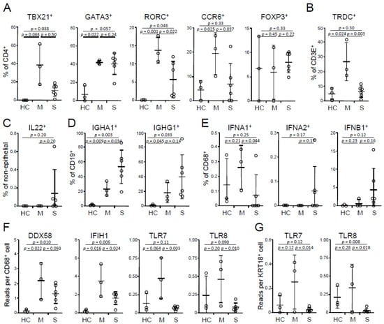
Figure 1.
Dysregulation of TH and B cell profiles and IFN-I pathway in BALFs. (A) Frequencies of TH1 (TBX21+), TH2 (GATA3+), TH17 (RORC+ or CCR6+) cells and regulatory T cells (FOXP3+) in BALF cells on a CD4+ CD14− gate. (B) Frequencies of γδT cells (TRDC+) on a CD3E+ gate. (C) Frequencies of IL22+ cells on a KRT18− gate. (D) Frequencies of IgA1 (IGHA1)- and IgG1 (IGHG1)-expressing B cells on a CD19+ gate. (E) Frequencies of IFN-I-expressing macrophages on a CD68+ gate. (F,G) Abundances of RNA recognition receptors in (F) macrophages and (G) epithelial cells. HC, healthy control (n = 3); M, mild (n = 3); S, severe (n = 6). Mean and s.d. are shown; p values; unpaired t-test.
3.2. Severe COVID-19 has an Impaired IFN-I Response
IFN-Is play an important role in antiviral immunity. Due to sequencing depth, IFN-Is were detected mainly in CD68+ macrophages and a few other cells, such as keratin 18 (KRT18)+ epithelial cells. Macrophages of severe cases had decreased frequencies of IFNA1, but not IFNA2- and IFNB1-expressing cells relative to those of mild cases (Figure 1E). Consistently, patients with severe disease had a decrease of expression of RNA sensors, TLR7 and TLR8, in both macrophages and epithelial cells and IFIH1 (encoding MDA5), but not DDX58 (encoding RIG-I) in macrophages (Figure 1F,G). There were no differences in IFIH1 and DDX58 in epithelial cells in both groups (data not shown). These results implicate an essential role of IFN-I pathway in the disease susceptibility, in agreement with previous observations on peripheral blood cells [22,23]. Although IFN-I only marginally declines with aging, and this decline can recover around the age of 55 [24], during virus challenges (such as influenza virus and West Nile virus), both plasmacytoid (pDCs) and myeloid (mDCs) dendritic cells of aged donors have a decreased capacity of IFN-I production, which subsequently impairs the secretion of IFNγ in CD4 T cells and IFNγ, perforin, and granzyme in CD8 T cells [25,26,27]. In addition, certain diseases, such as diabetes and hypertension, can also impair IFN-I production [28]. Furthermore, a recent study showed that women had higher IFNA2 levels than men [29]. In summary, the defect of IFN-I pathway, associated with advancing age, gender, and some diseases, emerges as a risk factor of severe COVID-19.
3.3. Severe COVID-19 Exhibits Enhanced Expression of MMPs
MMPs degrade extracellular matrix components of the interstitium and tight junction and, therefore, increase alveolar permeability that is observed in many destructive lung diseases, including ARDS, COPD, tuberculosis, sarcoidosis, and idiopathic pulmonary fibrosis (IPF), whereas tissue inhibitors of metalloproteinases (TIMPs) inhibit the enzymatic activity of MMPs [30,31]. Of abundant MMPs and TIMPs, BALF epithelial cells of severe COVID-19 had elevated frequencies of MMP7+ and MMP9+ cells and a trend of increasing MMP2+ and MMP13+ portions compared with mild cases, but no differences were found in MMP14+ cell percentages (Figure 2A). There were no alterations in the frequencies of either TIMP1+- or TIMP2+-expressing epithelial cells between mild vs. severe cases (Figure 2A). In addition to epithelial cells, macrophages also expressed MMPs, and the expression patterns were similar with those of epithelial cells, but only MMP9 reached significance (Figure 2B). Macrophages of severe cases had higher percentages of TIMP1+ cells, whereas a trend in decreasing TIMP2+ frequencies relative to those of mild cases (Figure 2B). Together, the overexpression of MMPs without the corresponding upregulation of TIMPs, in addition to macrophage and neutrophil products, such as reactive oxygen species (ROS), nitric oxide (NO), and enzymes, may promote intensive lung tissue destruction in severe COVID-19.
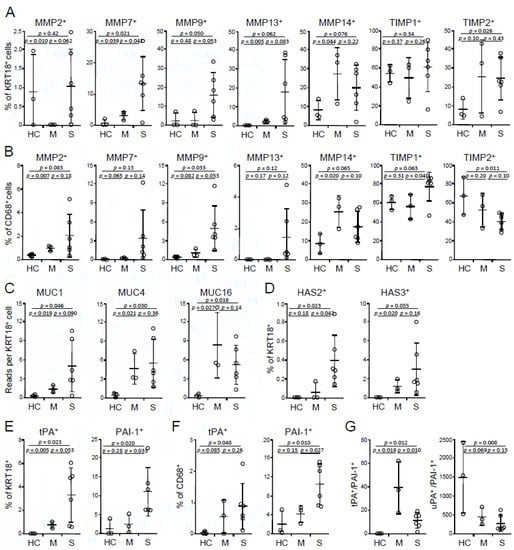
Figure 2.
Expression of tissue factors in BALF cells. (A,B) Frequencies of MMP- and TIMP-expressing epithelial cells (A) and macrophages (B). (C) Abundances of MUCs in epithelial cells. (D) Frequencies of HAS-expressing epithelial cells. (E,F) Frequencies of tPA- and PAI-1-expressing epithelial cells (E) and macrophages (F). (G) Ratios of total tPA vs. PAI-1 and total uPA- vs. PAI-1-expressing cells. HC, healthy control (n = 3); M, mild (n = 3); S, severe (n = 6). Mean and s.d. are shown; p values; unpaired t-test.
3.4. Severe COVID-19 Manifests Increased Expression of Mucin 1 (MUC1), HAS2, and PAI-1
Hypersecretion of MUCs is a common feature of many lung diseases, including viral infection, asthma, and COPD. Hypersecretion of MUCs was also reported in COVID-19 [32]. Of abundant mucins, MUC1 expression is upregulated (but did not reach significance) in epithelial cells of severe vs. mild COVID-19, whereas there were no differences in MUC4 and MUC16 between the two groups (Figure 2C). Hypersecretion of hyaluronic acid presents in severe COVID-19 [33,34]. Consistently, HAS2, but not HAS3, was upregulated in severe cases (Figure 2D), whereas there were very few HAS1-expressing cells (data not shown). In addition to hyperproduction of MUC1 and hyaluronic acid, there were more epithelial cells producing tissue plasminogen activator (tPA) and PAI-1 in severe disease compared with mild disease (Figure 2E). Severe cases also had higher frequencies of PAI-1+, but not tPA+ macrophages (Figure 2F). Total tPA+ to PAI-1+ (tPA/PAI-1) cell ratios were decreased in severe cases relative to mild cases, and there was no difference in urokinase-type plasminogen activator (uPA)/PAI-1 (Figure 2G). tPA and uPA promote fibrinolysis, whereas PAI-1 inhibits this process. Decreased tPA/PAI-1 in severe cases suggests that the dysregulation of coagulation plays an important role in the disease severity, in agreement with Tang et al.’s study that abnormal coagulation leads to a poor prognosis of COVID-19 [35]. In line with this, hyperexpression of tPA and PAI-1 was observed in the peripheral blood of death cases of COVID-19 compared with discharge cases [36]. Heightened expression of tPA and PAI-1 was also reported in highly lethal acute hantavirus cardiopulmonary syndrome [37]. PAI-1 levels are positively correlated, whereas tPA levels are inversely correlated, with D-dimer concentrations in COVID-19 patients [36]. PAI-1 expression is induced by a number of proinflammatory cytokines, such as IL1, IL6, TNF, and TGFβ, as well as hormones, such as insulin, glucocorticoids, and adrenaline (reviewed in References. [38,39]). Advancing age is considered as a major contributor to increased expression of PAI-I [40], which is correlated with aging-associated cardiovascular diseases and metabolic syndromes, such as atherosclerosis, hypertension, obesity, and diabetes (reviewed in References [38,39]). Moreover, men have higher serum PAI-1 levels than women [41,42]. Therefore, elevation of PAI-I expression can serve as a risk factor of severe COVID-19, especially in people with older age, male gender, and/or a group of preexisting conditions. In summary, augmented expression of MUC1, HAS2, and PAI-1 is associated with more severe disease and may contribute to the drowning edema and the coagulation disorder in severe COVID-19.
This study has several limitations. First, the existing dataset only contains small numbers of mild cases and healthy controls. Second, although severe cases had increased frequencies of IgA1 (IGHA1+), the neutralizing capability of these IgA antibodies is unknown. Third, this study analyzed mRNA expression of a variety of biomarkers in BALF cells, but lacked corresponding protein expression data, although there was some evidence at the protein levels from studies on peripheral blood (see references cited).
4. Conclusions
The dysregulation of pulmonary responses, including decreased TH17 cells (and CD8+ T cells [10]) and IFN-I (the latter is associated with impaired TLR7 and TLR8 expression), and the elevation of IgA, MMPs, MUC1, hyaluronic acid, and PAI-I (as well as myeloid cells [4,5,10]), is correlated with the disease severity (see more discussion in Figure 3). Amongst the above-mentioned factors, IFN-I and PAI-1 are dysregulated in older age, male gender, and preexisting diseases that are associated with increased risk to develop severe disease, and we propose the underexpression of IFN-I (and TLR7/TLR8) and the hypersecretion of PAI-1 as potential biomarkers to predict the susceptibility to severe COVID-19 (and maybe also other lung infections).
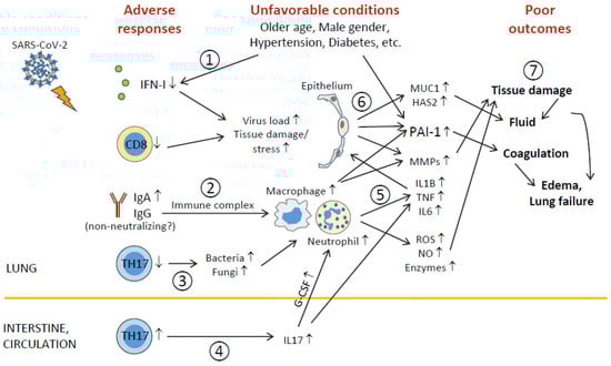
Figure 3.
Outline of unfavorable conditions and deleterious pulmonary responses. (1) Older age, male gender, underlying conditions (such as hypertension, diabetes, etc.), and unknown factors (including genetic background) impair antiviral immunity (including IFN-I deficiency and decreased CD8+ T), leading to higher virus loads and tissue damage/stress. (2) Elevation of humoral responses results in massive immune complexes that activate macrophages and neutrophils. (3) Decreased TH17 cell responses cause overgrowth of commensal bacteria and fungi, which further activate macrophages and neutrophils. (4) TH17 hyperactivation and/or expansion (in the intestine?) cause high levels of serum IL17, which induces G-CSF expression and, in turn, promotes neutrophilia. (5) Hyperactivated macrophages and neutrophils release immense amounts of proinflammatory cytokines, leading to cytokine release syndrome and subsequent ARDS, as well as tissue-destructive products, such as ROS, NO, MMPs, and other enzymes. (6) During ARDS, proinflammatory cytokines act on epithelial cells and induce MMPs, mucins, hyaluronic acids, antimicrobial peptides, and PAI-1 (unfavorable conditions also elevate PAI-1 expression). (7) ROS, NO, MMPs, and other enzymes cause epithelial and endothelial leakage, leading to tissue fluid/plasma accumulation in alveolar spaces. Mucins, hyaluronic acids, and antimicrobial peptides concentrate alveolar fluids and thicken mucosal lining, resulting in edema and even lung failure. Heightened PAI-1 facilitates coagulation and strengthens edema formation (and thrombosis). ARDS also has systemic consequences causing multiorgan injury.
Author Contributions
X.O.Y. designed the project. X.O.Y. and D.W. analyzed the dataset, depicted the plots, and wrote the manuscript. Both authors have read and agreed to the published version of the manuscript.
Funding
This work was supported in part by NIH [AI142200 and HL148337].
Institutional Review Board Statement
Not applicable.
Informed Consent Statement
Not applicable.
Data Availability Statement
No new data are available.
Acknowledgments
We acknowledge the trial of SeqGeq software (FlowJo LLC, Ashland, OR, USA).
Conflicts of Interest
The authors declare that there is no conflict of interest.
References
- Wu, Z.; McGoogan, J.M. Characteristics of and Important Lessons from the Coronavirus Disease 2019 (COVID-19) Outbreak in China: Summary of a Report of 72314 Cases from the Chinese Center for Disease Control and Prevention. J. Am. Med. Assoc. 2020, 323, 1239–1242. [Google Scholar] [CrossRef]
- Ruan, Q.; Yang, K.; Wang, W.; Jiang, L.; Song, J. Clinical predictors of mortality due to COVID-19 based on an analysis of data of 150 patients from Wuhan, China. Intensive Care Med. 2020, 46, 846–848. [Google Scholar] [CrossRef]
- Huang, C.; Wang, Y.; Li, X.; Ren, L.; Zhao, J.; Hu, Y.; Zhang, L.; Fan, G.; Xu, J.; Gu, X.; et al. Clinical features of patients infected with 2019 novel coronavirus in Wuhan, China. Lancet 2020, 395, 497–506. [Google Scholar] [CrossRef]
- Ren, X.; Wen, W.; Fan, X.; Hou, W.; Su, B.; Cai, P.; Li, J.; Liu, Y.; Tang, F.; Zhang, F.; et al. COVID-19 immune features revealed by a large-scale single-cell transcriptome atlas. Cell 2021, 184, 1895–1913. [Google Scholar] [CrossRef]
- Stephenson, E.; Reynolds, G.; Botting, R.A.; Calero-Nieto, F.J.; Morgan, M.D.; Tuong, Z.K.; Bach, K.; Sungnak, W.; Worlock, K.; Yoshida, M.; et al. Single-cell multi-omics analysis of the immune response in COVID-19. Nat. Med. 2021, 27, 904–916. [Google Scholar] [CrossRef]
- Bonam, S.R.; Kotla, N.G.; Bohara, R.A.; Rochev, Y.; Webster, T.J.; Bayry, J. Potential immuno-nanomedicine strategies to fight COVID-19 like pulmonary infections. Nano Today 2021, 36, 101051. [Google Scholar] [CrossRef]
- Grifoni, A.; Weiskopf, D.; Ramirez, S.I.; Mateus, J.; Dan, J.M.; Moderbacher, C.R.; Rawlings, S.A.; Sutherland, A.; Premkumar, L.; Jadi, R.S.; et al. Targets of T Cell Responses to SARS-CoV-2 Coronavirus in Humans with COVID-19 Disease and Unexposed Individuals. Cell 2020, 181, 1489–1501. [Google Scholar] [CrossRef]
- Braun, J.; Loyal, L.; Frentsch, M.; Wendisch, D.; Georg, P.; Kurth, F.; Hippenstiel, S.; Dingeldey, M.; Kruse, B.; Fauchere, F.; et al. SARS-CoV-2-reactive T cells in healthy donors and patients with COVID-19. Nature 2020, 587, 270–274. [Google Scholar] [CrossRef]
- Ng, K.W.; Faulkner, N.; Cornish, G.H.; Rosa, A.; Harvey, R.; Hussain, S.; Ulferts, R.; Earl, C.; Wrobel, A.G.; Benton, D.J.; et al. Preexisting and de novo humoral immunity to SARS-CoV-2 in humans. Science 2020, 370, 1339–1343. [Google Scholar] [CrossRef]
- Liao, M.; Liu, Y.; Yuan, J.; Wen, Y.; Xu, G.; Zhao, J.; Cheng, L.; Li, J.; Wang, X.; Wang, F.; et al. Single-cell landscape of bronchoalveolar immune cells in patients with COVID-19. Nat. Med. 2020, 26, 842–844. [Google Scholar] [CrossRef]
- Schulte-Schrepping, J.; Reusch, N.; Paclik, D.; Baßler, K.; Schlickeiser, S.; Zhang, B.; Krämer, B.; Krammer, T.; Brumhard, S.; Bonaguro, L.; et al. Severe COVID-19 Is Marked by a Dysregulated Myeloid Cell Compartment. Cell 2020, 182, 1419–1440.e23. [Google Scholar] [CrossRef]
- Wang, F.; Hou, H.; Yao, Y.; Wu, S.; Huang, M.; Ran, X.; Zhou, H.; Liu, Z.; Sun, Z. Systemically comparing host immunity between survived and deceased COVID-19 patients. Cell. Mol. Immunol. 2020, 17, 875–877. [Google Scholar] [CrossRef]
- Zhou, Z.; Ren, L.; Zhang, L.; Zhong, J.; Xiao, Y.; Jia, Z.; Guo, L.; Yang, J.; Wang, C.; Jiang, S.; et al. Heightened Innate Immune Responses in the Respiratory Tract of COVID-19 Patients. Cell Host Microbe 2020, 27, 883–890. [Google Scholar] [CrossRef]
- Er, J.Z.; Koean, R.A.G.; Ding, J.L. Loss of T-bet confers survival advantage to influenza–bacterial superinfection. EMBO J. 2019, 38, e99176. [Google Scholar] [CrossRef] [PubMed]
- Ma, W.-T.; Yao, X.-T.; Peng, Q.; Chen, D.-K. The protective and pathogenic roles of IL-17 in viral infections: Friend or foe? Open Biol. 2019, 9, 190109. [Google Scholar] [CrossRef] [PubMed]
- Ziegler, C.G.K.; Allon, S.J.; Nyquist, S.K.; Mbano, I.M.; Miao, V.N.; Tzouanas, C.N.; Cao, Y.; Yousif, A.S.; Bals, J.; Hauser, B.M.; et al. SARS-CoV-2 Receptor ACE2 Is an Interferon-Stimulated Gene in Human Airway Epithelial Cells and Is Detected in Specific Cell Subsets across Tissues. Cell 2020, 181, 1016–1035.e19. [Google Scholar] [CrossRef]
- Xu, Z.; Shi, L.; Wang, Y.; Zhang, J.; Huang, L.; Zhang, C.; Liu, S.; Zhao, P.; Liu, H.; Zhu, L.; et al. Pathological findings of COVID-19 associated with acute respiratory distress syndrome. Lancet Respir. Med. 2020, 8, 420–422. [Google Scholar] [CrossRef]
- Meckiff, B.J.; Ramírez-Suástegui, C.; Fajardo, V.; Chee, S.J.; Kusnadi, A.; Simon, H.; Grifoni, A.; Pelosi, E.; Weiskopf, D.; Sette, A.; et al. Single-cell transcriptomic analysis of SARS-CoV-2 reactive CD4+ T cells. BioRxiv 2020. [Google Scholar] [CrossRef]
- Chen, X.; Pan, Z.; Yue, S.; Yu, F.; Zhang, J.; Yang, Y.; Li, R.; Liu, B.; Yang, X.; Gao, L.; et al. Disease severity dictates SARS-CoV-2-specific neutralizing antibody responses in COVID-19. Signal Transduct. Target. Ther. 2020, 5, 180. [Google Scholar] [CrossRef] [PubMed]
- Ricke, D.O. Two Different Antibody-Dependent Enhancement (ADE) Risks for SARS-CoV-2 Antibodies. Front. Immunol. 2021, 12, 640093. [Google Scholar] [CrossRef]
- Lee, W.S.; Wheatley, A.K.; Kent, S.J.; DeKosky, B.J. Antibody-dependent enhancement and SARS-CoV-2 vaccines and therapies. Nat. Microbiol. 2020, 5, 1185–1191. [Google Scholar] [CrossRef] [PubMed]
- Hadjadj, J.; Yatim, N.; Barnabei, L.; Corneau, A.; Boussier, J.; Smith, N.; Péré, H.; Charbit, B.; Bondet, V.; Chenevier-Gobeaux, C.; et al. Impaired type I interferon activity and inflammatory responses in severe COVID-19 patients. Science 2020, 369, 718–724. [Google Scholar] [CrossRef] [PubMed]
- Arunachalam, P.S.; Wimmers, F.; Mok, C.K.P.; Perera, R.A.P.M.; Scott, M.; Hagan, T.; Sigal, N.; Feng, Y.; Bristow, L.; Tak-Yin Tsang, O.; et al. Systems biological assessment of immunity to mild versus severe COVID-19 infection in humans. Science 2020, 369, 1210–1220. [Google Scholar] [CrossRef] [PubMed]
- Uno, K.; Yagi, K.; Yoshimori, M.; Tanigawa, M.; Yoshikawa, T.; Fujita, S. IFN production ability and healthy ageing: Mixed model analysis of a 24 year longitudinal study in Japan. BMJ Open 2013, 3, e002113. [Google Scholar] [CrossRef] [PubMed]
- Sridharan, A.; Esposo, M.; Kaushal, K.; Tay, J.; Osann, K.; Agrawal, S.; Gupta, S.; Agrawal, A. Age-associated impaired plasmacytoid dendritic cell functions lead to decreased CD4 and CD8 T cell immunity. Age 2011, 33, 363–376. [Google Scholar] [CrossRef] [PubMed]
- Prakash, S.; Agrawal, S.; Cao, J.; Gupta, S.; Agrawal, A. Impaired secretion of interferons by dendritic cells from aged subjects to influenza. Age 2013, 35, 1785–1797. [Google Scholar] [CrossRef]
- Qian, F.; Wang, X.; Zhang, L.; Lin, A.; Zhao, H.; Fikrig, E.; Montgomery, R.R. Impaired Interferon Signaling in Dendritic Cells From Older Donors Infected In Vitro With West Nile Virus. J. Infect. Dis. 2011, 203, 1415–1424. [Google Scholar] [CrossRef]
- Tominaga, M.; Uno, K.; Yagi, K.; Fukui, M.; Hasegawa, G.; Yoshikawa, T.; Nakumura, N. Association Between Capacity of Interferon-α Production and Metabolic Parameters. J. Interf. Cytokine Res. 2010, 30, 451–454. [Google Scholar] [CrossRef]
- Takahashi, T.; Ellingson, M.K.; Wong, P.; Israelow, B.; Lucas, C.; Klein, J.; Silva, J.; Mao, T.; Oh, J.E.; Tokuyama, M.; et al. Sex differences in immune responses that underlie COVID-19 disease outcomes. Nature 2020, 588, 315–320. [Google Scholar] [CrossRef]
- Elkington, P.T.G. Matrix metalloproteinases in destructive pulmonary pathology. Thorax 2006, 61, 259–266. [Google Scholar] [CrossRef]
- McKeown, S.; Richter, A.G.; O’Kane, C.; McAuley, D.F.; Thickett, D.R. MMP expression and abnormal lung permeability are important determinants of outcome in IPF. Eur. Respir. J. 2009, 33, 77–84. [Google Scholar] [CrossRef] [PubMed]
- He, J.; Cai, S.; Feng, H.; Cai, B.; Lin, L.; Mai, Y.; Fan, Y.; Zhu, A.; Huang, H.; Shi, J.; et al. Single-cell analysis reveals bronchoalveolar epithelial dysfunction in COVID-19 patients. Protein Cell 2020, 11, 680–687. [Google Scholar] [CrossRef]
- Ding, M.; Zhang, Q.; Li, Q.; Wu, T.; Huang, Y. Correlation analysis of the severity and clinical prognosis of 32 cases of patients with COVID-19. Respir. Med. 2020, 167, 105981. [Google Scholar] [CrossRef]
- Mong, M.A.; Awkal, J.A.; Marik, P.E. Accelerated hyaluronan concentration as the primary driver of morbidity and mortality in high-risk COVID-19 patients: With therapeutic introduction of an oral hyaluronan inhibitor in the prevention of Induced Hyaluronan Storm Syndrome. MedRxiv 2020. [Google Scholar] [CrossRef]
- Tang, N.; Li, D.; Wang, X.; Sun, Z. Abnormal coagulation parameters are associated with poor prognosis in patients with novel coronavirus pneumonia. J. Thromb. Haemost. 2020, 18, 844–847. [Google Scholar] [CrossRef]
- Zuo, Y.; Warnock, M.; Harbaugh, A.; Yalavarthi, S.; Gockman, K.; Zuo, M.; Madison, J.A.; Knight, J.S.; Kanthi, Y.; Lawrence, D.A. Plasma tissue plasminogen activator and plasminogen activator inhibitor-1 in hospitalized COVID-19 patients. Sci. Rep. 2021, 11, 1580. [Google Scholar] [CrossRef]
- Bellomo, C.; Korva, M.; Papa, A.; Mäkelä, S.; Mustonen, J.; Avšič-Županc, T.; Vaheri, A.; Martinez, V.P.; Strandin, T. Differential Regulation of PAI-1 in Hantavirus Cardiopulmonary Syndrome and Hemorrhagic Fever With Renal Syndrome. Open Forum Infect. Dis. 2018, 5, ofy021. [Google Scholar] [CrossRef]
- Cesari, M.; Pahor, M.; Incalzi, R.A. REVIEW: Plasminogen Activator Inhibitor-1 (PAI-1): A Key Factor Linking Fibrinolysis and Age-Related Subclinical and Clinical Conditions. Cardiovasc. Ther. 2010, 28, e72–e91. [Google Scholar] [CrossRef]
- Yamamoto, K.; Takeshita, K.; Kojima, T.; Takamatsu, J.; Saito, H. Aging and plasminogen activator inhibitor-1 (PAI-1) regulation: Implication in the pathogenesis of thrombotic disorders in the elderly. Cardiovasc. Res. 2005, 66, 276–285. [Google Scholar] [CrossRef]
- Mari, D.; Coppola, R.; Provenzano, R. Hemostasis factors and aging. Exp. Gerontol. 2008, 43, 66–73. [Google Scholar] [CrossRef]
- Asselbergs, F.W.; Williams, S.M.; Hebert, P.R.; Coffey, C.S.; Hillege, H.L.; Navis, G.; Vaughan, D.E.; Van Gilst, W.H.; Moore, J.H. Gender-specific correlations of plasminogen activator inhibitor-1 and tissue plasminogen activator levels with cardiovascular disease-related traits. J. Thromb. Haemost. 2007, 5, 313–320. [Google Scholar] [CrossRef]
- Krishnamurti, C.; Tang, D.B.; Barr, C.F.; Alving, B.M. Plasminogen Activator and Plasminogen Activator Inhibitor Activities in a Reference Population. Am. J. Clin. Pathol. 1988, 89, 747–752. [Google Scholar] [CrossRef] [PubMed]


Publisher’s Note: MDPI stays neutral with regard to jurisdictional claims in published maps and institutional affiliations. |
© 2021 by the authors. Licensee MDPI, Basel, Switzerland. This article is an open access article distributed under the terms and conditions of the Creative Commons Attribution (CC BY) license (https://creativecommons.org/licenses/by/4.0/).