Article

Influence of Herbicide Exposure and Ranavirus Infection on Growth and Survival of Juvenile Red-Eared Slider Turtles (Trachemys scripta elegans)

by
Rachel M. Goodman
1,*,
Edward Davis Carter
2 and
Debra L. Miller
2,3
1
Biology Department, Hampden-Sydney College, Hampden-Sydney, VA 23943, USA
2
Center for Wildlife Health, University of Tennessee Institute of Agriculture, Knoxville, TN 37996, USA
3
Department of Biomedical and Diagnostic Sciences, College of Veterinary Medicine, University of Tennessee, Knoxville, TN 37996, USA
*
Author to whom correspondence should be addressed.
Viruses 2021, 13(8), 1440; https://doi.org/10.3390/v13081440
Submission received: 18 June 2021 / Revised: 14 July 2021 / Accepted: 19 July 2021 / Published: 23 July 2021
(This article belongs to the Special Issue Ranaviruses and Anti-ranaviral Immunity)

Abstract

:
Ranaviruses are an important wildlife pathogen of fish, amphibians, and reptiles. Previous studies have shown that susceptibility and severity of infection can vary with age, host species, virus strain, temperature, population density, and presence of environmental stressors. Experiments are limited with respect to interactions between this pathogen and environmental stressors in reptiles. In this study, we exposed hatchling red-eared slider turtles (Trachemys scripta elegans) to herbicide and ranavirus treatments to examine direct effects and interactions on growth, morbidity, and mortality. Turtles were assigned to one of three herbicide treatments or a control group. Turtles were exposed to atrazine, Roundup ProMax®, or Rodeo® via water bath during the first 3 weeks of the experiment. After 1 week, turtles were exposed to either a control (cell culture medium) or ranavirus-infected cell lysate via injection into the pectoral muscles. Necropsies were performed upon death or upon euthanasia after 5 weeks. Tissues were collected for histopathology and detection of ranavirus DNA via quantitative PCR. Only 57.5% of turtles exposed to ranavirus tested positive for ranaviral DNA at the time of death. Turtles exposed to ranavirus died sooner and lost more mass and carapace length, but not plastron length, than did controls. Exposure to environmentally relevant concentrations of herbicides did not impact infection rate, morbidity, or mortality of hatchling turtles due to ranavirus exposure. We also found no direct effects of herbicide or interactions with ranavirus exposure on growth or survival time. Results of this study should be interpreted in the context of the modest ranavirus infection rate achieved, the general lack of growth, and the unplanned presence of an additional pathogen in our study.

1. Introduction

In the last two decades, biologists have become alarmed about potential impacts of iridoviruses, which infect and can cause mass mortality events in reptiles, amphibians, and fishes [1]. Iridoviruses are double-stranded DNA viruses that replicate in temperatures ranging from 12–32 °C and may survive several months outside of any host in aquatic environments [2]. Studies on iridovirus pathogenesis and disease ecology have demonstrated that susceptibility and severity of infection vary with age, host species, virus strain, temperature, population density, and presence of environmental stressors [3,4,5,6,7,8,9,10]. Among fish, iridovirus infections have been reported on several continents and can cause economic damage in commercial freshwater fisheries [9,11,12]. In amphibians, ranaviruses (family Iridoviridae, genus Ranavirus) have caused more die-offs in North America than the better-known fungus, Batrachochytrium dendrobatidis (Bd) [13,14]. The impact of ranaviruses on reptilian population dynamics is largely unknown, although several cases of morbidity and mortality in captive and natural populations have been attributed to this pathogen (reviewed in [12,15,16,17]).
Studies on ranavirus in turtles have included isolation of ranaviruses from natural populations and reports of deaths and declines in captive and wild species [12,18,19]; experiments have examined susceptibility and transmission of ranavirus between species and the importance of exposure dose and rearing temperature to host susceptibility and mortality [18,20,21,22,23,24]. While ranavirus outbreaks with high mortality may result in disconcerting headlines, limited surveys have also found ranavirus infection occurring at low prevalence in populations without apparent die-offs [25,26,27]. Ranavirus is now well documented on six continents [12], but significant questions remain regarding distribution and factors influencing morbidity and mortality in wild reptile populations. Experimental studies examining the effect of environmental stressors on ranavirus infection are especially lacking in reptiles. Thus far, temperature is the only environmental variable that has been experimentally investigated for impact on ranavirus susceptibility and survival in reptiles [28].
The current study examined potential interactions among a common environmental stressor, herbicides, and ranavirus exposure in juveniles of a largely aquatic turtle, Trachemys scripta elegans. This species has a wide geographic distribution in central and eastern US, occupies diverse habitats including bogs, ponds, lakes, and rivers, spends ample time in the water [29], and can be a reservoir for ranaviruses that infect more susceptible species, including those from other vertebrate classes (reviewed in [12,20]). Research on infectious diseases in chelonians is needed because over half of species in this taxon are listed as vulnerable, threatened, or endangered according to the IUCN [30].
Pesticides are a multibillion-dollar annual industry and are used more heavily in the Unites States than in many other countries; these products are found in over half of the country’s streams and many other surface waters [31,32,33]. Herbicides and insecticides enter aquatic systems via runoff from agricultural and urban uses and via direct application to water bodies [34]. In aquatic systems, these chemicals can have profound impacts on the biodiversity and productivity of and interactions among organisms [35]. Chemical pollutants can impact host–pathogen interactions by deactivating or killing the pathogen, or by directly and indirectly weakening the host immune response, thus increasing susceptibility to disease [36,37]. Stress hormones such as cortisol and corticosterone can become elevated in response to environmental pollutants and decrease host immune response, which increases susceptibility to infectious diseases [38,39]. Commonly used pesticides applied at sublethal doses can reduce immune response and resistance to parasites [40,41,42,43]. Pesticides have also been shown to negatively impact growth and survival, and they can change sexual characteristics in amphibians and reptiles [44,45]. Thus far, the interaction between pesticides and ranavirus infection have been studied in amphibians [46,47,48] but not in reptiles.
We exposed juvenile turtles to three commonly used herbicides that contain two different main active ingredients, glyphosate and atrazine, to determine whether these chemicals altered growth rates and susceptibility, morbidity, and mortality due to ranavirus exposure. Glyphosate is the most heavily used herbicide in the United States, due largely to the increasing dominance of crops that are genetically engineered to be resistant to this herbicide [49,50]. Surfactants, which help the herbicide to penetrate plant leaves, are common ingredients of these products, yet are generally not tested or listed on labels although they can be equally or more toxic than glyphosate [44,51,52]. Therefore, we examined glyphosate exposure via water bath containing two glyphosate formulations: Roundup ProMax®, which is formulated for terrestrial use and, therefore, contains surfactants, and Rodeo®, which is formulated for aquatic use without surfactants. Atrazine is the second most commonly used herbicide in the United States and is the most common pesticide found in US streams and groundwater [32,53]. Because of its impacts of growth, development, and reproduction in various species and persistent half-life of up to 19 months in water, this herbicide has been banned from use in the European Union since 2004 [36,54]. However, controversy exists surrounding the nature and magnitude of atrazine impacts in ecological communities [55,56,57].
Because of the aforementioned impacts of glyphosate and atrazine on reptilian growth, development, and survival, as well as the interaction of pesticides and ranavirus in studies conducted on amphibians, we expected similar effects in our experiment with T. s. elegans. Specifically, we predicted that exposure to herbicides would negatively impact growth and survival and increase morbidity and mortality due to ranavirus infection relative to controls not exposed to herbicide treatments.

2. Materials and Methods
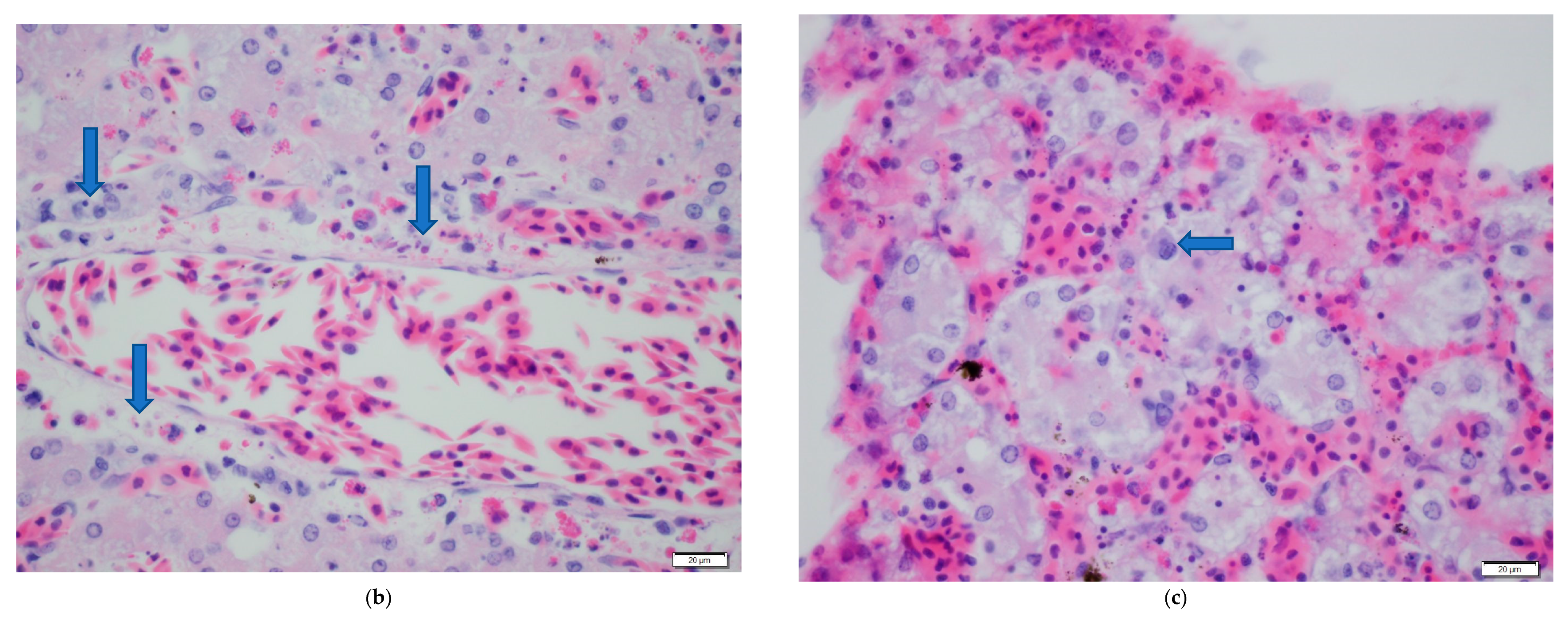
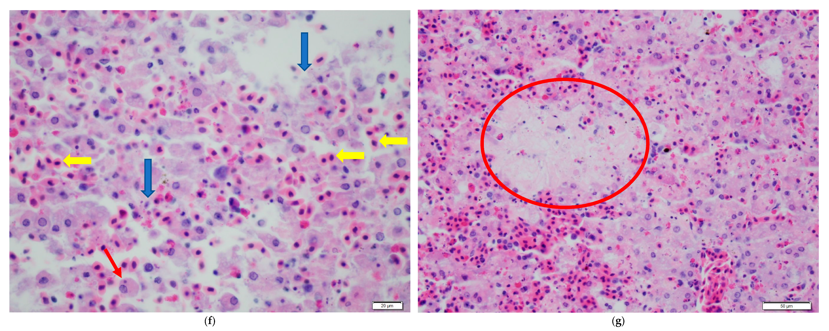
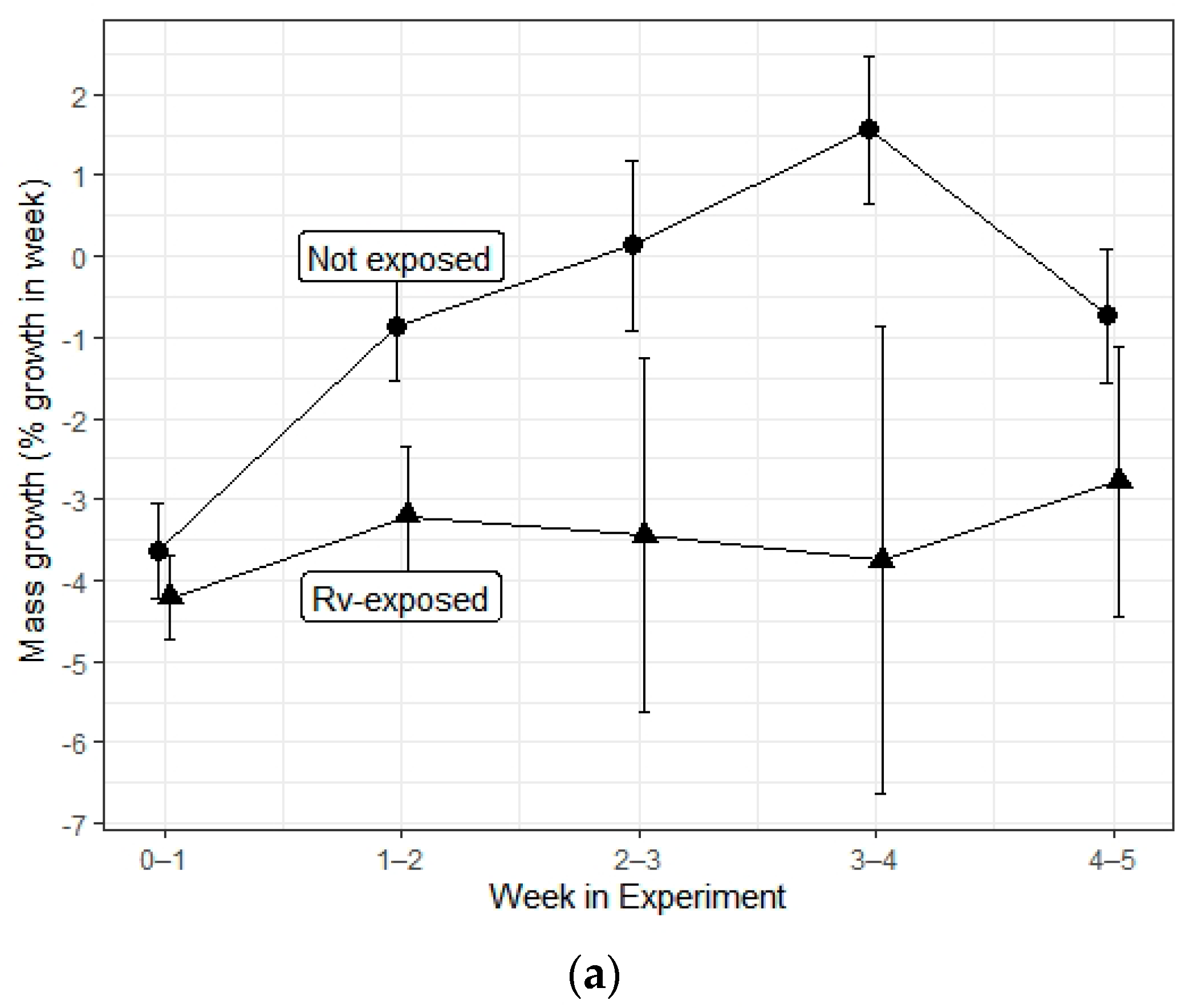
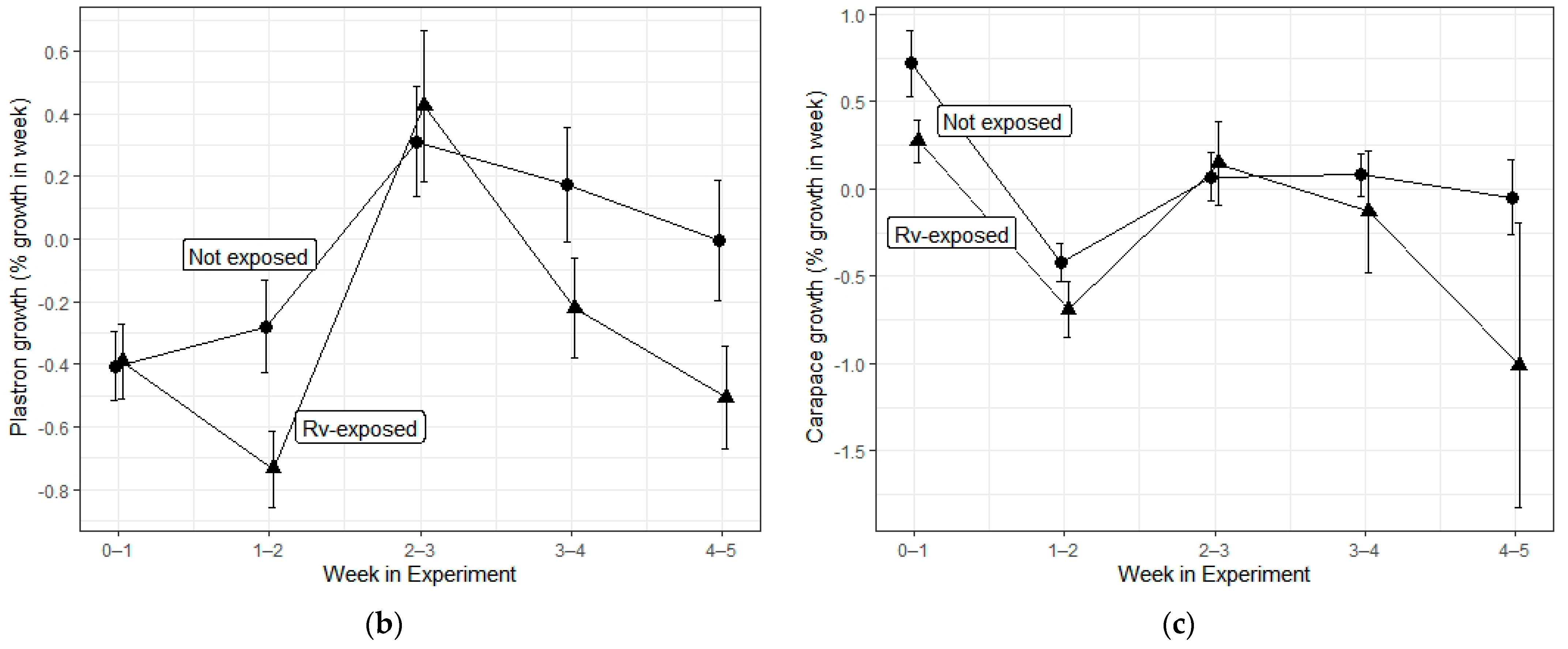
We obtained 170 juvenile red-eared slider turtles from a commercial supplier (Reptile City; Dallas, TX, USA) in May of 2014. Ten turtles were immediately euthanized by injection of >130 mg/kg sodium pentobarbital injection into the supravertebral (subcarapacial) vein, followed by decapitation 1 h after injection. These turtles were necropsied to obtain the following tissues which genomic DNA was then extracted from: approximately 4 cm of mid- and distal intestine, the left kidney, and the left lobe of liver. Testing via quantitative polymerase chain reaction (qPCR; described below) for ranaviral DNA was performed to ensure that supplied animals were not infected with ranavirus. Upon confirmation of negative results for the subsample of 10 turtles, the remaining 160 turtles were placed individually in 5 L plastic containers containing 350 mL of filtered, dechlorinated tap water.
Laboratory temperatures were maintained between 19.5–28 °C with an average temperature of 24 °C. A natural photoperiod (approximately 6:00 a.m.–8:30 p.m. for May–June in Virginia) supplied by large windows in the room was supplemented by overhead fluorescent lights during 9:00 a.m.–6:00 p.m. Throughout the experiment, we fed turtles approximately 10% of their weight in Zoo Med natural aquatic turtle feed every other day. Turtle enclosures were placed on shelves in our laboratory and rotated weekly to avoid any bias due to position within the laboratory. Paper dividers were placed between each container and on the outside edge, to reduce possible stress.
After 3 days of acclimation to the laboratory conditions, turtles were assigned to one of eight experimental treatments (N = 20 for each) in a full factorial design with two factors: ranavirus exposure (control, ranavirus-exposed) and herbicide exposure (control, Roundup ProMax®, Rodeo®, atrazine). At the initiation of the experiment, we collected initial weights and measurements of each turtle (mass in g; width and plastron and carapace length in mm using digital calipers) and collected 3–4 mm from each tail tip using disposable sterile scalpel blades and sealed the wound with KwikStop Styptic Powder (containing benzocaine anesthetic; Miracle Care, Dayton, OH, USA). These tissue were collected to sample initial conditions in the event that any turtles not exposed to ranavirus during the experiment turned out to be ranavirus-positive at the conclusion. However, this scenario did not occur; thus, these tail tissues were not tested for ranavirus.
Herbicide exposure consisted of herbicide addition (or lack thereof, for controls) in 200 mL of water in housing containers during the first 3 weeks of the experiment. Herbicide-free dechlorinated water was used for all turtles during the remaining 2 weeks of the 5 week experiment. Levels of herbicide exposure were based on the higher end of but not maximal concentrations reported in the literature: 2000 μg a.e./L glyphosate for Roundup ProMax® and Rodeo® [58,59] (Monsanto, Creve Coeur, MO, USA) and 20 μg/L for atrazine (Hi-Yield Voluntary Purchasing Groups, Bonham, TX, USA) [53,60,61].
To constitute treatment water for glyphosate herbicides, 33.4 μL of Rodeo and 29.6 μL of Roundup ProMax® were each added to 500 mL of water and mixed on a stir plate on high for 10 min. We then added each 500 mL stock solution to 7500 mL of water in a large bucket and stirred each by hand for 5 min. For the atrazine treatments, we added 20.1 μL of Hi-Yield Atrazine® to 600 mL and mixed this on a stir plate on high for 10 min. An aliquot of 100 mL of this stock solution was then added to 7900 mL of water in a large bucket and stirred by hand for 5 min. In two different weeks, we took samples from the three final herbicide solutions and shipped them overnight on ice to the Mississippi State University Chemical Laboratory to verify target concentrations. Actual concentrations of water samples were 2048–2223 ppb and 1952–2292 ppb for glyphosate in Roundup Pro Max® and Rodeo®, respectively, and 22–27 ppb for atrazine.
Ranavirus exposures were conducted 1 week after initiation of the experiment. Because water bath exposure to ranavirus can result in low infection rates (0–20% in three turtle species [62], we used injection of ranavirus as our exposure method to maximize the potential for infection [28,63,64,65]. Ranavirus exposure was executed at the start of the second week by injection of 100 μL of virus-infected cell lysate (6.3 × 10⁴ TCID50; FV3 ranavirus culture originally isolated from a box turtle isolate grown in fathead minnow cells (similar procedure described in [28]), with 50 μL injected into each pectoral muscle. Control turtles were each injected with 50 μL of Dulbecco’s modified Eagle medium (used for fathead minnow cell culture; Thermo Fisher Scientific, Waltham, MA, USA) in each pectoral muscle.
Each week, turtles were weighed, measured, and placed in cleaned, disinfected tubs with reconstitutions of herbicide treatments for the first 3 weeks or herbicide-free dechlorinated water for the last 2 weeks. After virus exposures, turtles were checked visually every 12 h for external signs of ranavirus infection: lethargy, respiratory stress, cutaneous erythema, and ocular/nasal discharge. During weekly measurements and water changes, turtles were examined while in hand for ranaviral disease signs. When turtles expired before the completion of the experiment, they were examined and sampled as described below following euthanasia done at the conclusion of the experiment. Turtles were considered deceased when unresponsive to stimulation of the limbs and corneas. We weighed and measured, took note of and photographed any external signs of disease, and then decapitated and necropsied the turtles. The color and condition of the liver was noted, and tissue samples were taken from the intestine, kidney, and liver, as previously described. Tissues were frozen at −80 °C until use in ranavirus testing. The remainder of the turtle was preserved in 10% buffered formalin for histopathological examination. The experiment was concluded after 5 weeks (4 weeks after virus exposures), when all remaining living turtles were euthanized, measured, and sampled as described above.
We extracted DNA from tissue samples with Qiagen DNeasy Blood and Tissue Kits (Qiagen, Hilden, Germany). We standardized the amount of genomic DNA used in each reaction with an Epoch spectrophotometer (Biotek, Winooski, VT, USA). We tested for presence of ranavirus DNA using quantitative polymerase chain reaction (qPCR) following the protocol of Picco et al. [64]. Each 25 μL PCR reaction contained: 7 μL volume of combined nuclease-free water and genomic DNA (volume specific to each individual for 50 ng DNA); 12.5 μL of TaqMan Universal PCR Master Mix (Applied Biosystems™, Foster City, CA, USA); 1.5 μL each of 10 μM primers: F 5′–ACA CCA CCG CCC AAA AGT AC–3′, R 5′–CCG TTC ATG ATG CGG ATA ATG–3′; 2.5 μL of 2.5 μM probe: 5′–/56-FAM/CCT CAT CGT /ZEN/TCT GGC CAT CAA CCA /3IABkFQ/–3′ (Integrated DNA Technologies, Coralville, IA, USA). All samples were tested in duplicate using Applied Biosystems™ StepOne Real-time PCR machine with two negative and two positive controls in each run (pure water and DNA extracted from cultured FV3 ranavirus). Samples with Ct values <30 for both runs were considered positive for ranavirus, according to standards established for this machine using known negative and positive controls from water, cultured ranavirus, and ranavirus-infected reptiles [25,26]. If Ct values from two samples for an individual were not both <30 or if one approximated 30, we ran two additional PCR reactions. Then, the consensus from three out of four total reactions was used.
We conducted histopathology on four turtles from each combination of herbicide and virus exposure (eight treatments, 32 turtles total). Four turtles with the lowest Ct values were selected from each of the ranavirus-exposed treatment groups, and we performed histological examinations of commonly infected tissues including the liver, spleen, pancreas, and gastrointestinal tract. All turtle tissues were processed at the University of Tennessee Veterinary Medical Center Diagnostic Laboratory where they were embedded in paraffin blocks, cut into 5 μm sections, mounted on glass slides, and stained with hematoxylin and eosin. We examined tissues under light microscopy for signs of ranaviral infection, as well as possible tissue lesions caused by herbicide exposure. All work in this study was approved by the Hampden-Sydney College Animal Care and Use Committee, under the protocol number 965 (approved 05 December 2014).
We used chi-squared tests of independence to compare ranavirus-positive and -negative PCR results among herbicide treatments and to compare incidence of external white growths (presumed fungus) and liver discoloration among herbicide treatments and ranavirus exposure treatments. To examine potential treatment effects of days survived and change in turtle mass, as well as plastron and carapace length, we used a weighted least squares regression model with herbicide and ranavirus exposure and an interaction effect as fixed factors, and time (day of measurement) as a fixed factor. We conducted our analyses in SPSS® Statistics version 23 (SPSS Inc., Chicago, IL, USA) and created Figure 1 and Figure 2 in R version 4.0.0 (R Foundation for Statistical Computing, Vienna, Austria) [66].

3. Results

Herbicide treatment did not affect incidence of ranavirus infection among turtles exposed to ranavirus (χ2 = 6.79, df = 3, p = 0.079), although there was a nonsignificant trend for higher infection rates in the herbicide control group relative to other groups infected with ranavirus (Table 1). Neither variable influenced liver color (tan or red), which was observed in 69 out of 160 turtles at the time of necropsy (χ2 = 1.13, df = 3, p = 0.770; Table 1; Figure 1). Neither herbicide treatments nor ranavirus exposure treatments affected the incidence of flocculent white growths (typically circular; 1–3 mm in diameter), which were observed in 65 out of 160 turtles at the time of necropsy (χ2 = 1.69, df = 3, p = 0.640; Table 1; Figure 2). These growths were not observed upon receipt of the shipment of turtles, but they were observed starting on the 10th day of the experiment and culminated in 1–23 growths per turtle, occurring on the skin but not the shells. We attempted to examine the white growths under microscopy but were unable to determine the causal agent. Histopathological changes in ranavirus-exposed turtles with the highest viral loads (lowest Ct values) included necrosis (Figure 3a,b,d,f,g) and hemorrhage in the liver (Figure 3f), hematopoietic necrosis in the pancreas (Figure 3e), and inclusion bodies consistent with ranavirus (Figure 3b–d,f).
Ranavirus exposure decreased the number of days survived; however, there was no effect of herbicide treatment nor interaction effect between ranavirus and herbicide exposures (Table 2; Figure 4). The average starting sizes of hatchlings turtles were 7.26 g (SD = 0.97), 33.32 mm in carapace length (SD = 1.54), and 32.07 mm in plastron length (SD = 1.53). Ranavirus exposure negatively impacted change in mass and carapace length; however, there was no effect of herbicide treatment nor interaction effect (Table 2; Figure 5a,c). Plastron length was not impacted by either treatment or interaction between them (Table 2). Turtles lost a smaller percentage of body mass in control groups (mean = −1.6, SE = 0.4% per week) relative to ranavirus-exposed groups (mean = −3.3, SE = 0.5% per week). A similar nonsignificant trend was apparent for plastron length (Table 2; Figure 5b) in control groups (mean = −0.2, SE = 0.1% per week) relative to ranavirus-exposed groups (mean = −0.4, SE = 0.01% per week). Turtle carapaces grew slightly in control groups (mean = 0.2, SE = 0.1% per week) but shrank slightly in ranavirus-exposed groups (mean = −0.2, SE = 0.1% per week; Table 2; Figure 5c). Growth in all three measures varied significantly between weeks (Table 2), although no temporal trends were apparent (Figure 5).

4. Discussion

Our study demonstrated decreased survival time and greater loss of mass and carapace but not plastron length in juvenile turtles exposed to ranavirus as compared to controls. For mass and carapace length, the effect size for ranavirus exposure was minimal and represented less loss in turtles in control as compared to treatment groups. Although we fed turtles regularly, we suspect they were mostly subsisting on residual yolk stores, since we observed residual yolks in many turtles at the time of necropsy. Yolk remains were only visible at necropsy in turtles who died at or before 21 days into the experiment, with one exception. This timing is similar to a previous study of the same species [67] in which fasted hatchling used up all yolk remains by the end of 21 days post hatching. However, our turtles may have been up to 1 week older than indicated by the days of our experiment, since we purchased juveniles rather than hatching them from eggs as in [67]. We observed some feeding by juvenile turtles, although it was limited and variable (we did not quantify this behavior). Another study using T. e. scripta found little evidence of eating in the first 14 days post hatching [68].
Only 57.5% of turtles in our study that were exposed to ranavirus tested positive for ranaviral DNA at the time of death. Differences in infection rate from previously published studies may be attributable to characteristics of viral strains, inoculation dosage, housing conditions of turtles, or differences in tissues samples for PCR testing. Wirth et al. [23] also used intramuscular inoculation to infect turtle hatchlings (Emydura macquarii krefftii) with dosage ranging from 1 × 101.33 to 1 × 105.33 TCID (versus 6.3 × 10⁴ TCID50 in our study). Median survival time in that study was 22 days across all dose groups, and turtles inoculated with 1 × 104.33 and 1 × 105.33 TCID had a 100% infection rate as indicated by testing liver tissue for ranaviral DNA. Comparison of infection and mortality rates with that study may be complicated by the fact that they infected an Australian freshwater turtle with BIV (Bohle iridovirus) isolate. In another study using BIV, Ariel et al. [22] exposed hatchlings of two species of Australian tortoise (Elseya latisternum and Emydura krefftii) with intracelomic injections of 104.5 TCID50. In the two species, respectively, three of five and eight of 12 exposed individuals died or were euthanized due to extreme lethargy at 10–29 days post inoculation. Among inoculated hatchlings, ranaviral DNA was only detected from two of five E. latisternum and four of 12 E. krefftii. Johnson et al. [21] injected 1 × 10⁵ TCID of Burmese star tortoise ranavirus (BSTRV) intramuscularly and found a 92% infection rate based on ranaviral DNA in kidney tissue (among eight hatchlings of T. scripta elegans and four hatchlings of T. carolina carolina). Allender et al. [24] injected hatching turtles with 5 × 10⁵ TCID50 of a FV3-like ranavirus isolated from an eastern box turtle (Terrapene carolina carolina); this represents a similar strain but higher dose relative to our study. In that study, which included our study subject T. scripta elegans, as well as three other species, ranavirus DNA was detected in all inoculated turtles, and deaths of all infected turtles occurred 6–16 days after inoculation. Allender et al. [28] injected 5 × 10⁵ TCID50 of a FV3-like virus intramuscularly into eight hatchings (T. scripta elegans) split into two rearing temperature treatments. All four turtles in the 28 °C treatment were euthanized due to severe clinical symptoms at 14–30 days post inoculation and contained ranaviral DNA in blood samples and oral and cloacal swabs. Only two of four hatchings in the 22 °C treatment developed clinical signs of infection and had detectable ranaviral DNA in blood samples and oral swabs. Comparing our infection rate of 57.5% with these studies, the most comparable studies were two studies by Allender et al. [24,28], which found infection rates of 100% and 75% using a similar ranaviral isolate, the same species, and hatchlings (10 days old or less) and adults. Our low rate of infection could be due to our inoculation dose of 6.3 × 10⁴ TCID50 which is roughly a magnitude lower than those studies or our average rearing temperature of 24 °C which is closer to the low-temperature treatment which resulted in a lower infection and mortality rate in [24,28]. Another difference from these studies is that we only sampled liver, kidney, and intestine tissues for PCR testing, whereas Allender et al. [24] sampled spleen, kidney, liver, and intestine tissues, and Allender et al. [28] sampled tongue, right forelimb skeletal muscle, liver, heart, lung, spleen, kidney, and ovary tissues.
In our experiment, we failed to detect any impact of environmentally relevant concentrations of atrazine, Roundup®, or Rodeo® on infection rate, morbidity, and mortality of hatchling turtles due to ranavirus exposure. We also found no direct effects of herbicide or interactions with ranavirus exposure on growth or survival time. However, our results should be interpreted with caution because of the modest ranavirus infection rate achieved and the general lack of growth (which may reflect overall health) in our study subjects.
Some studies indicated no detrimental health impacts of atrazine on fish, amphibians, or reptiles when organisms were exposed to environmentally relevant concentrations (reviewed in [56]). However, studies also found that atrazine can have subclinical impacts on immune function, including that of our study species T. scripta elegans [36,69]. In an experiment with tiger salamanders (Ambystoma tigrinum), atrazine caused higher ranavirus infection rates when Ambystoma tigrinum were challenged with Ambystoma tigrinum virus (ATV) at herbicide concentrations of 16 ug/L relative to 0, 1.6, and 160 ug/L [5]. For comparison, we used 20 ug/L for atrazine and found no impact on infection rate or other response variables despite using similar sample sizes per treatment group. Kerby and Storfer [48] exposed salamander larvae to atrazine (20 and 200 ug/L) and found that the herbicide slightly increased mortality rates in infected larvae but did not increase infection rates for ATV.
Although glyphosate toxicology has been studied in many wildlife species [70,71], little is known regarding interactions between glyphosate and ranavirus exposure. A recently published study found a higher percentage of mortality in juvenile hellbenders (Cryptobranchus alleganiensis) exposed to both ranavirus and Roundup in comparison to those only exposed to ranavirus; however, the differences in survival between these groups were not statistically significant [72]. Our study was limited to 4 weeks post exposure and measured only a few response variables: infection rate, survival, and growth. Although dramatic short-term impacts of glyphosate-based herbicides were not detected, this does not exclude the possibility of other changes that can have lasting impacts. For example, two environmentally realistic pulse exposures of Roundup WeatherMax® altered mRNA levels of thyroid- and stress-related genes in wood frog tadpoles [73]. Some studies evaluating the impacts of glyphosate on reptile species observed increased DNA damage caused by exposure to the herbicide [74,75,76]. Investigations with Caiman latirostris found that Roundup® reduced white blood cell counts, indicating that the herbicide can impact immune function in reptiles [77,78].
Impacts of glyphosate-based herbicides on amphibians may differ between chronic laboratory conditions or pulse exposure which better reflect environmental circumstances (reviewed in [73]). Chronic laboratory exposures, as in our study, have been criticized for representing a longer exposure period than in a natural setting; however, even under these circumstances, we failed to detect any negative impacts of Roundup ProMax® or Rodeo®. Some studies of herbicide exposures use higher concentrations than those in our study (2000 μg a.e./L glyphosate). We conservatively chose levels that are commonly reported in the literature rather than record or maximal concentrations to mimic a more realistic level of environmental exposure.
The lack of growth and even a slight decrease in mass and length in our experiment are comparable to a study in which T. e scripta hatchlings were fasted in the first 3 weeks post hatching [67]. Few turtles remained (42 of 160) in the last 2 weeks of our study, when hatchling growth most likely would have occurred. Furthermore, many of those that were remaining showed evidence of skin infection caused by another pathogen (discussed below). Therefore, our power to detect potential differences in growth rates in later weeks of the experiments was limited. In a similar study by Allender et al. [24], mass of hatchling T. scripta elegans increased by 7% in only 16 days and did not vary between turtles exposed to ranavirus versus controls. Unfortunately, no growth data are available from the other studies of ranavirus-exposed hatchling turtles to compare with the current study [21,22,23].
Our study unintentionally included the presence of another pathogen, presumably a fungus, which caused small fuzzy white growths on the epidermis of hatchling turtles. Several turtles eventually had these growths on their eyelids, which appeared to cause irritation and swelling. These white growths were distributed evenly among treatments, and neither ranavirus nor herbicide exposures were associated with increased occurrence. Not providing hides or perches for the turtles to dry off during the study may have facilitated the growth this pathogen, which may have been acquired in our laboratory or prior to shipment of the turtles. We chose to house our turtles with constant exposure to a shallow aquatic environment to ensure consistent herbicide exposure between individuals. This stress may have also contributed to lack of growth in turtles in our study. We purchased juvenile turtles from a reptile supplier that describes its breeding facility as 12 turtle ponds containing approximately 1800 adult breeders of multiple species. The juveniles were housed in a small natural pond; thus, exposure to parasites, as well as stress experienced en route to our laboratory, may have affected the initial health of turtles in our experiment. We received the turtles in a box with soft loose cushion material and cloth bags containing 10 turtles per bag without any barriers or separation between them. The aforementioned potential stressors could have impacted the scope of the study; however, they were not expected to produce any systematic bias between treatments.

5. Conclusions

Ranaviruses are increasingly recognized as significant pathogens worldwide, but their influence on health and survival in reptiles is understudied [79]. Our study provides an investigation of interactions between ranaviruses and herbicides, but the importance of many other pollutants and environmental stressors remains to be examined. As expected, ranavirus exposure decreased survival time and reduced mass and plastron length in juvenile turtles. However, contrary to our expectations, we did not detect any direct effects of atrazine or two formulations of glyphosate on growth or survival or any interaction with ranavirus exposure in terms of susceptibility, morbidity, or mortality. We urge caution in interpretation of our results, because of the low growth rates and infection rates and the unplanned occurrence of another pathogen in our study system. In conclusion, we urge replication of ranavirus exposure studies in different study species and with inclusion of other environmental pollutants and stressors.

Author Contributions

Conceptualization, R.M.G. and D.L.M.; data curation, R.M.G., E.D.C. and D.L.M.; formal analysis, R.M.G. and E.D.C.; funding acquisition, R.M.G. and D.L.M.; investigation, R.M.G., E.D.C., and D.L.M.; methodology, R.M.G. and E.D.C.; resources, R.M.G. and D.L.M.; writing—original draft, R.M.G. and E.D.C.; writing—review and editing, R.M.G., E.D.C. and D.L.M. All authors have read and agreed to the published version of the manuscript.

Funding

This work was largely funded by a grant from the Thomas F. and Kate Miller Jeffress Memorial Trust to R.M.G. Support, materials, and equipment were also provided by Hampden-Sydney College, particularly the Biology Department and Honors Program.

Institutional Review Board Statement

All work in this study was approved by the Hampden-Sydney College Animal Care and Use Committee, under the protocol number 965 (approved 05 December 2014).

Informed Consent Statement

Not applicable.

Data Availability Statement

The data presented in this study are available online at: dx.doi.org/10.6084/m9.figshare.15032154.

Acknowledgments

We are grateful to the Thomas F. and Kate Miller Jeffress Memorial Trust and Hampden-Sydney College for supporting this study. We thank Mark French for assisting with humane euthanasia, Rachel Hill, Becky Hardman, and University of Tennessee College of Veterinary Medicine Histology Laboratory for assistance with histopathology, and Becky Wilkes for virus isolation and quantification.

Conflicts of Interest

The authors declare no conflict of interest.

References

  1. Gray, M.J.; Chinchar, V.G. History and future of ranaviruses. In Ranaviruses: Lethal Pathogens of Ectothermic Vertebrates; Gray, M.J., Chinchar, V.G., Eds.; Springer: Berlin, Germany, 2015; pp. 1–7. [Google Scholar] [CrossRef]
  2. Chinchar, V.G. Ranaviruses (family Iridoviridae): Emerging cold-blooded killers. Arch. Virol. 2002, 147, 447–470. [Google Scholar] [CrossRef]
  3. Daszak, P.; Berger, L.; Cunningham, A.A.; Hyatt, A.D.; Green, D.E.; Speare, R. Emerging infectious diseases and amphibian population declines. Emerg. Infect. Dis. 1999, 5, 735–748. [Google Scholar] [CrossRef] [PubMed]
  4. Hoverman, J.T.; Gray, M.J.; Miller, D.L. Anuran susceptibilities to ranaviruses: Role of species identity, exposure route, and a novel virus isolate. Dis. Aquat. Org. 2010, 89, 97–107. [Google Scholar] [CrossRef] [PubMed]
  5. Forson, D.D.; Storfer, A. Atrazine increases ranavirus susceptibility in the Tiger Salamander, Ambystoma tigrinum. Ecol. Appl. 2006, 16, 2325–2332. [Google Scholar] [CrossRef] [Green Version]
  6. Schock, D.M.; Bollinger, T.K.; Collins, J.P. Mortality rates differ among amphibian populations exposed to three strains of a lethal ranavirus. Ecohealth 2009, 6, 438–448. [Google Scholar] [CrossRef] [PubMed]
  7. Brunner, J.L.; Richards, K.; Collins, J.P. Dose and host characteristics influence virulence of ranavirus infections. Oecologia 2005, 144, 399–406. [Google Scholar] [CrossRef]
  8. Brand, M.D.; Hill, R.D.; Brenes, R.; Chaney, J.C.; Wilkes, R.P.; Grayfer, L.; Miller, D.L.; Gray, M.J. Water temperature affects susceptibility to ranavirus. Ecohealth 2016, 13, 350–359. [Google Scholar] [CrossRef] [PubMed]
  9. Ahne, W.; Bremont, M.; Hedrick, R.P.; Hyatt, A.D.; Whittington, R.J. Iridoviruses associated with epizootic haematopoietic necrosis (EHN) in aquaculture. World J. Microbiol. Biotechnol. 1997, 13, 367–373. [Google Scholar] [CrossRef]
  10. Haislip, N.A.; Gray, M.J.; Hoverman, J.T.; Miller, D.L. Development and disease: How susceptibility to an emerging pathogen changes through anuran development. PLoS ONE 2011, 6, e22307. [Google Scholar] [CrossRef]
  11. Whittington, R.J.; Becker, J.A.; Dennis, M.M. Iridovirus infections in finfish—Critical review with emphasis on ranaviruses. J. Fish. Dis. 2010, 33, 95–122. [Google Scholar] [CrossRef]
  12. Duffus, A.L.J.; Waltzek, T.B.; Stöhr, A.C.; Allender, M.C.; Gotesman, M.; Whittington, R.J.; Hick, P.; Hines, M.K.; Marschang, R.E. Distribution and host range of ranaviruses. In Ranaviruses: Lethal Pathogens of Ectothermic Vertebrates; Gray, M.J., Chinchar, V.G., Eds.; Springer: Berlin, Germany, 2015; pp. 9–57. ISBN 9783319137551. [Google Scholar]
  13. Green, D.E.; Converse, K.A.; Schrader, A.K. Epizootiology of sixty-four amphibian morbidity and mortality events in the USA, 1996-2001. Ann. N. Y. Acad. Sci. 2002, 969, 323–339. [Google Scholar] [CrossRef]
  14. Daszak, P.; Cunningham, A.A.; Consortium, A.D.H. Infectious disease and amphibian population declines. Divers. Distrib. 2003, 9, 141–150. [Google Scholar] [CrossRef] [Green Version]
  15. Hyatt, A.D.; Williamson, M.; Coupar, B.E.H.; Middleton, D.; Hengstberger, S.G.; Gould, R.; Selleck, P.; Wise, T.G.; Kattenbelt, J.; Cunningham, A.A.; et al. First identification of a ranavirus from green pythons (Chondropython viridis). J. Wildl. Dis. 2002, 38, 239–252. [Google Scholar] [CrossRef] [PubMed]
  16. Stöhr, A.C.; Blahak, S.; Heckers, K.O.; Wiechert, J.; Behncke, H.; Mathes, K.; Günther, P.; Zwart, P.; Ball, I.; Rüschoff, B.; et al. Ranavirus infections associated with skin lesions in lizards. Vet. Res. 2013, 44, 84. [Google Scholar] [CrossRef] [Green Version]
  17. De Voe, R.; Geissler, K.; Elmore, S.; Rotstein, D.; Lewbart, G.; Guy, J. Ranavirus-associated morbidity and mortality in a group of captive eastern box turtles (Terrapene carolina carolina). J. Zoo Wildl. Med. 2004, 35, 534–543. [Google Scholar] [CrossRef] [PubMed]
  18. Sim, R.R.; Allender, M.C.; Crawford, L.K.; Wack, A.N.; Murphy, K.J.; Mankowski, J.L.; Bronson, E. Ranavirus epizootic in captive Eastern Box Turtles (Terrapene carolina carolina) with concurrent herpesvirus and mycoplasma infection: Management and monitoring. J. Zoo Wildl. Med. 2016, 47, 256–270. [Google Scholar] [CrossRef] [PubMed]
  19. McKenzie, C.M.; Piczak, M.L.; Snyman, H.N.; Joseph, T.; Theijin, C.; Chow-Fraser, P.; Jardine, C.M. First report of ranavirus mortality in a common snapping turtle Chelydra serpentina. Dis. Aquat. Org. 2019, 132, 221–227. [Google Scholar] [CrossRef]
  20. Brenes, R.; Gray, M.J.; Waltzek, T.B.; Wilkes, R.P.; Miller, D.L. Transmission of ranavirus between ectothermic vertebrate hosts. PLoS ONE 2014, 9, e92476. [Google Scholar] [CrossRef]
  21. Johnson, A.J.; Pessier, A.P.; Jacobson, E.R. Experimental transmission and induction of ranaviral disease in Western Ornate box turtles (Terrapene ornata ornata) and Red-eared Sliders (Trachemys scripta elegans). Vet. Pathol. 2007, 44, 285–297. [Google Scholar] [CrossRef] [Green Version]
  22. Ariel, E.; Wirth, W.; Burgess, G.; Scott, J.; Owens, L. Pathogenicity in six Australian reptile species following experimental inoculation with Bohle iridovirus. Dis. Aquat. Org. 2015, 115, 203–212. [Google Scholar] [CrossRef] [PubMed]
  23. Wirth, W.; Schwarzkopf, L.; Skerratt, L.F.; Tzamouzaki, A.; Ariel, E. Dose-dependent morbidity of freshwater turtle hatchlings, Emydura macquarii krefftii, inoculated with Ranavirus isolate (Bohle iridovirus, Iridoviridae). J. Gen. Virol. 2019, 100, 1431–1441. [Google Scholar] [CrossRef]
  24. Allender, M.C.; Barthel, A.C.; Rayl, J.M.; Terio, K.A. Experimental transmission of frog virus 3–like ranavirus in juvenile chelonians at two temperatures. J. Wildl. Dis. 2018, 54, 716–725. [Google Scholar] [CrossRef] [PubMed]
  25. Goodman, R.M.; Miller, D.L.; Ararso, Y.T. Prevalence of ranavirus in Virginia turtles as detected by tail-clip sampling versus oral-cloacal swabbing. Northeast. Nat. 2013, 20, 325–332. [Google Scholar] [CrossRef]
  26. Goodman, R.M.; Hargadon, K.M.; Carter, E.D. Detection of ranavirus in eastern fence lizards and eastern box turtles in central Virginia. Northeast. Nat. 2018, 25, 391–398. [Google Scholar] [CrossRef]
  27. Sutton, W.B.; Gray, M.J.; Hoverman, J.T.; Secrist, R.G.; Super, P.E.; Hardman, R.H.; Tucker, J.L.; Miller, D.L. Trends in ranavirus prevalence among Plethodontid salamanders in the Great Smoky Mountains National Park. Ecohealth 2014, 12, 320–329. [Google Scholar] [CrossRef]
  28. Allender, M.; Mitchell, M.; Torres, T.; Sekowska, J.; Driskell, E.A. Pathogenicity of frog virus 3-like virus in red-eared slider turtles (Trachemys scripta elegans) at two environmental temperatures. J. Comp. Pathol. 2013, 149, 356–367. [Google Scholar] [CrossRef]
  29. Powell, R.; Roger, C.; Collins, J.T. Peterson Field Guide to Reptiles and Amphibians of Eastern and Central North America, 4th ed.; Houghton Mifflin Co.: Boston, MA, USA, 2016. [Google Scholar]
  30. Rhodin, A.G.; Stanford, C.B.; Van Dijk, P.P.; Eisemberg, C.; Luiselli, L.; Mittermeier, R.A.; Hudson, R.; Horne, B.D.; Goode, E.V.; Kuchling, G.; et al. Global conservation status of turtles and tortoises (order Testudines). Chelonian Conserv. Biol. 2018, 17, 135–161. [Google Scholar] [CrossRef]
  31. Atwood, D.; Paisley-Jones, C. Pesticides Industry Sales and Usage: 2008–2012 Market Estimates; EPA: Washington, DC, USA, 2017.
  32. Stone, W.W.; Gilliom, R.J.; Ryberg, K.R. Pesticides in US streams and rivers: Occurrence and trends during 1992–2011. Environ. Sci. Technol. 2014, 48, 11025–11030. [Google Scholar] [CrossRef]
  33. Grube, A.; Donaldson, D.; Kiely, T.; Wu, L. Pesticide Industry Sales and Usage Report: 2006 and 2007 Market Estimates; EPA: Washington, DC, USA, 2007.
  34. Holland, J.; Sinclair, P. Environmental fate of pesticides and the consequences for residues in food and drinking water. In Pesticide Residues in Food and Drinking Water: Human Exposure and Risks; Hamilton, D., Crossley, S., Eds.; John Wiley & Sons, Ltd.: Chichester, UK, 2003; pp. 27–62. [Google Scholar]
  35. Relyea, R.A. The impact of insecticides and herbicides on the biodiversity and productivity of aquatic communities. Ecol. Appl. 2011, 15, 618–627. [Google Scholar] [CrossRef]
  36. Brodkin, M.A.; Madhoun, H.; Rameswaran, M.; Vatnick, I. Atrazine is an immune disruptor in adult Northern Leopard Frogs (Rana pipiens). Environ. Toxicol. Chem. 2007, 26, 80. [Google Scholar] [CrossRef] [PubMed]
  37. Gray, M.J.; Miller, D.L.; Hoverman, J.T. Ecology and pathology of amphibian ranaviruses. Dis. Aquat. Org. 2009, 87, 243–266. [Google Scholar] [CrossRef]
  38. Sures, B. How parasitism and pollution affect the physiological homeostasis of aquatic hosts. J. Helminthol. 2006, 80, 151–157. [Google Scholar] [CrossRef]
  39. Pickering, A.D.; Pottinger, T.G. Stress responses and disease resistance in salmonid fish: Effects of chronic elevation of plasma cortisol. Fish. Physiol. Biochem. 1989, 7, 253–258. [Google Scholar] [CrossRef]
  40. Galloway, T.S.; Depledge, M.H. Immunotoxicity in invertebrates: Measurement and ecotoxicological relevance. Ecotoxicology 2001, 10, 5–23. [Google Scholar] [CrossRef]
  41. Christin, M.S.; Gendron, A.D.; Brousseau, P.; Ménard, L.; Marcogliese, D.J.; Cyr, D.; Ruby, S.; Fournier, M. Effects of agricultural pesticides on the immune system of Rana pipiens and on its resistance to parasitic infection. Environ. Toxicol. Chem. Int. J. 2003, 22, 1127–1133. [Google Scholar] [CrossRef]
  42. Coors, A.; Decaestecker, E.; Jansen, M.; De Meester, L. Pesticide exposure strongly enhances parasite virulence in an invertebrate host model. Oikos 2008, 117, 1840–1846. [Google Scholar] [CrossRef]
  43. Taylor, S.K.; Williams, E.S.; Mills, K.W. Effects of malathion on disease susceptibility in Woodhouse’s toads. J. Wildl. Dis. 1999, 35, 536–541. [Google Scholar] [CrossRef] [PubMed] [Green Version]
  44. Howe, C.M.; Berrill, M.; Pauli, B.D.; Helbing, C.C.; Werry, K.; Veldhoen, N. Toxicity of glyphosate-based pesticides to four North American frog species. Environ. Toxicol. Chem. 2004, 23, 1928–1938. [Google Scholar] [CrossRef]
  45. Hayes, T.B.; Anderson, L.L.; Beasley, V.R.; De Solla, S.R.; Iguchi, T.; Ingraham, H.; Kestemont, P.; Kniewald, J.; Kniewald, Z.; Langlois, V.S.; et al. Demasculinization and feminization of male gonads by atrazine: Consistent effects across vertebrate classes. J. Steroid Biochem. Mol. Biol. 2001, 127, 64–73. [Google Scholar] [CrossRef] [Green Version]
  46. Pochini, K.M.; Hoverman, J.T. Reciprocal effects of pesticides and pathogens on amphibian hosts: The importance of exposure order and timing. Environ. Pollut. 2017, 221, 359–366. [Google Scholar] [CrossRef]
  47. Relyea, R.A.; Hoverman, J.T. Interactive effects of predators and a pesticide on aquatic communities. Oikos 2008, 117, 1647–1658. [Google Scholar] [CrossRef]
  48. Kerby, J.L.; Storfer, A. Combined effects of atrazine and chlorpyrifos on susceptibility of the tiger salamander to Ambystoma tigrinum virus. Ecohealth 2009, 6, 91–98. [Google Scholar] [CrossRef]
  49. Benbrook, C.M. Trends in glyphosate herbicide use in the United States and globally. Environ. Sci. Eur. 2016, 28, 3. [Google Scholar] [CrossRef] [PubMed] [Green Version]
  50. Gianessi, L.P. Economic and herbicide use impacts of glyphosate-resistant crops. Pest Manag. Sci. 2005, 61, 241–245. [Google Scholar] [CrossRef]
  51. Wan, M.T.; Watts, R.G.; Moul, D.J. Effects of different dilution water types on the acute toxicity to juvenile Pacific salmonids and rainbow trout of glyphosate and its formulated products. Bull. Environ. Contam. Toxicol. 1989, 43, 378–385. [Google Scholar] [CrossRef] [PubMed]
  52. Pérez, G.L.; Vera, M.S.; Miranda, L.A. Effects of herbicide glyphosate and glyphosate-based formulations on aquatic ecosystems. In Herbicides and Environment; Kortekamp, A., Ed.; IntechOpen: London, UK, 2011; Volume 16, pp. 343–368. [Google Scholar]
  53. Battaglin, W.A.; Rice, K.C.; Focazio, M.J.; Salmons, S.; Barry, R.X. The occurrence of glyphosate, atrazine, and other pesticides in vernal pools and adjacent streams in Washington, DC, Maryland, Iowa, and Wyoming, 2005–2006. Environ. Monit. Assess. 2009, 155, 281–307. [Google Scholar] [CrossRef] [PubMed]
  54. Sass, J.B.; Colangelo, A. European Union bans atrazine, while the United States negotiates continued use. Int. J. Occup. Environ. Health 2006, 12, 260–267. [Google Scholar] [CrossRef] [PubMed]
  55. Solomon, K.R.; Carr, J.A.; Du Preez, L.H.; Giesy, J.P.; Kendall, R.J.; Smith, E.E.; Van Der Kraak, G.J. Effects of atrazine on fish, amphibians, and aquatic reptiles: A critical review. Crit. Rev. Toxicol. 2008, 38, 721–772. [Google Scholar] [CrossRef]
  56. Van Der Kraak, G.J.; Hosmer, A.J.; Hanson, M.L.; Kloas, W.; Solomon, K.R. Effects of atrazine in fish, amphibians, and reptiles: An analysis based on quantitative weight of evidence. Crit. Rev. Toxicol. 2014, 44, 1–66. [Google Scholar] [CrossRef] [Green Version]
  57. Rohr, J.R.; McCoy, K.A. A qualitative meta-analysis reveals consistent effects of atrazine on freshwater fish and amphibians. Environ. Health Perspect. 2010, 118, 20–32. [Google Scholar] [CrossRef] [Green Version]
  58. Solomon, K.R.; Thompson, D.G. Ecological risk assessment for aquatic organisms from over-water uses of glyphosate. J. Toxicol. Environ. Health Part B 2003, 6, 289–324. [Google Scholar] [CrossRef]
  59. Giesy, J.P.; Dobson, S.; Solomon, K.R. Ecotoxicological risk assessment for Roundup (R) herbicide. Rev. Environ. Contam. Toxicol. 2000, 167, 35–120. [Google Scholar] [CrossRef]
  60. Battaglin, W.A.; Thurman, E.M.; Kalkhoff, S.J.; Porter, S.D. Herbicides and transformation products in surface waters of the Midwestern United States. J. Am. Water Resour. Assoc. 2004, 80225, 743–756. [Google Scholar] [CrossRef]
  61. Solomon, K.R.; Baker, D.B.; Richards, R.P.; Dixon, K.R.; Klaine, S.J.; La Point, T.W.; Kendall, R.J.; Weisskopf, C.P.; Giddins, J.M.; Giesy, J.P.; et al. Ecological risk assessment of atrazine in North American surface waters. Environ. Toxicol. Chem. 1996, 15, 31–76. [Google Scholar] [CrossRef]
  62. Brenes, R.; Miller, D.L.; Waltzek, T.B.; Wilkes, R.P.; Tucker, J.L.; Chaney, J.C.; Hardman, R.H.; Brand, M.D.; Huether, R.R.; Gray, M.J. Susceptibility of fish and turtles to three ranaviruses isolated from different ectothermic vertebrate classes. J. Aquat. Anim. Health 2014, 26, 118–126. [Google Scholar] [CrossRef]
  63. Schock, D.M.; Bollinger, T.K.; Gregory Chinchar, V.; Jancovich, J.K.; Collins, J.P. Experimental evidence that amphibian ranaviruses are multi-host. Copeia 2008, 2008, 133–143. [Google Scholar] [CrossRef]
  64. Picco, A.M.; Brunner, J.L.; Collins, J.P. Susceptibility of the endangered California tiger salamander, Ambystoma californiense, to ranavirus infection. J. Wildl. Dis. 2007, 43, 286–290. [Google Scholar] [CrossRef] [PubMed] [Green Version]
  65. Sutton, W.B.; Gray, M.J.; Hardman, R.H.; Wilkes, R.P.; Kouba, A.J.; Miller, D.L. High susceptibility of the endangered dusky gopher frog to ranavirus. Dis. Aquat. Org. 2014, 112, 9–16. [Google Scholar] [CrossRef] [PubMed]
  66. R Core Team. R: A Language and Environment for Statistical Computing; R Foundation for Statistical Computing: Vienna, Austria, 2021. [Google Scholar]
  67. Willingham, E. Embryonic exposure to low-dose pesticides: Effects on growth rate in the hatchling Red-eared Slider Turtle. J. Toxicol. Environ. Health Part A 2001, 64, 257–272. [Google Scholar] [CrossRef] [PubMed]
  68. Sparling, D.W.; Matson, C.; Bickham, J.; Doelling-Brown, P. Toxicity of glyphosate as Glypro and LI700 to Red-eared Slider (Trachemys scripta elegans) embryos and early hatchlings. Environ. Toxicol. Chem. 2006, 25, 2768–2774. [Google Scholar] [CrossRef]
  69. Soltanian, S. Effect of atrazine on immunocompetence of Red-eared Slider Turtle (Trachemys scripta). J. Immunotoxicol. 2016, 13, 804–809. [Google Scholar] [CrossRef] [Green Version]
  70. Gill, J.P.K.; Sethi, N.; Mohan, A.; Datta, S.; Girdhar, M. Glyphosate toxicity for animals. Environ. Chem. Lett. 2018, 16, 401–426. [Google Scholar] [CrossRef]
  71. Mensah, P.K.; Palmer, C.G.; Odume, O.N. Ecotoxicology of glyphosate and glyphosate-based herbicides—Toxicity to wildlife and humans. In Toxicity and Hazard of Agrochemicals; Larramendy, M.L., Soloneski, S., Eds.; IntechOpen: London, UK, 2015. [Google Scholar] [CrossRef] [Green Version]
  72. Cusaac, J.; Carter, E.; Woodhams, D.; Robert, J.; Spatz, J.; Howard, J.; Lillard, C.; Graham, A.; Hill, R.; Reinsch, S.; et al. Emerging pathogens and a current-use pesticide: Potential impacts on Eastern Hellbenders. J. Aquat. Anim. Health 2021, 33, 24–32. [Google Scholar] [CrossRef]
  73. Lanctôt, C.; Robertson, C.; Navarro-Martín, L.; Edge, C.; Melvin, S.D.; Houlahan, J.; Trudeau, V.L. Effects of the glyphosate-based herbicide Roundup WeatherMax® on metamorphosis of wood frogs (Lithobates sylvaticus) in natural wetlands. Aquat. Toxicol. 2013, 140–141, 48–57. [Google Scholar] [CrossRef] [PubMed]
  74. Schaumburg, L.; Siroski, P.; Poletta, G.; Mudry, M. Genotoxicity induced by Roundup® (Glyphosate) in tegu lizard (Salvator merianae) embryos. Pestic. Biochem. Physiol. 2016, 130, 71–78. [Google Scholar] [CrossRef] [PubMed]
  75. Poletta, G.; Larriera, A.; Kleinsorge, E.; Mudry, M. Genotoxicity of the herbicide formulation Roundup® (glyphosate) in broad- snouted caiman (Caiman latirostris) evidenced by the Comet assay and the Micronucleus test. Mutat. Res. Genet. Toxicol. Environ. Mutagen. 2009, 672, 95–102. [Google Scholar] [CrossRef]
  76. González, E.; Latorre, M.; Larriera, A.; Siroski, P.; Poletta, G. Induction of micronuclei in broad snouted caiman (Caiman latirostris) hatchlings exposed in vivo to Roundup® (glyphosate) concentrations used in agriculture. Pestic. Biochem. Physiol. 2013, 105, 131–134. [Google Scholar] [CrossRef]
  77. Latorre, M.; López González, E.; Larriera, A.; Poletta, G.; Siroski, P. Effects of in vivo exposure to Roundup® on immune system of Caiman latirostris. J. Immunotoxicol. 2013, 10, 349–354. [Google Scholar] [CrossRef] [PubMed] [Green Version]
  78. Siroski, P.; Poletta, G.; Latorre, M.; Merchant, M.; Ortega, H.; Mudry, M. Immunotoxicity of commercial-mixed glyphosate in Broad Snouted Caiman (Caiman latirostris). Chem. Biol. Interact. 2016, 244, 64–70. [Google Scholar] [CrossRef]
  79. Duffus, A.; Bartlett, P.; Stilwell, N.; Goodman, R. Ranaviruses in Wild Reptiles in the USA. Available online: https://static1.squarespace.com/static/574455b9f8baf3fd9156d863/t/607dc2939e25a5085c7c3f34/1618854548016/Ranaviruses+in+Wild+North+American+Reptiles+-+17R1.pdf (accessed on 18 June 2021).
Figure 1. Representative images from necropsies of hatching turtles (Trachemys scripta elegans), showing liver discoloration due to ranavirus infection and herbicide exposure versus control conditions: (a) Turtle 41 (no herbicide, no ranavirus) control; (b) Turtle 78 (Rodeo, ranavirus-exposed) with petechial hemorrhaging on serosal surfaces (blue arrows) and edema fluid expanding soft tissue (yellow arrows); (c) Turtle 18 (no herbicide, ranavirus-exposed) with petechial hemorrhaging on serosal surfaces (blue arrows) and edema fluid expanding soft tissue (yellow arrows); (d) Turtle 13 (Rodeo, no ranavirus) control.
Viruses 13 01440 g001
Figure 2. Representative images of white growths occurring on skin of hatching turtles (Trachemys scripta elegans), suspected as an unidentified fungal pathogen. Two different individuals are shown, with growths visible on the limbs and neck.
Viruses 13 01440 g002
Figure 3. Representative tissues from hatching turtles (Trachemys scripta elegans) experimentally exposed to herbicides and ranavirus. Hematoxylin and eosin stain. Images are large and labels are on each for the purpose of manuscript review. (a) Turtle 40 (atrazine, ranavirus-exposed), liver showing areas of necrosis; (b) Turtle 48 (atrazine, ranavirus-exposed), liver showing necrosis of hematopoietic tissue and intracytoplasmic inclusions in granulocytes; (c) Turtle 62 (Rodeo, ranavirus-exposed), liver showing inclusion body consistent with ranavirus; (d) Turtle 66 (no herbicide, ranavirus-exposed), showing inclusions in hepatocytes (blue arrows) and hematopoietic necrosis (yellow arrows); (e) Turtle 76 (Roundup, ranavirus-exposed), showing hematopoietic necrosis in pancreas; (f) Turtle 94 (Rodeo, ranavirus-exposed), liver showing hematopoietic and hepatocellular necrosis (blue arrow), intracytoplasmic inclusion body (red arrow), and hemorrhage (yellow arrows); (g) Turtle 122 (no herbicide, ranavirus-exposed), liver showing hepatocellular necrosis.
Viruses 13 01440 g003aViruses 13 01440 g003bViruses 13 01440 g003cViruses 13 01440 g003d
Figure 4. Days survived for hatchling turtles (Trachemys scripta elegans) exposed to herbicide and ranavirus treatments. Rv-exposed turtles were injected with 100 uL of ranavirus-infected cell lysate (FV3; 6.3 × 10⁴ TCID50) into the pectoral muscles after 1 week of the experiment; controls were injected similarly with cell culture medium. Turtles were exposed to herbicides via water bath during the first 3 weeks of the experiment. All remaining turtles were euthanized at the conclusion of the 5-week experiment.
Viruses 13 01440 g004
Figure 5. Response variables for hatchling turtles (Trachemys scripta elegans) in a 5-week experiment wherein they were exposed to ranavirus (Rv-exposed), or not, for controls. Rv-exposed turtles were injected with 100 uL of ranavirus-infected cell lysate (FV3; 6.3 × 10⁴ TCID50) into the pectoral muscles after 1 week of the experiment; controls were injected similarly with cell culture medium used for culturing the virus. Turtles were exposed to herbicides via water bath during the first 3 weeks of the experiment; all groups are combined here because these treatments had no impact on the response variables shown here on the y-axes: change in mass (a), plastron (b), and carapace (c). Points represent the mean (±1 SE) for Rv-exposed and control hatchlings for each week of experiment.
Viruses 13 01440 g005aViruses 13 01440 g005b
Table 1. Incidence of response variables at time of death among hatchling turtles (Trachemys scripta elegans) exposed to herbicide and ranavirus treatments. Rv-exposed turtles were injected with 100 uL of ranavirus-infected cell lysate (FV3; 6.3 × 10⁴ TCID50) into the pectoral muscles after 1 week of the experiment; controls were injected similarly with cell culture medium. Turtles were exposed to herbicides via water bath during the first 3 weeks of the experiment. The criterion for ranavirus infection (incidence in table) was Ct values <30 for duplicate quantitative PCR wells using intestine, kidney, and liver tissue. Liver discoloration and external appearance of white growths were observed upon necropsy. Necropsies were performed upon death prior to the end of the experiment or upon euthanasia at the conclusion of the 5 week experiment.
Ranavirus Treatment Herbicide TreatmentNIncidence of InfectionLiver DiscolorationWhite Growths
Rv-exposed Atrazine2012129
Rv-exposed Roundup20968
Rv-exposed Rodeo209811
Rv-exposed Control20161012
ControlAtrazine20-99
ControlRoundup20-73
ControlRodeo20-106
ControlControl20-77
Table 2. Results of GLM analysis with herbicide treatments, ranavirus exposure, and interaction effect between the two variables as fixed factors, and time (weeks 1–5) as a covariate. For herbicide treatments, hatchling turtles (Trachemys scripta elegans) were constantly exposed to four chemicals (atrazine, Roundup, Rodeo, and control) during the first 3 weeks of the experiment via water bath exposure. Ranavirus exposure consisted of injecting turtles with 100 uL of virus-infected cell lysate (FV3; 6.3 × 10⁴ TCID50) into the pectoral muscles after 1 week of the experiment; controls were injected similarly with cell culture medium.
Response VariablesIndependent VariablesdfFp
Days survivedHerbicide3, 3471.1810.319
Rv exposure1, 34731.366<0.001 *
Interaction3, 3470.2350.872
Mass growthHerbicide3, 3470.680.565
Rv exposure1, 3477.9330.005 *
Interaction3, 3470.5820.627
Time1, 34717.886<0.001 *
Carapace growthHerbicide3, 3470.4010.752
Rv exposure1, 3476.8610.009 *
Interaction3, 3470.1490.931
Time1, 34713.539<0.001 *
Plastron growthHerbicide3, 3470.2860.836
Rv exposure1, 3472.7610.098
Interaction3, 3470.0530.984
Time1, 3478.5290.004 *
*—Each asterisk indicates a significant relationship between the independent variable and response variable (p < 0.05).
Publisher’s Note: MDPI stays neutral with regard to jurisdictional claims in published maps and institutional affiliations.

