
The Microvillar and Solitary Chemosensory Cells as the Novel Targets of Infection of SARS-CoV-2 in Syrian Golden Hamsters


Abstract
:1. Introduction
2. Materials and Methods
2.1. Viruses and Animals
2.2. Tissue Preparation and Histopathological Assessment
2.3. Immunostaining
2.4. Histopathological Assessments and Statistical Analysis
3. Results
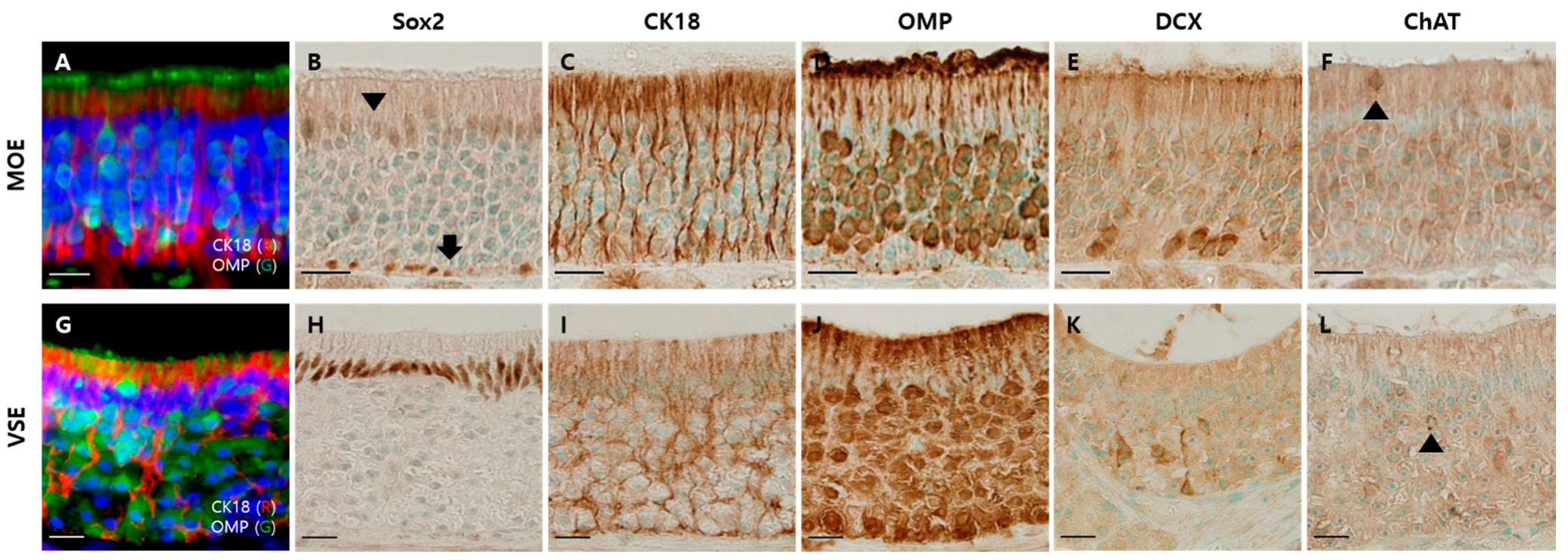
3.1. Components of the Olfactory Epithelium of the Hamsters
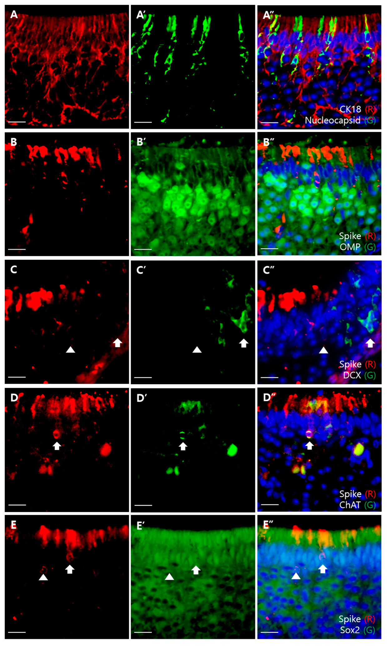
3.2. SARS-CoV-2 Infection in the Olfactory Epithelium
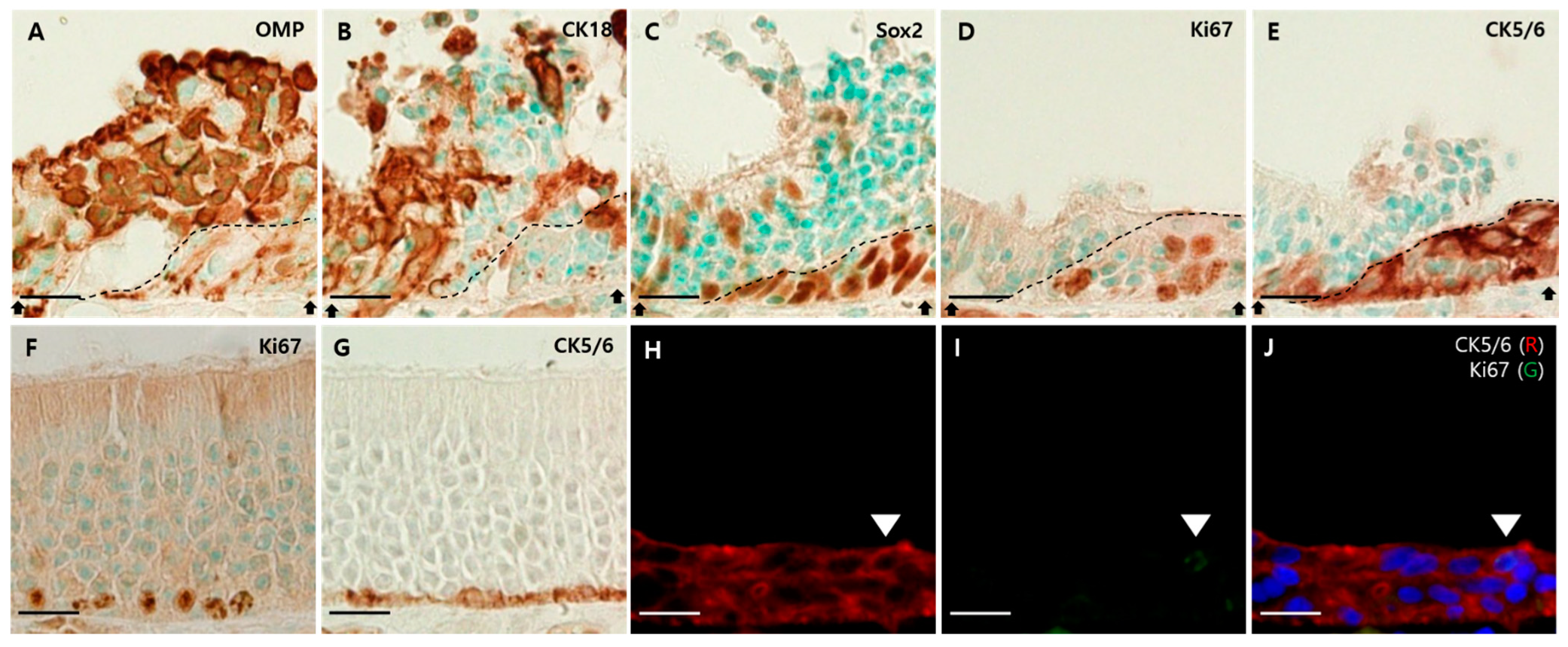
3.3. Pathologic Changes in the Olfactory Epithelium and Its Regeneration
4. Discussion
Author Contributions
Funding
Institutional Review Board Statement
Informed Consent Statement
Data Availability Statement
Conflicts of Interest
References
- Centers for Disease Control and Prevention, Symptom of Coronavirus. Available online: http://www.cdc.gov/coronavirus/2019-ncov/symptoms-testing/symptoms.html (accessed on 22 February 2021).
- Kaye, R.; Chang, C.D.; Kazahaya, K.; Brereton, J.; Denneny, J.C., III. COVID-19 anosmia reporting tool: Initial findings. Otolaryngol. Head Neck Surg. 2020, 163, 132–134. [Google Scholar] [CrossRef]
- Menni, C.; Valdes, A.M.; Freidin, M.B.; Sudre, C.; Nguyen, L.H.; Drew, D.A.; Ganesh, S.; Varsavsky, T.; Cardoso, M.J.; Moustafa, J.S.E.-S.; et al. Real-time tracking of self-reported symptoms to predict potential COVID-19. Nat. Med. 2020, 26, 1037–1040. [Google Scholar] [CrossRef] [PubMed]
- Moein, S.T.; Hashemian, S.M.; Mansourafshar, B.; Khorram-Tousi, A.; Tabarsi, P.; Doty, R.L. Smell dys-function: A biomarker for COVID-19. Int. Forum Allergy Rhinol. Otolaryngol. Head Neck Surg. 2020, 10, 944–950. [Google Scholar] [CrossRef] [PubMed]
- Whitcroft, K.L.; Hummel, T. Olfactory dysfunction in COVID-19: Diagnosis and management. JAMA 2020, 323, 2512–2514. [Google Scholar] [CrossRef] [PubMed]
- Butowt, R.; von Bartheld, C.S. Anosmia in COVID-19: Underlying Mechanisms and Assessment of an Olfactory Route to Brain Infection. Neuroscientist 2020. [Google Scholar] [CrossRef]
- Hoffmann, M.; Kleine-Weber, H.; Schroeder, S.; Krüger, N.; Herrler, T.; Erichsen, S.; Pöhlmann, S. SARS-CoV-2 cell entry depends on ACE2 and TMPRSS2 and is blocked by a clinically proven protease inhibitor. Cell 2020, 181, 271–280. [Google Scholar] [CrossRef]
- Zou, X.; Chen, K.; Zou, J.; Han, P.; Hao, J.; Han, Z. Single-cell RNA-seq data analysis on the receptor ACE2 expression reveals the potential risk of different human organs vulnerable to 2019-nCoV infection. Front. Med. 2020, 14, 185–192. [Google Scholar] [CrossRef] [Green Version]
- Hikmet, F.; Méar, L.; Edvinsson, A.; Micke, P.; Uhlén, M.; Lindskog, C. The protein expression profile of ACE2 in human tissues. Mol. Syst. Biol. 2020, 16, e9610. [Google Scholar] [CrossRef]
- Zhang, A.J.; Lee, A.C.-Y.; Chu, H.; Chan, J.F.-W.; Fan, Z.; Li, C.; Liu, F.; Chen, Y.; Yuan, S.; Poon, V.K.-M.; et al. Severe Acute Respiratory Syndrome Coronavirus 2 Infects and Damages the Mature and Immature Olfactory Sensory Neurons of Hamsters. Clin. Infect. Dis. 2020, 73, e503–e512. [Google Scholar] [CrossRef]
- Fodoulian, L.; Tuberosa, J.; Rossier, D.; Boillat, M.; Kan, C.; Pauli, V.; Rodriguez, I. Sars-Cov-2 receptors and entry genes are expressed in the human olfactory neuroepithelium and brain. iScience 2020, 23, 101839. [Google Scholar] [CrossRef]
- Brann, D.H.; Tsukahara, T.; Weinreb, C.; Lipovsek, M.; Van den Berge, K.; Gong, B.; Datta, S.R. Non-neuronal expression of SARS-CoV-2 entry genes in the olfactory system suggests mechanisms underlying COVID-19-associated anosmia. Sci. Adv. 2020, 6, eabc5801. [Google Scholar] [CrossRef] [PubMed]
- Bilińska, K.; Jakubowska, P.; Von Bartheld, C.S.; Butowt, R. Expression of the SARS-CoV-2 Entry Proteins, ACE2 and TMPRSS2, in Cells of the Olfactory Epithelium: Identification of Cell Types and Trends with Age. ACS Chem. Neurosci. 2020, 11, 1555–1562. [Google Scholar] [CrossRef]
- Butowt, R.; Bilińska, K. SARS-CoV-2: Olfaction, Brain Infection, and the Urgent Need for Clinical Samples Allowing Earlier Virus Detection. ACS Chem. Neurosci. 2020, 11, 1200–1203. [Google Scholar] [CrossRef] [PubMed] [Green Version]
- Chen, M.; Shen, W.; Rowan, N.R.; Kulaga, H.; Hillel, A.; Ramanathan, M.; Lane, A.P. Elevated ACE-2 expression in the olfactory neuroepithelium: Implications for anosmia and upper respiratory SARS-CoV-2 entry and replication. Eur. Respir. J. 2020, 56, 3. [Google Scholar] [CrossRef]
- Ueha, R.; Kondo, K.; Kagoya, R.; Shichino, S.; Shichino, S.; Yamasoba, T. ACE2, TMPRSS2, and Furin expression in the nose and olfactory bulb in mice and humans. Rhinology 2020, 59, 105–109. [Google Scholar]
- Borgmann-Winter, K.; Willard, S.L.; Sinclair, D.; Mirza, N.; Turetsky, B.; Berretta, S.; Hahn, C.G. Translational potential of olfactory mucosa for the study of neuropsychiatric illness. Transl. Psychiatry 2015, 5, e527. [Google Scholar] [CrossRef] [Green Version]
- Sokpor, G.; Abbas, E.; Rosenbusch, J.; Staiger, J.F.; Tuoc, T. Transcriptional and Epigenetic Control of Mammalian Olfactory Epithelium Development. Mol. Neurobiol. 2018, 55, 8306–8327. [Google Scholar] [CrossRef]
- Muñoz-Fontela, C.; Dowling, W.E.; Funnell, S.G.P.; Gsell, P.-S.; Riveros-Balta, A.X.; Albrecht, R.A.; Andersen, H.; Baric, R.S.; Carroll, M.W.; Cavaleri, M.; et al. Animal models for COVID-19. Nature 2020, 586, 509–515. [Google Scholar] [CrossRef]
- Rosenke, K.; Meade-White, K.; Letko, M.; Clancy, C.; Hansen, F.; Liu, Y.; Feldmann, H. Defining the Syrian hamster as a highly susceptible preclinical model for SARS-CoV-2 infection. Emerg. Microb. Infect. 2020, 9, 2673–2684. [Google Scholar] [CrossRef]
- Bryche, B.; St Albin, A.; Murri, S.; Lacôte, S.; Pulido, C.; Gouilh, M.A.; Meunier, N. Massive transient damage of the olfactory epithelium associated with infection of sustentacular cells by SARS-CoV-2 in golden Syrian hamsters. Brain Behav. Immun. 2020, 89, 579–586. [Google Scholar] [CrossRef] [PubMed]
- Sia, S.F.; Yan, L.M.; Chin, A.W.; Fung, K.; Choy, K.T.; Wong, A.Y.; Yen, H.L. Pathogenesis and transmission of SARS-CoV-2 in golden hamsters. Nature 2020, 583, 834–838. [Google Scholar] [CrossRef] [PubMed]
- Holbrook, E.H.; Wu, E.; Curry, W.T.; Lin, D.T.; Schwob, J.E. Immunohistochemical characterization of human ol-factory tissue. Laryngoscope 2011, 121, 1687–1701. [Google Scholar] [CrossRef] [PubMed] [Green Version]
- Kupke, A.; Wenisch, S.; Failing, K.; Herden, C. Intranasal Location and Immunohistochemical Characterization of the Equine Olfactory Epithelium. Front. Neuroanat. 2016, 10, 97. [Google Scholar] [CrossRef] [Green Version]
- Genovese, F.; Tizzano, M. Microvillous cells in the olfactory epithelium express elements of the solitary chemosensory cell transduction signaling cascade. PLoS ONE 2018, 13, e0202754. [Google Scholar] [CrossRef] [Green Version]
- Ramos, M.F.; Baker, J.; Atzpodien, E.-A.; Bach, U.; Brassard, J.; Cartwright, J.; Farman, C.; Fishman, C.; Jacobsen, M.; Junker-Walker, U.; et al. Nonproliferative and Proliferative Lesions of the Ratand Mouse Special Sense Organs (Ocular [eye and glands], Olfactory and Otic). J. Toxicol. Pathol. 2018, 31, 97S–214S. [Google Scholar] [CrossRef] [PubMed] [Green Version]
- Rodewald, A.; Gisder, D.; Gebhart, V.; Oehring, H.; Jirikowski, G. Distribution of olfactory marker protein in the rat vomeronasal organ. J. Chem. Neuroanat. 2016, 77, 19–23. [Google Scholar] [CrossRef]
- Klingenstein, M.; Klingenstein, S.; Neckel, P.H.; Mack, A.F.; Wagner, A.P.; Kleger, A.; Liebau, S.; Milazzo, A. Evidence of SARS-CoV2 Entry Protein ACE2 in the Human Nose and Olfactory Bulb. Cells Tissues Organs 2020, 209, 155–164. [Google Scholar] [CrossRef]
- Meinhardt, J.; Radke, J.; Dittmayer, C.; Franz, J.; Thomas, C.; Mothes, R.; Heppner, F.L. Olfactory transmucosal SARS-CoV-2 invasion as a port of central nervous system entry in individuals with COVID-19. Nat. Neurosci. 2020, 24, 168–175. [Google Scholar] [CrossRef]
- Baxter, B.D.; Larson, E.D.; Merle, L.; Feinstein, P.; Polese, A.G.; Bubak, A.N.; Niemeyer, C.S.; Hassell, J.; Shepherd, D.; Ramakrishnan, V.R.; et al. Transcriptional profiling reveals potential involvement of microvillous TRPM5-expressing cells in viral infection of the olfactory epithelium. BMC Genom. 2021, 22, 1–20. [Google Scholar] [CrossRef]
- Cooper, K.; Brann, D.H.; Farruggia, M.C.; Bhutani, S.; Pellegrino, R.; Tsukahara, T.; Weinreb, C.; Joseph, P.V.; Larson, E.D.; Parma, V.; et al. COVID-19 and the Chemical Senses: Supporting Players Take Center Stage. Neuron 2020, 107, 219–233. [Google Scholar] [CrossRef]
- Ogura, T.; Szebenyi, S.A.; Krosnowski, K.; Sathyanesan, A.; Jackson, J.; Lin, W. Cholinergic microvillous cells in the mouse main olfactory epithelium and effect of acetylcholine on olfactory sensory neurons and supporting cells. J. Neurophysiol. 2011, 106, 1274–1287. [Google Scholar] [CrossRef] [PubMed] [Green Version]
- Lemons, K.; Fu, Z.; Aoude, I.; Ogura, T.; Sun, J.; Chang, J.; Mbonu, K.; Matsumoto, I.; Arakawa, H.; Lin, W. Lack of TRPM5-Expressing Microvillous Cells in Mouse Main Olfactory Epithelium Leads to Impaired Odor-Evoked Responses and Olfactory-Guided Behavior in a Challenging Chemical Environment. eNeuro 2017, 4. [Google Scholar] [CrossRef] [Green Version]
- Ogura, T.; Krosnowski, K.; Zhang, L.; Bekkerman, M.; Lin, W. Chemoreception Regulates Chemical Access to Mouse Vomeronasal Organ: Role of Solitary Chemosensory Cells. PLoS ONE 2010, 5, e11924. [Google Scholar] [CrossRef] [Green Version]
- Saunders, C.J.; Christensen, M.; Finger, T.; Tizzano, M. Cholinergic neurotransmission links solitary chemosensory cells to nasal inflammation. Proc. Natl. Acad. Sci. USA 2014, 111, 6075–6080. [Google Scholar] [CrossRef] [PubMed] [Green Version]
- Deng, J.; Tan, L.H.; Kohanski, M.A.; Kennedy, D.W.; Bosso, J.V.; Adappa, N.D.; Palmer, J.N.; Shi, J.; Cohen, N.A. Solitary chemosensory cells are innervated by trigeminal nerve endings and autoregulated by cholinergic receptors. Int. Forum Allergy Rhinol. 2020, 11, 877–884. [Google Scholar] [CrossRef] [PubMed]
- Perlman, S.; Jacobsen, G.; Afifi, A. Spread of a neurotropic murine coronavirus into the CNS via the trigeminal and olfactory nerves. Virology 1989, 170, 556–560. [Google Scholar] [CrossRef]
- Miura, T.A.; Travanty, E.A.; Oko, L.; Bielefeldt-Ohmann, H.; Weiss, S.R.; Beauchemin, N.; Holmes, K.V. The Spike Glycoprotein of Murine Coronavirus MHV-JHM Mediates Receptor-Independent Infection and Spread in the Central Nervous Systems of Ceacam1a−/− Mice. J. Virol. 2008, 82, 755–763. [Google Scholar] [CrossRef] [Green Version]
- Kachramanoglou, C.; Li, D.; Andrews, P.; Choi, D.; Chen, C.R. Anatomy and Cellular Constituents of the Human Olfactory Mucosa: A Review. J. Neurol. Surg. Part B Skull Base 2014, 75, 293–300. [Google Scholar] [CrossRef] [Green Version]
- Urata, S.; Maruyama, J.; Kishimoto-Urata, M.; Sattler, R.A.; Cook, R.; Lin, N.; Paessler, S. Regeneration profiles of olfactory epithelium after SARS-CoV-2 infection in golden syrian hamsters. ACS Chem. Neurosci. 2021, 12, 589–595. [Google Scholar] [CrossRef]
- Brann, J.H.; Firestein, S.J. A lifetime of neurogenesis in the olfactory system. Front. Neurosci. 2014, 8, 182. [Google Scholar] [CrossRef]
- Schwob, J.E.; Jang, W.; Holbrook, E.H.; Lin, B.; Herrick, D.; Peterson, J.N.; Coleman, J.H. Stem and progenitor cells of the mammalian olfactory epithelium: Taking poietic license. J. Comp. Neurol. 2016, 525, 1034–1054. [Google Scholar] [CrossRef] [PubMed] [Green Version]
- Packard, A.; Schnittke, N.; Romano, R.A.; Sinha, S.; Schwob, J.E. ΔNp63 regulates stem cell dynamics in the mammalian olfactory epithelium. J. Neurosci. 2011, 31, 8748–8759. [Google Scholar] [CrossRef] [PubMed]
- Harrison, A.G.; Lin, T.; Wang, P. Mechanisms of SARS-CoV-2 transmission and pathogenesis. Trends Immunol. 2020, 41, 1100–1115. [Google Scholar] [CrossRef] [PubMed]
- Bannister, L.H.; Dodson, H.C. Endocytic pathways in the olfactory and vomeronasal epithelia of the mouse: Ultra-structure and uptake of tracers. Microsc. Res. Tech. 1992, 23, 128–141. [Google Scholar] [CrossRef] [PubMed]
- McBride, R.; Van Zyl, M.; Fielding, B.C. The Coronavirus Nucleocapsid Is a Multifunctional Protein. Viruses 2014, 6, 2991–3018. [Google Scholar] [CrossRef] [Green Version]
- Witt, M.; Hummel, T. Vomeronasal Versus Olfactory Epithelium: Is There a Cellular Basis for Human Vomeronasal Perception? Int. Rev. Cytol. 2006, 248, 209–259. [Google Scholar] [CrossRef] [PubMed]
- D’Aniello, B.; Semin, G.R.; Scandurra, A.; Pinelli, C. The Vomeronasal Organ: A Neglected Organ. Front. Neuroanat. 2017, 11, 70. [Google Scholar] [CrossRef] [Green Version]
- Stoyanov, G.S.; Matev, B.K.; Valchanov, P.; Sapundzhiev, N.; Young, J.R. The human vomeronasal (Jacobson’s) organ: A short review of current conceptions, with an English translation of Potiquet’s original text. Cureus 2018, 10, 5. [Google Scholar] [CrossRef] [Green Version]
- Daly, J.L.; Simonetti, B.; Klein, K.; Chen, K.E.; Williamson, M.K.; Antón-Plágaro, C.; Yamauchi, Y. Neuropilin-1 is a host factor for SARS-CoV-2 infection. Science 2020, 370, 861–865. [Google Scholar] [CrossRef]
- Cantuti-Castelvetri, L.; Ojha, R.; Pedro, L.D.; Djannatian, M.; Franz, J.; Kuivanen, S.; Simons, M. Neuropilin-1 facilitates SARS-CoV-2 cell entry and infectivity. Science 2020, 370, 856–860. [Google Scholar] [CrossRef]
- Hopkins, C.; Lechien, J.R.; Saussez, S. More that ACE2? NRP1 may play a central role in the underlying patho-physiological mechanism of olfactory dysfunction in COVID-19 and its association with enhanced survival. Med. Hypotheses 2021, 146, 110406. [Google Scholar] [CrossRef] [PubMed]


| Antibody | Species | Dilution | Target | Company | Reference |
|---|---|---|---|---|---|
| ACE2 | Rabbit | 1:200 | SARS-CoV-2 receptor | Novusbio | NBP2-67692 |
| Spike | Mouse | 1:200 | SARS-CoV-2 | GeneTex | GTX632604 |
| Nucleocapsid | Rabbit | 1:200 | SARS-CoV-2 | Novusbio | NB100-56576 |
| OMP | Rabbit | 1:1000 | mOSN and mVSN | Abcam | ab183947 |
| DCX | Rabbit | 1:2000 | iOSN and iVSN | Abcam | ab18723 |
| SOX2 | Rabbit | 1:1000 | SC and some BC | Abcam | ab97959 |
| Ki67 | Rabbit | 1:1000 | Proliferating cell | Abcam | ab15580 |
| CK5/6 | Mouse | 1:100 | HBC | Dako | M7237 |
| CK18 | Mouse | 1:1000 | SC | Abcam | ab668 |
| ChAT | Rabbit | 1:2000 | MC and SCC | Abcam | ab178850 |
| Iba1 | Rabbit | 1:1000 | Monocyte and macrophage | Wako | 019-19741 |
| BAX | Mouse | 1:200 | Apoptotic cell | Santa Cruz | sc-7480 |
| Cleaved caspase 3 | Rabbit | 1:1000 | Apoptotic cell | Abcam | ab2302 |
| Antibody | Species | Company | Reference |
|---|---|---|---|
| Biotinylated anti-mouse IgG | Horse | Vector | BA-2000 |
| Biotinylated anti-rabbit IgG | Horse | Vector | BA-1100 |
| Texas red conjugated anti-mouse IgG | Horse | Vector | TI-2000 |
| FITC-conjugated anti-rabbit IgG | Goat | Santa Cruz | sc-2012 |
| Reference | ACE2 | TMPRSS2 | SARS-CoV-2 |
|---|---|---|---|
| Anna Jinxia Zhang et al. (2020) [10] | In the apical and middle layer (immunostaining in Syrian hamsters) | - | SCs, iOSNs, and mOSNs (immunostaining in Syrian hamsters) |
| Leon Fodoulian et al. (2020) [11] | SCs (single cell RNA sequencing in humans, immunohistochemistry in mice and humans) | SCs (single cell RNA sequencing in humans, immunohistochemistry in mice and humans) | - |
| David H. Brann et al. (2020) [12] | SCs and BCs (bulk and single cell RNA sequencing and immunostaining in mice and humans) | SCs and BCs (bulk and single cell RNA sequencing in mice and humans) | - |
| Katarzyna Bilinska et al. (2020) [13] | SCs (immunostaining in mice) | SCs and iOSNs (ISH) | - |
| Rafal Butowt and Katarzyna Bilinska (2020) [14] | Non-neuronal cells (RNA-seq in mice and humans) | Neuronal and non-neuronal cells (RNA-seq in mice and humans) | - |
| Chen et al. (2020) [15] | SCs (immunostaining in humans) | - | - |
| Ueha et al. (2020) [16] | SCs, OSNs, and BCs (immunostaining in mice), OSNs (immunostaining in humans) | SCs (immunostaining in mice) | |
| Betrand Bryche et al. (2020) [21] | - | - | SCs (immunostaining in Syrian hamsters) |
| Sin Fun Sia et al. (2020) [22] | - | - | OSNs (immunostaining in Syrian hamsters) |
| Klingenstein et al. (2020) [28] | SCs (immunostaining in humans) | SCs (immunostaining in humans) | - |
| Jenny Meinhardt et al. (2020) [29] | - | - | Neural/neuronal cells (immunostaining, ISH, and EM in humans) |
Publisher’s Note: MDPI stays neutral with regard to jurisdictional claims in published maps and institutional affiliations. |
© 2021 by the authors. Licensee MDPI, Basel, Switzerland. This article is an open access article distributed under the terms and conditions of the Creative Commons Attribution (CC BY) license (https://creativecommons.org/licenses/by/4.0/).
Share and Cite
Seo, J.-S.; Yoon, S.-W.; Hwang, S.-H.; Nam, S.-M.; Nahm, S.-S.; Jeong, J.-H.; Lee, J.; Youn, H.-N.; Kim, J.-B.; Kim, W. The Microvillar and Solitary Chemosensory Cells as the Novel Targets of Infection of SARS-CoV-2 in Syrian Golden Hamsters. Viruses 2021, 13, 1653. https://doi.org/10.3390/v13081653
Seo J-S, Yoon S-W, Hwang S-H, Nam S-M, Nahm S-S, Jeong J-H, Lee J, Youn H-N, Kim J-B, Kim W. The Microvillar and Solitary Chemosensory Cells as the Novel Targets of Infection of SARS-CoV-2 in Syrian Golden Hamsters. Viruses. 2021; 13(8):1653. https://doi.org/10.3390/v13081653
Chicago/Turabian StyleSeo, Jin-Seok, Sun-Woo Yoon, Seung-Hyeon Hwang, Sung-Min Nam, Sang-Soep Nahm, Jei-Hyun Jeong, Jiho Lee, Ha-Na Youn, Jun-Beom Kim, and Woosuk Kim. 2021. "The Microvillar and Solitary Chemosensory Cells as the Novel Targets of Infection of SARS-CoV-2 in Syrian Golden Hamsters" Viruses 13, no. 8: 1653. https://doi.org/10.3390/v13081653
APA StyleSeo, J.-S., Yoon, S.-W., Hwang, S.-H., Nam, S.-M., Nahm, S.-S., Jeong, J.-H., Lee, J., Youn, H.-N., Kim, J.-B., & Kim, W. (2021). The Microvillar and Solitary Chemosensory Cells as the Novel Targets of Infection of SARS-CoV-2 in Syrian Golden Hamsters. Viruses, 13(8), 1653. https://doi.org/10.3390/v13081653