Article

Borna Disease Virus 1 Phosphoprotein Forms a Tetramer and Interacts with Host Factors Involved in DNA Double-Strand Break Repair and mRNA Processing

1
Institut de Biologie Structurale (IBS), CEA, CNRS, Université Grenoble Alpes, 38058 Grenoble, France
2
Laboratory of RNA Viruses, Department of Virus Research, Institute for Life and Medical Sciences, Kyoto University, Kyoto 606-8507, Japan
3
Laboratory of RNA Viruses, Department of Mammalian Regulatory Network, Graduate School of Biostudies, Kyoto University, Kyoto 606-8507, Japan
4
Department of Biology, Univ. Lille, Inserm, CHU Lille, U1192—Protéomique Réponse Inflammatoire Spectrométrie de Masse—PRISM, 59000 Lille, France
5
Synchrotron SOLEIL, L’Orme des Merisiers, 91192 Gif-sur-Yvette, France
6
Institut Toulousain des Maladies Infectieuses et Inflammatoires (Infinity), Université de Toulouse, Inserm, CNRS, UPS, 31024 Toulouse, France
7
Department of Molecular Virology, Graduate School of Medicine, Kyoto University, Kyoto 606-8507, Japan
8
Laboratory of Veterinary Microbiology, Graduate School of Veterinary Science, Osaka Metropolitan University, Izumisano 598-8531, Japan
9
Osaka International Research Center for Infectious Diseases, Osaka Metropolitan University, Izumisano 598-8531, Japan
*
Author to whom correspondence should be addressed.
These authors contributed equally to this work.
Viruses 2022, 14(11), 2358; https://doi.org/10.3390/v14112358
Submission received: 23 September 2022 / Revised: 21 October 2022 / Accepted: 24 October 2022 / Published: 26 October 2022
(This article belongs to the Special Issue Bornaviridae)

Abstract

:
Determining the structural organisation of viral replication complexes and unravelling the impact of infection on cellular homeostasis represent important challenges in virology. This may prove particularly useful when confronted with viruses that pose a significant threat to human health, that appear unique within their family, or for which knowledge is scarce. Among Mononegavirales, bornaviruses (family Bornaviridae) stand out due to their compact genomes and their nuclear localisation for replication. The recent recognition of the zoonotic potential of several orthobornaviruses has sparked a surge of interest in improving our knowledge on this viral family. In this work, we provide a complete analysis of the structural organisation of Borna disease virus 1 (BoDV-1) phosphoprotein (P), an important cofactor for polymerase activity. Using X-ray diffusion and diffraction experiments, we revealed that BoDV-1 P adopts a long coiled-coil α-helical structure split into two parts by an original β-strand twist motif, which is highly conserved across the members of whole Orthobornavirus genus and may regulate viral replication. In parallel, we used BioID to determine the proximal interactome of P in living cells. We confirmed previously known interactors and identified novel proteins linked to several biological processes such as DNA repair or mRNA metabolism. Altogether, our study provides important structure/function cues, which may improve our understanding of BoDV-1 pathogenesis.

1. Introduction

Nonsegmented negative-strand RNA viruses (or Mononegavirales) are a class of viruses that infect plants and animals, with many of them causing significant diseases or even death in humans. These enveloped viruses share common strategies in viral replication and/or gene expression [1]. Their genome, encapsulated by the nucleoprotein (N), is the biologically active template for both transcription and replication by the polymerase complex [2] resulting from the association of the viral RNA-dependent RNA polymerase (RdRp or L) with its essential cofactor, phosphoprotein (P). P is a highly versatile protein involved in numerous functions, with a high degree of structural flexibility. Through its modular architecture consisting of long disordered regions that alternate with structured domains, P interacts with the main proteins of the viral RNA synthesis machine. It self-associates in oligomers through a central oligomerisation domain (POD) [3,4,5,6,7,8,9,10,11,12] and bridges the RdRp [13,14,15,16,17,18,19] and the nucleocapsid [20]. It also chaperones N to maintain it in an RNA-free form needed for replication [21,22,23,24,25,26,27,28]. In addition, P is an essential actor of viral replication by recruiting a plethora of cellular partners with proviral activities [10,29,30,31,32,33,34,35,36,37,38,39,40]. The disordered regions of P seem to be of primary importance for these additional functions.
Bornaviruses (family Bornaviridae) occupy a special place within Mononegavirales. They infect the widest range of species of the animal kingdom, ranging from fishes to mammals [41,42,43]. While most mononegaviruses mainly perform cytoplasmic replication, bornaviruses replicate in the nucleus, where they assemble viral factories [44] designated as “viral Speckles Of Transcripts” (vSPOTs). It has been recently shown that some mammalian orthobornaviruses (e.g., Borna disease virus 1 (BoDV-1) and variegated squirrel bornavirus 1 (VSBV-1)) can infect humans, predominantly affecting neuronal tissues and causing fatal encephalitis [45,46,47]. The genome of bornaviriruses consists of approximately 8900 nucleotides and represents the smallest known genome amongst Mononegavirales. BoDV-1 vSPOTs formed by P protein-driven liquid–liquid phase separation [48], are present in the nucleus, in close interaction with the chromatin, where they dock on neuronal DNA double-strand breaks (DSBs), thereby affecting neuronal epigenetics and activity [49]. In particular, both N and P seem to modulate epigenetic signalling in neurons [50,51].
BoDV-1 replicative machinery, a complex between the RdRp (L) and P, produces different mRNAs from the genome via polar sequential transcription, encoding for a total of six viral proteins. Except for the RNA-free N and the matrix protein [52,53], nothing is known concerning the structure of these viral proteins. Here, we used X-ray diffusion and diffraction experiments to provide a complete structural analysis of BoDV-1 P. We observed that P possesses a modular architecture, consisting of a central oligomerisation domain (POD) surrounded by two disordered regions, similar to other Mononegavirales Ps. We also showed that BoDV-1 POD has the most elongated multimerisation domain of all mononegaviruses, with one monomer composed of two consecutive α-helices linked by a short three-amino acid stretch, making a 140 Å long coiled-coil α-helical structure split into two parts by a β-strand twist motif. This particular motif appears to be important for viral replication and is highly conserved across the members of the whole Orthobornavirus genus. Moreover, we determined the BoDV-1 P proximal interactome in human cells, confirming previous interactors and identifying novel proteins linked to DNA breaks and mRNA metabolism. Interestingly, the strong overlap between the proximal interactomes in living cells of N- and C-terminal biotin ligase (BirAR118G, or BirA*)-tagged BoDV-1 P data in living cell suggests that this structure is highly flexible at the β-strand twist motif and/or that the disordered domains confer enough freedom of movement to the BirA* moiety to biotinylate a highly similar protein environment regardless of its position in the primary protein sequence. Altogether, our results provide important advances in our understanding of the structural organisation of BoDV-1 P protein and strongly support the relevance of our structural model in a living cellular context.

2. Materials and Methods

2.1. Cloning and Plasmid Preparation

The bacterial expression-optimised DNA coding sequence of Borna disease virus 1 (BoDV-1) P was purchased from GeneArt (Thermofischer Scientific, Regensburg, Germany). Primers were designed for the amplification of the different constructs of BoDV-1 (residues 1 to 201 and 65 to 172), munia bornavirus 1 (MuBV-1; residues 65 to 172), and Gaboon viper virus 1 (GaVV-1; residues 67 to 178). Each DNA fragment was cloned into pETM11 bacterial expression vector (EMBL) using 5′ end Nco1 and 3′ end Xho1 restriction sites introduced by PCR amplification. All constructs were expressed as N-terminally TEV-removable His-tagged proteins. Sequencing verifications were performed by Eurofins Genomics.

2.2. Protein Expression and Protein Purification

Plasmids with the corresponding constructs were transformed into Escherichia coli BL21 RIL (DE3) cells (Life Technologies, Thermo Fischer Scientific, Villebon-sur-Yvette, France). LB cultures were induced for 12 h with 0.3 mM isopropyl-β-D-thiogalactopyranoside (IPTG) at 18 °C, collected by centrifugation, and stored at −80 °C. Briefly, Bacterial pellets were resuspended in 50 mM Tris-HCl (pH 7.5), 500 mM NaCl, 1 mM β-mercaptoethanol (β-ME), and cOmpleteTM ethylenediaminetetraacetic acid (EDTA)-free protease inhibitor cocktail (Roche, Meylan, France). After lysis by sonication and centrifugation, the supernatant was complemented with 15 mM imidazole and the recombinant proteins were purified by nickel affinity chromatography (resin Ni-NTA; Qiagen, Courtabœuf, France). Based on SDS-PAGE analysis, fractions of interest were pooled together before the addition of 1% tobacco etch virus (TEV) protease and dialysed overnight in 20 mM HEPES (pH 7.5), 200 mM NaCl, and 1 mM β-ME. A second nickel affinity chromatography was performed to remove both the uncleaved protein of interest and TEV protease before the size-exclusion chromatography (SEC). The flow-through was concentrated by centrifugation (Millipore-Amicon Ultra centrifugal filters 10 K) and injected into a HiLoadTM S200 16/600 column (Cytiva, Velizy-Villacoublay, France) with a NGC system (Bio-Rad, Marnes-La-Coquette, France). All proteins were in the same final buffer composed of 20 mM HEPES (pH 7.5), 150 mM NaCl, and 1mM β-ME.

2.3. Crystallisation and X-ray Structure Determination

Crystallisation experiments were initiated using the HTX platform (EMBL, Grenoble, France) before a final manual screening to obtain single crystals. BoDV-1 POD crystals were obtained from a 10 mg·mL−1 protein solution using a precipitant composed of 100 mM HEPES (pH 7.5), 14–18% polyethylene glycol (PEG) 8K, and 200 mM CaCl2. MuBV-1 POD crystals were obtained from a 4 mg·mL−1 protein solution using a precipitant composed of 15–18% PEG 3350, 150 mM Mg(NO₃)₂, and 10 mM phenol. GaVV-1 POD crystals were obtained from a 5 mg·mL−1 protein solution using a precipitant composed of 100 mM HEPES (pH 7.5), 18–22% PEG 3350, and 200 mM NaSCN. Crystals were cryo-protected in their mother liquor solution containing 20% glycerol, mounted on cryoloops (Hampton Research, Aliso Viejo, CA, USA), and frozen in liquid nitrogen.
BoDV-1 POD diffraction experiments were undertaken on ID-29 at the European Synchrotron Radiation Facility (ESRF, Grenoble, France). Data were collected using a Dectris Pilatus 6M detector. Indexing and integration were performed using the XDS program suite [54,55]. Thereafter, data were processed using STARANISO [56], as implemented in autoPROC [57], which applies non-elliptical anisotropic limits based on a locally averaged mean I/σ(I) cut-off, performs a Bayesian estimation of structure amplitudes, and applies an anisotropic correction to the data. Detailed crystallographic statistics are provided in Table S1. Molecular replacement solutions were obtained with Phaser [58] using the measles virus POD structure [6]. The initial solution was then used through repeated cycles of autobuilding using Autobuild [59] within the Phenix suite [60]. The final building phase used both manual model building with COOT [61], together with Autobuild cycles and ArpWarp [62,63]. Refinement of the model was carried out using Refmac5 [64].
MuBV-1 POD diffraction experiments were undertaken on PROXIMA-1 at the SOLEIL Synchrotron (Saint-Aubin, France). Data were collected using a Dectris Eiger-X 16M detector. Indexing and integration were performed using the XDS program suite. Molecular replacement solutions were obtained with Phaser using the BoDV-1 POD tetrameric structure separated into two ensembles: one containing residues 86 to 125 and the other containing residues 127 to 166. The final building phase used both a manual model building with COOT and refinement of the model using Refmac5.
GaVV-1 POD diffraction experiments were undertaken on PROXIMA-1 for the native dataset and on PROXIMA-2A for the Se-SAD dataset at the SOLEIL Synchrotron. PROXIMA-2A is equipped with a Dectris Eiger-X 9M detector. Indexing and integration were performed using the XDS program suite. Heavy atoms sites were determined using the SHELX suite [65], their positions were refined using PHASER in EP mode, and the phases were improved using PARROT [66] for density modification. The resulting electron density allowed the positioning of an AlphaFold2 [67] model, which was improved and refined against the native dataset with COOT and Refmac5.
The figures were drawn using PyMOL [68].

2.4. Small-Angle X-ray Scattering (SAXS) Analysis

All datasets were collected on the SWING beamline at the SOLEIL synchrotron. The collection parameters are listed in Table S2. The samples were loaded onto a SuperdexTM 200 increase 5/150 GL (Cytiva) SEC column and SAXS measurements were performed throughout elution, operating at 20 °C with a flow rate of 0.3 mL·min−1 and a mobile phase containing 20 mM HEPES (pH 7.5), 150 mM NaCl, and 5 mM β-ME. The data were integrated and subtracted with Foxtrot [69], providing the first estimation of I(0) and Rg for each frame of the SEC profile. This allowed the identification along the elution peak of the frames with constant Rg values. The corresponding frames were merged into a final scatter curve used for further analysis. Final Rg values were determined using ATSAS [70] and RAW [71]. Dmax values were determined using GNOM [72] in the ATSAS suite and compared to the values obtained with BIFT [73] implemented in RAW. Molecular weights of the proteins were estimated by using the Qr ratio derived from the invariant volume of correlation [74].

2.5. Ab Initio Molecular Shape Analysis of POD

Ab initio shape analyses were performed using DAMMIF [75] from the ATSAS package. Twenty independent DAMMIF calculations were performed both with and without four-fold (P4) symmetry. In each case, to create the most probable molecular shape, an average shape was determined with DAMAVER [76], with and without P4 symmetry. The filtered models were then reprocessed against the experimental data using DAMMIN [77].

2.6. Full Atom Modelling of BoDV-1 PFULL

An initial pool of 10,000 random conformation models of BoDV PFULL was generated using the Ensemble Optimisation Method (EOM 2.1 [78]). The models generated for the disordered N-terminal (residues 1 to 71) and C-terminal segments (residues 167 to 201) were attached to the POD X-ray structure. As the disordered parts generated by EOM are Cα, we used an in-house procedure to obtain the full atom structures of the pool. First, PD2 ca2main [79] was used to reconstruct the backbone, followed by SCWRL4 [80] to add the side chains. This pool was then used to select a final ensemble with the EOM algorithm based on the experimental scattering curve. This process was repeated three times.

2.7. Minireplicon Assays

Minireplicon assays were conducted as described previously [81]. Briefly, 293T cells seeded onto 48-well plates were transfected with 50 ng of minigenome plasmid containing Gaussia luciferase as a reporter gene [82], 50 ng of pCAGGS-N, 5 ng of pCAGGS-P (or point-mutated P), and 50 ng of pCAGGS-L using TransIT293 (Mirus) according to the manufacturer’s instructions. The culture supernatants were collected at 72 h post transfection and subjected to luciferase assay using a Biolux Gaussia luciferase assay kit (New England BioLabs, Evry, France) according to the manufacturer’s instructions. The assays were performed in technical duplicates for each experiment.

2.8. Determination of Proximal Host–Viral Protein Interactions in Living Cells (BioID)

These experiments were performed as in [83]. Briefly, Flp-In™ T-REx™ HEK293 cells (Life Technologies) cells were co-transfected with pOG44 (encoding the Flp recombinase) and pcDNA5 FRT/TO FlagBirA*- or BirA*Flag PFULL (N- and C-ter tag, respectively) of BoDV-1. Cells were selected with Hygromycin B to generate new stable tetracycline-inducible cell lines expressing single copies of N- and C-terminal tagged PFULL-BirA* fusion proteins. Cultures were expanded to ~100 million cells per sample, before being placed into a medium of 1 µg·mL−1 tetracycline to induce bait protein expression, along with 50 µM biotin. After 24 h, induction cells were collected, washed twice with PBS, spun down, and stored at −80 °C. Each pellet was resuspended in a 5 mL of modified RIPA buffer (50 mM Tris-HCl (pH 7.5), 150 mM NaCl, 1 mM EDTA, 1 mM EGTA, 1% Triton X-100, 0.1% SDS, 1:500 protease inhibitor cocktail (Sigma-Aldrich, Saint-Quentin Fallavier, France), and 1:1000 Turbonuclease (BPS Bioscience, Nanterre, France)), incubated for one hour on an end-over-end rotator at 4 °C, sonicated until no visible aggregates remained, and centrifuged at 45,000× g for 20 min. Cleared supernatants containing all compartment solubilized proteins were transferred to new tubes and incubated with 30 µL of packed and pre-equilibrated streptavidin resin (Ultralink, Pierce, Thermo Fischer Scientific, Villebon-sur-Yvette, France; 2 h under rotation at 4 °C) to capture biotinylated species. Beads were pelleted (300 g, 1 min) and washed six times with 50 mM ammonium bicarbonate (AB). Trypsin (1 µg) was added to each 200 µL sample to digest on beads (overnight at 37 °C). Tryptic peptides were collected in the supernatant, lyophilized, and desalted on C18 ziptips. One-quarter of each sample was analysed by nLC MS-MS (Easy1000 in line with a Thermo Q-Exactive; see Supplemental Methods). The MS raw files were then analysed using MaxQuant (version 1.5.8.3) against the UniProt Reference database to generate the protein identification list with a 1% FDR. High confidence proximal interactors were defined using Perseus (version 1.6.15.0) with the following filters for each given ID: peptides detected in 3/3 bait protein runs; an average Log2 LFQ abundance against average top 3/20 control over 1; and a corresponding q-value < 0.01 (Table S3).

3. Results

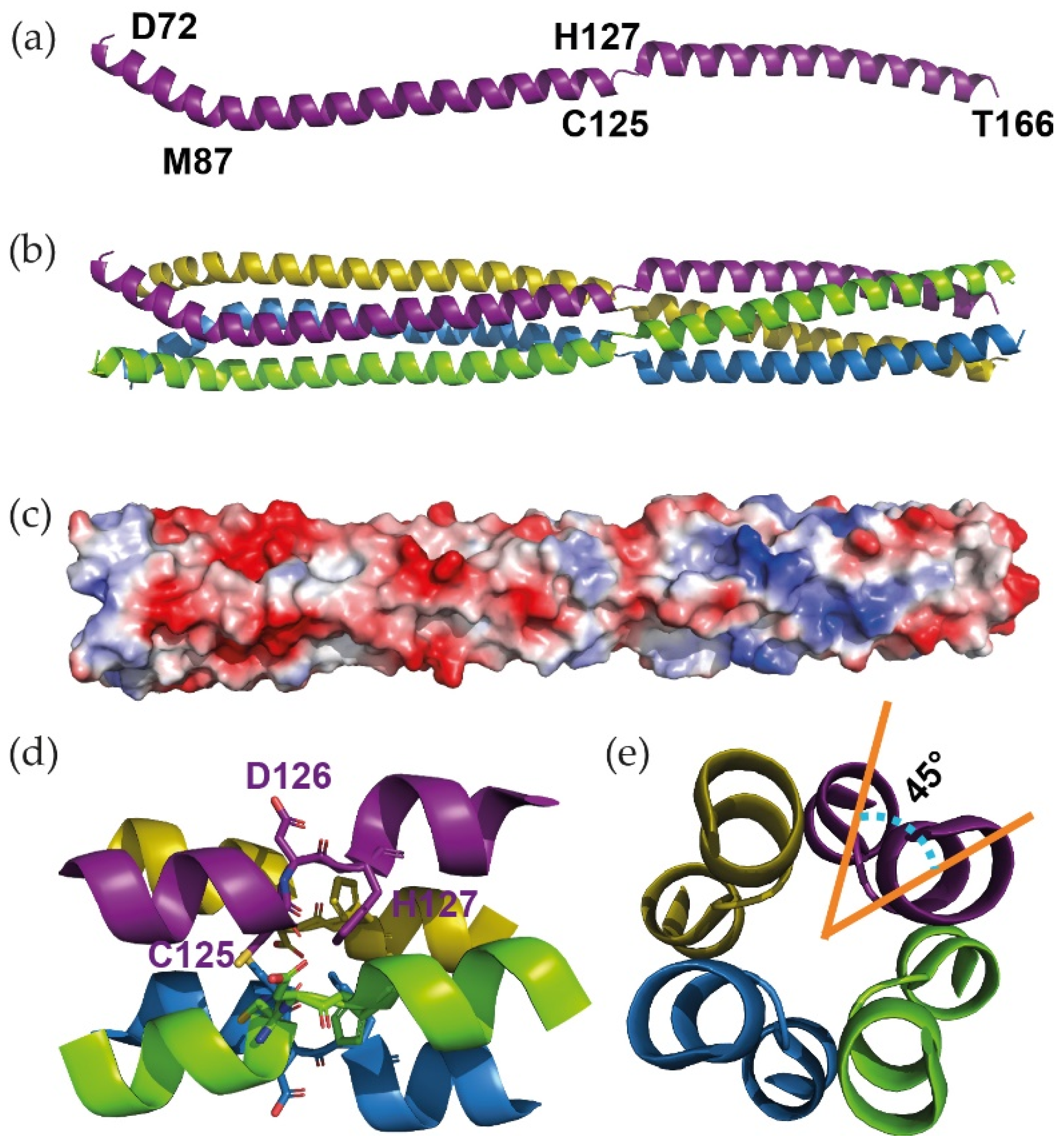
3.1. X-ray Structure of the Oligomerisation Domain of BoDV-1 Phosphoprotein

With Mononegavirales Ps being known as multimodular nonglobular molecules, we first performed a bioinformatic analysis of the BoDV-1 P amino acid sequence using 16 web servers, all of them predicting the location of disordered regions and secondary structure elements. We then combined all of the results in a single representation (D-score; Figure 1a). On this graph, residues with a D-score <0.50 were assigned as disordered, while residues with a D-score >0.5 were assigned as structured. We observed that BoDV-1 P presented an organisation similar to other Mononegavirales Ps, with central folded domains flanked by N- and C-terminal disordered regions. Structure prediction using AlphaFold2 is compatible with the D-score analysis (Figure S11). Based on this prediction, two constructs were designed (Figure 1b): the full-length P (PFULL) and the central domain (from residue 65 to 172) that we hypothesized to be the oligomerisation domain (POD), even if its size represented nearly half of the protein length and was much longer than POD from other viruses.
BoDV-1 POD crystals (fine plates growing in urchins) were obtained in PEG 8000, only in the presence of calcium chloride. Crystals belonged to space group P4212, with cell parameters a = b = 35 Å and c = 166 Å. The analysis of the asymmetric unit indicated that it could only contain a single-amino acid chain (Matthew coefficient of 2.1 Å3·Da−1 with a solvent content of 42% for a 108 amino acid-long chain), meaning that it was likely to contain a single narrow elongated molecule. This informed our choice to use a single coiled-coil chain of the measles virus tetrameric POD [6] as a starting model to solve the structure by molecular replacement. An initial solution was obtained from Phaser [58] and improved using Autobuild [59], before being classically built and refined. The final structure is shown in Figure 2a.
The monomeric POD is composed of two consecutive aligned α-helices (from residue E73 to Q123 and S128 to A164, respectively) linked by a short stretch of three residues, making a ≈ 140 Å long molecule. As most Mononegavirales P assemble in oligomers through their central domain, we analysed POD by SAXS (Table S2) in order to compare its behaviour in the crystal and in solution. With a Dmax of 170 ± 9 Å, it confirmed one dimension of the protein. Furthermore, the volume of correlation (Vc) determination method [74] estimated a Mw of 40 kDa, corresponding to a tetramer (discussed later in Section 3.3). A tetrameric POD could then be reconstituted by applying the inherent 4-fold symmetry of this tetragonal space group (Figure 2b). It was then composed of two α-helical coiled-coils with all the monomers being parallel, with a break centred at position 126 in the middle of the whole structure (Figure 2d and Figure S9). Considering the electrostatic surface of the domain, the break seemed to separate the N-terminal acidic region from the C-terminal more neutral region (Figure 2c). A careful analysis of the Crick parameters [85,86] using CCCP [87] and TWISTER [88] demonstrated a decrease in the helical and superhelical radius together with an increase in the rise per residue and a decrease in the periodicity. In addition, the Ramachandran plot showed that residue D126 fell into the β-area, whereas neighbouring residues (C125 and H127) were in intermediate positions, between the α- and the β-areas (Figure S1a). Altogether, our data point towards the formation of a β-strand twist motif within the helices, as described for trimeric oligomerisation domains [89]. As opposed to the trimers, the tetrameric motif of POD identified herein did not form regular β-sheet main chain hydrogen bonds, and the shift in the helical axis was limited to 45° (Figure 2e) instead of the 120° observed in trimers.

3.2. The β-Strand Twist of BoDV-1 P Is Important for Viral Replication

In order to assess the involvement of the β-strand twist motif in BoDV-1 replication, we performed minireplicon assays using a series of P mutants, each one bearing a single amino acid substitution at the twist or in its proximal regions (from position 124 to 129). 293T cells were then transfected with a minigenome plasmid containing the Gaussia luciferase as a reporter gene, together with plasmids encoding for N, L, and different versions of P. The Gaussia luciferase activity levels were then measured (Figure 3a). Correct expression of each P mutant was confirmed by western blotting (Figure 3b). Interestingly, substitutions by alanine of R124 and D129, two proximal residues of the motif, did not affect the polymerase activity. Rather, the activity was abolished or markedly reduced by mutating C125, D126, or H127. Note that the quantity of protein for this mutant, as well as for the second mutant tested for this position (i.e., C125D), appeared strongly reduced in the western blot analysis. Altogether, these results suggest that the β-strand twist motif is important for BoDV-1 replication, notably concerning D126 and H127 residues, whereas C125 would have an effect on the stability and/or the folding of P. Cysteine residues are known to form disulfide bridges and stabilise the structuration of secreted proteins. Surprisingly for a nuclear protein, such a disulfide bridge has been previously proposed at position 125 of BoDV-1 P [90]. Our experimental conditions did not allow us to detect them, especially in the crystal structure. However, compared to D126, which is strictly conserved (Figure 4j and Figure S8), the amino acid in position 125 is a cysteine only in mammalian 1 orthobornaviruses (i.e., BoDV-1 and -2).

3.3. The β-Strand Twist Motif Is Conserved in Orthobornavirus POD

The comparison with tetrameric oligomerisation domains of P from other Mononegavirales (metapneumovirus, parainfluenza 5, measles, mumps, Nipah and murine respirovirus) showed that BoDV-1 POD was indeed the longest (the size of one monomer in the corresponding X-ray structures is 141, 100, and 93 Å long for BoDV-1, murine respirovirus, and measles POD, respectively). Above all, it is the only one with such a β-strand twist-like motif that breaks the continuity of its α-helical coiled-coil organisation. We therefore investigated its conservation in other members of the Bornaviridae family. Several POD were cloned, expressed, and purified. Crystals were obtained for the POD of munia bornavirus 1 (MuBV-1; 77% sequence identity with BoDV-1) and Gaboon viper virus 1 (GaVV-1; 41 and 43% sequence identity with BoDV-1 and MuBV-1, respectively). The complete SEC-SAXS and X-ray analysis of the three POD were performed (Figure 4).
In solution, the three POD presented the same behaviour. They were all eluted as a single straight symmetric peak from the SEC-SAXS column, without any aggregate signal. Using the Vc determination on the diffusion data, the MW were estimated between 40 and 45 kDa, 12–16% lower than the theoretical values (Table S2 and Figures S2–S4), all corresponding to tetramers. The Guinier transforms measured a hydrodynamic radius (Rg) between 44 and 46 Å and GNOM produced the pair distribution functions that fit with Dmax between 169 and 178 Å. Independently to the X-ray crystallography analysis, ab initio modelling was performed using twenty DAMMIF models averaged by DAMAVER. Averaged models’ correlation with the diffusion curves were then checked using the damstart file as a starting envelope for DAMMIN (Figure 4a–c and Table S2). Each DAMAVER envelope appeared as an elongated tube (Figure 4d–f). In parallel, the X-ray structures of MuBV-1 and GaVV-1 were solved using different determination procedures (Table S1). The structure of MuBV-1 POD was solved by molecular replacement using BoDV-1 POD as a starting model, whereas GaVV-1 POD was obtained de novo using Se-Met derivative crystals. For both MuBV-1 and GaVV-1, the asymmetric units contained a tetrameric POD, whereas the tetramer for BoDV-1 could be obtained after applying the inherent symmetry operations of the tetragonal space group. Figure 4g–i shows the three biological POD (i.e., as tetramers) in the same orientation. The three X-ray structures fit perfectly with the SAXS data. MuBV-1 and GaVV-1 POD presented similar architectures composed of two α-helical coiled-coils (the monomers being parallel) linked by a kink. Globally, the only differences concern the relative spatial position of the two coiled-coils, due to the inherent flexibility induced by such a break (Figure S10) and the crystal packing. MuBV-1 and GaVV-1 breaks were also composed of three residues, with a strictly conserved aspartate in the middle (Figure 4j and Figure S9). The Ramachandran analysis showed that all of them fall into the β-area, with neighbouring residues at intermediate positions (Figure S1); this confirms that this specificity is conserved in all Orthobornavirus P.

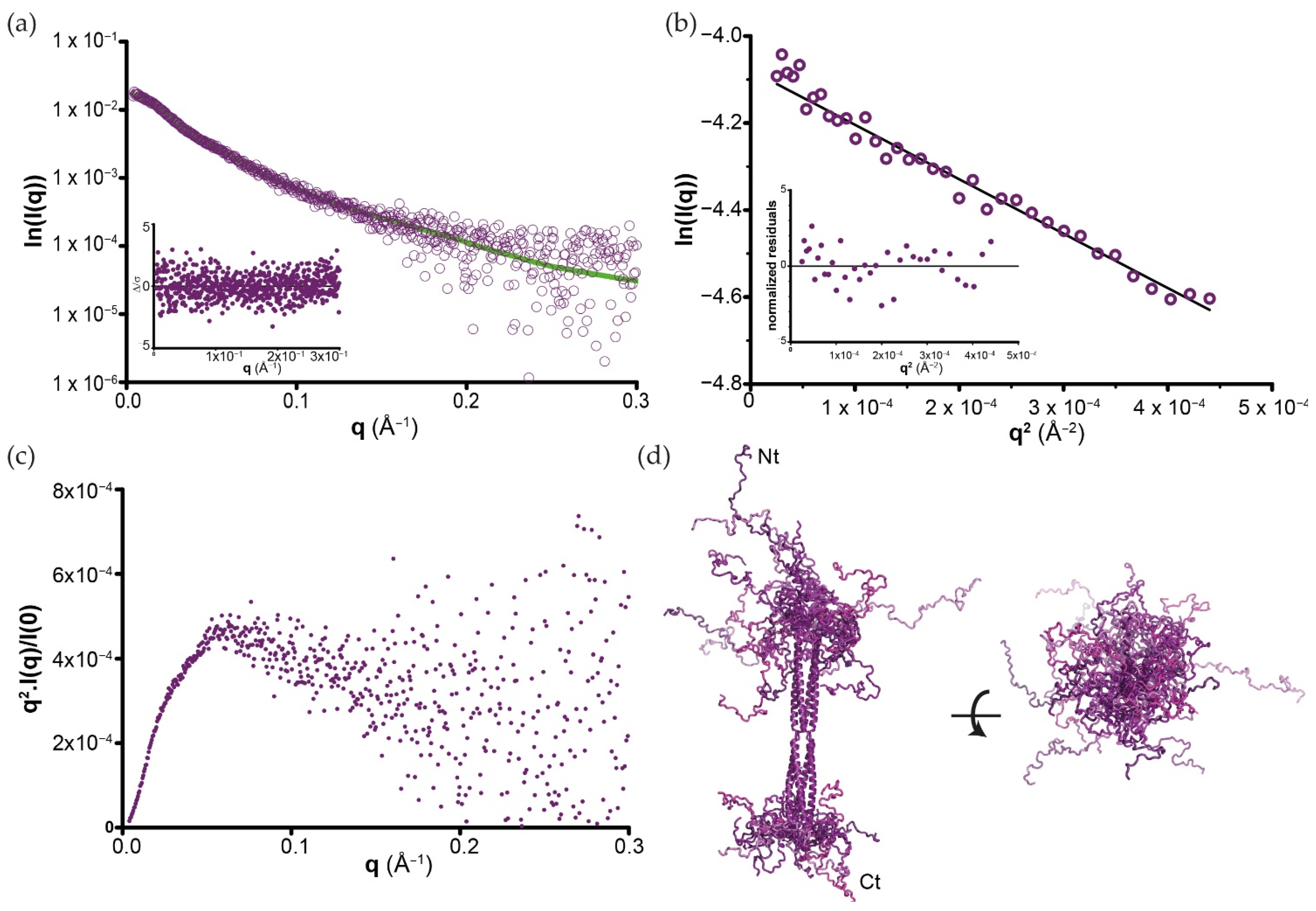
3.4. SAXS Analysis of Full-Length BoDV-1 Phosphoprotein

Having shown that POD assembles as a tetramer, we next analysed BoDV-1 PFULL by SAXS in order to provide a complete view of the protein (Table S2). A PFULL diffusion curve is shown in Figure 5a. Data processing (Figure 5b and Figure S5) indicated that it is elongated for a 201 amino acid-long protein, with a Dmax value of 215 ± 10 Å, and Vc determination gives a value of 90 kDa, in accordance with a tetramer in solution, similar to the behaviour of POD. The main difference between POD and PFULL became apparent when examining the Kratky plots. Indeed, while a nice bell-shaped (Gaussian) peak was observed for POD (Figure S3b), we obtained a curve with a plateau that slowly decays for PFULL (Figure 5c). These two profiles are characteristic of compact/globular and partially unfolded proteins, respectively, thereby validating our bioinformatic analysis. These data also indicate that the biological unit of BoDV-1 P forms a tetramer in solution, with a central domain responsible for this oligomerisation.
Being partially unfolded, the inherent flexibility of BoDV-1 P complicated the analysis of its structural organisation as a single particle. In order to progress in the complete structural analysis of PFULL, we used a modelling procedure based on EOM [78] and the POD X-ray structure discussed in Section 3.2. A total of 10,000 Cα chains corresponding to the missing N- and C-terminal parts were selected to satisfy the SAXS data. Each protein backbone chain was reconstructed from these Cα models [79], and the missing side-chains were finally added with SCWRL4 [80]. Figure 5d shows an ensemble of 6 (out of the 10,000) independent models that fit the SAXS diffusion curves (black line in Figure 5a). This model revealed that despite its rigid central part, BoDV-1 P displays significant flexibility, as illustrated in the figure by the superimposition of the different conformers. However, transitory and/or stable small secondary-structure elements within both N- and C-terminal extremities cannot be excluded in our modelling.

3.5. Determination of BoDV-1 P–Host Proximal Interactomics in Living Cells

To gather insights in PFULL behaviour in human living cells, we identified its proximity interactors using the BioID technique [91]. BioID uniquely provides information on physical and proximal protein-of-interest partners through the conjugation of biotin on the vicinal free ε-amine groups of surrounding proteins (within a 10 nm radius [92]). This covalent modification renders possible the thorough solubilisation of all cell compartments using harsh lysis conditions since protein interactions no longer need to be preserved during the purification process. Briefly, we performed BioID using stable and tetracycline-inducible N- and C-terminal BirA* tagged PFULL HEK293 TREx Flp-In cells and purified the biotinylated proteins on streptavidin columns.
We then identified the proximal interactomes by nLC-MS/MS. Following background removal using a set of FlagBirA* alone samples (localising both in the cytoplasm and the nucleus [93]), we identified a total of 207 high-confidence interactors (Table S3a,b; 147 with FlagBirA*-PFULL and 154 with PFULL-BirA*Flag). As expected, we identified previously reported PFULL interactions (e.g., HMGB1 [44] and DNA break repair machineries), but the vast majority of our dataset was novel and links PFULL to new mechanisms/complexes. In the set of more than 207 interactors (either nuclear or cytoplasmic), 123 were strictly nuclear, 55 could be in both compartments, and 6 were cytoplasmic (Table S3c). These findings are in agreement with the intrinsic ability of PFULL to localise in the nucleus. The relevance of the non-nuclear interactors identified here is subject to caution and could be model-dependent, as discussed hereafter. Word clouds of Gene Ontology enrichment analyses indicated that P could associate with diverse nuclear subcompartments (Figure 6a; e.g., telomeres, nuclear speck, kinetochores) and with factors involved in transcription and DNA repair processes (Figure 6b). Further exploring our data, PFULL BioID identified 44 chromatin-associated partners, with 10 and 22 of them being linked to either replication forks or double-strand break repair, respectively (Table S3d; Figure 6c). PFULL BioID also detected 52 partners linked to RNA metabolism, including 13 components of the U2-type spliceosomal complex and 6 members involved in mRNA polyadenylation. A comparison between N- and C-terminal BirA* tagged PFULL (Table S3a,e; Figure 6d) revealed that N-terminal BirA* preferentially identified the HMGB1 and HMGB2 proteins, with HMGB3 being just below the threshold (p = 0.014) [94,95], as well as dynactin proteins (DCTN2 and DCTN4). This observation suggests two possibilities: (i) HMGB1/2 and DCTN2/4 proteins interact at the N-terminal region of the BoDV-1 phosphoprotein, with a putative structural “locking” of the twist or of the N-terminal disordered region; (ii) C-terminal tagging of P could hinder these interactions. Conversely, C-terminal tagged PFULL preferentially identified two components of the Commander complex (COMMD3 and CCDC22), which are involved in the regulation of NFκB signalling regulation [96]. Similarly, it could indicate an association of these factors with the C-terminal region of PFULL, potentially concomitant with an induced structural stiffness, or that the N-terminal tagging impairs their binding or proximity to PFULL. Finally, several additional proximal partners were associated with the regulation of innate immune responses (i.e., PQBP1 [97] and its paralog CACTIN [98], HEXIM1 [99], and METTL3 [100]), suggesting a putative function of P in BoDV-1-driven innate immune escape.

4. Discussion

By interacting with all the proteins of the replication machinery and recruiting many cellular partners, Mononegavirales P can be considered as the cornerstone of viral proliferation. Their peculiar architecture consisting of long disordered regions that alternate with structured domains is largely related to all these functions. Our structural findings reveal that orthobornaviral P does not deviate from that model, with an extended central oligomerisation domain that represents half of the size of the protein, surrounded by N- and C-terminal flexible parts. In the past, it was speculated that BoDV-1 P assembled in oligomers, from dimers stabilized by an internal disulfide bridge to tetramers [90,101]. With our data, we confirm that orthobornaviral P forms tetramers in solution, which is in complete agreement with these previous works. There were uncertainties regarding the formation of a trimeric or a tetrameric form in these previous studies, which could be related to the fact that the qualitative analysis of such a protein with disordered propensities was performed using a Superose 12 column [101]. Indeed, disordered and globular proteins are known to be eluted differently from SEC, and the column used at that time was less suited to efficiently discriminate proteins with such small differences in composition. Because of our experimental procedures, in particular during the crystallisation step, we cannot totally exclude the presence of a disulfide bridge at position 125 for a non-secreted protein [90]. However, the presence of a disulfide bridge at position 125 may not be of primary importance, since C125 is only conserved in mammalian 1 orthobornaviruses P (Figure S8). For the other orthobornaviruses, position 125 (or similar) is occupied by amino acids with different physico-chemical properties (from serine/threonine to leucine/isoleucine). In contrast, D126 is strictly conserved amongst the members of genus Orthobornavirus, and our minireplicon assays suggested that it plays an important role in viral replication, without any explanation yet. Future studies examining the role of D126 position in the assembly of the polymerase complex and vSPOT formation will help to better understand the role of this key position.
In Mononegavirales, Bornaviridae P is the shortest regarding amino acid composition. However, it bears the most elongated oligomerisation domain, although to date, this feature has only been proven in solution, far from the cellular context. Structural data on the Bornaviridae L-P complex may be particularly relevant for information about P structure within the replication complex. For instance, P may conserve its elongated structuration, similarly to PIV5, where the tetrameric POD protrudes from the globular RdRp [15]. Alternatively, the twist present in BoDV-1 POD may correspond to a hinge that would favour a structural rearrangement of the cofactor to reach an octopus-like organisation intertwined with L, as seen for RSV [14,102]. However, such a rearrangement would involve the destabilisation of the N- or the C-terminal coiled-coil, which would be energetically consuming. The L binding site for P has been described to be located between residues 135 and 183 [101]. This part of the POD corresponds to the most conserved of the two helical coiled-coils (the sequence conservations of the N- and C-terminal α-helixes are 30% and 55%, respectively). The C-terminal α-helix ending at position 162 in our crystal structure, the interaction between L and P could be only mediated by the end of the second α-helix and a stretch of flexible residues up to 183, without any major rearrangement of the cofactor.
Our results also show that BoDV-1 P presents significant inherent flexibility through its N- and C-terminal parts. This is stressed by the high overlap between the N- and C-terminal BioID tagged P proximal interactomes. Comparing the N- and C-terminal BioID tagged proximal interactomes, we observed that only 45% (94/207) of factors are in common. However, when comparing the relative abundance of the high-confidence interactors, over 90% (189/207) did not show significant differences depending on the tag positioning. This apparent discrepancy is due to stringent filtering used to define high-confidence proximal interactors, and the LFQ comparison between merged hits strongly supports the ability of the BirA* moiety to conjugate biotin on proteins located in similar places, whether it is positioned at the N- or C-terminal of P. Given that the biotinylation radius of BirA* has been estimated to be ~10 nm [92], and that that of POD alone is ~14 nm, we hypothesise that the disordered domains remain unstructured in living cells and confer to BirA* enough freedom to move around the POD, thereby biotinylating similar species. Our analysis of the proximal interactomes confirmed the intrinsic ability of BoDV-1 P to localise at DNA DSBs regions. The interactome analysis particularly pointed out a proximity with sensing factors (TP53BP1) or resolution by repair of the DSB (i.e., NBN, BRIP, PPP4). It also supported its role in recruiting numerous mRNA processing factors, which could serve as complementation factors for vRNA production and maturation in the context of BoDV-1 infection (e.g., mRNA polyadenylation machinery). Considering that BioID identifies proximal partners of the fusion bait protein over 24 h (labelling time roughly equal to one cell cycle in HEK293), our data hence represent a collection of all detectable proteins that were at some point in contact with or in the close vicinity of PFULL. Considering that differentiated neurons (the cells targeted by BoDV-1) are postmitotic cells and thus do not enter in mitosis, it is important to stress that our approach must be seen as a discovery step with several limitations to keep in mind. Besides lacking the co-expression of N and L BoDV-1 proteins, which could trigger synergic interactions, labelling over a cell cycle time could explain the presence of a few non-nuclear factors that could interact with BoDV-1 P upon nuclear envelope breakdown (prophase). If this hypothesis is true, non-nuclear interactors identified here should be considered with caution since P expressed alone would remain strictly nuclear. However, since P is translated in the cytoplasm, these interactions could represent P processing factors before its delivery to the nucleoplasm. In line with the previous remark, we also identified replication fork factors. In view of the tropism of BoDV-1 towards neurons, our data regarding the replication fork components must be considered with caution as well. Given the recruitment of DNA repair machineries on stalled forks during replication stress to repair DNA damage and resume replication ([103] for review), we consider the interactions with the replication fork factor to be model-dependent and not necessarily relevant in an infectious context. However, the frequent occurrence of DNA DSBs and the peculiar centrality of the DNA repair machineries in driving neurons somatic mosaicism and diverse neurodegenerative diseases are well documented (reviewed in [104]). We see our data as a discovery step sketching the ability of BoDV-1 phosphoprotein to interact with ubiquitous DNA repair factors. Moreover, these results may be particularly important in the context of novel findings showing that neuronal DSB responses play key roles in learning and memory [105,106]. For instance, our data could serve as a first step to supervised studies in infected neurons aiming at understanding which of these factors are essential for the positioning of vSPOTs at DNA DSBs. In addition, the identification of proximal partners involved in the regulation of innate immune responses suggests that P could interfere with the host cell’s ability to defend against BoDV-1 infection.
Beyond supporting the relevance of our structural model in living cells, our proximal interactomics data represent—to our knowledge—the first published BioID study of any Mononegavirales P. Making sense of what is suspected in the field and providing dozens of nuclear ubiquitous factors, this work hence represents an unprecedented wealth of data, which could direct numerous studies aiming at deciphering BoDV-1 P functions in human cells. Repeating such experiments in an infectious context (i.e., complementing with the other BoDV-1 proteins ideally in neurons) would provide information on the cooperation between viral proteins and better resolve the issue of vSPOT composition.

Supplementary Materials

The following supporting information can be downloaded at https://www.mdpi.com/article/10.3390/v14112358/s1. Experimental procedures: generation of Bio-ID samples; Experimental procedures: BioID data acquisition; Experimental procedures: BioID data analysis; Figure S1: Ramachandran plot of the three POD X-ray structures; Figure S2: Guinier analysis of the POD SAXS analysis; Figure S3: P(R) functions and Krakty plots of the POD SAXS analysis; Figure S4: Orthobornaviral POD SAXS envelops; Figure S5: PFULL P(r) function; Figure S6: Validity of the six PFULL models presented in Figure 2d; Figure S7: Detection of BoDV-1 P mutants; Figure S8: Sequence alignment of P from all representative viruses of the genus Orthobornavirus; Figure S9: Detailed electron density maps; Figure S10: Orthobornaviral POD flexibility; Figure S11: BoDV-1 P structure prediction by AlphaFold2; Table S1: X-ray data collection and refinement parameters; Table S2: SAXS data collection and scattering-derived parameters; Table S3: BioID data and enrichment analyses. References [108,109,110,111] are cited in the supplementary materials.

Author Contributions

Conceptualization, T.C., M.H., D.G.-D. and E.C.; methodology, N.T., F.C., J.K., K.B., J.-M.B., E.M.N.L., P.L., L.L.F. and T.C.; formal analysis, N.T., J.-M.B., M.H., E.C. and T.C.; writing—original draft preparation, E.C., M.H. and T.C.; writing—review and editing, N.T., D.G.-D., M.H., E.C., E.S., R.W.H.R., K.T. and T.C.; funding acquisition, T.C., D.G.-D., E.C. and M.H. All authors have read and agreed to the published version of the manuscript.

Funding

This research was funded by the Agence Nationale de la Recherche (grant Bavarian; ANR-21-CE15-0026) and the Japan Society for the Promotion of Science (Grant-in-Aid for Scientific Research (B): JP21H01199). For this project, F.C. received funding from GRAL (Grenoble Alliance for Integrated Structural and Cell Biology, a program of the Chemistry Biology Health Graduate School of Université Grenoble Alpes (ANR-17-EURE-0003)), K.B. was co-funded by the Ministère de l’Enseignement Supérieur et de la Recherche and the Université de Lille Nord Europe (project MONET), and E.MN.L. was funded by the Métropole européenne de Lille (“Accueil de Talents” funding scheme).

Institutional Review Board Statement

Not applicable.

Informed Consent Statement

Not applicable.

Data Availability Statement

The X-ray diffraction data are available in the protein databank (https://www.rcsb.org/; accessed on 1 September 2022) with the accession numbers 8B8A, 8B8B, and 8B8D for BoDV-1, MuBV-1, and GaVV-1 POD, respectively; the SAXS data can be found in the small-angle scattering biological databank (https://www.sasbdb.org/; accessed on 1 September 2022) with the accession numbers SASDQP5, SASDQQ5, SASDQR5, and SASDQS5 for BoDV-1 PFULL, BoDV-1, MuBV-1, and GaVV-1 POD, respectively; the mass spectrometry proteomics data have been deposited to the ProteomeXchange Consortium via the PRIDE [107] partner repository with the dataset identifier PDX036750 at http://www.ebi.ac.uk/pride; accessed on 1 September 2022 (login: [email protected]; pw: OXUWT9se).

Acknowledgments

We acknowledge Patrice Velicite, Alexandra Mousset, Ambre Valenti, Marwan Hamoudi, and Jean-Pascal Gimeno for technical support, with special thanks to Ada Roy for her involvement. We thank all members of our laboratories for their contributions and helpful discussions. This work used the platforms of the Grenoble Instruct-ERIC centre (ISBG; UAR 3518 CNRS-CEA-UGA-EMBL) within the Grenoble Partnership for Structural Biology (PSB), supported by FRISBI (ANR-10-INBS-0005-02) and GRAL, financed within the University Grenoble Alpes graduate school (Ecoles Universitaires de Recherche) CBH-EUR-GS (ANR-17-EURE-0003). We acknowledge SOLEIL (proposal numbers 20170827 and 20191314) and the ESRF (proposal number MX2285) for provision of synchrotron radiation facilities, and we would like to thank Aurélien Thureau, Pierre Montaville, Andrew Thompson, William Shepard, and Daniele De Sanctis for assistance in using the beamlines SWING, PROXIMA-1, PROXIMA-2A, and ID29, respectively.

Conflicts of Interest

The authors declare no conflict of interest.

References

  1. Whelan, S.P.; Barr, J.N.; Wertz, G.W. Transcription and replication of nonsegmented negative-strand RNA viruses. Curr. Top. Microbiol. Immunol. 2004, 283, 61–119. [Google Scholar]
  2. Kolakofsky, D.; Le Mercier, P.; Nishio, M.; Blackledge, M.; Crepin, T.; Ruigrok, R.W.H. Sendai virus and a unified model of mononegavirus RNA synthesis. Viruses 2021, 13, 2466. [Google Scholar] [CrossRef]
  3. Tarbouriech, N.; Curran, J.; Ruigrok, R.W.; Burmeister, W.P. Tetrameric coiled-coil domain of Sendai virus phosphoprotein. Nat. Struct. Biol. 2000, 7, 777–781. [Google Scholar]
  4. Ding, H.; Green, T.J.; Lu, S.; Luo, M. Crystal structure of the oligomerization domain of the phosphoprotein of vesicular stomatitis virus. J. Virol. 2006, 80, 2808–2814. [Google Scholar] [CrossRef] [PubMed] [Green Version]
  5. Ivanov, I.; Crépin, T.; Jamin, M.; Ruigrok, R.W. Structure of the dimerization domain of the rabies virus phosphoprotein. J. Virol. 2010, 84, 3707–3710. [Google Scholar] [CrossRef] [Green Version]
  6. Communie, G.; Crépin, T.; Maurin, D.; Jensen, M.R.; Blackledge, M.; Ruigrok, R.W. Structure of the tetramerization domain of measles virus phosphoprotein. J. Virol. 2013, 87, 7166–7169. [Google Scholar] [CrossRef] [PubMed] [Green Version]
  7. Cox, R.; Green, T.J.; Purushotham, S.; Deivanayagam, C.; Bedwell, G.J.; Prevelige, P.E.; Luo, M. Structural and functional characterization of the mumps virus phosphoprotein. J. Virol. 2013, 87, 7558–7568. [Google Scholar] [CrossRef] [PubMed] [Green Version]
  8. Bruhn, J.F.; Hotard, A.L.; Spiropoulou, C.F.; Lo, M.K.; Saphire, E.O. A conserved basic patch and central kink in the Nipah virus phosphoprotein multimerization domain are essential for polymerase function. Structure 2019, 27, 660–668 e4. [Google Scholar] [CrossRef] [Green Version]
  9. Zinzula, L.; Nagy, I.; Orsini, M.; Weyher-Stingl, E.; Bracher, A.; Baumeister, W. Structures of Ebola and Reston virus VP35 oligomerization domains and comparative biophysical characterization in all ebolavirus species. Structure 2019, 27, 39–54 e6. [Google Scholar] [CrossRef] [Green Version]
  10. Jensen, M.R.; Yabukarski, F.; Communie, G.; Condamine, E.; Mas, C.; Volchkova, V.; Tarbouriech, N.; Bourhis, J.M.; Volchkov, V.; Blackledge, M.; et al. Structural description of the Nipah virus phosphoprotein and its interaction with STAT1. Biophys. J. 2020, 118, 2470–2488. [Google Scholar] [CrossRef]
  11. Wang, Z.X.; Liu, S.B.; Guan, H.; Lu, L.F.; Tu, J.G.; Ouyang, S.; Zhang, Y.A. Structural and functional characterization of the phosphoprotein central domain of spring viremia of carp virus. J. Virol. 2020, 94. [Google Scholar] [CrossRef] [PubMed]
  12. Cardone, C.; Caseau, C.M.; Bardiaux, B.; Thureaux, A.; Galloux, M.; Bajorek, M.; Eleouet, J.F.; Litaudon, M.; Bontems, F.; Sizun, C. A structural and dynamic analysis of the partially disordered polymerase-binding domain in RSV phosphoprotein. Biomolecules 2021, 11, 1225. [Google Scholar] [CrossRef] [PubMed]
  13. Gilman, M.S.A.; Liu, C.; Fung, A.; Behera, I.; Jordan, P.; Rigaux, P.; Ysebaert, N.; Tcherniuk, S.; Sourimant, J.; Eleouet, J.F.; et al. Structure of the respiratory syncytial virus polymerase complex. Cell 2019, 179, 193–204.e14. [Google Scholar] [CrossRef] [PubMed] [Green Version]
  14. Cao, D.; Gao, Y.; Roesler, C.; Rice, S.; D’Cunha, P.; Zhuang, L.; Slack, J.; Domke, M.; Antonova, A.; Romanelli, S.; et al. Cryo-EM structure of the respiratory syncytial virus RNA polymerase. Nat. Commun. 2020, 11, 368. [Google Scholar] [CrossRef] [Green Version]
  15. Abdella, R.; Aggarwal, M.; Okura, T.; Lamb, R.A.; He, Y. Structure of a paramyxovirus polymerase complex reveals a unique methyltransferase-CTD conformation. Proc. Natl. Acad. Sci. USA 2020, 117, 4931–4941. [Google Scholar] [CrossRef]
  16. Jenni, S.; Bloyet, L.M.; Diaz-Avalos, R.; Liang, B.; Whelan, S.P.J.; Grigorieff, N.; Harrison, S.C. Structure of the vesicular stomatitis virus L protein in complex with its phosphoprotein cofactor. Cell Rep. 2020, 30, 53–60.e5. [Google Scholar] [CrossRef] [Green Version]
  17. Horwitz, J.A.; Jenni, S.; Harrison, S.C.; Whelan, S.P.J. Structure of a rabies virus polymerase complex from electron cryo-microscopy. Proc. Natl. Acad. Sci. USA 2020, 117, 2099–2107. [Google Scholar] [CrossRef] [Green Version]
  18. Pan, J.; Qian, X.; Lattmann, S.; El Sahili, A.; Yeo, T.H.; Jia, H.; Cressey, T.; Ludeke, B.; Noton, S.; Kalocsay, M.; et al. Structure of the human metapneumovirus polymerase phosphoprotein complex. Nature 2020, 577, 275–279. [Google Scholar] [CrossRef]
  19. Gould, J.R.; Qiu, S.; Shang, Q.; Ogino, T.; Prevelige, P.E., Jr.; Petit, C.M.; Green, T.J. The connector domain of vesicular stomatitis virus large protein interacts with the viral phosphoprotein. J. Virol. 2020, 94. [Google Scholar] [CrossRef]
  20. Green, T.J.; Luo, M. Structure of the vesicular stomatitis virus nucleocapsid in complex with the nucleocapsid-binding domain of the small polymerase cofactor, P. Proc. Natl. Acad. Sci. USA 2009, 106, 11713–11718. [Google Scholar] [CrossRef] [Green Version]
  21. Leyrat, C.; Yabukarski, F.; Tarbouriech, N.; Ribeiro, E.A., Jr.; Jensen, M.R.; Blackledge, M.; Ruigrok, R.W.; Jamin, M. Structure of the vesicular stomatitis virus N(0)-P complex. PLoS Pathog. 2011, 7, e1002248. [Google Scholar] [CrossRef] [PubMed]
  22. Yabukarski, F.; Lawrence, P.; Tarbouriech, N.; Bourhis, J.M.; Delaforge, E.; Jensen, M.R.; Ruigrok, R.W.; Blackledge, M.; Volchkov, V.; Jamin, M. Structure of Nipah virus unassembled nucleoprotein in complex with its viral chaperone. Nat. Struct. Mol. Biol. 2014, 21, 754–759. [Google Scholar] [CrossRef] [PubMed]
  23. Guryanov, S.G.; Liljeroos, L.; Kasaragod, P.; Kajander, T.; Butcher, S.J. Crystal structure of the measles virus nucleoprotein core in complex with an N-terminal region of phosphoprotein. J. Virol. 2015, 90, 2849–2857. [Google Scholar] [CrossRef] [PubMed] [Green Version]
  24. Leung, D.W.; Borek, D.; Luthra, P.; Binning, J.M.; Anantpadma, M.; Liu, G.; Harvey, I.B.; Su, Z.; Endlich-Frazier, A.; Pan, J.; et al. An intrinsically disordered peptide from Ebola virus VP35 controls viral RNA synthesis by modulating nucleoprotein-RNA interactions. Cell Rep. 2015, 11, 376–389. [Google Scholar] [CrossRef] [Green Version]
  25. Renner, M.; Bertinelli, M.; Leyrat, C.; Paesen, G.C.; Saraiva de Oliveira, L.F.; Huiskonen, J.T.; Grimes, J.M. Nucleocapsid assembly in pneumoviruses is regulated by conformational switching of the N protein. Elife 2016, 5, e12627. [Google Scholar] [CrossRef]
  26. Zhu, T.; Song, H.; Peng, R.; Shi, Y.; Qi, J.; Gao, G.F. Crystal structure of the Marburg virus nucleoprotein core domain chaperoned by a VP35 peptide reveals a conserved drug target for Filovirus. J. Virol. 2017, 91. [Google Scholar] [CrossRef]
  27. Aggarwal, M.; Leser, G.P.; Kors, C.A.; Lamb, R.A. Structure of the paramyxovirus parainfluenza virus 5 nucleoprotein in complex with an amino-terminal peptide of the phosphoprotein. J. Virol. 2018, 92. [Google Scholar] [CrossRef] [Green Version]
  28. Dong, X.; Wang, X.; Xie, M.; Wu, W.; Chen, Z. Structural basis of human parainfluenza virus 3 unassembled nucleoprotein in complex with its viral chaperone. J. Virol. 2022, 96, e0164821. [Google Scholar] [CrossRef]
  29. Barik, S.; Banerjee, A.K. Phosphorylation by cellular casein kinase II is essential for transcriptional activity of vesicular stomatitis virus phosphoprotein P. Proc. Natl. Acad. Sci. USA 1992, 89, 6570–6574. [Google Scholar] [CrossRef] [Green Version]
  30. Schwemmle, M.; De, B.; Shi, L.; Banerjee, A.; Lipkin, W.I. Borna disease virus P-protein is phosphorylated by protein kinase Cepsilon and casein kinase II. J. Biol. Chem. 1997, 272, 21818–21823. [Google Scholar] [CrossRef] [Green Version]
  31. Dupuy, L.C.; Dobson, S.; Bitko, V.; Barik, S. Casein kinase 2-mediated phosphorylation of respiratory syncytial virus phosphoprotein P is essential for the transcription elongation activity of the viral polymerase; phosphorylation by casein kinase 1 occurs mainly at Ser(215) and is without effect. J. Virol. 1999, 73, 8384–8392. [Google Scholar] [CrossRef] [PubMed] [Green Version]
  32. Raux, H.; Flamand, A.; Blondel, D. Interaction of the rabies virus P protein with the LC8 dynein light chain. J. Virol. 2000, 74, 10212–10216. [Google Scholar] [CrossRef] [PubMed]
  33. Sun, M.; Fuentes, S.M.; Timani, K.; Sun, D.; Murphy, C.; Lin, Y.; August, A.; Teng, M.N.; He, B. Akt plays a critical role in replication of nonsegmented negative-stranded RNA viruses. J. Virol. 2008, 82, 105–114. [Google Scholar] [CrossRef] [PubMed] [Green Version]
  34. Kubota, T.; Matsuoka, M.; Chang, T.H.; Bray, M.; Jones, S.; Tashiro, M.; Kato, A.; Ozato, K. Ebolavirus VP35 interacts with the cytoplasmic dynein light chain 8. J. Virol. 2009, 83, 6952–6956. [Google Scholar] [CrossRef] [PubMed] [Green Version]
  35. Leung, D.W.; Ginder, N.D.; Fulton, D.B.; Nix, J.; Basler, C.F.; Honzatko, R.B.; Amarasinghe, G.K. Structure of the Ebola VP35 interferon inhibitory domain. Proc. Natl. Acad. Sci. USA 2009, 106, 411–416. [Google Scholar] [CrossRef] [Green Version]
  36. Lahaye, X.; Vidy, A.; Fouquet, B.; Blondel, D. Hsp70 protein positively regulates rabies virus infection. J. Virol. 2012, 86, 4743–4751. [Google Scholar] [CrossRef] [Green Version]
  37. Fouquet, B.; Nikolic, J.; Larrous, F.; Bourhy, H.; Wirblich, C.; Lagaudrière-Gesbert, C.; Blondel, D. Focal adhesion kinase is involved in rabies virus infection through its interaction with viral phosphoprotein P. J. Virol. 2015, 89, 1640–1651. [Google Scholar] [CrossRef] [Green Version]
  38. Jespersen, N.E.; Leyrat, C.; Gerard, F.C.; Bourhis, J.M.; Blondel, D.; Jamin, M.; Barbar, E. The LC8-RavP ensemble structure svinces a role for LC8 in regulating lyssavirus polymerase functionality. J. Mol. Biol. 2019, 431, 4959–4977. [Google Scholar] [CrossRef]
  39. Briggs, K.; Wang, L.; Nagashima, K.; Zengel, J.; Tripp, R.A.; He, B. Regulation of mumps virus replication and transcription by kinase RPS6KB1. J. Virol. 2020, 94. [Google Scholar] [CrossRef]
  40. Lim, D.; Shin, H.C.; Choi, J.S.; Kim, S.J.; Ku, B. Crystal structure of human LC8 bound to a peptide from Ebola virus VP35. J. Microbiol. 2021, 59, 410–416. [Google Scholar] [CrossRef]
  41. Kuhn, J.H.; Durrwald, R.; Bao, Y.; Briese, T.; Carbone, K.; Clawson, A.N.; deRisi, J.L.; Garten, W.; Jahrling, P.B.; Kolodziejek, J.; et al. Taxonomic reorganization of the family Bornaviridae. Arch. Virol. 2015, 160, 621–632. [Google Scholar] [CrossRef] [PubMed] [Green Version]
  42. Hyndman, T.H.; Shilton, C.M.; Stenglein, M.D.; Wellehan, J.F.X., Jr. Divergent bornaviruses from Australian carpet pythons with neurological disease date the origin of extant Bornaviridae prior to the end-Cretaceous extinction. PLoS Pathog. 2018, 14, e1006881. [Google Scholar] [CrossRef] [PubMed]
  43. Shi, M.; Lin, X.D.; Chen, X.; Tian, J.H.; Chen, L.J.; Li, K.; Wang, W.; Eden, J.S.; Shen, J.J.; Liu, L.; et al. The evolutionary history of vertebrate RNA viruses. Nature 2018, 556, 197–202. [Google Scholar] [CrossRef] [PubMed]
  44. Matsumoto, Y.; Hayashi, Y.; Omori, H.; Honda, T.; Daito, T.; Horie, M.; Ikuta, K.; Fujino, K.; Nakamura, S.; Schneider, U.; et al. Bornavirus closely associates and segregates with host chromosomes to ensure persistent intranuclear infection. Cell Host Microbe 2012, 11, 492–503. [Google Scholar] [CrossRef] [Green Version]
  45. Hoffmann, B.; Tappe, D.; Hoper, D.; Herden, C.; Boldt, A.; Mawrin, C.; Niederstrasser, O.; Muller, T.; Jenckel, M.; van der Grinten, E.; et al. A variegated squirrel bornavirus associated with fatal human encephalitis. N. Engl. J. Med. 2015, 373, 154–162. [Google Scholar] [CrossRef] [PubMed]
  46. Korn, K.; Coras, R.; Bobinger, T.; Herzog, S.M.; Lucking, H.; Stohr, R.; Huttner, H.B.; Hartmann, A.; Ensser, A. Fatal encephalitis associated with Borna disease virus 1. N. Engl. J. Med. 2018, 379, 1375–1377. [Google Scholar] [CrossRef]
  47. Cadar, D.; Allendorf, V.; Schulze, V.; Ulrich, R.G.; Schlottau, K.; Ebinger, A.; Hoffmann, B.; Hoffmann, D.; Rubbenstroth, D.; Ismer, G.; et al. Introduction and spread of variegated squirrel bornavirus 1 (VSBV-1) between exotic squirrels and spill-over infections to humans in Germany. Emerg. Microbes Infect. 2021, 10, 602–611. [Google Scholar] [CrossRef]
  48. Hirai, Y.; Tomonaga, K.; Horie, M. Borna disease virus phosphoprotein triggers the organization of viral inclusion bodies by liquid-liquid phase separation. Int. J. Biol. Macromol. 2021, 192, 55–63. [Google Scholar] [CrossRef]
  49. Marty, F.H.; Bettamin, L.; Thouard, A.; Bourgade, K.; Allart, S.; Larrieu, G.; Malnou, C.E.; Gonzalez-Dunia, D.; Suberbielle, E. Borna disease virus docks on neuronal DNA double-strand breaks to replicate and dampens neuronal activity. iScience 2022, 25, 103621. [Google Scholar] [CrossRef]
  50. Suberbielle, E.; Stella, A.; Pont, F.; Monnet, C.; Mouton, E.; Lamouroux, L.; Monsarrat, B.; Gonzalez-Dunia, D. Proteomic analysis reveals selective impediment of neuronal remodeling upon Borna disease virus infection. J. Virol. 2008, 82, 12265–12279. [Google Scholar] [CrossRef] [Green Version]
  51. Bonnaud, E.M.; Szelechowski, M.; Betourne, A.; Foret, C.; Thouard, A.; Gonzalez-Dunia, D.; Malnou, C.E. Borna disease virus phosphoprotein modulates epigenetic signaling in neurons to control viral replication. J. Virol. 2015, 89, 5996–6008. [Google Scholar] [CrossRef] [PubMed] [Green Version]
  52. Rudolph, M.G.; Kraus, I.; Dickmanns, A.; Eickmann, M.; Garten, W.; Ficner, R. Crystal structure of the borna disease virus nucleoprotein. Structure 2003, 11, 1219–1226. [Google Scholar] [CrossRef] [PubMed]
  53. Neumann, P.; Lieber, D.; Meyer, S.; Dautel, P.; Kerth, A.; Kraus, I.; Garten, W.; Stubbs, M.T. Crystal structure of the Borna disease virus matrix protein (BDV-M) reveals ssRNA binding properties. Proc. Natl. Acad. Sci. USA 2009, 106, 3710–3715. [Google Scholar] [CrossRef] [PubMed] [Green Version]
  54. Kabsch, W. Automatic processing of rotation diffraction data from crystals of initially unknown symmetry and cell constants. J. Appl. Crystallogr. 1993, 26, 795–800. [Google Scholar] [CrossRef]
  55. Kabsch, W. Integration, scaling, space-group assignment and post-refinement. Acta Crystallogr. D Biol. Crystallogr. 2010, 66 Pt 2, 133–144. [Google Scholar] [CrossRef] [Green Version]
  56. Tickle, I.J.; Flensburg, C.; Keller, P.; Paciorek, W.; Sharff, A.; Vonrhein, C.; Bricogne, G. STARANISO; Global Phasing Ltd.: Cambridge, UK, 2017. [Google Scholar]
  57. Vonrhein, C.; Flensburg, C.; Keller, P.; Sharff, A.; Smart, O.; Paciorek, W.; Womack, T.; Bricogne, G. Data processing and analysis with the autoPROC toolbox. Acta Crystallogr. D Biol. Crystallogr. 2011, 67 Pt 4, 293–302. [Google Scholar] [CrossRef] [Green Version]
  58. McCoy, A.J.; Grosse-Kunstleve, R.W.; Adams, P.D.; Winn, M.D.; Storoni, L.C.; Read, R.J. Phaser crystallographic software. J. Appl. Crystallogr. 2007, 40 Pt 4, 658–674. [Google Scholar] [CrossRef] [Green Version]
  59. Terwilliger, T.C.; Grosse-Kunstleve, R.W.; Afonine, P.V.; Moriarty, N.W.; Zwart, P.H.; Hung, L.W.; Read, R.J.; Adams, P.D. Iterative model building, structure refinement and density modification with the PHENIX AutoBuild wizard. Acta Crystallogr. D Biol. Crystallogr. 2008, 64 Pt 1, 61–69. [Google Scholar] [CrossRef] [Green Version]
  60. Adams, P.D.; Afonine, P.V.; Bunkoczi, G.; Chen, V.B.; Davis, I.W.; Echols, N.; Headd, J.J.; Hung, L.W.; Kapral, G.J.; Grosse-Kunstleve, R.W.; et al. PHENIX: A comprehensive Python-based system for macromolecular structure solution. Acta Crystallogr. D Biol. Crystallogr. 2010, 66 Pt 2, 213–221. [Google Scholar] [CrossRef] [Green Version]
  61. Emsley, P.; Lohkamp, B.; Scott, W.G.; Cowtan, K. Features and development of Coot. Acta Crystallogr. D Biol. Crystallogr. 2010, 66 Pt 4, 486–501. [Google Scholar] [CrossRef] [Green Version]
  62. Cohen, S.X.; Ben Jelloul, M.; Long, F.; Vagin, A.; Knipscheer, P.; Lebbink, J.; Sixma, T.K.; Lamzin, V.S.; Murshudov, G.N.; Perrakis, A. ARP/wARP and molecular replacement: The next generation. Acta Crystallogr. D Biol. Crystallogr. 2008, 64 Pt 1, 49–60. [Google Scholar] [CrossRef] [Green Version]
  63. Langer, G.; Cohen, S.X.; Lamzin, V.S.; Perrakis, A. Automated macromolecular model building for X-ray crystallography using ARP/wARP version 7. Nat. Protoc. 2008, 3, 1171–1179. [Google Scholar] [CrossRef] [PubMed]
  64. Murshudov, G.N. Refinement of macromolecular structures by the maximum-likelihood method. Acta Crystallogr. D Biol. Crystallogr. 1997, 53 Pt 3, 240–255. [Google Scholar] [CrossRef]
  65. Sheldrick, G.M. A short history of SHELX. Acta Crystallogr. A 2008, 64 Pt 1, 112–122. [Google Scholar] [CrossRef] [Green Version]
  66. Cowtan, K. Recent developments in classical density modification. Acta Crystallogr. D Biol. Crystallogr. 2010, 66 Pt 4, 470–478. [Google Scholar] [CrossRef] [Green Version]
  67. Jumper, J.; Evans, R.; Pritzel, A.; Green, T.; Figurnov, M.; Ronneberger, O.; Tunyasuvunakool, K.; Bates, R.; Zidek, A.; Potapenko, A.; et al. Highly accurate protein structure prediction with AlphaFold. Nature 2021, 596, 583–589. [Google Scholar] [CrossRef]
  68. Schrodinger, LLC. The PyMOL Molecular Graphics System, Version 1.8; Schrödinger, Inc.: New York, NY, USA, 2015.
  69. Thureau, A.; Roblin, P.; Perez, J. BioSAXS on the SWING beamline at Synchrotron SOLEIL. J. Appl. Crystallogr. 2021, 54, 1698–1710. [Google Scholar] [CrossRef]
  70. Manalastas-Cantos, K.; Konarev, P.V.; Hajizadeh, N.R.; Kikhney, A.G.; Petoukhov, M.V.; Molodenskiy, D.S.; Panjkovich, A.; Mertens, H.D.T.; Gruzinov, A.; Borges, C.; et al. ATSAS 3.0: Expanded functionality and new tools for small-angle scattering data analysis. J. Appl. Crystallogr. 2021, 54, 343–355. [Google Scholar] [CrossRef] [PubMed]
  71. Hopkins, J.B.; Gillilan, R.E.; Skou, S. BioXTAS RAW: Improvements to a free open-source program for small-angle X-ray scattering data reduction and analysis. J. Appl. Crystallogr. 2017, 50, 1545–1553. [Google Scholar] [CrossRef] [PubMed] [Green Version]
  72. Svergun, D. Determination of the regularization parameter in indirect-transform methods using perceptual criteria. J. Appl. Crystallogr. 1992, 25, 495–503. [Google Scholar] [CrossRef]
  73. Hansen, S. Bayesian estimation of hyperparameters for indirect Fourier transformation in small-angle scattering. J. Appl. Crystallogr. 2000, 33, 1415–1421. [Google Scholar] [CrossRef]
  74. Rambo, R.P.; Tainer, J.A. Accurate assessment of mass, models and resolution by small-angle scattering. Nature 2013, 496, 477–481. [Google Scholar] [CrossRef] [PubMed]
  75. Franke, D.; Svergun, D.I. DAMMIF, a program for rapid ab initio shape determination in small-angle scattering. J. Appl. Crystallogr. 2009, 42 Pt 2, 342–346. [Google Scholar] [CrossRef] [Green Version]
  76. Volkov, V.V.; Svergun, D.I. Uniqueness of ab initio shape determination in small-angle scattering. J. Appl. Crystallogr. 2003, 36, 860–864. [Google Scholar] [CrossRef] [Green Version]
  77. Svergun, D.I. Restoring low resolution structure of biological macromolecules from solution scattering using simulated annealing. Biophys. J. 1999, 76, 2879–2886. [Google Scholar] [CrossRef] [Green Version]
  78. Tria, G.; Mertens, H.D.; Kachala, M.; Svergun, D.I. Advanced ensemble modelling of flexible macromolecules using X-ray solution scattering. IUCrJ 2015, 2 Pt 2, 207–217. [Google Scholar] [CrossRef] [Green Version]
  79. Moore, B.L.; Kelley, L.A.; Barber, J.; Murray, J.W.; MacDonald, J.T. High-quality protein backbone reconstruction from alpha carbons using Gaussian mixture models. J. Comput. Chem. 2013, 34, 1881–1889. [Google Scholar] [CrossRef]
  80. Krivov, G.G.; Shapovalov, M.V.; Dunbrack, R.L., Jr. Improved prediction of protein side-chain conformations with SCWRL4. Proteins 2009, 77, 778–795. [Google Scholar] [CrossRef] [Green Version]
  81. Kanda, T.; Horie, M.; Komatsu, Y.; Tomonaga, K. The Borna disease virus 2 (BoDV-2) nucleoprotein is a conspecific protein that enhances BoDV-1 RNA-dependent RNA polymerase activity. J. Virol. 2021, 95, e0093621. [Google Scholar] [CrossRef] [PubMed]
  82. Reuter, A.; Horie, M.; Höper, D.; Ohnemus, A.; Narr, A.; Rinder, M.; Beer, M.; Staeheli, P.; Rubbenstroth, D. Synergistic antiviral activity of ribavirin and interferon-α against parrot bornaviruses in avian cells. J. Gen. Virol. 2016, 97, 2096–2103. [Google Scholar] [CrossRef]
  83. Tu, Y.X.I.; Sydor, A.M.; Coyaud, E.; Laurent, E.M.N.; Dyer, D.; Mellouk, N.; St-Germain, J.; Vernon, R.M.; Forman-Kay, J.D.; Li, T.; et al. Global proximity interactome of the human macroautophagy pathway. Autophagy 2022, 18, 1174–1186. [Google Scholar] [CrossRef] [PubMed]
  84. Gerard, F.C.; Ribeiro Ede, A., Jr.; Leyrat, C.; Ivanov, I.; Blondel, D.; Longhi, S.; Ruigrok, R.W.; Jamin, M. Modular organization of rabies virus phosphoprotein. J. Mol. Biol. 2009, 388, 978–996. [Google Scholar] [CrossRef] [PubMed]
  85. Crick, F.H.C. The Fourier transform of a coiled-coil. Acta Crystallogr. 1953, 6, 685–689. [Google Scholar] [CrossRef] [Green Version]
  86. Crick, F.H.C. The packing of α-helices: Simple coiled-coils. Acta Crystallogr. 1953, 6, 689–697. [Google Scholar] [CrossRef] [Green Version]
  87. Grigoryan, G.; Degrado, W.F. Probing designability via a generalized model of helical bundle geometry. J. Mol. Biol. 2011, 405, 1079–1100. [Google Scholar] [CrossRef] [Green Version]
  88. Strelkov, S.V.; Burkhard, P. Analysis of alpha-helical coiled-coils with the program TWISTER reveals a structural mechanism for stutter compensation. J. Struct. Biol. 2002, 137, 54–64. [Google Scholar] [CrossRef] [Green Version]
  89. Hartmann, M.D.; Mendler, C.T.; Bassler, J.; Karamichali, I.; Ridderbusch, O.; Lupas, A.N.; Hernandez Alvarez, B. alpha/beta coiled-coils. Elife 2016, 5, 351–358. [Google Scholar] [CrossRef]
  90. Kliche, S.; Stitz, L.; Mangalam, H.; Shi, L.; Binz, T.; Niemann, H.; Briese, T.; Lipkin, W.I. Characterization of the Borna disease virus phosphoprotein, p23. J. Virol. 1996, 70, 8133–8137. [Google Scholar] [CrossRef] [Green Version]
  91. Roux, K.J.; Kim, D.I.; Raida, M.; Burke, B. A promiscuous biotin ligase fusion protein identifies proximal and interacting proteins in mammalian cells. J. Cell Biol. 2012, 196, 801–810. [Google Scholar] [CrossRef] [Green Version]
  92. Kim, D.I.; Birendra, K.C.; Zhu, W.; Motamedchaboki, K.; Doye, V.; Roux, K.J. Probing nuclear pore complex architecture with proximity-dependent biotinylation. Proc. Natl. Acad. Sci. USA 2014, 111, E2453–E2461. [Google Scholar] [CrossRef] [Green Version]
  93. Mehus, A.A.; Anderson, R.H.; Roux, K.J. BioID Identification of Lamin-Associated Proteins. Methods Enzymol. 2016, 569, 3–22. [Google Scholar]
  94. Kamitani, W.; Shoya, Y.; Kobayashi, T.; Watanabe, M.; Lee, B.J.; Zhang, G.; Tomonaga, K.; Ikuta, K. Borna disease virus phosphoprotein binds a neurite outgrowth factor, amphoterin/HMG-1. J. Virol. 2001, 75, 8742–8751. [Google Scholar] [CrossRef] [PubMed] [Green Version]
  95. Zhang, G.; Kobayashi, T.; Kamitani, W.; Komoto, S.; Yamashita, M.; Baba, S.; Yanai, H.; Ikuta, K.; Tomonaga, K. Borna disease virus phosphoprotein represses p53-mediated transcriptional activity by interference with HMGB1. J. Virol. 2003, 77, 12243–12251. [Google Scholar] [CrossRef] [PubMed]
  96. Starokadomskyy, P.; Gluck, N.; Li, H.; Chen, B.; Wallis, M.; Maine, G.N.; Mao, X.; Zaidi, I.W.; Hein, M.Y.; McDonald, F.J.; et al. CCDC22 deficiency in humans blunts activation of proinflammatory NF-kappaB signaling. J. Clin. Investig. 2013, 123, 2244–2256. [Google Scholar] [CrossRef] [PubMed] [Green Version]
  97. Yoh, S.M.; Schneider, M.; Seifried, J.; Soonthornvacharin, S.; Akleh, R.E.; Olivieri, K.C.; De Jesus, P.D.; Ruan, C.; de Castro, E.; Ruiz, P.A.; et al. PQBP1 Is a proximal sensor of the cGAS-dependent innate response to HIV-1. Cell 2015, 161, 1293–1305. [Google Scholar] [CrossRef] [Green Version]
  98. Atzei, P.; Gargan, S.; Curran, N.; Moynagh, P.N. Cactin targets the MHC class III protein IkappaB-like (IkappaBL) and inhibits NF-kappaB and interferon-regulatory factor signaling pathways. J. Biol. Chem. 2010, 285, 36804–36817. [Google Scholar] [CrossRef] [Green Version]
  99. Morchikh, M.; Cribier, A.; Raffel, R.; Amraoui, S.; Cau, J.; Severac, D.; Dubois, E.; Schwartz, O.; Bennasser, Y.; Benkirane, M. HEXIM1 and NEAT1 long non-coding RNA form a multi-subunit complex that regulates DNA-mediated innate immune response. Mol. Cell 2017, 67, 387–399.e5. [Google Scholar] [CrossRef] [PubMed]
  100. Qiu, W.; Zhang, Q.; Zhang, R.; Lu, Y.; Wang, X.; Tian, H.; Yang, Y.; Gu, Z.; Gao, Y.; Yang, X.; et al. N(6)-methyladenosine RNA modification suppresses antiviral innate sensing pathways via reshaping double-stranded RNA. Nat. Commun. 2021, 12, 1582. [Google Scholar] [CrossRef] [PubMed]
  101. Schneider, U.; Blechschmidt, K.; Schwemmle, M.; Staeheli, P. Overlap of interaction domains indicates a central role of the P protein in assembly and regulation of the Borna disease virus polymerase complex. J. Biol. Chem. 2004, 279, 55290–55296. [Google Scholar] [CrossRef] [PubMed] [Green Version]
  102. Gilman, M.S.A.; Furmanova-Hollenstein, P.; Pascual, G.; B van’t Wout, A.; Langedijk, J.P.M.; McLellan, J.S. Transient opening of trimeric prefusion RSV F proteins. Nat. Commun. 2019, 10, 2105. [Google Scholar] [CrossRef] [Green Version]
  103. Cortez, D. Replication-Coupled DNA Repair. Mol. Cell 2019, 74, 866–876. [Google Scholar] [CrossRef] [PubMed] [Green Version]
  104. Qing, X.; Zhang, G.; Wang, Z.Q. DNA damage response in neurodevelopment and neuromaintenance. FEBS J. 2022. [Google Scholar] [CrossRef] [PubMed]
  105. Suberbielle, E.; Sanchez, P.E.; Kravitz, A.V.; Wang, X.; Ho, K.; Eilertson, K.; Devidze, N.; Kreitzer, A.C.; Mucke, L. Physiologic brain activity causes DNA double-strand breaks in neurons, with exacerbation by amyloid-beta. Nat. Neurosci. 2013, 16, 613–621. [Google Scholar] [CrossRef] [PubMed]
  106. Suberbielle, E.; Djukic, B.; Evans, M.; Kim, D.H.; Taneja, P.; Wang, X.; Finucane, M.; Knox, J.; Ho, K.; Devidze, N.; et al. DNA repair factor BRCA1 depletion occurs in Alzheimer brains and impairs cognitive function in mice. Nat. Commun. 2015, 6, 8897. [Google Scholar] [CrossRef] [Green Version]
  107. Perez-Riverol, Y.; Bai, J.; Bandla, C.; Garcia-Seisdedos, D.; Hewapathirana, S.; Kamatchinathan, S.; Kundu, D.J.; Prakash, A.; Frericks-Zipper, A.; Eisenacher, M.; et al. The PRIDE database resources in 2022: A hub for mass spectrometry-based proteomics evidences. Nucleic Acids Res. 2022, 50, D543–D552. [Google Scholar] [CrossRef]
  108. Goedhart, J.; Luijsterburg, M.S. VolcaNoseR is a web app for creating, exploring, labeling and sharing volcano plots. Sci Rep. 2020, 10, 20560. [Google Scholar] [CrossRef] [PubMed]
  109. Williams, C.J.; Headd, J.J.; Moriarty, N.W.; Prisant, M.G.; Videau, L.L.; Deis, L.N.; Verma, V.; Keedy, D.A.; Hintze, B.J.; Chen, V.B.; et al. MolProbity: More and better reference data for improved all-atom structure validation. Protein Sci. 2018, 27, 293–315. [Google Scholar] [CrossRef]
  110. Larkin, M.A.; Blackshields, G.; Brown, N.P.; Chenna, R.; McGettigan, P.A.; McWilliam, H.; Valentin, F.; Wallace, I.M.; Wilm, A.; Lopez, R.; et al. Clustal W and Clustal X version 2.0. Bioinformatics 2007, 23, 2947–2948. [Google Scholar] [CrossRef] [Green Version]
  111. Gouet, P.; Courcelle, E.; Stuart, D.I.; Metoz, F. ESPript: Analysis of multiple sequence alignments in PostScript. Bioinformatics 1999, 15, 305–308. [Google Scholar] [CrossRef]
Figure 1. Predicted architecture of BoDV-1 phosphoprotein. (a) D-score (score for disorder as a function of residue) of BoDV-1 P. The prediction was based on 16 predictor web servers and the D-score was calculated by adding the values for each residue and dividing by the number of used algorithms. We arbitrarily defined a threshold level at 0.50. Residues with a D-score <0.50 were assigned as disordered [84]. (b) Scheme of the BoDV-1 P constructs designed for this work based on the bioinformatics analysis.
Viruses 14 02358 g001
Figure 2. X-ray structure of BoDV-1 POD. (a) Structure of BoDV-1 monomeric POD as obtained in the asymmetric unit. (b) Structure of the tetrameric POD after applying the inherent symmetry operations of the tetragonal space group. The four parallel monomers are coloured in purple, yellow, blue, and green, respectively. (c) Electrostatic surface potential of POD. The potential scale ranges from −5 kT/e (red) to 5 kT/e (blue); the orientation is as in (b). (d) Detail of the β-strand twist motif that separates the two α-helical coiled-coils. (e) Orthogonal view of the β-strand twist motif as shown in (d), showing the 45° rotation of the helical axis around the super helical axis.
Viruses 14 02358 g002
Figure 3. Mutations within the β-strand twist motif of BoDV-1 P modulates viral replication. (a) Minireplicon assay using a series of P mutants. The relative luciferase activities are shown as the ratios relative to wild-type P transfected cells (WT). The indicated BoDV-1 P mutants were used as helper plasmids. The dots show mean values (biological replicates; n = 2). (b) Detection of BoDV-1 P mutants by western blotting. The whole western blot is shown in Figure S7.
Viruses 14 02358 g003
Figure 4. Structural analysis of orthobornaviral POD. Experimental SAXS diffusion curves (with residuals (small plots)) of (a) BoDV-1, (b) MuBV-1, and (c) GaVV-1 POD overlaid with the averaged SAXS curves (black) resulting from the ab initio models shown in panels df. Low-resolution envelops of (d) BoDV-1, (e) MuBV-1, and (f) GaVV-1 POD based on the ab initio modelling from the SAXS data. X-ray structure of (g) BoDV-1, (h) MuBV-1, and (i) GaVV-1 POD with the same orientation (i.e., N-terminal on the left). The SAXS envelops (df) and the X-ray structures (gi) are not at the same scale. Figure S4 presents the dockings of the X-ray structures in the SAXS envelops. (j) Sequence alignment of the corresponding POD with the secondary structures of BoDV-1 and GaVV-1, shown above and below the sequence alignment, respectively. Residues in a solid red background are identical between the three sequences. The green triangle indicates the position of the conserved aspartate residue within the β-strand twist motif.
Viruses 14 02358 g004
Figure 5. Flexibility analysis of BoDV-1 full-length P. (a) Experimental SAXS diffusion curve (deep-purple circles) of PFULL overlaid with the averaged SAXS curves (green) calculated from the EOM ensemble shown in panel (d). (b) Guinier extrapolation for the determination of the Rg. The smallest plot corresponds to the normalised residual. (c) Kratky plot showing the partially unfolded propensity of PFULL samples. (d) Conformational ensemble (6 of 10,000 conformers are shown; these models are perfectly representative of the whole population (Figure S6)) of PFULL obtained from the modelling procedure based on EOM. POD X-ray structure is coloured deep purple, whereas the N- and C-terminal intrinsically disordered regions are coloured with six different shades of purples.
Viruses 14 02358 g005
Figure 6. Proximal interactomes of PFULL in human cells. Word clouds of Gene Ontology enrichment analysis (https://toppgene.cchmc.org/; accessed on 1 September 2022) of Cellular Component (a) and Biological Process (b) (assembled on http://www.bioinformatics.com.cn/plot_basic_wordcloud_118_en; accessed on 1 September 2022). (c) Illustration of a subset of PFULL high-confidence interactors. Proteins are grouped and coloured by selected functions or compartments, as indicated on the diagram; node diameter is proportional to the Log2 fold change against control. (d) Volcano plot comparing the N- and C-terminal BirA* tagged BioID data. Blue and red dots represent N- and C-terminal BirA* tagged preferential partners, respectively (p-value < 0.01 and Log2 fold change > 1 between both fusions). Assembled using https://huygens.science.uva.nl/VolcaNoseR/; accessed on 1 September 2022). All data are based on three biological replicates and have been filtered for background against a set of 20 control runs (see Methods and Supplemental Methods for details).
Viruses 14 02358 g006
Publisher’s Note: MDPI stays neutral with regard to jurisdictional claims in published maps and institutional affiliations.

