
Genomic Diversity of Bacteriophages Infecting the Genus Acinetobacter

 , , , and
Abstract
:1. Introduction
2. Materials and Methods
2.1. Genome Annotation
2.2. Global Profiling of Acinetobacter Phage Genomes
2.3. Genome Clustering
2.4. Cluster Assignment
2.5. Transmission Electron Microscopy
3. Results
3.1. Taxonomy of the Acinetobacter Phages
3.2. T4-like Phages Infecting Acinetobacter sp.
3.3. Phages of the Obolenskvirus Represent a New Subfamily of Myoviruses
3.4. The Cluster Comprising Saclayvirus, Acibel004, and phiAbaA1 Represent a New Subfamily of Myoviruses
3.5. AM24 and R2096 Represent a New Genus of Myoviruses
3.6. Expansion of the Subfamily Beijerinckvirinae
3.7. Temperate Acinetobacter Phages Encode Diverse Complements of Accessory Proteins
3.8. New Strains in the Genus Lokivirus
3.9. Phages DMU1 and SH-Ab15497 Comprise a New Genus of Siphoviruses
3.10. Singleton Phages
3.10.1. Singletons Presley and XC38 Belong to the Family Schitoviridae
3.10.2. Singletons BS46 and B9 Represent a New Subfamily of Myoviruses
3.10.3. Singleton ABPH49 Is a New Species of Vequintavirinae
3.10.4. Singleton SH-Ab 15599 Is a New Species of Ackermannviridae
3.10.5. Arae Is a Genomic Singleton
3.10.6. ME3, TRS5 and BFG Are Unique ‘Jumbo’ Singleton Phages of Acinetobacter
3.11. Select Features of Acinetobacter Phage Proteins
3.11.1. Nucleotide Modification Systems
3.11.2. Lysis Cassettes
3.11.3. Virion Structural Polysaccharide Depolymerases
4. Discussion
Supplementary Materials
Author Contributions
Funding
Institutional Review Board Statement
Informed Consent Statement
Data Availability Statement
Conflicts of Interest
References
- Yutin, N.; Makarova, K.S.; Gussow, A.B.; Krupovic, M.; Segall, A.; Edwards, R.A.; Koonin, E.V. Discovery of an expansive bacteriophage family that includes the most abundant viruses from the human gut. Nat. Microbiol. 2018, 3, 38–46. [Google Scholar] [CrossRef] [PubMed]
- Adriaenssens, E.M. Phage diversity in the human gut microbiome: A taxonomist’s perspective. MSystems 2021, 6, e0079921. [Google Scholar] [CrossRef] [PubMed]
- Adriaenssens, E.M.; Brister, J.R. How to name and classify your phage: An informal guide. Viruses 2017, 9, 70. [Google Scholar] [CrossRef] [Green Version]
- Kyriakidis, I.; Vasileiou, E.; Pana, Z.D.; Tragiannidis, A. Acinetobacter baumannii antibiotic resistance mechanisms. Pathogens 2021, 10, 373. [Google Scholar] [CrossRef] [PubMed]
- Potron, A.; Poirel, L.; Nordmann, P. Emerging broad-spectrum resistance in Pseudomonas aeruginosa and Acinetobacter baumannii: Mechanisms and epidemiology. Int. J. Antimicrob. Agents 2015, 45, 568–585. [Google Scholar] [CrossRef] [Green Version]
- Mea, H.J.; Yong, P.V.C.; Wong, E.H. An overview of Acinetobacter baumannii pathogenesis: Motility, adherence and biofilm formation. Microbiol. Res. 2021, 247, 126722. [Google Scholar] [CrossRef] [PubMed]
- Almasaudi, S.B. Acinetobacter spp. as nosocomial pathogens: Epidemiology and resistance features. Saudi J. Biol. Sci. 2018, 25, 586–596. [Google Scholar] [CrossRef] [PubMed] [Green Version]
- Nemec, A.; Krizova, L.; Maixnerova, M.; van der Reijden, T.J.K.; Deschaght, P.; Passet, V.; Vaneechoutte, M.; Brisse, S.; Dijkshoorn, L. Genotypic and phenotypic characterization of the Acinetobacter calcoaceticus-Acinetobacter baumannii complex with the proposal of Acinetobacter pittii sp. nov (formerly Acinetobacter genomic species 3) and Acinetobacter nosocomialis sp. nov (formerly Acinetobacter genomic species 13TU). Res. Microbiol. 2011, 162, 393–404. [Google Scholar] [CrossRef]
- Rodrigues, D.L.N.; Morais-Rodrigues, F.; Hurtado, R.; dos Santos, R.G.; Costa, D.C.; Barh, D.; Ghosh, P.; Alzahrani, K.J.; Soares, S.C.; Ramos, R.; et al. Pan-resistome insights into the multidrug resistance of Acinetobacter baumannii. Antibiotics 2021, 10, 596. [Google Scholar] [CrossRef]
- Alvarez, V.E.; Quiroga, M.P.; Galan, A.V.; Vilacoba, E.; Quiroga, C.; Ramirez, M.S.; Centron, D. Crucial role of the accessory genome in the evolutionary trajectory of Acinetobacter baumannii global clone 1. Front. Microbiol. 2020, 11, 342. [Google Scholar] [CrossRef] [Green Version]
- Holt, K.; Kenyon, J.J.; Hamidian, M.; Schultz, M.B.; Pickard, D.J.; Dougan, G.; Hall, R. Five decades of genome evolution in the globally distributed, extensively antibiotic-resistant Acinetobacter baumannii global clone 1. Microb. Genom. 2016, 2, e000052. [Google Scholar] [CrossRef]
- Vahhabi, A.; Hasani, A.; Rezaee, M.A.; Baradaran, B.; Samadi Kafil, H.; Abbaszadeh, F.; Dehghani, L. A plethora of carbapenem resistance in Acinetobacter baumannii: No end to a long insidious genetic journey. J. Chemother. 2021, 33, 137–155. [Google Scholar] [CrossRef]
- Turner, D.; Ackermann, H.W.; Kropinski, A.M.; Lavigne, R.; Sutton, J.M.; Reynolds, D.M. Comparative analysis of 37 Acinetobacter bacteriophages. Viruses 2017, 10, 5. [Google Scholar] [CrossRef] [Green Version]
- Seemann, T. Prokka: Rapid prokaryotic genome annotation. Bioinformatics 2014, 30, 2068–2069. [Google Scholar] [CrossRef] [PubMed]
- Grazziotin, A.L.; Koonin, E.V.; Kristensen, D.M. Prokaryotic virus orthologous groups (pVOGs): A resource for comparative genomics and protein family annotation. Nucleic Acids Res. 2017, 45, D491–D498. [Google Scholar] [CrossRef] [PubMed]
- Cook, R.; Brown, N.; Redgwell, T.; Rihtman, B.; Barnes, M.; Clokie, M.; Stekel, D.J.; Hobman, J.; Jones, M.A.; Millard, A. INfrastructure for a PHAge REference Database: Identification of large-scale biases in the current collection of phage genomes. Ther. Appl. Res. 2021, 2, 214–223. [Google Scholar] [CrossRef]
- Shannon, P.; Markiel, A.; Ozier, O.; Baliga, N.S.; Wang, J.T.; Ramage, D.; Amin, N.; Schwikowski, B.; Ideker, T. Cytoscape: A software environment for integrated models of biomolecular interaction networks. Genome Res. 2003, 13, 2498–2504. [Google Scholar] [CrossRef] [PubMed]
- Moraru, C.; Varsani, A.; Kropinski, A.M. VIRIDC—A novel tool to calculate the intergenomic similarities of prokaryote-infecting viruses. Viruses 2020, 12, 1268. [Google Scholar] [CrossRef]
- Bayliss, S.C.; Thorpe, H.A.; Coyle, N.M.; Sheppard, S.K.; Feil, E.J. PIRATE: A fast and scalable pangenomics toolbox for clustering diverged orthologues in bacteria. Gigascience 2019, 8, giz119. [Google Scholar] [CrossRef]
- Jones, P.; Binns, D.; Chang, H.Y.; Fraser, M.; Li, W.; McAnulla, C.; McWilliam, H.; Maslen, J.; Mitchell, A.; Nuka, G.; et al. Interproscan 5: Genome-scale protein function classification. Bioinformatics 2014, 30, 1236–1240. [Google Scholar] [CrossRef] [PubMed] [Green Version]
- Sievers, F.; Higgins, D.G. Clustal Omega for making accurate alignments of many protein sequences. Protein Sci. 2018, 27, 135–145. [Google Scholar] [CrossRef] [PubMed] [Green Version]
- Cresawn, S.G.; Bogel, M.; Day, N.; Jacobs-Sera, D.; Hendrix, R.W.; Hatfull, G.F. Phamerator: A bioinformatic tool for comparative bacteriophage genomics. BMC Bioinform. 2011, 12, 395. [Google Scholar] [CrossRef] [Green Version]
- Huson, D.H.; Bryant, D. Application of phylogenetic networks in evolutionary studies. Mol. Biol. Evol. 2006, 23, 254–267. [Google Scholar] [CrossRef] [PubMed]
- Nguyen, L.T.; Schmidt, H.A.; von Haeseler, A.; Minh, B.Q. IQ-TREE: A fast and effective stochastic algorithm for estimating maximum-likelihood phylogenies. Mol. Biol. Evol. 2015, 32, 268–274. [Google Scholar] [CrossRef] [PubMed]
- Minh, B.Q.; Schmidt, H.A.; Chernomor, O.; Schrempf, D.; Woodhams, M.D.; von Haeseler, A.; Lanfear, R. IQ-TREE 2: New models and efficient methods for phylogenetic inference in the genomic era. Mol. Biol. Evol. 2020, 37, 1530–1534. [Google Scholar] [CrossRef] [Green Version]
- Hoang, D.T.; Chernomor, O.; von Haeseler, A.; Minh, B.Q.; Vinh, L.S. UFBoot2: Improving the ultrafast bootstrap approximation. Mol. Biol. Evol. 2018, 35, 518–522. [Google Scholar] [CrossRef] [PubMed]
- Kalyaanamoorthy, S.; Minh, B.Q.; Wong, T.K.F.; von Haeseler, A.; Jermiin, L.S. ModelFinder: Fast model selection for accurate phylogenetic estimates. Nat. Methods 2017, 14, 587–589. [Google Scholar] [CrossRef] [PubMed] [Green Version]
- Letunic, I.; Bork, P. Interactive Tree Of Life (iTOL) v5: An online tool for phylogenetic tree display and annotation. Nucleic Acids Res. 2021, 49, W293–W296. [Google Scholar] [CrossRef]
- Gilchrist, C.L.M.; Chooi, Y.H. Clinker & clustermap. js: Automatic generation of gene cluster comparison figures. Bioinformatics 2021, 37, 2473–2475. [Google Scholar] [CrossRef]
- Turner, D.; Kropinski, A.M.; Adriaenssens, E.M. A roadmap for genome-based phage taxonomy. Viruses 2021, 13, 506. [Google Scholar] [CrossRef] [PubMed]
- Merabishvili, M.; Vandenheuvel, D.; Kropinski, A.M.; Mast, J.; De Vos, D.; Verbeken, G.; Noben, J.P.; Lavigne, R.; Vaneechoutte, M.; Pirnay, J.P. Characterization of newly isolated lytic bacteriophages active against Acinetobacter baumannii. PLoS ONE 2014, 9, e104853. [Google Scholar] [CrossRef] [Green Version]
- Essoh, C.; Vernadet, J.P.; Vergnaud, G.; Coulibaly, A.; Kakou-N’Douba, A.; N’Guetta, A.S.; Resch, G.; Pourcel, C. Complete genome sequences of five Acinetobacter baumannii phages from Abidjan, Côte d’ivoire. Microbiol. Resour. Announc. 2019, 8, e01358-18. [Google Scholar] [CrossRef] [Green Version]
- Jeon, J.; Park, J.H.; Yong, D. Efficacy of bacteriophage treatment against carbapenem-resistant Acinetobacter baumannii in Galleria mellonella larvae and a mouse model of acute pneumonia. BMC Microbiol. 2019, 19, 70. [Google Scholar] [CrossRef]
- Popova, A.V.; Shneider, M.M.; Myakinina, V.P.; Bannov, V.A.; Edelstein, M.V.; Rubalskii, E.O.; Aleshkin, A.V.; Fursova, N.K.; Volozhantsev, N.V. Characterization of myophage AM24 infecting Acinetobacter baumannii of the K9 capsular type. Arch. Virol. 2019, 164, 1493–1497. [Google Scholar] [CrossRef]
- Loh, B.; Chen, J.; Manohar, P.; Yu, Y.; Hua, X.; Leptihn, S. A biological inventory of prophages in A. baumannii genomes reveal distinct distributions in classes, length, and genomic positions. Front. Microbiol. 2020, 11, 579802. [Google Scholar] [CrossRef]
- Costa, A.R.; Monteiro, R.; Azeredo, J. Genomic analysis of Acinetobacter baumannii prophages reveals remarkable diversity and suggests profound impact on bacterial virulence and fitness. Sci. Rep. 2018, 8, 15346. [Google Scholar] [CrossRef] [Green Version]
- Turner, D.; Wand, M.E.; Briers, Y.; Lavigne, R.; Sutton, J.M.; Reynolds, D.M. Characterisation and genome sequence of the lytic Acinetobacter baumannii bacteriophage vB_AbaS_Loki. PLoS ONE 2017, 12, e0172303. [Google Scholar] [CrossRef]
- Wittmann, J.; Turner, D.; Millard, A.D.; Mahadevan, P.; Kropinski, A.M.; Adriaenssens, E.M. From orphan phage to a proposed new family-the diversity of N4-like viruses. Antibiotics 2020, 9, 663. [Google Scholar] [CrossRef]
- Soothill, J.S. Treatment of experimental infections of mice with bacteriophages. J. Med. Microbiol. 1992, 37, 258–261. [Google Scholar] [CrossRef] [Green Version]
- Popova, A.V.; Shneider, M.M.; Mikhailova, Y.V.; Shelenkov, A.A.; Shagin, D.A.; Edelstein, M.V.; Kozlov, R.S. Complete genome sequence of Acinetobacter baumannii phage BS46. Microbiol. Resour. Announc. 2020, 9, e00398-20. [Google Scholar] [CrossRef]
- Oliveira, H.; Costa, A.R.; Ferreira, A.; Konstantinides, N.; Santos, S.B.; Boon, M.; Noben, J.P.; Lavigne, R.; Azeredo, J. Functional analysis and antivirulence properties of a new depolymerase from a myovirus that infects Acinetobacter baumannii capsule K45. J. Virol. 2019, 93, e01163-18. [Google Scholar] [CrossRef] [PubMed] [Green Version]
- Hua, Y.F.; Luo, T.T.; Yang, Y.Q.; Dong, D.; Wang, R.; Wang, Y.J.; Xu, M.S.; Guo, X.K.; Hu, F.P.; He, P. Phage therapy as a promising new treatment for lung infection caused by carbapenem-resistant Acinetobacter baumannii in mice. Front. Microbiol. 2018, 8, 2659. [Google Scholar] [CrossRef] [PubMed] [Green Version]
- Al-Shayeb, B.; Sachdeva, R.; Chen, L.X.; Ward, F.; Munk, P.; Devoto, A.; Castelle, C.J.; Olm, M.R.; Bouma-Gregson, K.; Amano, Y.; et al. Clades of huge phages from across earth’s ecosystems. Nature 2020, 578, 425–431. [Google Scholar] [CrossRef] [Green Version]
- Hutinet, G.; Kot, W.; Cui, L.; Hillebrand, R.; Balamkundu, S.; Gnanakalai, S.; Neelakandan, R.; Carstens, A.B.; Fa Lui, C.; Tremblay, D.; et al. 7-deazaguanine modifications protect phage DNA from host restriction systems. Nat. Commun. 2019, 10, 5442. [Google Scholar] [CrossRef]
- Kropinski, A.M.; Bose, R.J.; Warren, R.A. 5-(4-aminobutylaminomethyl)uracil, an unusual pyrimidine from the deoxyribonucleic acid of bacteriophage phiW-14. Biochemistry 1973, 12, 151–157. [Google Scholar] [CrossRef]
- Witmer, H. Synthesis of deoxythymidylate and the unusual deoxynucleotide in mature DNA of Bacillus subtilis bacteriophage SP10 occurs by postreplicational modification of 5-hydroxymethyldeoxyuridylate. J. Virol. 1981, 39, 536–547. [Google Scholar] [CrossRef] [Green Version]
- Lee, Y.J.; Dai, N.; Walsh, S.E.; Muller, S.; Fraser, M.E.; Kauffman, K.M.; Guan, C.; Correa, I.R., Jr.; Weigele, P.R. Identification and biosynthesis of thymidine hypermodifications in the genomic DNA of widespread bacterial viruses. Proc. Natl. Acad. Sci. USA 2018, 115, E3116–E3125. [Google Scholar] [CrossRef] [PubMed] [Green Version]
- Weigele, P.; Raleigh, E.A. Biosynthesis and function of modified bases in bacteria and their viruses. Chem. Rev. 2016, 116, 12655–12687. [Google Scholar] [CrossRef] [PubMed]
- Timoshina, O.Y.; Shneider, M.M.; Evseev, P.V.; Shchurova, A.S.; Shelenkov, A.A.; Mikhaylova, Y.V.; Sokolova, O.S.; Kasimova, A.A.; Arbatsky, N.P.; Dmitrenok, A.S.; et al. Novel Acinetobacter baumannii bacteriophage Aristophanes encoding structural polysaccharide deacetylase. Viruses 2021, 13, 1688. [Google Scholar] [CrossRef] [PubMed]
- Roucourt, B.; Lavigne, R. The role of interactions between phage and bacterial proteins within the infected cell: A diverse and puzzling interactome. Environ. Microbiol. 2009, 11, 2789–2805. [Google Scholar] [CrossRef] [PubMed]
- Wan, X.; Hendrix, H.; Skurnik, M.; Lavigne, R. Phage-based target discovery and its exploitation towards novel antibacterial molecules. Curr. Opin. Biotechnol. 2021, 68, 1–7. [Google Scholar] [CrossRef] [PubMed]
- Leskinen, K.; Blasdel, B.G.; Lavigne, R.; Skurnik, M. RNA-sequencing reveals the progression of phage-host interactions between ɸR1-37 and Yersinia enterocolitica. Viruses 2016, 8, 111. [Google Scholar] [CrossRef] [PubMed] [Green Version]
- Luo, P.; Lui, Q.; Deng, Y.; Tian, Y.; Yun, L.; Hu, C. Strand-specific RNA-Seq analysis provides first insight into transcriptome response of Vibrio alginolyticus to phage infection. Mar. Genom. 2018, 38, 5–8. [Google Scholar] [CrossRef]
- Blasdel, B.G.; Chevallereau, A.; Monot, M.; Lavigne, R.; Debarbieux, L. Comparative transcriptomics analyses reveal the conservation of an ancestral infectious strategy in two bacteriophage genera. ISME J. 2017, 11, 1988–1996. [Google Scholar] [CrossRef] [Green Version]
- Wicke, L.; Ponath, P.; Coppens, L.; Gerovac, M.; Lavigne, R.; Vogel, J. Introducing differential RNA-seq mapping to track the early infection phase for Pseudomonas phage ɸKZ. RNA Biol. 2021, 18, 1099–1110. [Google Scholar] [CrossRef]
- Yang, Z.C.; Yi, S.P.; Li, G.; Wang, J.; Huang, G.T.; Jiang, B.; You, B.; Gong, Y.L.; Zhang, C.; Luo, X.Q.; et al. Global transcriptomic analysis of the interactions between phage phi abp1 and extensively drug-resistant Acinetobacter baumannii. Msystems 2019, 4, e00068-19. [Google Scholar] [CrossRef] [Green Version]
- Theuretzbacher, U.; Bush, K.; Harbarth, S.; Paul, M.; Rex, J.H.; Tacconelli, E.; Thwaites, G.E. Critical analysis of antibacterial agents in clinical development. Nat. Rev. Microbiol. 2020, 18, 286–298. [Google Scholar] [CrossRef] [Green Version]
- WHO. 2020 Antibacterial Agents in Clinical and Preclinical Development: An Overview and Analysis; World Health Organization: Geneva, Switzerland, 2021. [Google Scholar]
- Knecht, L.E.; Veljkovic, M.; Fieseler, L. Diversity and function of phage encoded depolymerases. Front. Microbiol. 2019, 10, 2949. [Google Scholar] [CrossRef] [PubMed]
- Singh, J.K.; Adams, F.G.; Brown, M.H. Diversity and function of capsular polysaccharide in Acinetobacter baumannii. Front. Microbiol. 2018, 9, 3301. [Google Scholar] [CrossRef]
- Santos, S.B.; Melo, L.D.R.; Oliveira, H. Phage structural antimicrobial proteins. In Exploitation for Biocontrol and Therapeutics; Coffey, A., Buttimer, C., Eds.; Caister Academic: Poole, UK, 2020; pp. 419–476. [Google Scholar]
- Greenfield, J.; Shang, X.; Luo, H.; Zhou, Y.; Heselpoth, R.D.; Nelson, D.C.; Herzberg, O. Structure and tailspike glycosidase machinery of ORF212 from E. coli O157:H7 phage CBA120 (TSP3). Sci. Rep. 2019, 9, 7349. [Google Scholar] [CrossRef]
- Oliveira, H.; Costa, A.R.; Konstantinides, N.; Ferreira, A.; Akturk, E.; Sillankorva, S.; Nemec, A.; Shneider, M.; Dotsch, A.; Azeredo, J. Ability of phages to infect Acinetobacter calcoaceticus-Acinetobacter baumannii complex species through acquisition of different pectate lyase depolymerase domains. Environ. Microbiol. 2017, 19, 5060–5077. [Google Scholar] [CrossRef] [Green Version]
- Prokhorov, N.S.; Riccio, C.; Zdorovenko, E.L.; Shneider, M.M.; Browning, C.; Knirel, Y.A.; Leiman, P.G.; Letarov, A.V. Function of bacteriophage G7C esterase tailspike in host cell adsorption. Mol. Microbiol. 2017, 105, 385–398. [Google Scholar] [CrossRef] [PubMed]
- Oliveira, H.; Mendes, A.; Fraga, A.G.; Ferreira, A.; Pimenta, A.I.; Mil-Homens, D.; Fialho, A.M.; Pedrosa, J.; Azeredo, J. K2 capsule depolymerase is highly stable, is refractory to resistance, and protects larvae and mice from Acinetobacter baumannii sepsis. Appl. Environ. Microbiol. 2019, 85, e00934-19. [Google Scholar] [CrossRef] [Green Version]
- Popova, A.V.; Lavysh, D.G.; Klimuk, E.I.; Edelstein, M.V.; Bogun, A.G.; Shneider, M.M.; Goncharov, A.E.; Leonov, S.V.; Severinov, K.V. Novel Fri1-like viruses infecting Acinetobacter baumannii-vB_AbaP_AS11 and vB_AbaP_AS12-characterization, comparative genomic analysis, and host-recognition strategy. Viruses 2017, 9, 188. [Google Scholar] [CrossRef] [Green Version]
- Liu, Y.; Leung, S.S.Y.; Guo, Y.; Zhao, L.; Jiang, N.; Mi, L.; Li, P.; Wang, C.; Qin, Y.; Mi, Z.; et al. The capsule depolymerase Dpo48 rescues Galleria mellonella and mice from Acinetobacter baumannii systemic infections. Front. Microbiol. 2019, 10, 545. [Google Scholar] [CrossRef]
- Popova, A.V.; Shneider, M.M.; Arbatsky, N.P.; Kasimova, A.A.; Senchenkova, S.N.; Shashkov, A.S.; Dmitrenok, A.S.; Chizhov, A.O.; Mikhailova, Y.V.; Shagin, D.A.; et al. Specific interaction of novel Friunavirus phages encoding tailspike depolymerases with corresponding Acinetobacter baumannii capsular types. J. Virol. 2020, 95, e01714-20. [Google Scholar] [CrossRef]
- Knirel, Y.A.; Shneider, M.M.; Popova, A.V.; Kasimova, A.A.; Senchenkova, S.N.; Shashkov, A.S.; Chizhov, A.O. Mechanisms of Acinetobacter baumannii capsular polysaccharide cleavage by phage depolymerases. Biochemistry 2020, 85, 567–574. [Google Scholar] [CrossRef]
- Domingues, R.; Barbosa, A.; Santos, S.B.; Pires, D.P.; Save, J.; Resch, G.; Azeredo, J.; Oliveira, H. Unpuzzling Friunavirus-host interactions one piece at a time: Phage recognizes Acinetobacter pittii via a new k38 capsule depolymerase. Antibiotics 2021, 10, 1304. [Google Scholar] [CrossRef] [PubMed]
- Latka, A.; Lemire, S.; Grimon, D.; Dams, D.; Maciejewska, B.; Lu, T.; Drulis-Kawa, Z.; Briers, Y. Engineering the modular receptor-binding proteins of Klebsiella phages switches their capsule serotype specificity. Mbio 2021, 12, e00455-21. [Google Scholar] [CrossRef] [PubMed]
- Kongari, R.; Rajaure, M.; Cahill, J.; Rasche, E.; Mijalis, E.; Berry, J.; Young, R. Phage spanins: Diversity, topological dynamics and gene convergence. BMC Bioinform. 2018, 19, 326. [Google Scholar] [CrossRef] [PubMed] [Green Version]
- Lai, M.J.; Lin, N.T.; Hu, A.; Soo, P.C.; Chen, L.K.; Chen, L.H.; Chang, K.C. Antibacterial activity of Acinetobacter baumannii phage ɸAB2 endolysin (LysAB2) against both gram-positive and gram-negative bacteria. Appl. Microbiol. Biotechnol. 2011, 90, 529–539. [Google Scholar] [CrossRef] [PubMed]
- Lood, R.; Winer, B.Y.; Pelzek, A.J.; Diez-Martinez, R.; Thandar, M.; Euler, C.W.; Schuch, R.; Fischetti, V.A. Novel phage lysin capable of killing the multidrug-resistant gram-negative bacterium Acinetobacter baumannii in a mouse bacteremia model. Antimicrob. Agents Chemother. 2015, 59, 1983–1991. [Google Scholar] [CrossRef] [PubMed] [Green Version]
- Holt, A.; Cahill, J.; Ramsey, J.; Martin, C.; O’Leary, C.; Moreland, R.; Maddox, L.T.; Galbadage, T.; Sharan, R.; Sule, P.; et al. Phage-encoded cationic antimicrobial peptide required for lysis. J. Bacteriol. 2021, JB0021421. [Google Scholar] [CrossRef] [PubMed]
- Oliveira, P.H.; Touchon, M.; Rocha, E.P. The interplay of restriction-modification systems with mobile genetic elements and their prokaryotic hosts. Nucleic Acids Res. 2014, 42, 10618–10631. [Google Scholar] [CrossRef] [PubMed]
- Pingoud, A.; Wilson, G.G.; Wende, W. Type II restriction endonucleases-a historical perspective and more. Nucleic Acids Res. 2014, 42, 7489–7527. [Google Scholar] [CrossRef] [PubMed]
- Loenen, W.A.; Dryden, D.T.; Raleigh, E.A.; Wilson, G.G.; Murray, N.E. Highlights of the DNA cutters: A short history of the restriction enzymes. Nucleic Acids Res. 2014, 42, 3–19. [Google Scholar] [CrossRef] [PubMed] [Green Version]
- Sanchez-Romero, M.A.; Casadesus, J. The bacterial epigenome. Nat. Rev. Microbiol. 2020, 18, 7–20. [Google Scholar] [CrossRef]
 A;
A;  B;
B;  C;
C;  D;
D;  E;
E;  F;
F;  G;
G;  H;
H;  I;
I;  J;
J;  K;
K;  L;
L;  M.
A; B; C; D; E; F; G; H; I; J; K; L; M.
M.
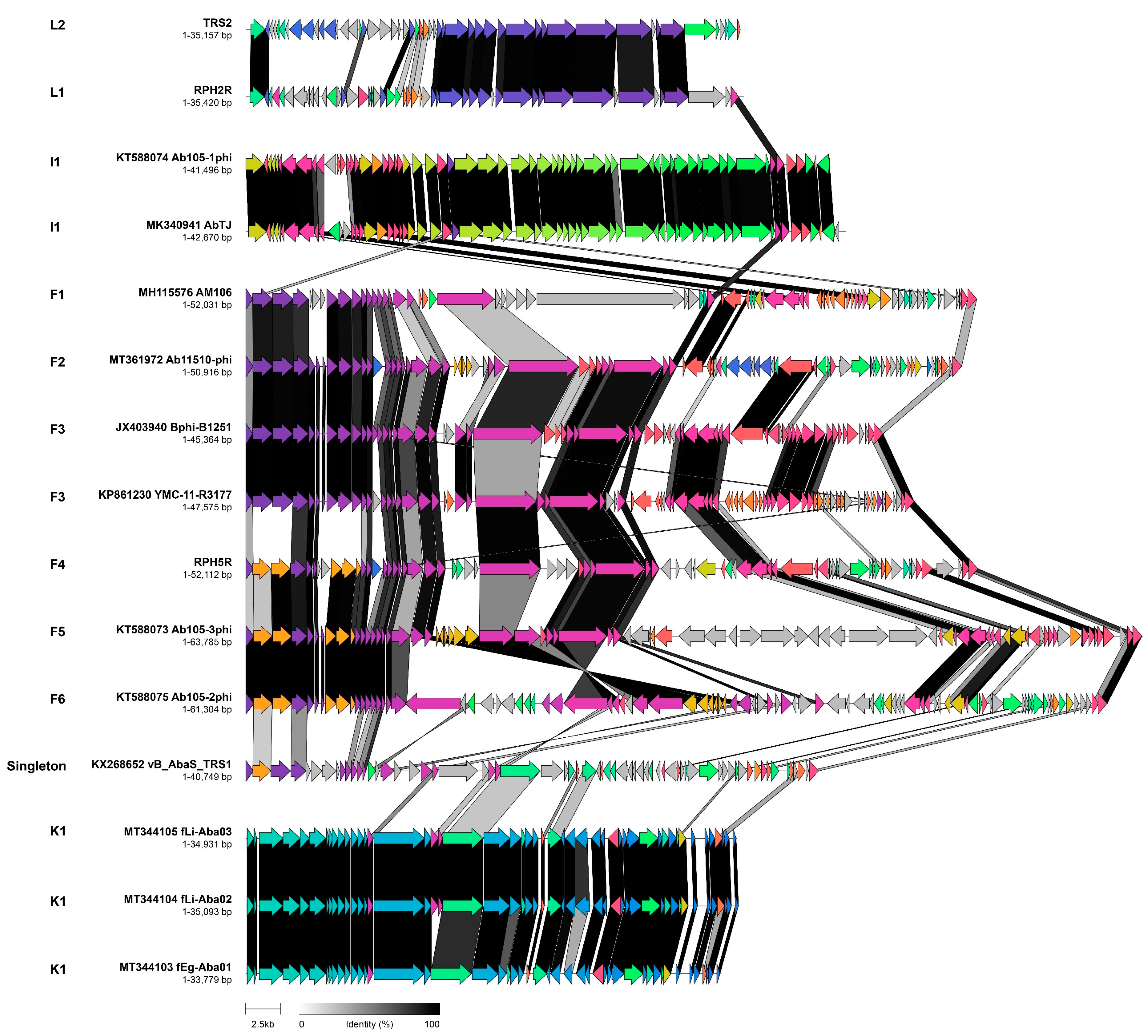
A; B; C; D; E; F; G; H; I; J; K; L; M. A; B; C; D; E; F; G; H; I; J; K; L; M. Singleton phages Arae, ABP49, ME3, TRS5, BFG and SH-Ab 15599 are named in (B).
A; B; C; D; E; F; G; H; I; J; K; L; M. Singleton phages Arae, ABP49, ME3, TRS5, BFG and SH-Ab 15599 are named in (B).
A; B; C; D; E; F; G; H; I; J; K; L; M. Singleton phages Arae, ABP49, ME3, TRS5, BFG and SH-Ab 15599 are named in (B). A; B; C; D; E; F; G; H; I; J; K; L; M). The scale bar represents substitutions per site.
A; B; C; D; E; F; G; H; I; J; K; L; M). The scale bar represents substitutions per site.
A; B; C; D; E; F; G; H; I; J; K; L; M). The scale bar represents substitutions per site.
 A;
A;  B;
B;  C;
C;  D1;
D1;  E;
E;  F;
F;  G;
G;  H;
H;  I;
I;  J1;
J1;  K;
K;  L;
L;  M; Singleton phages:
  BFG;
BFG;  TRS5;
TRS5;  5W;
5W;  ABPH49;
  TRS1;
TRS1;  SH-Ab 15599;
  Arae;
Arae;  ME3.
A; B; C; D1; E; F; G; H; I; J1; K; L; M; Singleton phages: BFG; TRS5; 5W; ABPH49; TRS1; SH-Ab 15599; Arae; ME3.
ME3.
A; B; C; D1; E; F; G; H; I; J1; K; L; M; Singleton phages: BFG; TRS5; 5W; ABPH49; TRS1; SH-Ab 15599; Arae; ME3.


| Cluster | Taxonomy | Representative Phage | No. of Genomes | No. of Core Protein Groups | No. of Accessory Protein Groups | No. of Unique Proteins |
|---|---|---|---|---|---|---|
| A | Twarogvirinae | - | 25 | 107 (81) | 255 (225) | 252 |
| A1 | Zedzedvirus | ZZ1 | 2 | 241 (52) | N/A | 4 |
| A2 | Lasarusvirus | Lazarus | 12 | 218 (11) | 32 (22) | 2 |
| A3 | Hassadvirus | PhT2 | 7 | 227 (13) | 26 (17) | 3 |
| A4 | Lasallevirus | Acj61 | 1 | N/A | N/A | 46 |
| A5 | Acajnonavirus | Acj9 | 1 | N/A | N/A | 46 |
| A6 | “Freretvirus” | Ac42 | 1 | N/A | N/A | 74 |
| A7 | “Audubonvirus” | 133 | 1 | N/A | N/A | 77 |
| B | “Zhukovskyvirinae” | - | 17 | 23 (22) | 117 (100) | 63 |
| B1 | “Solivirus” | phiAC-1 | 1 | N/A | N/A | 33 |
| B2 | “Fengtaivirus” | IME-AB2 | 8 | 56 (4) | 36 (4) | 6 |
| B3 | “Shapingbavirus” | AbP2 | 5 | 58 (1) | 42 (33) | 5 |
| B4 | Obolenskvirus | AP22 | 1 | N/A | N/A | 3 |
| B5 | “Wenzhouvirus” | AB1 | 1 | N/A | N/A | 8 |
| B6 | “Dongdavirus” | IME284 | 1 | N/A | N/A | 9 |
| C | “Astridvirinae” | - | 8 | 69 (42) | 117 (106) | 88 |
| C1 | “Acibelquatrovirus” | Acibel004 | 1 | N/A | N/A | 58 |
| C2 | “Powislevirus” | phiAbaA1 | 1 | N/A | N/A | 15 |
| C3 | Saclayvirus | TAC1 | 6 | 129 (97) | 32 (28) | 15 |
| D1 | “Caradocvirus” | AM24 | 2 | 115 (87) | N/A | 45 |
| E * | Beijernickvirinae | - | 49 | 21 (20) | 57 (45) | 69 |
| E1 | “Aristophanvirus” | Aristophanes | 1 | N/A | N/A | 15 |
| E2 * | Friunavirus | Fri1 | 45 | 27 (0) | 70 (61) | 15 |
| E3 | Pettyvirus | Petty | 1 | N/A | N/A | 12 |
| E4 | Daemvirus | Acibel007 | 1 | N/A | N/A | 13 |
| E5 | “Lisbonvirus” | F1245-05 | 1 | N/A | N/A | 14 |
| F | “Junivirinae” | - | 7 | 11 (2) | 87 | 96 |
| F1 | “Shemyakinvirus” | AM106 | 1 | N/A | N/A | 16 |
| F2 | “Bogotavirus” | Ab11510-phi | 1 | N/A | N/A | 8 |
| F3 | Vieuvirus | Bphi-B1251 | 2 | 44 (12) | N/A | 17 |
| F4 | “Breacavirus” | RPH5R | 1 | N/A | N/A | 19 |
| F5 | “Geihvirus” | Ab105-2phi | 1 | N/A | N/A | 18 |
| F6 | “Reipivirus” | Ab105-3phi | 1 | N/A | N/A | 18 |
| G1 | Lokivirus | Loki | 4 | 43 (38) | 10 (9) | 5 |
| H | Schitoviridae | - | 2 | 28 (28) | N/A | 104 |
| H1 | Presleyvirus | Presley | 1 | N/A | N/A | 54 |
| H2 | Xceevirus | XC38 | 1 | N/A | N/A | 50 |
| I1 | “Xubiasvirus” | Ab105-1phi | 2 | 58 (33) | N/A | 4 |
| J1 | “Stillvirus” | DMU1 | 2 | 53 (41) | N/A | 2 |
| K1 | “Haartmanvirus” | fEg-Aba01 | 3 | 47 (32) | 2 (1) | 0 |
| L | “Grainevirinae” | - | 2 | 21 (18) | N/A | 26 |
| L1 | “Corvusvirus” | RPH2R | 1 | N/A | N/A | 14 |
| L2 | “Boudicavirus” | TRS2 | 1 | N/A | N/A | 14 |
| M | “Soothillvirinae” | - | 2 | 79 (53) | N/A | 129 |
| M1 | “Bragavirus” | B9 | 1 | N/A | N/A | 56 |
| M2 | “Bathrowvirus” | BS46 | 1 | N/A | N/A | 73 |
| Singleton | Metrivirus | ME3 | 1 | N/A | N/A | 261 |
| Singleton | “Gogmagogvirus” | TRS5 | 1 | N/A | N/A | 405 |
| Singleton | “Comoranvirus” | BFG | 1 | N/A | N/A | 422 |
| Singleton | “Lianhecunvirus” | 5W | 1 | N/A | N/A | 22 |
| Singleton | “Fengtaivirus” | ABPH49 | 1 | N/A | N/A | 246 |
| Singleton | “Camillusvirus” | TRS1 | 1 | N/A | N/A | 23 |
| Singleton | “Jiatongvirus” | SH-Ab 15599 | 1 | N/A | N/A | 127 |
| Singleton | “Lucanusvirus” | Arae | 1 | N/A | N/A | 55 |
Publisher’s Note: MDPI stays neutral with regard to jurisdictional claims in published maps and institutional affiliations. |
© 2022 by the authors. Licensee MDPI, Basel, Switzerland. This article is an open access article distributed under the terms and conditions of the Creative Commons Attribution (CC BY) license (https://creativecommons.org/licenses/by/4.0/).
Share and Cite
Oliveira, H.; Domingues, R.; Evans, B.; Sutton, J.M.; Adriaenssens, E.M.; Turner, D. Genomic Diversity of Bacteriophages Infecting the Genus Acinetobacter. Viruses 2022, 14, 181. https://doi.org/10.3390/v14020181
Oliveira H, Domingues R, Evans B, Sutton JM, Adriaenssens EM, Turner D. Genomic Diversity of Bacteriophages Infecting the Genus Acinetobacter. Viruses. 2022; 14(2):181. https://doi.org/10.3390/v14020181
Chicago/Turabian StyleOliveira, Hugo, Rita Domingues, Benjamin Evans, J. Mark Sutton, Evelien M. Adriaenssens, and Dann Turner. 2022. "Genomic Diversity of Bacteriophages Infecting the Genus Acinetobacter" Viruses 14, no. 2: 181. https://doi.org/10.3390/v14020181