
Molecular Characterization and Pathogenesis of H6N6 Low Pathogenic Avian Influenza Viruses Isolated from Mallard Ducks (Anas platyrhynchos) in South Korea

 ,
,  , , and
Abstract
:1. Introduction
2. Materials and Methods
2.1. Sample Collection
2.2. Virus Isolation from Feces
2.3. Bird Species Identification Using the Mitochondrial Gene Cytochrome c Oxidase I (COI) as a DNA Barcode
2.4. Extraction of Viral RNA for Sequencing
2.5. Next Generation Sequencing (NGS) Analysis
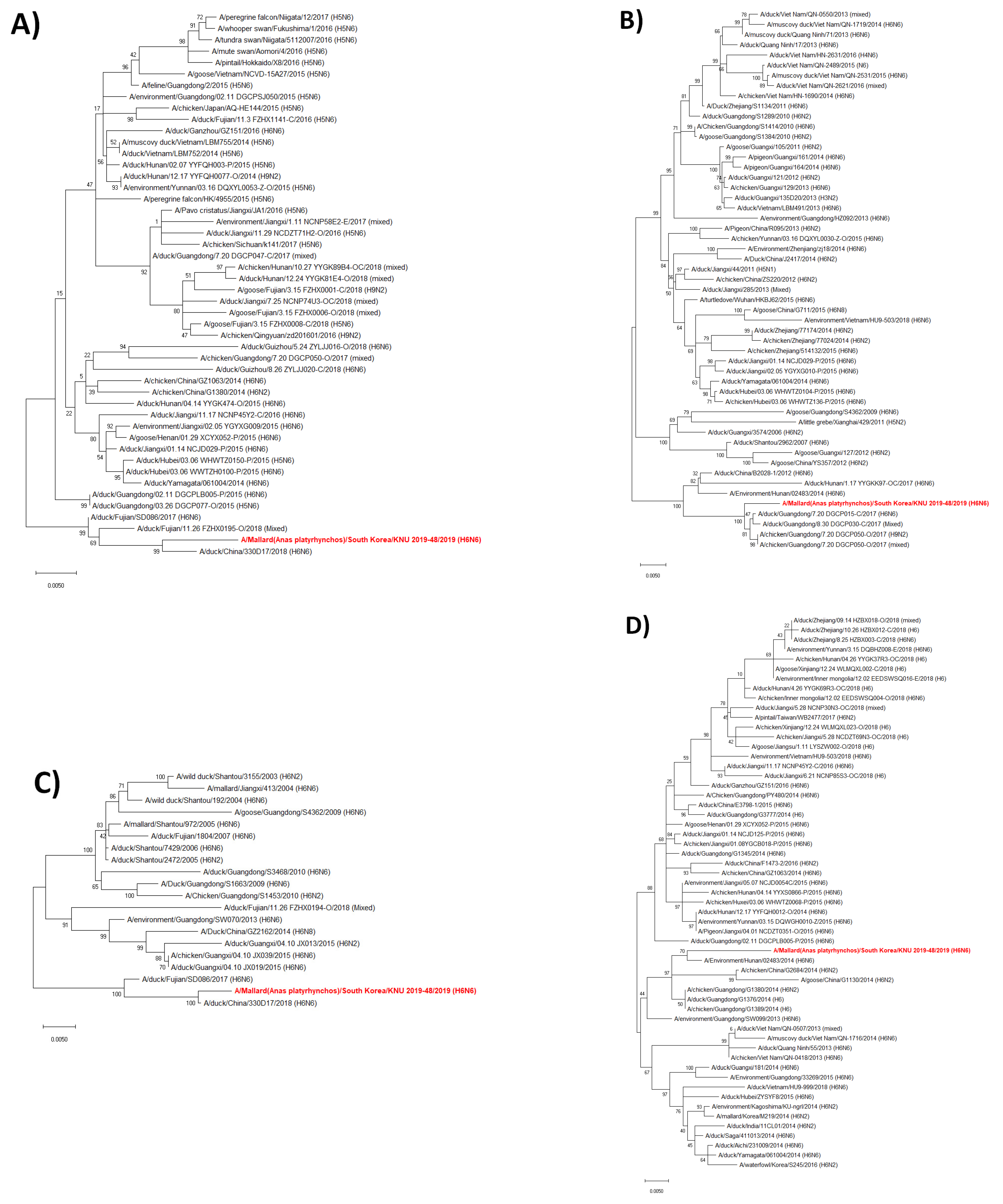
2.6. Phylogenetic Analysis and Molecular Characterization
2.7. Determination of 50% Tissue Culture Infectious Dose (TCID50) and 50% Egg Infectious Dose (EID50)
2.8. Viral Growth Kinetics in MDCK Cells
2.9. Animal Experiment
2.10. Statistics
3. Results
3.1. Genome Characterization of the KNU2019-48 (H6N6) Isolate
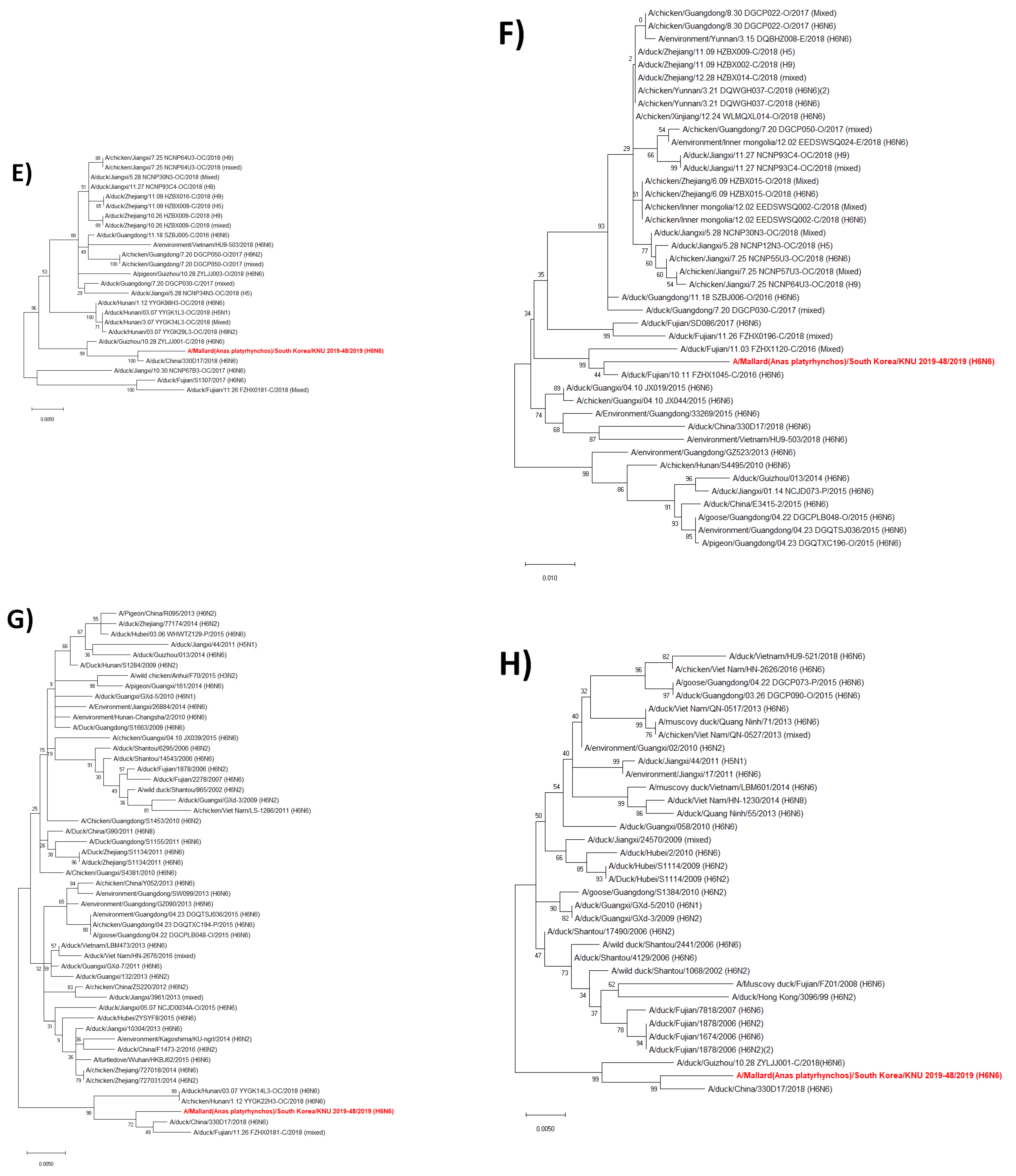
3.2. Hypothesis for the Reassortment Event of Each Gene Segment
3.3. Molecular Characterization of the KNU2019-48 (H6N6) Isolate
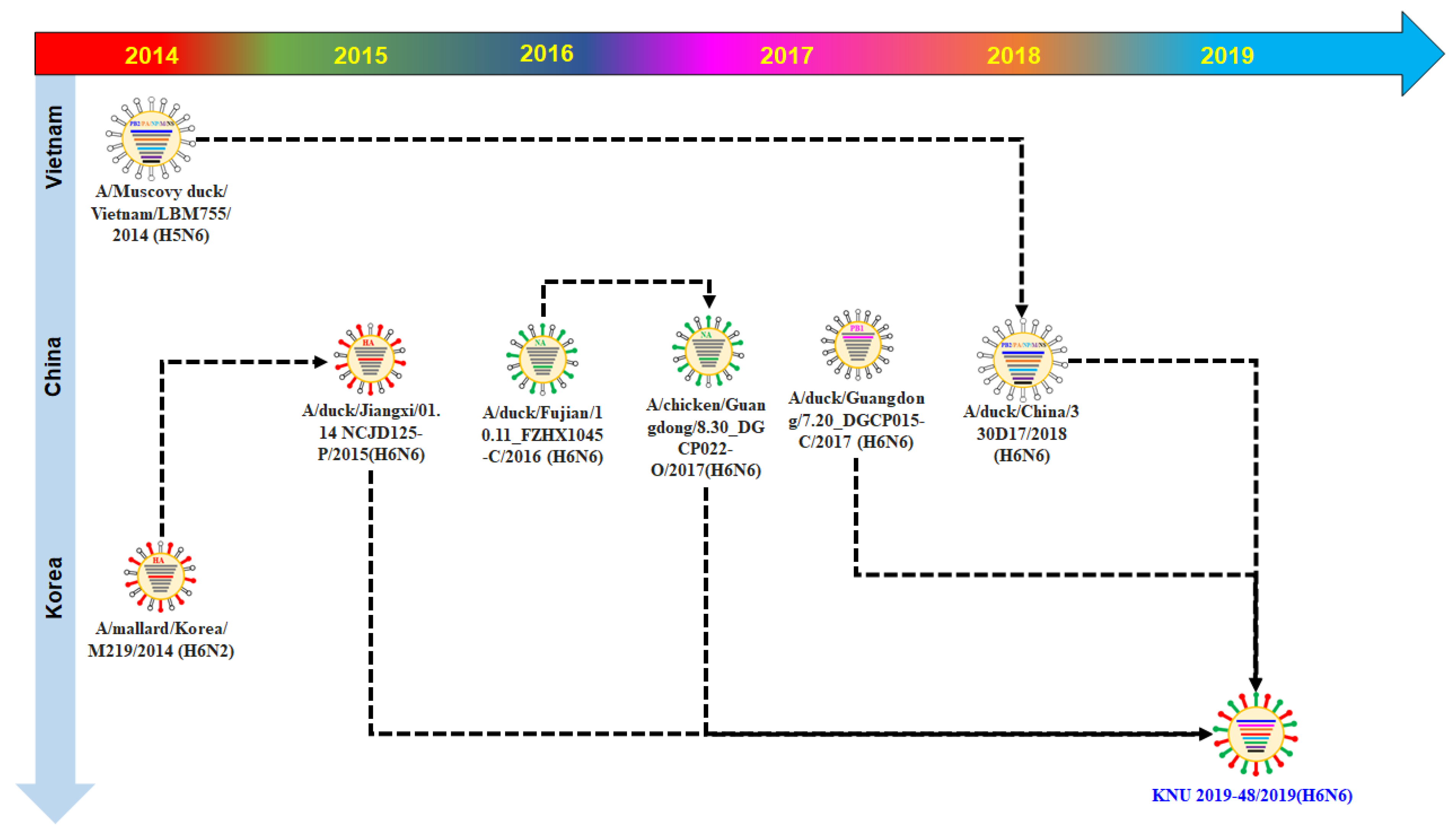
3.4. Growth Kinetics of KNU2019-48 (H6N6) Isolate in Mammalian Cell Culture
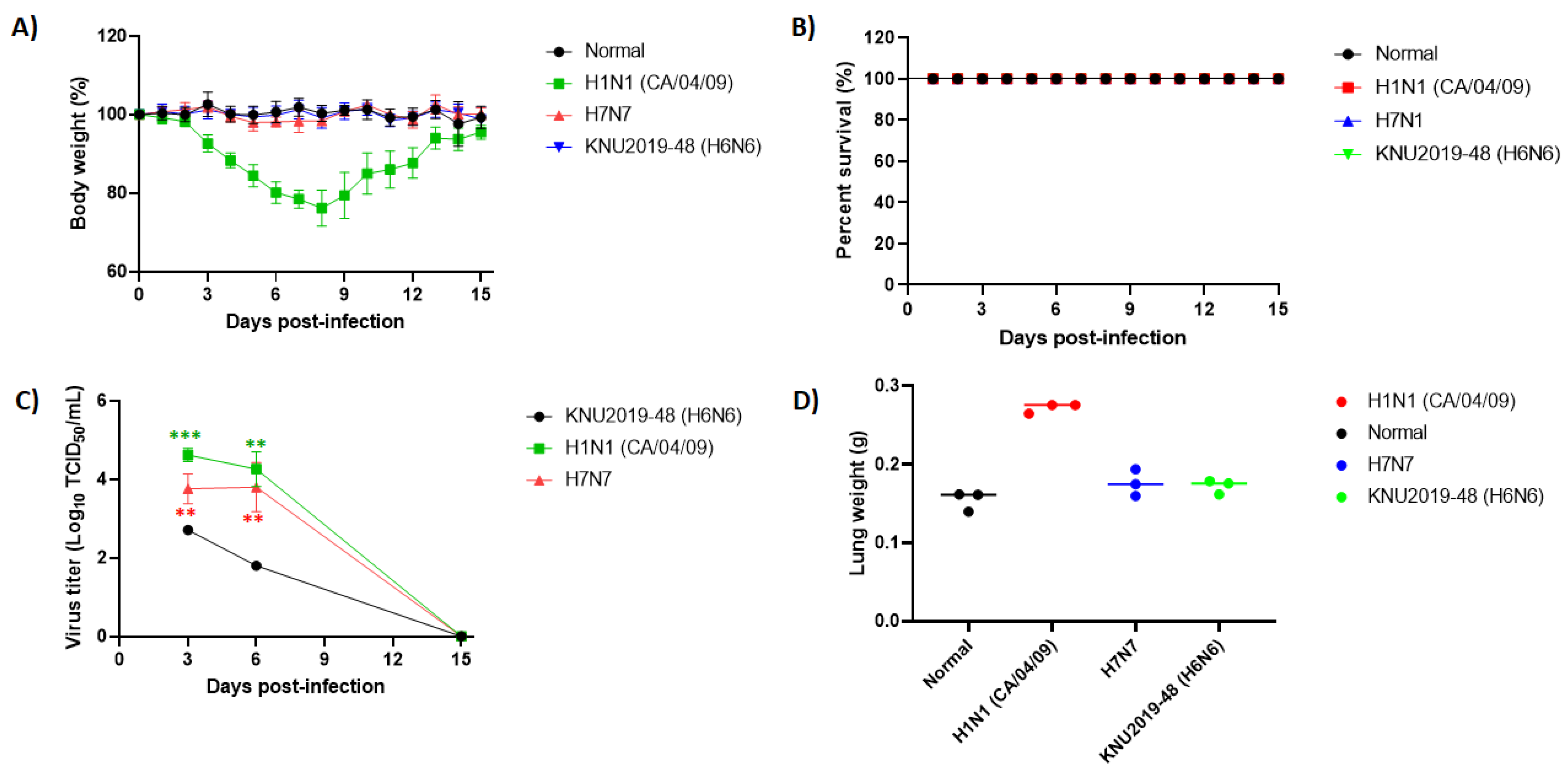
3.5. Pathogenicity in Mice
4. Discussion
Supplementary Materials
Author Contributions
Funding
Institutional Review Board Statement
Informed Consent Statement
Data Availability Statement
Conflicts of Interest
References
- Tong, S.; Li, Y.; Rivailler, P.; Conrardy, C.; Castillo, D.A.A.; Chen, L.-M.; Recuenco, S.; Ellison, J.A.; Davis, C.T.; York, I.A.; et al. A distinct lineage of influenza A virus from bats. Proc. Natl. Acad. Sci. USA 2012, 109, 4269–4274. [Google Scholar] [CrossRef] [PubMed] [Green Version]
- Tong, S.; Zhu, X.; Li, Y.; Shi, M.; Zhang, J.; Bourgeois, M.; Yang, H.; Chen, X.; Recuenco, S.; Gomez, J.; et al. New World Bats Harbor Diverse Influenza A Viruses. PLoS Pathog. 2013, 9, e1003657. [Google Scholar] [CrossRef] [PubMed] [Green Version]
- Li, Y.; Li, M.; Tian, J.; Bai, X.; Li, Y. Genetic characteristics and pathogenicity of novel reassortant H6 viruses isolated from wild birds in China. Vet. Microbiol. 2021, 254, 108978. [Google Scholar] [CrossRef] [PubMed]
- Jin, Y.; Ren, H.; Teng, Y.; Hu, M.; Peng, X.; Yue, J.; Liang, L. Novel reassortment of avian influenza A(H7N9) virus with subtype H6N6 and H5N6 viruses circulating in Guangdong Province, China. J. Infect. 2017, 75, 179–182. [Google Scholar] [CrossRef]
- El Taweel, A.; Kandeil, A.; Barakat, A.; Alfaroq Rabiee, O.; Kayali, G.; Ali, M.A. Diversity of Astroviruses Circulating in Humans, Bats, and Wild Birds in Egypt. Viruses 2020, 12, 485. [Google Scholar] [CrossRef]
- Downie, J.C.; Laver, W.G. Isolation of a type A influenza virus from an Australian pelagic bird. Virology 1973, 51, 259–269. [Google Scholar] [CrossRef]
- Abolnik, C.; Strydom, C.; Rauff, D.L.; Wandrag, D.B.R.; Petty, D. Continuing evolution of H6N2 influenza a virus in South African chickens and the implications for diagnosis and control. BMC Vet. Res. 2019, 15, 455. [Google Scholar] [CrossRef] [Green Version]
- Araujo, J.; Petry, M.V.; Fabrizio, T.; Walker, D.; Ometto, T.; Thomazelli, L.M.; Scherer, A.L.; Serafini, P.P.; Neto, I.S.; Krauss, S.; et al. Migratory birds in southern Brazil are a source of multiple avian influenza virus subtypes. Influenza Other Respi. Viruses 2018, 12, 220–231. [Google Scholar] [CrossRef] [Green Version]
- Cheon, S.-H.; Lee, Y.-N.; Kang, S.-I.; Kye, S.-J.; Lee, E.-K.; Heo, G.-B.; Lee, M.-H.; Kim, J.-W.; Lee, K.-N.; Son, H.-M.; et al. Genetic evidence for the intercontinental movement of avian influenza viruses possessing North American-origin nonstructural gene allele B into South Korea. Infect. Genet. Evol. 2018, 66, 18–25. [Google Scholar] [CrossRef]
- Zou, S.; Gao, R.; Zhang, Y.; Li, X.; Chen, W.; Bai, T.; Dong, L.; Wang, D.; Shu, Y. Molecular characterization of H6 subtype influenza viruses in southern China from 2009 to 2011. Emerg. Microbes Infect. 2016, 5, 1–8. [Google Scholar] [CrossRef] [Green Version]
- Cui, J.; Cui, P.; Shi, J.; Fan, W.; Xing, X.; Gu, W.; Zhang, Y.; Zhang, Y.; Zeng, X.; Jiang, Y.; et al. Continued evolution of H6 avian influenza viruses isolated from farms in China between 2014 and 2018. Transbound. Emerg. Dis. 2021. [CrossRef]
- Wang, G.; Deng, G.; Shi, J.; Luo, W.; Zhang, G.; Zhang, Q.; Liu, L.; Jiang, Y.; Li, C.; Sriwilaijaroen, N.; et al. H6 Influenza Viruses Pose a Potential Threat to Human Health. J. Virol. 2014, 88, 3953–3964. [Google Scholar] [CrossRef] [Green Version]
- Everest, H.; Hill, S.; Daines, R.; Sealy, J.; James, J.; Hansen, R.; Iqbal, M. The Evolution, Spread and Global Threat of H6Nx Avian Influenza Viruses. Viruses 2020, 12, 673. [Google Scholar] [CrossRef] [PubMed]
- Huang, K.; Zhu, H.; Fan, X.; Wang, J.; Cheung, C.-L.; Duan, L.; Hong, W.; Liu, Y.; Li, L.; Smith, D.K.; et al. Establishment and Lineage Replacement of H6 Influenza Viruses in Domestic Ducks in Southern China. J. Virol. 2012, 86, 6075–6083. [Google Scholar] [CrossRef] [PubMed] [Green Version]
- Ni, F.; Kondrashkina, E.; Wang, Q. Structural and Functional Studies of Influenza Virus A/H6 Hemagglutinin. PLoS One 2015, 10, e0134576. [Google Scholar] [CrossRef] [Green Version]
- Trinh, T.-T.T.; Tiwari, I.; Durairaj, K.; Duong, B.T.; Nguyen, A.T.V.; Tuong, H.T.; Hoang, V.T.; Than, D.D.; Nam, S.; Yeo, S.-J.; et al. Genetic Characterization and Pathogenesis of Avian Influenza Virus H7N3 Isolated from Spot-Billed Ducks in South Korea, Early 2019. Viruses 2021, 13, 856. [Google Scholar] [CrossRef] [PubMed]
- Hebert, P.D.N.; Stoeckle, M.Y.; Zemlak, T.S.; Francis, C.M. Identification of Birds through DNA Barcodes. PLoS Biol. 2004, 2, e312. [Google Scholar] [CrossRef] [PubMed] [Green Version]
- Lee, D.-H.; Lee, H.-J.; Lee, Y.-J.; Kang, H.-M.; Jeong, O.-M.; Kim, M.-C.; Kwon, J.-S.; Kwon, J.-H.; Kim, C.-B.; Lee, J.-B.; et al. DNA Barcoding Techniques for Avian Influenza Virus Surveillance in Migratory Bird Habitats. J. Wildl. Dis. 2010, 46, 649–654. [Google Scholar] [CrossRef] [PubMed]
- Yeo, S.-J.; Than, D.-D.; Park, H.-S.; Sung, H.W.; Park, H. Molecular Characterization of a Novel Avian Influenza A (H2N9) Strain Isolated from Wild Duck in Korea in 2018. Viruses 2019, 11, 1046. [Google Scholar] [CrossRef] [Green Version]
- Reed, L.J.; Muench, H. A simple method of estimating fifty per cent endpoints. Am. J. Epidemiol. 1938, 27, 493–497. [Google Scholar] [CrossRef]
- Killian, M.L. Hemagglutination Assay for Influenza Virus. In Animal influenza virus; Humana Press: New York, NY, USA, 2014; pp. 3–9. [Google Scholar]
- Nguyen, N.M.; Sung, H.W.; Yun, K.-J.; Park, H.; Yeo, S.-J. Genetic Characterization of a Novel North American-Origin Avian Influenza A (H6N5) Virus Isolated from Bean Goose of South Korea in 2018. Viruses 2020, 12, 774. [Google Scholar] [CrossRef]
- World Health Organization. Manual for the laboratory diagnosis and virological surveillance of influenza; World Health Organization: Geneva, Switzerland, 2011; p. xii. 140p. [Google Scholar]
- Fan, S.; Hatta, M.; Kim, J.H.; Halfmann, P.; Imai, M.; Macken, C.A.; Le, M.Q.; Nguyen, T.; Neumann, G.; Kawaoka, Y. Novel residues in avian influenza virus PB2 protein affect virulence in mammalian hosts. Nat. Commun. 2014, 5, 5021. [Google Scholar] [CrossRef] [Green Version]
- Li, J.; Li, Y.; Hu, Y.; Chang, G.; Sun, W.; Yang, Y.; Kang, X.; Wu, X.; Zhu, Q. PB1-mediated virulence attenuation of H5N1 influenza virus in mice is associated with PB2. J. Gen. Virol. 2011, 92, 1435–1444. [Google Scholar] [CrossRef] [PubMed]
- Li, J.; Ishaq, M.; Prudence, M.; Xi, X.; Hu, T.; Liu, Q.; Guo, D. Single mutation at the amino acid position 627 of PB2 that leads to increased virulence of an H5N1 avian influenza virus during adaptation in mice can be compensated by multiple mutations at other sites of PB2. Virus Res. 2009, 144, 123–129. [Google Scholar] [CrossRef] [PubMed]
- Prokopyeva, E.A.; Sobolev, I.A.; Prokopyev, M.V.; Shestopalov, A.M. Adaptation of influenza A(H1N1)pdm09 virus in experimental mouse models. Infect. Genet. Evol. 2016, 39, 265–271. [Google Scholar] [CrossRef] [PubMed]
- Kong, H.; Ma, S.; Wang, J.; Gu, C.; Wang, Z.; Shi, J.; Deng, G.; Guan, Y.; Chen, H. Identification of Key Amino Acids in the PB2 and M1 Proteins of H7N9 Influenza Virus That Affect Its Transmission in Guinea Pigs. J. Virol. 2019, 94. [Google Scholar] [CrossRef] [Green Version]
- Govorkova, E.A.; Rehg, J.E.; Krauss, S.; Yen, H.-L.; Guan, Y.; Peiris, M.; Nguyen, T.D.; Hanh, T.H.; Puthavathana, P.; Long, H.T.; et al. Lethality to Ferrets of H5N1 Influenza Viruses Isolated from Humans and Poultry in 2004. J. Virol. 2005, 79, 2191–2198. [Google Scholar] [CrossRef] [Green Version]
- Salomon, R.; Franks, J.; Govorkova, E.A.; Ilyushina, N.A.; Yen, H.-L.; Hulse-Post, D.J.; Humberd, J.; Trichet, M.; Rehg, J.E.; Webby, R.J.; et al. The polymerase complex genes contribute to the high virulence of the human H5N1 influenza virus isolate A/Vietnam/1203/04. J. Exp. Med. 2006, 203, 689–697. [Google Scholar] [CrossRef] [Green Version]
- Tsurumura, T.; Qiu, H.; Yoshida, T.; Tsumori, Y.; Tsuge, H. Crystallization and preliminary X-ray diffraction studies of a surface mutant of the middle domain of PB2 from human influenza A (H1N1) virus. Acta Crystallogr. Sect. F Struct. Biol. Commun. 2014, 70, 72–75. [Google Scholar] [CrossRef] [Green Version]
- Li, Z.; Chen, H.; Jiao, P.; Deng, G.; Tian, G.; Li, Y.; Hoffmann, E.; Webster, R.G.; Matsuoka, Y.; Yu, K. Molecular Basis of Replication of Duck H5N1 Influenza Viruses in a Mammalian Mouse Model. J. Virol. 2005, 79, 12058–12064. [Google Scholar] [CrossRef] [Green Version]
- Subbarao, E.K.; London, W.; Murphy, B.R. A single amino acid in the PB2 gene of influenza A virus is a determinant of host range. J. Virol. 1993, 67, 1761–1764. [Google Scholar]
- Dreier, C.; Resa-Infante, P.; Thiele, S.; Stanelle-Bertram, S.; Walendy-Gnirß, K.; Speiseder, T.; Preuss, A.; Müller, Z.; Klenk, H.-D.; Stech, J.; et al. Mutations in the H7 HA and PB1 genes of avian influenza a viruses increase viral pathogenicity and contact transmission in guinea pigs. Emerg. Microbes Infect. 2019, 8, 1324–1336. [Google Scholar] [CrossRef] [Green Version]
- Hulse-Post, D.J.; Franks, J.; Boyd, K.; Salomon, R.; Hoffmann, E.; Yen, H.L.; Webby, R.J.; Walker, D.; Nguyen, T.D.; Webster, R.G. Molecular Changes in the Polymerase Genes (PA and PB1) Associated with High Pathogenicity of H5N1 Influenza Virus in Mallard Ducks. J. Virol. 2007, 81, 8515–8524. [Google Scholar] [CrossRef] [Green Version]
- Taubenberger, J.K.; Reid, A.H.; Lourens, R.M.; Wang, R.; Jin, G.; Fanning, T.G. Characterization of the 1918 influenza virus polymerase genes. Nature 2005, 437, 889–893. [Google Scholar] [CrossRef]
- Suttie, A.; Deng, Y.-M.; Greenhill, A.R.; Dussart, P.; Horwood, P.F.; Karlsson, E.A. Inventory of molecular markers affecting biological characteristics of avian influenza A viruses. Virus Genes 2019, 55, 739–768. [Google Scholar] [CrossRef] [PubMed] [Green Version]
- Xu, C.; Hu, W.-B.; Xu, K.; He, Y.-X.; Wang, T.-Y.; Chen, Z.; Li, T.-X.; Liu, J.-H.; Buchy, P.; Sun, B. Amino acids 473V and 598P of PB1 from an avian-origin influenza A virus contribute to polymerase activity, especially in mammalian cells. J. Gen. Virol. 2012, 93, 531–540. [Google Scholar] [CrossRef] [PubMed]
- Feng, X.; Wang, Z.; Shi, J.; Deng, G.; Kong, H.; Tao, S.; Li, C.; Liu, L.; Guan, Y.; Chen, H. Glycine at Position 622 in PB1 Contributes to the Virulence of H5N1 Avian Influenza Virus in Mice. J. Virol. 2016, 90, 1872–1879. [Google Scholar] [CrossRef] [PubMed] [Green Version]
- Shaw, M.; Cooper, L.; Xu, X.; Thompson, W.; Krauss, S.; Guan, Y.; Zhou, N.; Klimov, A.; Cox, N.; Webster, R.; et al. Molecular changes associated with the transmission of avian influenza a H5N1 and H9N2 viruses to humans. J. Med. Virol. 2002, 66, 107–114. [Google Scholar] [CrossRef]
- Song, J.; Xu, J.; Shi, J.; Li, Y.; Chen, H. Synergistic Effect of S224P and N383D Substitutions in the PA of H5N1 Avian Influenza Virus Contributes to Mammalian Adaptation. Sci. Rep. 2015, 5, 10510. [Google Scholar] [CrossRef] [PubMed] [Green Version]
- Li, J.; Zheng, W.; Hou, L.; Chen, C.; Fan, W.; Qu, H.; Jiang, J.; Liu, J.; Gao, G.F.; Zhou, J.; et al. Differential nucleocytoplasmic shuttling of the nucleoprotein of influenza a viruses and association with host tropism. Cell. Microbiol. 2017, 19, e12692. [Google Scholar] [CrossRef] [Green Version]
- Leung, B.W.; Chen, H.; Brownlee, G.G. Correlation between polymerase activity and pathogenicity in two duck H5N1 influenza viruses suggests that the polymerase contributes to pathogenicity. Virology 2010, 401, 96–106. [Google Scholar] [CrossRef] [Green Version]
- Finkelstein, D.B.; Mukatira, S.; Mehta, P.K.; Obenauer, J.C.; Su, X.; Webster, R.G.; Naeve, C.W. Persistent Host Markers in Pandemic and H5N1 Influenza Viruses. J. Virol. 2007, 81, 10292–10299. [Google Scholar] [CrossRef] [Green Version]
- Yamaji, R.; Yamada, S.; Le, M.Q.; Ito, M.; Sakai-Tagawa, Y.; Kawaoka, Y. Mammalian Adaptive Mutations of the PA Protein of Highly Pathogenic Avian H5N1 Influenza Virus. J. Virol. 2015, 89, 4117–4125. [Google Scholar] [CrossRef] [Green Version]
- Su, Y.; Yang, H.-Y.; Zhang, B.-J.; Jia, H.-L.; Tien, P. Analysis of a point mutation in H5N1 avian influenza virus hemagglutinin in relation to virus entry into live mammalian cells. Arch. Virol. 2008, 153, 2253–2261. [Google Scholar] [CrossRef]
- Gao, Y.; Zhang, Y.; Shinya, K.; Deng, G.; Jiang, Y.; Li, Z.; Guan, Y.; Tian, G.; Li, Y.; Shi, J.; et al. Identification of Amino Acids in HA and PB2 Critical for the Transmission of H5N1 Avian Influenza Viruses in a Mammalian Host. PLOS Pathog. 2009, 5, e1000709. [Google Scholar] [CrossRef] [Green Version]
- Tan, L.; Su, S.; Smith, D.K.; He, S.; Zheng, Y.; Shao, Z.; Ma, J.; Zhu, H.; Zhang, G. A Combination of HA and PA Mutations Enhances Virulence in a Mouse-Adapted H6N6 Influenza A Virus. J. Virol. 2014, 88, 14116–14125. [Google Scholar] [CrossRef] [PubMed] [Green Version]
- Zhu, W.; Zou, X.; Zhou, J.; Tang, J.; Shu, Y. Residues 41V and/or 210D in the NP protein enhance polymerase activities and potential replication of novel influenza (H7N9) viruses at low temperature. Virol. J. 2015, 12, 71. [Google Scholar] [CrossRef] [PubMed] [Green Version]
- Liu, Q.; Chen, H.; Huang, J.; Chen, Y.; Gu, M.; Wang, X.; Hu, S.; Liu, X.; Liu, X. A nonpathogenic duck-origin H9N2 influenza A virus adapts to high pathogenicity in mice. Arch. Virol. 2014, 159, 2243–2252. [Google Scholar] [CrossRef]
- Ma, S.; Zhang, B.; Shi, J.; Yin, X.; Wang, G.; Cui, P.; Liu, L.; Deng, G.; Jiang, Y.; Li, C.; et al. Amino Acid Mutations A286V and T437M in the Nucleoprotein Attenuate H7N9 Viruses in Mice. J. Virol. 2020, 94. [Google Scholar] [CrossRef] [Green Version]
- Zhao, Y.; Yu, Z.; Liu, L.; Wang, T.; Sun, W.; Wang, C.; Xia, Z.; Gao, Y.; Zhou, B.; Qian, J.; et al. Adaptive amino acid substitutions enhance the virulence of a novel human H7N9 influenza virus in mice. Vet. Microbiol. 2016, 187, 8–14. [Google Scholar] [CrossRef]
- Chen, H.; Bright, R.A.; Subbarao, K.; Smith, C.; Cox, N.J.; Katz, J.M.; Matsuoka, Y. Polygenic virulence factors involved in pathogenesis of 1997 Hong Kong H5N1 influenza viruses in mice. Virus Res. 2007, 128, 159–163. [Google Scholar] [CrossRef] [PubMed]
- Lee, M.S.; Deng, M.C.; Lin, Y.J.; Chang, C.Y.; Shieh, H.K.; Shiau, J.Z.; Huang, C.C. Characterization of an H5N1 avian influenza virus from Taiwan. Vet. Microbiol. 2007, 124, 193–201. [Google Scholar] [CrossRef]
- Fan, S.; Deng, G.; Song, J.; Tian, G.; Suo, Y.; Jiang, Y.; Guan, Y.; Bu, Z.; Kawaoka, Y.; Chen, H. Two amino acid residues in the matrix protein M1 contribute to the virulence difference of H5N1 avian influenza viruses in mice. Virology 2009, 384, 28–32. [Google Scholar] [CrossRef] [Green Version]
- Jiao, P.; Tian, G.; Li, Y.; Deng, G.; Jiang, Y.; Liu, C.; Liu, W.; Bu, Z.; Kawaoka, Y.; Chen, H. A Single-Amino-Acid Substitution in the NS1 Protein Changes the Pathogenicity of H5N1 Avian Influenza Viruses in Mice. J. Virol. 2008, 82, 1146–1154. [Google Scholar] [CrossRef] [Green Version]
- Zheng, W.; Cao, S.; Chen, C.; Li, J.; Zhang, S.; Jiang, J.; Niu, Y.; Fan, W.; Li, Y.; Bi, Y.; et al. Threonine 80 phosphorylation of non-structural protein 1 regulates the replication of influenza A virus by reducing the binding affinity with RIG-I. Cell. Microbiol. 2017, 19, e12643. [Google Scholar] [CrossRef] [Green Version]
- Min, J.-Y.; Li, S.; Sen, G.C.; Krug, R.M. A site on the influenza A virus NS1 protein mediates both inhibition of PKR activation and temporal regulation of viral RNA synthesis. Virology 2007, 363, 236–243. [Google Scholar] [CrossRef] [Green Version]
- Li, Z.; Jiang, Y.; Jiao, P.; Wang, A.; Zhao, F.; Tian, G.; Wang, X.; Yu, K.; Bu, Z.; Chen, H. The NS1 Gene Contributes to the Virulence of H5N1 Avian Influenza Viruses. J. Virol. 2006, 80, 11115–11123. [Google Scholar] [CrossRef] [Green Version]
- Imai, H.; Shinya, K.; Takano, R.; Kiso, M.; Muramoto, Y.; Sakabe, S.; Murakami, S.; Ito, M.; Yamada, S.; Le, M. thi Q.; et al. The HA and NS Genes of Human H5N1 Influenza A Virus Contribute to High Virulence in Ferrets. PLoS Pathog. 2010, 6, e1001106. [Google Scholar] [CrossRef] [PubMed] [Green Version]
- Lin, W.; Cui, H.; Teng, Q.; Li, L.; Shi, Y.; Li, X.; Yang, J.; Liu, Q.; Deng, J.; Li, Z. Evolution and pathogenicity of H6 avian influenza viruses isolated from Southern China during 2011 to 2017 in mice and chickens. Sci. Rep. 2020, 10, 1–12. [Google Scholar] [CrossRef] [PubMed]
- Tsai, S.-K.; Shih, C.-H.; Chang, H.-W.; Teng, K.-H.; Hsu, W.-E.; Lin, H.-J.; Lin, H.-Y.; Huang, C.-H.; Chen, H.-W.; Wang, L.-C. Replication of a Dog-Origin H6N1 Influenza Virus in Cell Culture and Mice. Viruses 2020, 12, 704. [Google Scholar] [CrossRef] [PubMed]
- Duong, B.T.; Bal, J.; Sung, H.W.; Yeo, S.-J.; Park, H. Molecular Analysis of the Avian H7 Influenza Viruses Circulating in South Korea during 2018–2019: Evolutionary Significance and Associated Zoonotic Threats. Viruses 2021, 13, 2260. [Google Scholar] [CrossRef] [PubMed]
- Kang, Y.; Liu, L.; Feng, M.; Yuan, R.; Huang, C.; Tan, Y.; Gao, P.; Xiang, D.; Zhao, X.; Li, Y.; et al. Highly pathogenic H5N6 influenza A viruses recovered from wild birds in Guangdong, southern China, 2014–2015. Sci. Rep. 2017, 7, 44410. [Google Scholar] [CrossRef] [PubMed] [Green Version]
- Bi, Y.; Li, J.; Li, S.; Fu, G.; Jin, T.; Zhang, C.; Yang, Y.; Ma, Z.; Tian, W.; Xiao, S.; et al. Dominant subtype switch in avian influenza viruses during 2016–2019 in China. Nat. Commun 2020, 11, 5909. [Google Scholar] [CrossRef]
- Zhang, G.; Kong, W.; Qi, W.; Long, L.-P.; Cao, Z.; Huang, L.; Qi, H.; Cao, N.; Wang, W.; Zhao, F.; et al. Identification of an H6N6 swine influenza virus in southern China. Infect. Genet. Evol. 2011, 11, 1174–1177. [Google Scholar] [CrossRef] [Green Version]
- Shanmuganatham, K.K.; Jones, J.C.; Marathe, B.M.; Feeroz, M.M.; Jones-Engel, L.; Walker, D.; Turner, J.; Rabiul Alam, S.M.; Kamrul Hasan, M.; Akhtar, S.; et al. The replication of Bangladeshi H9N2 avian influenza viruses carrying genes from H7N3 in mammals. Emerg. Microbes Infect. 2016, 5, 1–12. [Google Scholar] [CrossRef] [Green Version]
- Miura, H.; Ozeki, Y.; Omatsu, T.; Katayama, Y.; Imai, K.; Mizutani, T.; Ogawa, H.; Takeda, Y. The Single E627K Amino Acid Substitution in PB2 Enhances the Pathogenicity of Wild-Bird-Origin H6N6 Subtype Avian Influenza Virus in Mice. Austin J. Infect. Dis. 2020, 7, 1041. [Google Scholar]


| Gene | GenBank ID | Reference Strain Accession ID | Origin | Per Ident (%) |
|---|---|---|---|---|
| PB2 | MW380639 | EPI_ISL_501514 | A/duck/China/330D17/2018 (H6N6) | 98.75 (2341/2328) |
| EPI_ISL_285466 | A/duck/Fujian/SD086/2017 (H6N6) | 98.29 (2280/2328) | ||
| EPI666098 | A/duck/Guangdong/02.11_DGCPLB005-P/2015 (H6N6) | 97.16 (2335/2328) | ||
| PB1 | MW380640 | EPI_ISL_707456 | A/duck/Guangdong/7.20_DGCP015-C/2017 (H6N6) | 99.12 (2274/2304) |
| MW104102 | A/chicken/Guangdong/7.20_DGCP050-O/2017(mixed) | 99.03 (2274/2304) | ||
| EPI_ISL_698000 | A/chicken/Guangdong/7.20_DGCP050-O/2017 (H9N2) | 99.03 (2274/2304) | ||
| PA | MW380641 | EPI_ISL_501514 | A/duck/China/330D17/2018 (H6N6) | 99.44 (2233/2151) |
| EPI_ISL_285466 | A/duck/Fujian/SD086/2017 (H6N6) | 98.14 (2151/2151) | ||
| EPI_ISL_76327 | A/duck/Shantou/2472/2005 (H6N2) | 96.09 (2151/2151) | ||
| HA | MW380642 | EPI_ISL_199312 | A/duck/Jiangxi/01.14 NCJD125-P/2015(H6N6) | 97.18 (1740/1701) |
| MH130170 | A/mallard/Korea/M219/2014 (H6N2) | 96.47 (1726/1701) | ||
| EPI_ISL_219853 | A/Environment/Hunan/02483/2014 (H6N6) | 98 (1701/1701) | ||
| NP | MW380643 | EPI_ISL_501514 | A/duck/China/330D17/2018 (H6N6) | 99.21 (1565/1527) |
| EPI_ISL_696839 | A/duck/Guizhou/10.28_ZYLJJ001-C/2018 (H6N6) | 98.33 (1497/1527) | ||
| MW098939 | A/duck/Guangdong/7.20_DGCP030-C/2017(mixed) | 97.33 (1497/1527) | ||
| NA | MW380644 | EPI_ISL_696964 | A/duck/Fujian/10.11_FZHX1045-C/2016 (H6N6) | 97.38 (1412/1465) |
| EPI666988 | A/duck/Guangxi/04.10_JX019/2015 (H6N6) | 96.10 (1412/1465) | ||
| MW100376 | A/chicken/Inner_mongolia/12.02_EEDSWSQ002-C/2018 (H6N6) | 95.47 (1413/1465) | ||
| M | MW380645 | MN088783 | A/duck/China/330D17/2018 (H6N6) | 97.59 (1027/979) |
| MW101275 | A/duck/Fujian/11.26_FZHX0181-C/2018(mixed) | 99.18 (976/979) | ||
| LC028304 | A/muscovy duck/Vietnam/LBM755/2014(H5N6) | 99.18 (976/979) | ||
| NS | MW380646 | MN088790 | A/duck/China/330D17/2018(H6N6) | 98.86 (890/889) |
| MW101859 | A/duck/Guizhou/10.28_ZYLJJ001-C/2018(H6N6) | 97.75 (844/889) | ||
| CY109470 | A/duck/Shantou/17490/2006(H6N2) | 97.16 (844/889) |
| Virus Strains | HA Receptor-Binding Residues (H3 Numbering) | NA | |||||||||
|---|---|---|---|---|---|---|---|---|---|---|---|
| Cleavage Sites 340-348 | A138S | P186L | E190V | Q226L | G228S | Stalk Region Deletion | E119V | H275Y | R293K | N295S | |
| KNU2019-48 (H6N6) | PRIETR↓GLF | A | P | E | Q | G | NO | E | H | R | N |
| H10/2010 (H6N6) | PQIETR↓GLF | A | P | E | Q | G | NO | E | H | R | N |
| K6/2010 (H6N6) | PQIETR↓GLF | S | I | E | Q | S | NO | E | H | R | N |
| A729-2/2011 (H6N6) | PQIETR↓GLF | A | P | E | Q | G | YES (59–69) | E | H | R | N |
| KNU18-6/2018 (H6N5) | PQIETR↓GLF | A | P | E | Q | G | NO | E | H | R | N |
| Viral Protein | Amino Acid | KNU2019-48 (H6N6) | H10/2010 (H6N6) | K6/2010 (H6N6) | A72-2/2011 (H6N6) | KNU18-6/2018 (H6N5) | Phenotype | Reference |
|---|---|---|---|---|---|---|---|---|
| PB2 | K147T, M147L | I | I | I | I | I | - | [24] |
| T63I (with PB1 M677T) | I | I | I | I | I | Pathogenic in mice | [25] | |
| L89V | V | V | V | V | V | Enhanced polymerase activity; Increased virulence in mice | [26] | |
| K251R | R | R | R | R | R | Increased virulence in mice | [27] | |
| I292V | I | I | I | I | I | Increase the polymerase activity of H7N9 viruses in both avian and human cells and facilitate the transmission | [28] | |
| G309D | D | D | D | D | D | Enhanced polymerase activity; Increased virulence in mice | [26] | |
| T339K | K | K | K | K | K | Enhanced polymerase activity; Increased virulence in mice | ||
| Q368R | R | R | R | R | R | Increased polymerase activity; Increased virulence in mammals | [29,30] | |
| H447Q | Q | Q | Q | Q | Q | Increased polymerase activity; Increased virulence in mammals | ||
| I471T (with PB2 P453H) | T | T | T | T | T | Change the surface electrostatic potential drastically | [31] | |
| R477G | G | G | G | G | G | Enhanced polymerase activity; Increased virulence in mice | [26] | |
| I495V | V | V | V | V | V | Enhanced polymerase activity; Increased virulence in mice | ||
| A676T | T | T | T | T | T | Enhanced polymerase active; Increased virulence in mice | ||
| E627K | E | E | E | E | E | Mammalian adaptation marker | [32,33] | |
| D701N | D | D | D | D | D | Mammalian adaptation marker | ||
| PB1 | D/A3V | V | V | V | V | V | Increased polymerase activity; Increased virulence in mammals | [29,30] |
| L13P | P | P | - | P | P | Increased polymerase activity; Increased virulence in mammals, Mammalian host marker | [34] | |
| R207K | K | K | K | K | K | Increased polymerase activity in mammalian cells | [35] | |
| K328N | N | N | N | N | N | Increased polymerase activity; Increased virulence in mammals | [29,30] | |
| S375N/T | N | N | N | N | N | Increased polymerase activity; Increased virulence in mammals, Human host marker | ||
| H436Y | Y | Y | Y | Y | Y | Increased polymerase activity and virulence in mallards, ferrets, and mice | [36] | |
| A469T (with NS1 N205K; NEP T48N) | T | T | T | T | T | Conferred in contact transmissibility in guinea pigs | [35] | |
| L473V | V | V | V | V | V | Increased polymerase activity and replication efficiency | ||
| V652A | A | A | A | A | A | Increased virulence in mice | [27] | |
| M677T (with PB2 T63I) | I | I | I | V | I | Pathogenic in mice | [25] | |
| V598P | L | L | L | L | L | Decreased polymerase activity and replication efficiency in mammalian cells | [37,38] | |
| D622G | G | G | G | G | G | Increased polymerase activity and virulence in mice | [39] | |
| PA | N383D | D | D | D | D | D | Increased polymerase activity in mammalian and avian cell lines | [40,41] |
| S37A | A | A | A | A | A | Significantly increased viral growth and polymerase activity in mammalian cells | [42] | |
| H266R | R | R | R | R | R | Increased polymerase activity; Increased virulence in mammals and birds | [43] | |
| F277S | S | S | S | S | S | Adapt to mammalian hosts | ||
| C278Q | Q | Q | Q | Q | Q | Adapt to mammalian hosts | ||
| I357K | T | T | T | T | T | Increased polymerase activity; Increased virulence in mammals and birds | ||
| N383D (with S224P) | D | D | D | D | D | Enhanced the pathogenicity and viral replication of H5N1 virus in mice | [40,41] | |
| A404S | S | S | S | S | A | Human host marker | [44] | |
| S409N | N | N | N | N | S | Enhanced Transmission; Human host marker | ||
| S/A515T | T | T | T | T | T | Increased polymerase activity; Increased virulence in mammals and birds | [43,45] | |
| L653P | P | P | P | P | P | Adapt to mammalian hosts | [43] | |
| HA | V110A | A | A | A | A | A | Host specificity shift to Enhance binding of HA to human-type SAα2,6Gal receptor | [46] |
| T160A | E | G | S | A | E | Increased binding to human-type influenza receptor | [47] | |
| T/E173G/D/V | D | D | D | D | T | Increased virus binding to α-2,6-linked sialic acid | [22,48] | |
| NP | V41I | I | I | I | I | I | Might contribute to viral transmissibility | [49] |
| V105M | M | I | M | M | M | Contribute to the increased virulence of the H9N2 | [50] | |
| D210E | E | E | E | E | E | Might contribute to viral transmissibility | [49] | |
| F253I | I | I | I | I | I | Results in attenuated pathogenicity of the virus in mice | [42] | |
| A286V | A | A | A | A | A | Affect the pathogenicity of the virus in mice | [51] | |
| I353V | V | V | V | V | V | Increased virulence in mice | [27] | |
| T437M | T | T | T | T | T | Affect the pathogenicity of the virus in mice | [51] | |
| NA | M26I | V | I | I | I | Increased virulence in mice | [52] | |
| T223I | I | I | I | I | Increased virulence in mammals | [53,54] | ||
| M1 | N30D | D | D | D | D | D | Increased virulence in mammals | [55] |
| V15I/T | I | I | I | I | I | Increased virulence in mammals | [53,54] | |
| A166V | V | V | V | V | V | Contribute to the increased virulence of the H9N2. | [49] | |
| T215A | A | A | A | A | A | Increased virulence in mammals | [55] | |
| NS1 | A/P42S | S | S | S | S | S | Increased virulence in mammals; Antagonism of IFN induction | [56] |
| T80E | N | T | T | T | T | Reduced influenza virus replication through controlling RIG-I-mediated IFN production and vRNP activity | [57] | |
| T/D/V/R/A127N | D | N | N | N | N | Increased virulence in mammals | [58] | |
| V149A | A | A | A | A | A | Pathogenicity in mice; Antagonism of IFN induction | [59] | |
| NS2 | T47A (with NS1 N200S) | E | E | E | E | E | Decreased IFN antagonism | [60] |
| M51I (with NS1 G205R) | R | R | R | R | R | Decreased IFN antagonism |
Publisher’s Note: MDPI stays neutral with regard to jurisdictional claims in published maps and institutional affiliations. |
© 2022 by the authors. Licensee MDPI, Basel, Switzerland. This article is an open access article distributed under the terms and conditions of the Creative Commons Attribution (CC BY) license (https://creativecommons.org/licenses/by/4.0/).
Share and Cite
Durairaj, K.; Trinh, T.-T.T.; Yun, S.-Y.; Yeo, S.-J.; Sung, H.-W.; Park, H. Molecular Characterization and Pathogenesis of H6N6 Low Pathogenic Avian Influenza Viruses Isolated from Mallard Ducks (Anas platyrhynchos) in South Korea. Viruses 2022, 14, 1001. https://doi.org/10.3390/v14051001
Durairaj K, Trinh T-TT, Yun S-Y, Yeo S-J, Sung H-W, Park H. Molecular Characterization and Pathogenesis of H6N6 Low Pathogenic Avian Influenza Viruses Isolated from Mallard Ducks (Anas platyrhynchos) in South Korea. Viruses. 2022; 14(5):1001. https://doi.org/10.3390/v14051001
Chicago/Turabian StyleDurairaj, Kaliannan, Thuy-Tien Thi Trinh, Su-Yeon Yun, Seon-Ju Yeo, Haan-Woo Sung, and Hyun Park. 2022. "Molecular Characterization and Pathogenesis of H6N6 Low Pathogenic Avian Influenza Viruses Isolated from Mallard Ducks (Anas platyrhynchos) in South Korea" Viruses 14, no. 5: 1001. https://doi.org/10.3390/v14051001
APA StyleDurairaj, K., Trinh, T.-T. T., Yun, S.-Y., Yeo, S.-J., Sung, H.-W., & Park, H. (2022). Molecular Characterization and Pathogenesis of H6N6 Low Pathogenic Avian Influenza Viruses Isolated from Mallard Ducks (Anas platyrhynchos) in South Korea. Viruses, 14(5), 1001. https://doi.org/10.3390/v14051001