Review

Overview of Neutralization Assays and International Standard for Detecting SARS-CoV-2 Neutralizing Antibody

1
Research Center for Emerging Viral Infections, College of Medicine, Chang Gung University, Taoyuan 33302, Taiwan
2
Graduate Institute of Biomedical Science, College of Medicine, Chang Gung University, Taoyuan 33302, Taiwan
3
Division of Infectious Diseases, Department of Pediatrics, Linkou Chang Gung Memorial Hospital, Taoyuan 333, Taiwan
4
International Master Degree Program for Molecular Medicine in Emerging Viral Infections, Chang Gung University, Taoyuan 33302, Taiwan
*
Author to whom correspondence should be addressed.
These authors contributed equally to the study.
Viruses 2022, 14(7), 1560; https://doi.org/10.3390/v14071560
Submission received: 24 June 2022 / Revised: 12 July 2022 / Accepted: 15 July 2022 / Published: 18 July 2022
(This article belongs to the Special Issue SARS-CoV-2 Neutralizing Antibodies)

Abstract

:
We aimed to review the existing literature on the different types of neutralization assays and international standards for severe acute respiratory syndrome coronavirus 2 (SARS-CoV-2). We comprehensively summarized the serological assays for detecting neutralizing antibodies against SARS-CoV-2 and demonstrated the importance of an international standard for calibrating the measurement of neutralizing antibodies. Following the coronavirus disease outbreak in December 2019, there was an urgent demand to detect neutralizing antibodies in patients or vaccinated people to monitor disease outcomes and determine vaccine efficacy. Therefore, many approaches were developed to detect neutralizing antibodies against SARS-CoV-2, such as microneutralization assay, SARS-CoV-2 pseudotype virus assay, enzyme-linked immunosorbent assay (ELISA), and rapid lateral flow assay. Given the many types of serological assays for quantifying the neutralizing antibody titer, the comparison of different assay results is a challenge. In 2020, the World Health Organization proposed the first international standard as a common unit to define neutralizing antibody titer and antibody responses against SARS-CoV-2. These standards are useful for comparing the results of different assays and laboratories.

1. Introduction

Severe acute respiratory syndrome coronavirus 2 (SARS-CoV-2) originated in Wuhan, China, in December 2019 [1,2]. This virus belongs to the Nidovirales order of the Coronaviridae family, which includes SARS-CoV and MERS-CoV, which caused outbreaks in 2003 and 2012, respectively. SARS-CoV-2 causes coronavirus disease (COVID-19) that can manifest as a severe acute respiratory syndrome. In March 2020, the World Health Organization (WHO) declared COVID-19 a global pandemic. By April 2022, the total disease burden of COVID-19 was more than 500 million people, and over six million fatalities had been recorded worldwide.
The symptoms of COVID-19 include fever, dry cough, and tiredness, as well as more severe symptoms, such as acute respiratory distress syndrome (ARDS), coagulation disorders, multiorgan dysfunction, and central nervous system infection. The gold standard for diagnosing SARS-CoV-2 is the real-time reverse transcription polymerase chain reaction (qRT-PCR), which involves the detection of viral RNA [3]. However, in patients with an asymptomatic and mild infection, a low PCR positivity rate has been reported in samples collected 8 days after onset of symptoms. A recent study found an increase in the number of asymptomatic cases of COVID-19, suggesting an unmet need for the serodiagnosis of COVID-19. The currently available enzyme immunoassays for detecting exposure to SARS-CoV-2 or vaccine immunization are based on the detection of serum immunoglobulin (Ig) A, IgM, IgG, or total antibodies against the virus [4,5,6].
SARS-CoV-2 has four structural proteins—spike (S), nucleocapsid (N), envelope (E), and membrane (M), with S and N viral proteins being the most immunogenic [6]. The N protein facilitates viral replication, assembly, and release, while the S protein mediates the binding of the virus to the angiotensin-converting enzyme 2 (ACE-2) cellular receptors for virus entry [6,7]. The S protein comprises two subunits, S1 and S2, responsible for binding to host cell receptor (ACE-2) and fusion of viral and cellular membranes, respectively [8,9]. Many serological assays for SARS-CoV-2 have S or N proteins as their target antigens.
The serodiagnosis of the SARS-CoV-2 neutralization antibody (NAbs) needs to be explored for an accurate and reliable diagnosis. Several enzyme-linked immunosorbent assays (ELISAs) and other NAbs testing assays, such as chemiluminescence-based immunoassays and lateral flow (rapid diagnostic) assays, are now available from different manufacturers. The detection accuracy of neutralization IgG ELISA may vary considerably among the test kits, highlighting the need for validation before clinical and research use. Here, we have described SARS-CoV-2 NAbs detection assays, which can be used to address different diagnostic requirements. We analyzed their performance in relation to the neutralization assay, which is the gold standard for assessing immunity against SARS-CoV-2.

2. Gold Standard for Neutralizing Antibody Detection and High-Throughput Neutralizing Test

The COVID-19 pandemic triggered a global health emergency, and tremendous efforts have been made to control the pandemic, among which NAbs are of specific interest to researchers. NAbs are generated within weeks after immunization or infection and can provide protective immunity. Thus, the production of NAbs is the main goal of SARS-CoV-2 vaccination and NAbs may be used for patient treatment in the form of monoclonal antibodies. Until now, NAbs detection has been important in vaccine development and determining seroprevalence to assist the government in adjusting policy decisions. The plaque reduction neutralization test (PRNT) is considered the gold standard for measuring neutralization antibodies against SARS-CoV-2 [10,11,12]. Briefly, serial dilutions of the patient specimen (sera) are incubated with the target virus to form an immune complex. The immune complex is then incubated with a cell monolayer and covered with agar to prevent virus diffusion. The last step is to visualize the plaques, which constitute cytopathic effects on the monolayer cells. However, this method is time-consuming and is limited to 6- or 24-well plate format. Thus, it is not suitable for emergency responses, as well as large-scale serology studies.
To accelerate the detection rates, the focus reduction neutralization test (FRNT) was developed and utilizes a specific antigen antibody conjugated with horseradish peroxidase (HRP) to visualize the foci of infected cells. Compared to PRNT, FRNT has a shorter process and can be completed in 3 days. In addition, it uses a 96-well plate, which allows the processing of more samples, and can be read out using the Enzyme-linked immunospot (ELISPOT) reader [13]. Besides the FRNT, the reporter virus is another strategy to detect neutralization titer by visualizing the foci. Due to the characteristics of the reporter virus, no antibodies are used, and no time is spent on blocking, antibody probing, and washing. Thus, the diagnostic procedure is rapid and takes approximately 24 h. Results are read out using the ELISPOT reader [14]. Above, the microneutralization assay is an alternative high-throughput method based on PRNT; it uses the 96-well plate and requires less sample volume to quantify neutralizing antibodies [15,16,17]. Despite several limitations of PRNT, including the use of more reagents and a large sample volume, it is a high-sensitivity assay for detecting neutralization antibodies against live viruses.

3. Application of Pseudovirus in SARS-CoV-2 Neutralization Assay

SARS-CoV-2 is a high-risk transmission pathogen, which means that serologic investigations of neutralization antibodies that require handling of live viruses can only be performed in biosafety level 3 facilities. This limitation can delay the development of new insights into vaccine or drug development, especially in a pandemic situation that requires an urgent response. A pseudovirus is a chimeric virus comprising a core skeleton surrounded by a surface protein of the virus of interest. The internal genes of the pseudovirus are modified to hinder them from synthesizing their own surface proteins and thus deter second-round replication [18,19]. The pseudovirus can be handled in biosafety level 2 laboratories, which are available in many facilities. Various SARS-CoV-2 variants have emerged and scientists can use molecular technology to generate point mutations in the pseudovirus consistent with the dominant circulating strain [20]. For high throughput purposes, the pseudovirus harbors the reported gene (NanoLuc luciferase or green fluorescent genes) for the detection of viral-infected cells. Retroviruses are candidates for the construction of pseudovirus vectors. Vesicular stomatitis virus (VSV) and human immunodeficiency virus type 1 (HIV-1) are the commonly used viruses in the SARS-CoV-2 pseudovirus packaging system, based on their high delivery efficiency and allowing insertion of 8 to 9 kb of the interested genes [21,22]. It has been reported that increased S protein expression or increased infectious particle production in the VSV system can be achieved by deleting 18 or 19 amino acids of the cytoplasmic tail and modifying the amino acids at R282Q or D614G [23,24,25]. The VSV harboring the S protein has been applied to pseudovirus neutralization assay for quantifying SARS-CoV-2 neutralization antibodies. In addition, many cell lines have been tested in the VSV-based pseudovirus neutralization assay, including human ACE2 293T (renal epithelia), human Calu-3 (lung epithelial), Vero-E6 (renal epithelial), Ips-CMs (iPS-derived cardiomyocytes), and Huh-7 (human hepatoma) cells, and have shown substantial efficiency [26,27]. Of note, VSV systems that truncate spike proteins in the cytoplasmic tail of SARS-CoV-2 can obtain higher antibody titer in the HIV-1 containing a Gaussia luciferase (HIV-Gluc) pseudovirus system. The HIV-Gluc systems harboring the spike protein modification of D614G and R682Q could increase the production of infectious particles [24]. Except for a mono reporter system, a dual reporter system that combines NanoLucluciferase and GFP reporter genes encoded in the HIV-1 pseudovirus can be quantitatively measured through microscopy or flow cytometry. The dual reporter system ensures flexibility in measuring neutralization antibodies [28]. Although the pseudovirus system facilitates S protein studies, some concerns remain. In the pseudovirus-based experiment, cell lines express exogenous human ACE2 only or co-express transmembrane Serine Protease 2 (TMPRSS2) to facilitate the pseudovirus infectivity [29,30]. The S protein density in the viral surface may influence cell entry and neutralization ability [31,32]. The accuracy of the pseudovirus system in neutralization assays still relies on the plaque reduction neutralization test for comparison with the live virus. Before establishing a pseudovirus neutralization assay, cell line susceptibility, optimal cell number, and viral dose range should be carefully investigated.

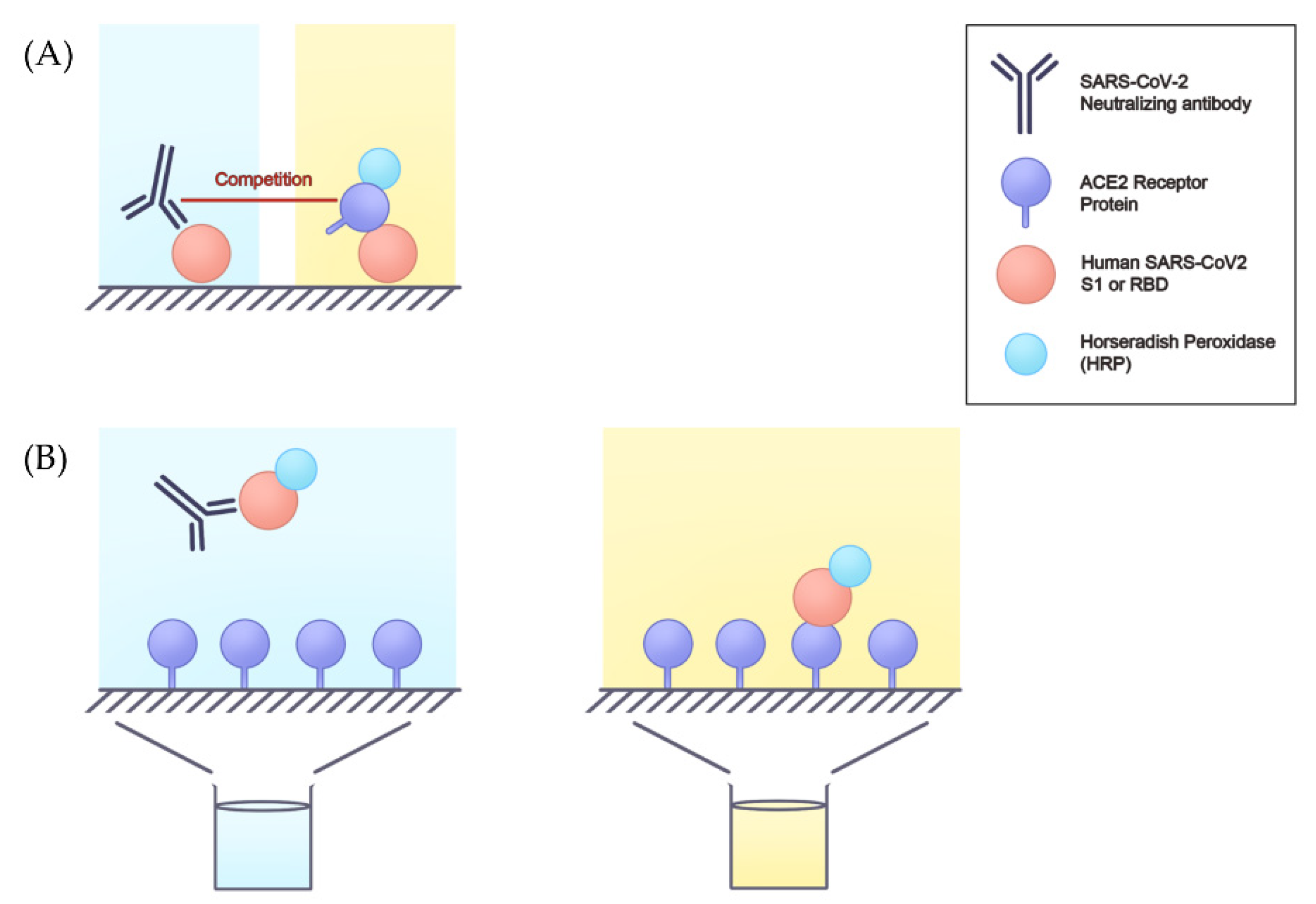
4. Detect Neutralizing Antibodies Using Surrogate Assays

Besides the viral neutralization test (VNT), a non-viral neutralizing antibody detection platform can be utilized. Using the surrogate viral neutralization test (sVNT), biosafety level 1 or 2 (BSL-1 or BSL-2) facilities are adequate for sample processing and antibody detection. Therefore, many immunoassays are used for serum neutralizing activity. The application of surrogate assays not only predicted humoral protection, but also vaccine efficacy during clinical trials and after large-scale vaccination [33]. ELISA, which is based on antigen-antibody reaction, has been applied to detect many biological molecules and as a diagnostic tool [34]. Since the COVID-19 outbreak, many ELISAs have been developed to detect SARS-CoV-2 antibodies, especially neutralizing antibodies. ELISAs are cost-effective and less time-consuming compared to the pseudovirus neutralization test. Based on the interaction between ACE2 and receptor binding domain (RBD), ELISAs usually use recombinant RBD protein or ACE2 as the coating antigen, and HRP-labeled ACE2 or HRP-labeled RBD is used to produce the detection signal [35]. Because the most potent neutralizing antibodies recognize the spike protein RBD, followed by the S1 domain, the spike protein trimer, and the S2 subunit [36], most ELISAs utilize RBD as a target antigen. In addition, four major types of ELISAs, including direct, indirect, sandwich, and competitive, are used. Competitive ELISAs are commonly used for detecting SARS-CoV-2 neutralizing antibodies. The principle of the assay entails the competitive inhibition enzyme immunoassay technique, in which the neutralizing antibodies in serum compete with the HRP-ACE2 or coated ACE2 to bind to the coated RBD or HRP-RBD, respectively. If the serum sample contains SARS-CoV-2 neutralizing antibodies, the intensity of the color produced is inversely proportional to the amount of SARS-CoV-2 neutralizing antibodies [33,37] (Figure 1).
It is also possible to detect the specific type of neutralizing antibody, including IgG, IgA, and IgM, using indirect ELISA [38]. Besides ELISA, chemiluminescence immunoassay (CLIA), chemiluminescence microparticle immunoassay, enzyme-linked fluorescent assay, and fluorescent microsphere immunoassay are also used for neutralizing antibody detection, and related 59 SARS-CoV-2 serology/antibody tests have been granted Emergency Use Authorization (EUA) by the Food and Drug Administration [39]. Most of them are qualitative, but some are semi-quantitative or quantitative [40]. The sensitivity and specificity of ELISA-based serological assay detection are approximately 65% to 98% and 71% to 100%, respectively. CLIA-based serological assay detection shows higher sensitivity and specificity of 77% to 100% and 90% to 100%, respectively, and takes approximately 30 min [41]. However, chemiluminescence equipment is required for CLIA. Therefore, numerous assays of neutralizing antibody detection are available and can be chosen according to need. Neutralizing antibody detection can help determine whether the measured antibody is correlated to the functional antibodies that can keep a population safe during a pandemic [42].

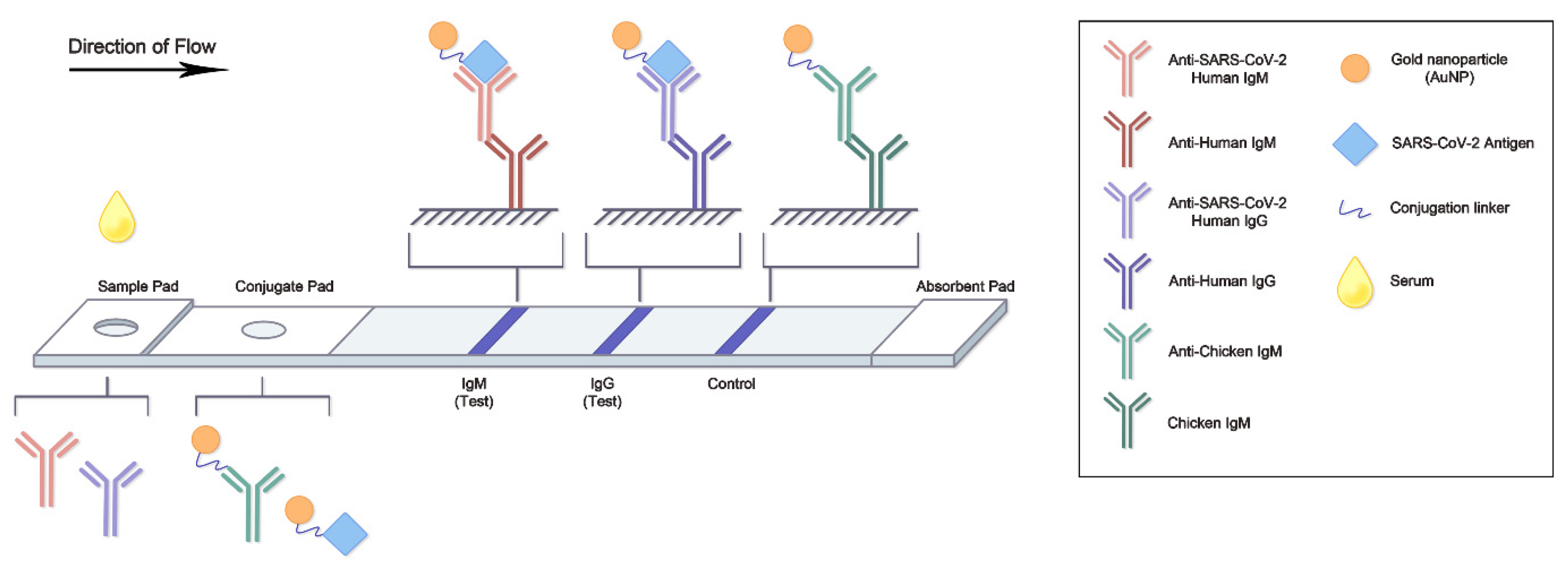
5. Lateral Flow Assay of Neutralizing Antibody

Given the time-consuming nature of ELISA/CLIA-based serological assay and the increasing number of COVID-19 cases, the lateral flow assay (LFA) is an ideal alternative for rapid diagnosis. LFA can detect either SARS-CoV-2 antigens or antibodies and provides results within 15–20 min; the results can be observed directly and visually or with a signal detector [43]. Due to its low cost and high availability, the antigen rapid diagnostic test is commonly used for the detection of SARS-CoV-2 protein [44]. At the same time, a serological antibody rapid diagnostic test has been developed for continuous monitoring of the neutralizing antibodies; it can identify individuals with an adaptive immune response to SARS-CoV-2, indicating recent or prior infection [35,43,45]. It is also useful as a necessary adjunct to all available neutralization tests [46]. In addition to diagnosis, LFA can potentially be used in vaccine development by tracing the level of neutralizing antibodies from clinical subjects. As of May 2022, 24 serological antibody rapid diagnostic tests from various manufacturers have been granted emergency use authorization (EUA) by the FDA [39]. The target biomarkers of serological antibody LFA are often COVID-19-specific IgG and/or IgM antibodies. Structurally, each LFA is based on a support card that spontaneously delivers the liquid sample by capillary action. The sample pad is typically at the proximal end of the assay cartridge and is soaked with sufficient sample. The soaked liquid sample enters a second zone, the conjugate pad, which contains a dry conjugate that captures the target analytes in the migrating liquid sample [46]. Neutralizing antibodies in the sample are detected by antigen-gold nanoparticles (Ag-NPs) conjugates within the conjugate pad. The antigens are typically the S and/or RBD, which bind their respective serum antibodies [47]. In the control line region, a signal-transducing colloidal gold nanoparticle (AuNP) antibody binds its capture antibody, while in the test line region, the coated antibody (anti-Human IgG or IgM) captures the AuNP-Ab conjugate-antigen complex (Figure 2). In addition, dedicated instruments, such as fluorescence analysis devices, can be used to provide qualitative or semi-quantitative results.
Recently, a novel fluorescent-based LFA for rapid detection and quantification of total binding antibodies has been reported [48]. Although LFA has the advantages of low cost, high speed, and simple operation, the combined sensitivity of LFAs for COVID-19 was only 66% and showed lower specificity compared to ELISA or CLIA-based detection [49]. Also, LFA was limited to admitted patients only [50] and thus may not be sensitive enough for diagnosing COVID-19 patients in the early stages of illness, as well as in determining vaccine efficacy [43,51]. Nevertheless, LFA can be a preliminary test to hint at potential immune response for further and more robust investigation using surrogate assays.

6. Limitations of Surrogate Assays and Rapid Lateral Flow Tests for SARS-CoV-2 Neutralization Antibodies

Surrogate assays for measuring neutralizing antibodies have been indispensable during the COVID-19 pandemic and can be performed at lower biosafety level facilities. A comparison of sVNT and microneutralization assay showed a correlation coefficient (R2) above 0.7, indicating a relatively well performance [33,52,53]. Besides, sVNTs are faster and simpler than traditional detection methods. However, the sVNT cannot detect all neutralizing antibodies in the patient sample. As previously demonstrated, neutralizing antibodies bind not only to the RBD but also to the N-terminal of spike proteins or other surface proteins on SARS-CoV-2 [54]. Notably, a previous study did not find a high correlation between the surrogate assay and the neutralization titer, possibly because of some neutralizing antibodies against other surface proteins on SARS-CoV-2 in addition to RBD [52,55]. Thus, surrogate assays can bias the detection of neutralizing antibodies. Further, most reports have used linear regression to analyze surrogate assay results. However, huge variations were discovered in the lower 25th and the higher 75th percentile [55].
A rapid lateral flow test is another approach for detecting SARS-CoV-2 neutralizing antibodies [35,56]. It is based on determining the color intensity in a test line to show the level of the neutralizing antibody. Although it can be highly correlated with the microneutralization titer, it cannot accurately reflect the level of the titer value. Hence, the rapid lateral flow test is not universally used to measure neutralizing antibodies. Despite these limitations of sVNT and rapid lateral flow test, they are still powerful approaches to accelerate vaccine development and satisfy the requirement for mass detection of neutralizing antibodies.

7. International Standard for SARS-CoV-2 and Its Importance for Calibrating Measurements in Serological Assays

An international standard, comprising a common unit and value of neutralizing antibody titer and antibody responses, is important for comparable diagnosis. Despite the development of many neutralization antibody tests, it is difficult to compare the results of different assays. Besides, the variation of experimental results in different laboratories may be due to disparity in interpretation, detection equipment, and protocol. Therefore, international standards for SARS-CoV-2 diagnosis are required to calibrate measurements from different methods or laboratories.
The significance of international standards in emerging viral infections has been proposed previously [57,58,59]. During, the outbreak of Zika virus disease in Latin America in 2015–2016 [60], the WHO released the international standard for immunologic assays for Zika virus to accelerate vaccine development and improve the consistency of quantification [61]. In addition, the first international standard for antiserum to Respiratory Syncytial Virus has been reported previously [62].
The first WHO International Standard and International Reference Panel for anti-SARS-CoV-2 immunoglobulin were adopted by the WHO Expert Committee on Biological Standardization in December 2020 [63,64]. The International Standard was based on pooled human plasma from SARS-CoV-2 convalescent patients, and antibodies were tested for use as a reference panel. The reference panel included sample F (20/150), high titer; sample J (20/148), mid titer, sample E (20/144), low anti-S and relatively high anti-N protein antibodies; sample I (20/140) low titer, and sample H (20/142), negative (Table 1).
In addition to the international standard, the binding antibody unit (BAU) is a unit proposed by WHO and represents the amounts of antigen-specific antibodies in a sample [65]. For instance, the antibody responses in the WHO reference panel include anti-S1, anti-spike, anti-RBD, and anti-N, and their levels can be expressed in BAU. Scientists can use BAU in many serological assays, such as microneutralization, pseudotype, and surrogate assays, for detecting antibody responses against SARS-CoV-2. Recently, many studies have used the international standard as a unit to represent the amount of neutralizing antibodies in the sample, and this has enabled comparisons to the reference value [66,67].
During the COVID-19 pandemic, many candidate vaccines underwent clinical trials [68,69]. With the high demand for COVID-19 vaccines worldwide, more licensed vaccines should be availed, particularly in the low- and middle-developed countries. However, a candidate vaccine must complete the clinical trial before it is licensed for public use; this process is time-consuming, and it is sometimes difficult to find eligible clinical trial participants. Therefore, immunological correlates of protection are proposed to replace traditional large-scale clinical trials [70,71,72]. Correlates of protection are based on quantification of neutralizing antibody responses and comparison of neutralizing antibody titers between candidate and listed vaccines. Previous studies reported that the neutralizing antibody level is highly proportional to vaccine efficacy and protection [73], indicating why the correlates of protection are feasible approaches for accelerating the development of vaccines and their certification. Given the suggested importance of the parallel neutralizing level comparison between candidate and listed vaccines, the international standard becomes a powerful reference and enabler of the correlates of protection. Undoubtedly, international standards for SARS-CoV-2 are vital biological references for serological assays and common units for calibrating neutralizing antibody measurements.

8. Conclusions

NAbs titer is a key factor for predicting immunity against SARS-CoV-2. Neutralization assays are powerful tools that have been used for COVID-19 diagnosis and vaccine evaluation. Various mature methods for detecting NAbs have been useful in the development of vaccines. However, most of these methods have advantages and shortcomings that need to be improved in the future. For instance, PRNT has safety issues, as it requires exposure to live viruses in the biosafety level 3 laboratory. Also, it remains unclear how the dynamic changes in the NAbs titers are correlated with patient clinical outcomes. Some sVNTs have been developed with high-throughput and time-saving features. The high-throughput assay may become a potential alternative gold standard for detecting and measuring NAbs. Scientists and virologists need to be innovative and develop more convenient methods for NAbs measurement in the future. Additionally, the heterogeneous design of the virus neutralization and binding assays limits the quantitative comparison of the results. These technical hurdles can be alleviated by the incorporation of an international standard.
Moreover, the natural effective level of NAbs required to prevent SARS-CoV-2 infection is unknown. A precise evaluation of the protective immunity effective at both the individual and population levels is difficult. Nevertheless, the NAbs titer is crucial for guiding decision-makers in easing the strict COVID-19 restrictions. An effective and time-effective neutralization assay can monitor in real-time the changes in the protective neutralization titers of SARS-CoV-2-infected patients and vaccinated individuals. Additionally, it can help in screening effective vaccine candidates. With the high mutation rate of SASR-CoV-2, the circulating strains keep changing and make the antigenicity alteration. The antigenicity alteration affects the interaction between RBD and ACE2 where the neutralizing antibodies most frequently bind. Hence, the next generation of neutralization assays should be designed against the mutated viruses in the future. In sum, scientists should make effort to further improve the specificity and sensitivity of novel neutralization assays, which are key in preparing for future pandemics, such as COVID-19.

Author Contributions

K.-T.L., Y.-J.H. and G.-H.W. writing the manuscript; K.-Y.A.H. and P.-N.H. review and editing the manuscript. All authors have read and agreed to the published version of the manuscript.

Funding

This research received funding from the Chang Gung Medical Research Program (CORPD1J0061-63). This work was financially supported by the Research Center for Emerging Viral Infections from The Featured Areas Research Center Program within the framework of the Higher Education Sprout Project by the Ministry of Education (MOE) in Taiwan and the Ministry of Science and Technology (MOST), Taiwan (MOST 111-2634-F-182-001).

Institutional Review Board Statement

Not applicable.

Informed Consent Statement

Not applicable.

Data Availability Statement

Not applicable.

Acknowledgments

The authors would like to thank Chung-Jung Wu for providing us with Figure 1 and Figure 2.

Conflicts of Interest

The authors declare no conflict of interest.

References

  1. Zhou, P.; Yang, X.L.; Wang, X.G.; Hu, B.; Zhang, L.; Zhang, W.; Si, H.R.; Zhu, Y.; Li, B.; Huang, C.L.; et al. A pneumonia outbreak associated with a new coronavirus of probable bat origin. Nature 2020, 579, 270–273. [Google Scholar] [CrossRef] [PubMed] [Green Version]
  2. Lai, C.C.; Shih, T.P.; Ko, W.C.; Tang, H.J.; Hsueh, P.R. Severe acute respiratory syndrome coronavirus 2 (SARS-CoV-2) and coronavirus disease-2019 (COVID-19): The epidemic and the challenges. Int. J. Antimicrob. Agents 2020, 55, 105924. [Google Scholar] [CrossRef] [PubMed]
  3. Jiang, C.; Wang, Y.; Hu, M.; Wen, L.; Wen, C.; Wang, Y.; Zhu, W.; Tai, S.; Jiang, Z.; Xiao, K.; et al. Antibody seroconversion in asymptomatic and symptomatic patients infected with severe acute respiratory syndrome coronavirus 2 (SARS-CoV-2). Clin. Transl. Immunol. 2020, 9, e1182. [Google Scholar] [CrossRef] [PubMed]
  4. Catry, E.; Jacqmin, H.; Dodemont, M.; Saad Albichr, I.; Lardinois, B.; de Fays, B.; Delaere, B.; Closset, M.; Laurent, T.; Denis, O.; et al. Analytical and clinical evaluation of four commercial SARS-CoV-2 serological immunoassays in hospitalized patients and ambulatory individuals. J. Virol. Methods 2021, 289, 114060. [Google Scholar] [CrossRef]
  5. Li, C.; Zhao, C.; Bao, J.; Tang, B.; Wang, Y.; Gu, B. Laboratory diagnosis of coronavirus disease-2019 (COVID-19). Clin. Chim. Acta 2020, 510, 35–46. [Google Scholar] [CrossRef]
  6. Kontou, P.I.; Braliou, G.G.; Dimou, N.L.; Nikolopoulos, G.; Bagos, P.G. Antibody Tests in Detecting SARS-CoV-2 Infection: A Meta-Analysis. Diagnostics 2020, 10, 319. [Google Scholar] [CrossRef]
  7. Zeng, W.; Liu, G.; Ma, H.; Zhao, D.; Yang, Y.; Liu, M.; Mohammed, A.; Zhao, C.; Yang, Y.; Xie, J.; et al. Biochemical characterization of SARS-CoV-2 nucleocapsid protein. Biochem. Biophys. Res. Commun. 2020, 527, 618–623. [Google Scholar] [CrossRef]
  8. Wang, Q.; Zhang, Y.; Wu, L.; Niu, S.; Song, C.; Zhang, Z.; Lu, G.; Qiao, C.; Hu, Y.; Yuen, K.Y.; et al. Structural and Functional Basis of SARS-CoV-2 Entry by Using Human ACE2. Cell 2020, 181, 894–904.e9. [Google Scholar] [CrossRef]
  9. Walls, A.C.; Park, Y.J.; Tortorici, M.A.; Wall, A.; McGuire, A.T.; Veesler, D. Structure, Function, and Antigenicity of the SARS-CoV-2 Spike Glycoprotein. Cell 2020, 181, 281–292.e286. [Google Scholar] [CrossRef]
  10. Zhu, Z.; Chakraborti, S.; He, Y.; Roberts, A.; Sheahan, T.; Xiao, X.; Hensley, L.E.; Prabakaran, P.; Rockx, B.; Sidorov, I.A.; et al. Potent cross-reactive neutralization of SARS coronavirus isolates by human monoclonal antibodies. Proc. Natl. Acad. Sci. USA 2007, 104, 12123–12128. [Google Scholar] [CrossRef] [Green Version]
  11. Perera, R.A.; Mok, C.K.; Tsang, O.T.; Lv, H.; Ko, R.L.; Wu, N.C.; Yuan, M.; Leung, W.S.; Chan, J.M.; Chik, T.S.; et al. Serological assays for severe acute respiratory syndrome coronavirus 2 (SARS-CoV-2), March 2020. Eurosurveillance 2020, 25, 2000421. [Google Scholar] [CrossRef] [Green Version]
  12. Okba, N.M.A.; Muller, M.A.; Li, W.; Wang, C.; GeurtsvanKessel, C.H.; Corman, V.M.; Lamers, M.M.; Sikkema, R.S.; de Bruin, E.; Chandler, F.D.; et al. Severe Acute Respiratory Syndrome Coronavirus 2-Specific Antibody Responses in Coronavirus Disease Patients. Emerg. Infect. Dis. 2020, 26, 1478–1488. [Google Scholar] [CrossRef]
  13. Vanderheiden, A.; Edara, V.V.; Floyd, K.; Kauffman, R.C.; Mantus, G.; Anderson, E.; Rouphael, N.; Edupuganti, S.; Shi, P.Y.; Menachery, V.D.; et al. Development of a Rapid Focus Reduction Neutralization Test Assay for Measuring SARS-CoV-2 Neutralizing Antibodies. Curr. Protoc. Immunol. 2020, 131, e116. [Google Scholar] [CrossRef]
  14. Muruato, A.E.; Fontes-Garfias, C.R.; Ren, P.; Garcia-Blanco, M.A.; Menachery, V.D.; Xie, X.; Shi, P.Y. A high-throughput neutralizing antibody assay for COVID-19 diagnosis and vaccine evaluation. Nat. Commun. 2020, 11, 4059. [Google Scholar] [CrossRef]
  15. Bewley, K.R.; Coombes, N.S.; Gagnon, L.; McInroy, L.; Baker, N.; Shaik, I.; St-Jean, J.R.; St-Amant, N.; Buttigieg, K.R.; Humphries, H.E.; et al. Quantification of SARS-CoV-2 neutralizing antibody by wild-type plaque reduction neutralization, microneutralization and pseudotyped virus neutralization assays. Nat. Protoc. 2021, 16, 3114–3140. [Google Scholar] [CrossRef]
  16. Amanat, F.; White, K.M.; Miorin, L.; Strohmeier, S.; McMahon, M.; Meade, P.; Liu, W.C.; Albrecht, R.A.; Simon, V.; Martinez-Sobrido, L.; et al. An In Vitro Microneutralization Assay for SARS-CoV-2 Serology and Drug Screening. Curr. Protoc. Microbiol. 2020, 58, e108. [Google Scholar] [CrossRef]
  17. Deshpande, G.R.; Sapkal, G.N.; Tilekar, B.N.; Yadav, P.D.; Gurav, Y.; Gaikwad, S.; Kaushal, H.; Deshpande, K.S.; Kaduskar, O.; Sarkale, P.; et al. Neutralizing antibody responses to SARS-CoV-2 in COVID-19 patients. Indian J. Med. Res. 2020, 152, 82–87. [Google Scholar] [CrossRef]
  18. Zhang, L.; Li, Q.; Liu, Q.; Huang, W.; Nie, J.; Wang, Y. A bioluminescent imaging mouse model for Marburg virus based on a pseudovirus system. Hum. Vaccines Immunother. 2017, 13, 1811–1817. [Google Scholar] [CrossRef] [Green Version]
  19. Sanders, D.A. No false start for novel pseudotyped vectors. Curr. Opin. Biotechnol. 2002, 13, 437–442. [Google Scholar] [CrossRef]
  20. Li, Q.; Wu, J.; Nie, J.; Zhang, L.; Hao, H.; Liu, S.; Zhao, C.; Zhang, Q.; Liu, H.; Nie, L.; et al. The Impact of Mutations in SARS-CoV-2 Spike on Viral Infectivity and Antigenicity. Cell 2020, 182, 1284–1294.e9. [Google Scholar] [CrossRef]
  21. Kumar, M.; Keller, B.; Makalou, N.; Sutton, R.E. Systematic determination of the packaging limit of lentiviral vectors. Hum. Gene Ther. 2001, 12, 1893–1905. [Google Scholar] [CrossRef]
  22. Counsell, J.R.; Asgarian, Z.; Meng, J.; Ferrer, V.; Vink, C.A.; Howe, S.J.; Waddington, S.N.; Thrasher, A.J.; Muntoni, F.; Morgan, J.E.; et al. Lentiviral vectors can be used for full-length dystrophin gene therapy. Sci. Rep. 2017, 7, 44775. [Google Scholar] [CrossRef] [Green Version]
  23. Xiong, H.L.; Wu, Y.T.; Cao, J.L.; Yang, R.; Liu, Y.X.; Ma, J.; Qiao, X.Y.; Yao, X.Y.; Zhang, B.H.; Zhang, Y.L.; et al. Robust neutralization assay based on SARS-CoV-2 S-protein-bearing vesicular stomatitis virus (VSV) pseudovirus and ACE2-overexpressing BHK21 cells. Emerg. Microbes Infect. 2020, 9, 2105–2113. [Google Scholar] [CrossRef]
  24. Johnson, M.C.; Lyddon, T.D.; Suarez, R.; Salcedo, B.; LePique, M.; Graham, M.; Ricana, C.; Robinson, C.; Ritter, D.G. Optimized Pseudotyping Conditions for the SARS-COV-2 Spike Glycoprotein. J. Virol. 2020, 94, e01062-20. [Google Scholar] [CrossRef]
  25. Meyer, B.; Torriani, G.; Yerly, S.; Mazza, L.; Calame, A.; Arm-Vernez, I.; Zimmer, G.; Agoritsas, T.; Stirnemann, J.; Spechbach, H.; et al. Validation of a commercially available SARS-CoV-2 serological immunoassay. Clin. Microbiol. Infect. 2020, 26, 1386–1394. [Google Scholar] [CrossRef]
  26. Condor Capcha, J.M.; Lambert, G.; Dykxhoorn, D.M.; Salerno, A.G.; Hare, J.M.; Whitt, M.A.; Pahwa, S.; Jayaweera, D.T.; Shehadeh, L.A. Generation of SARS-CoV-2 Spike Pseudotyped Virus for Viral Entry and Neutralization Assays: A 1-Week Protocol. Front. Cardiovasc Med. 2020, 7, 618651. [Google Scholar] [CrossRef]
  27. Nie, J.; Li, Q.; Wu, J.; Zhao, C.; Hao, H.; Liu, H.; Zhang, L.; Nie, L.; Qin, H.; Wang, M.; et al. Quantification of SARS-CoV-2 neutralizing antibody by a pseudotyped virus-based assay. Nat. Protoc. 2020, 15, 3699–3715. [Google Scholar] [CrossRef]
  28. Schmidt, F.; Weisblum, Y.; Muecksch, F.; Hoffmann, H.H.; Michailidis, E.; Lorenzi, J.C.C.; Mendoza, P.; Rutkowska, M.; Bednarski, E.; Gaebler, C.; et al. Measuring SARS-CoV-2 neutralizing antibody activity using pseudotyped and chimeric viruses. J. Exp. Med. 2020, 217, e20201181. [Google Scholar] [CrossRef]
  29. Kalkeri, R.; Cai, Z.; Lin, S.; Farmer, J.; Kuzmichev, Y.V.; Koide, F. SARS-CoV-2 Spike Pseudoviruses: A Useful Tool to Study Virus Entry and Address Emerging Neutralization Escape Phenotypes. Microorganisms 2021, 9, 1744. [Google Scholar] [CrossRef]
  30. Hoffmann, M.; Kleine-Weber, H.; Schroeder, S.; Kruger, N.; Herrler, T.; Erichsen, S.; Schiergens, T.S.; Herrler, G.; Wu, N.H.; Nitsche, A.; et al. SARS-CoV-2 Cell Entry Depends on ACE2 and TMPRSS2 and Is Blocked by a Clinically Proven Protease Inhibitor. Cell 2020, 181, 271–280.e278. [Google Scholar] [CrossRef] [PubMed]
  31. Dieterle, M.E.; Haslwanter, D.; Bortz, R.H., 3rd; Wirchnianski, A.S.; Lasso, G.; Vergnolle, O.; Abbasi, S.A.; Fels, J.M.; Laudermilch, E.; Florez, C.; et al. A Replication-Competent Vesicular Stomatitis Virus for Studies of SARS-CoV-2 Spike-Mediated Cell Entry and Its Inhibition. Cell Host Microbe 2020, 28, 486–496.e6. [Google Scholar] [CrossRef] [PubMed]
  32. Case, J.B.; Rothlauf, P.W.; Chen, R.E.; Kafai, N.M.; Fox, J.M.; Smith, B.K.; Shrihari, S.; McCune, B.T.; Harvey, I.B.; Keeler, S.P.; et al. Replication-Competent Vesicular Stomatitis Virus Vaccine Vector Protects against SARS-CoV-2-Mediated Pathogenesis in Mice. Cell Host Microbe 2020, 28, 465–474.e4. [Google Scholar] [CrossRef] [PubMed]
  33. Tan, C.W.; Chia, W.N.; Qin, X.; Liu, P.; Chen, M.I.; Tiu, C.; Hu, Z.; Chen, V.C.; Young, B.E.; Sia, W.R.; et al. A SARS-CoV-2 surrogate virus neutralization test based on antibody-mediated blockage of ACE2-spike protein-protein interaction. Nat. Biotechnol. 2020, 38, 1073–1078. [Google Scholar] [CrossRef] [PubMed]
  34. Gan, S.D.; Patel, K.R. Enzyme immunoassay and enzyme-linked immunosorbent assay. J. Investig. Dermatol. 2013, 133, e12. [Google Scholar] [CrossRef] [Green Version]
  35. Wang, J.J.; Zhang, N.; Richardson, S.A.; Wu, J.V. Rapid lateral flow tests for the detection of SARS-CoV-2 neutralizing antibodies. Expert Rev. Mol. Diagn. 2021, 21, 363–370. [Google Scholar] [CrossRef]
  36. Andreano, E.; Nicastri, E.; Paciello, I.; Pileri, P.; Manganaro, N.; Piccini, G.; Manenti, A.; Pantano, E.; Kabanova, A.; Troisi, M.; et al. Extremely potent human monoclonal antibodies from COVID-19 convalescent patients. Cell 2021, 184, 1821–1835.e16. [Google Scholar] [CrossRef]
  37. Taylor, S.C.; Hurst, B.; Charlton, C.L.; Bailey, A.; Kanji, J.N.; McCarthy, M.K.; Morrison, T.E.; Huey, L.; Annen, K.; DomBourian, M.G.; et al. A New SARS-CoV-2 Dual-Purpose Serology Test: Highly Accurate Infection Tracing and Neutralizing Antibody Response Detection. J. Clin. Microbiol. 2021, 59, e02438-20. [Google Scholar] [CrossRef]
  38. Tre-Hardy, M.; Wilmet, A.; Beukinga, I.; Favresse, J.; Dogne, J.M.; Douxfils, J.; Blairon, L. Analytical and clinical validation of an ELISA for specific SARS-CoV-2 IgG, IgA, and IgM antibodies. J. Med. Virol. 2021, 93, 803–811. [Google Scholar] [CrossRef]
  39. In Vitro Diagnostics EUAs Serology and Other Adaptive Immune Response Tests for SARS-CoV-2. Available online: https://www.fda.gov/medical-devices/coronavirus-disease-2019-covid-19-emergency-use-authorizations-medical-devices/in-vitro-diagnostics-euas-serology-and-other-adaptive-immune-response-tests-sars-cov-2 (accessed on 23 June 2022).
  40. VITROS. Immunodiagnostic Products SARS-CoV-2 Antigen Reagent Pack. Available online: https://www.fda.gov/media/150675/download (accessed on 23 June 2022).
  41. Mohit, E.; Rostami, Z.; Vahidi, H. A comparative review of immunoassays for COVID-19 detection. Expert Rev. Clin. Immunol. 2021, 17, 573–599. [Google Scholar] [CrossRef]
  42. GeurtsvanKessel, C.H.; Okba, N.M.A.; Igloi, Z.; Bogers, S.; Embregts, C.W.E.; Laksono, B.M.; Leijten, L.; Rokx, C.; Rijnders, B.; Rahamat-Langendoen, J.; et al. An evaluation of COVID-19 serological assays informs future diagnostics and exposure assessment. Nat. Commun. 2020, 11, 3436. [Google Scholar] [CrossRef]
  43. Biby, A.; Wang, X.; Liu, X.; Roberson, O.; Henry, A.; Xia, X. Rapid testing for coronavirus disease 2019 (COVID-19). MRS Commun. 2022, 12, 12–23. [Google Scholar] [CrossRef]
  44. Porte, L.; Legarraga, P.; Vollrath, V.; Aguilera, X.; Munita, J.M.; Araos, R.; Pizarro, G.; Vial, P.; Iruretagoyena, M.; Dittrich, S.; et al. Evaluation of a novel antigen-based rapid detection test for the diagnosis of SARS-CoV-2 in respiratory samples. Int. J. Infect. Dis. 2020, 99, 328–333. [Google Scholar] [CrossRef]
  45. Peeling, R.W.; Heymann, D.L.; Teo, Y.Y.; Garcia, P.J. Diagnostics for COVID-19: Moving from pandemic response to control. Lancet 2022, 399, 757–768. [Google Scholar] [CrossRef]
  46. Banga Ndzouboukou, J.L.; Zhang, Y.D.; Fan, X.L. Recent Developments in SARS-CoV-2 Neutralizing Antibody Detection Methods. Curr. Med. Sci. 2021, 41, 1052–1064. [Google Scholar] [CrossRef]
  47. Ogata, A.F.; Maley, A.M.; Wu, C.; Gilboa, T.; Norman, M.; Lazarovits, R.; Mao, C.P.; Newton, G.; Chang, M.; Nguyen, K.; et al. Ultra-Sensitive Serial Profiling of SARS-CoV-2 Antigens and Antibodies in Plasma to Understand Disease Progression in COVID-19 Patients with Severe Disease. Clin. Chem. 2020, 66, 1562–1572. [Google Scholar] [CrossRef]
  48. Shurrab, F.M.; Younes, N.; Al-Sadeq, D.W.; Liu, N.; Qotba, H.; Abu-Raddad, L.J.; Nasrallah, G.K. Performance evaluation of novel fluorescent-based lateral flow immunoassay (LFIA) for rapid detection and quantification of total anti-SARS-CoV-2 S-RBD binding antibodies in infected individuals. Int. J. Infect. Dis. 2022, 118, 132–137. [Google Scholar] [CrossRef]
  49. Lisboa Bastos, M.; Tavaziva, G.; Abidi, S.K.; Campbell, J.R.; Haraoui, L.P.; Johnston, J.C.; Lan, Z.; Law, S.; MacLean, E.; Trajman, A.; et al. Diagnostic accuracy of serological tests for covid-19: Systematic review and meta-analysis. BMJ 2020, 370, m2516. [Google Scholar] [CrossRef]
  50. Ong, D.S.Y.; de Man, S.J.; Lindeboom, F.A.; Koeleman, J.G.M. Comparison of diagnostic accuracies of rapid serological tests and ELISA to molecular diagnostics in patients with suspected coronavirus disease 2019 presenting to the hospital. Clin. Microbiol. Infect. 2020, 26, 1094.e7–1094.e10. [Google Scholar] [CrossRef]
  51. Imai, K.; Tabata, S.; Ikeda, M.; Noguchi, S.; Kitagawa, Y.; Matuoka, M.; Miyoshi, K.; Tarumoto, N.; Sakai, J.; Ito, T.; et al. Clinical evaluation of an immunochromatographic IgM/IgG antibody assay and chest computed tomography for the diagnosis of COVID-19. J. Clin. Virol. 2020, 128, 104393. [Google Scholar] [CrossRef]
  52. Embregts, C.W.E.; Verstrepen, B.; Langermans, J.A.M.; Boszormenyi, K.P.; Sikkema, R.S.; de Vries, R.D.; Hoffmann, D.; Wernike, K.; Smit, L.A.M.; Zhao, S.; et al. Evaluation of a multi-spe.ecies SARS-CoV-2 surrogate virus neutralization test. One Health 2021, 13, 100313. [Google Scholar] [CrossRef]
  53. Liu, K.T.; Gong, Y.N.; Huang, C.G.; Huang, P.N.; Yu, K.Y.; Lee, H.C.; Lee, S.C.; Chiang, H.J.; Kung, Y.A.; Lin, Y.T.; et al. Quantifying Neutralizing Antibodies in Patients with COVID-19 by a Two-Variable Generalized Additive Model. mSphere 2022, 7, e0088321. [Google Scholar] [CrossRef]
  54. Chi, X.; Yan, R.; Zhang, J.; Zhang, G.; Zhang, Y.; Hao, M.; Zhang, Z.; Fan, P.; Dong, Y.; Yang, Y.; et al. A neutralizing human antibody binds to the N-terminal domain of the Spike protein of SARS-CoV-2. Science 2020, 369, 650–655. [Google Scholar] [CrossRef]
  55. Tang, M.S.; Case, J.B.; Franks, C.E.; Chen, R.E.; Anderson, N.W.; Henderson, J.P.; Diamond, M.S.; Gronowski, A.M.; Farnsworth, C.W. Association between SARS-CoV-2 Neutralizing Antibodies and Commercial Serological Assays. Clin. Chem. 2020, 66, 1538–1547. [Google Scholar] [CrossRef]
  56. Fulford, T.S.; Van, H.; Gherardin, N.A.; Zheng, S.; Ciula, M.; Drummer, H.E.; Redmond, S.; Tan, H.X.; Boo, I.; Center, R.J.; et al. A point-of-care lateral flow assay for neutralising antibodies against SARS-CoV-2. eBioMedicine 2021, 74, 103729. [Google Scholar] [CrossRef] [PubMed]
  57. Mattiuzzo, G.; Bentley, E.M.; Page, M. The Role of Reference Materials in the Research and Development of Diagnostic Tools and Treatments for Haemorrhagic Fever Viruses. Viruses 2019, 11, 781. [Google Scholar] [CrossRef] [PubMed] [Green Version]
  58. Rampling, T.; Page, M.; Horby, P. International Biological Reference Preparations for Epidemic Infectious Diseases. Emerg. Infect. Dis. 2019, 25, 205–211. [Google Scholar] [CrossRef] [PubMed] [Green Version]
  59. He, Q.; Mao, Q.; Zhang, J.; Bian, L.; Gao, F.; Wang, J.; Xu, M.; Liang, Z. COVID-19 Vaccines: Current Understanding on Immunogenicity, Safety, and Further Considerations. Front. Immunol. 2021, 12, 669339. [Google Scholar] [CrossRef] [PubMed]
  60. De Oliveira, W.K.; de Franca, G.V.A.; Carmo, E.H.; Duncan, B.B.; de Souza Kuchenbecker, R.; Schmidt, M.I. Infection-related microcephaly after the 2015 and 2016 Zika virus outbreaks in Brazil: A surveillance-based analysis. Lancet 2017, 390, 861–870. [Google Scholar] [CrossRef] [Green Version]
  61. Mattiuzzo, G.; Knezevic, I.; Hassall, M.; Ashall, J.; Myhill, S.; Faulkner, V.; Hockley, J.; Rigsby, P.; Wilkinson, D.E.; Page, M.; et al. Harmonization of Zika neutralization assays by using the WHO International Standard for anti-Zika virus antibody. NPJ Vaccines 2019, 4, 42. [Google Scholar] [CrossRef]
  62. McDonald, J.U.; Rigsby, P.; Dougall, T.; Engelhardt, O.G.; Study, P. Establishment of the first WHO International Standard for antiserum to Respiratory Syncytial Virus: Report of an international collaborative study. Vaccine 2018, 36, 7641–7649. [Google Scholar] [CrossRef]
  63. Kristiansen, P.A.; Page, M.; Bernasconi, V.; Mattiuzzo, G.; Dull, P.; Makar, K.; Plotkin, S.; Knezevic, I. WHO International Standard for anti-SARS-CoV-2 immunoglobulin. Lancet 2021, 397, 1347–1348. [Google Scholar] [CrossRef]
  64. Knezevic, I.; Mattiuzzo, G.; Page, M.; Minor, P.; Griffiths, E.; Nuebling, M.; Moorthy, V. WHO International Standard for evaluation of the antibody response to COVID-19 vaccines: Call for urgent action by the scientific community. Lancet Microbe 2022, 3, e235–e240. [Google Scholar] [CrossRef]
  65. Mattiuzzo, G.; Bentley, E.M.; Hassall, M.; Routley, S.; Richardson, S.; Bernasconi, V.; Kristiansen, P.; Harvala, H.; Roberts, D.; Semple, M.G.; et al. Establishment of the WHO International Standard and Reference Panel for anti-SARS-CoV-2 antibody. Available online: https://www.who.int/publications/m/item/WHO-BS-2020.2403 (accessed on 23 June 2022).
  66. Ruetalo, N.; Flehmig, B.; Schindler, M.; Pridzun, L.; Haage, A.; Reichenbacher, M.; Kirchner, T.; Kirchner, T.; Klingel, K.; Ranke, M.B.; et al. Long-Term Humoral Immune Response against SARS-CoV-2 after Natural Infection and Subsequent Vaccination According to WHO International Binding Antibody Units (BAU/mL). Viruses 2021, 13, 2336. [Google Scholar] [CrossRef]
  67. Zhu, F.; Althaus, T.; Tan, C.W.; Costantini, A.; Chia, W.N.; Van Vinh Chau, N.; Van Tan, L.; Mattiuzzo, G.; Rose, N.J.; Voiglio, E.; et al. WHO international standard for SARS-CoV-2 antibodies to determine markers of protection. Lancet Microbe 2022, 3, e81–e82. [Google Scholar] [CrossRef]
  68. Krammer, F. SARS-CoV-2 vaccines in development. Nature 2020, 586, 516–527. [Google Scholar] [CrossRef]
  69. Kyriakidis, N.C.; Lopez-Cortes, A.; Gonzalez, E.V.; Grimaldos, A.B.; Prado, E.O. SARS-CoV-2 vaccines strategies: A comprehensive review of phase 3 candidates. NPJ Vaccines 2021, 6, 28. [Google Scholar] [CrossRef]
  70. Krammer, F. A correlate of protection for SARS-CoV-2 vaccines is urgently needed. Nat. Med. 2021, 27, 1147–1148. [Google Scholar] [CrossRef]
  71. Plotkin, S.A. Correlates of protection induced by vaccination. Clin. Vaccine Immunol. 2010, 17, 1055–1065. [Google Scholar] [CrossRef] [Green Version]
  72. Earle, K.A.; Ambrosino, D.M.; Fiore-Gartland, A.; Goldblatt, D.; Gilbert, P.B.; Siber, G.R.; Dull, P.; Plotkin, S.A. Evidence for antibody as a protective correlate for COVID-19 vaccines. Vaccine 2021, 39, 4423–4428. [Google Scholar] [CrossRef]
  73. Khoury, D.S.; Cromer, D.; Reynaldi, A.; Schlub, T.E.; Wheatley, A.K.; Juno, J.A.; Subbarao, K.; Kent, S.J.; Triccas, J.A.; Davenport, M.P. Neutralizing antibody levels are highly predictive of immune protection from symptomatic SARS-CoV-2 infection. Nat. Med. 2021, 27, 1205–1211. [Google Scholar] [CrossRef]
Figure 1. The principle of neutralizing antibody detection using ELISA. Based on the competitive ELISA, one of the methods (A), the HRP-ACE2 competes with SARS-CoV-2 neutralizing antibody to bind to the antigen-like S1 or RBD. The light signal is strong when the level of the neutralizing antibody is low. In the other method (B), the neutralizing antibody is pre-incubated with antigen-like S1 or RBD and then added to the well that is coated with ACE2. After the wash step, the antigens not recognized by the neutralizing antibodies can bind to the well plate (ACE2), and a bright color can also be detected. ELISA, enzyme-linked immunosorbent assay; HRP-ACE2, horseradish peroxidase-angiotensin-converting enzyme 2; RBD, receptor binding domain; SARS-CoV-2, severe acute respiratory syndrome coronavirus 2; S1, spike protein 1.
Viruses 14 01560 g001
Figure 2. The scheme of neutralizing antibody LFA. After the drop of serum sample is placed on the sample pad, the sample moves to the conjugate pad by capillary action. In this part, the AuNP-antigen can be recognized by the neutralizing antibody. Antibody detection, including IgM and IgG, occurs in the next part; the antibody and AuNP-antigen complex show a signal in the test region. AuNP, gold nanoparticle; Ig, immunoglobulin; LFA, lateral flow assay; SARS-CoV-2, severe acute respiratory syndrome corona virus 2.
Viruses 14 01560 g002
Table 1. WHO international reference standards for anti-SARS-CoV-2 immunoglobulin and neutralizing antibody.
WHO Reference Standards
sample code20/13020/13620/15020/14820/14420/14020/142
Neutralizing antibody (IU/mL)1300100014732109544-
Anti-RBD IgG (BAU/mL)50210008172056645-
Anti-S1 IgG (BAU/mL)58810007662465046-
Anti-Spike IgG (BAU/mL)47610008322418653-
Anti-N IgG (BAU/mL)747100071329514612-
IU: international unit; BAU: binding antibody unit; Ig, immunoglobulin; S1, spike protein subunit 1; RBD, receptor binding domain; N, nucleocapsid; WHO, World Health Organization. -: Negative.
Publisher’s Note: MDPI stays neutral with regard to jurisdictional claims in published maps and institutional affiliations.

