Article

Towards the Laboratory Maintenance of Haemagogus janthinomys (Dyar, 1921), the Major Neotropical Vector of Sylvatic Yellow Fever

by
Adam Hendy
1,*,
Nelson Ferreira Fé
2,
Danielle Valério
3,
Eduardo Hernandez-Acosta
4,
Bárbara A. Chaves
3,
Luís Felipe Alho da Silva
3,
Rosa Amélia Gonçalves Santana
3,
Andréia da Costa Paz
3,
Matheus Mickael Mota Soares
5,
Flamarion Prado Assunção
3,
José Tenaçol Andes, Jr.
3,
Chiara Andolina
6,
Vera Margarete Scarpassa
7,
Marcus Vinícius Guimarães de Lacerda
3,8,
Kathryn A. Hanley
4 and
Nikos Vasilakis
1,9,10,11,12,*
1
Department of Pathology, University of Texas Medical Branch, 301 University Blvd, Galveston, TX 77555-0609, USA
2
Centro de Entomologia, Fundação de Medicina Tropical Doutor Heitor Vieira Dourado (FMT-HVD), Manaus 69040-000, AM, Brazil
3
Instituto de Pesquisa Clínica Carlos Borborema (IPCCB), Fundação de Medicina Tropical Doutor Heitor Vieira Dourado (FMT-HVD), Manaus 69040-000, AM, Brazil
4
Department of Biology, New Mexico State University, Las Cruces, NM 88003-8801, USA
5
Graduate Program in Entomology, Instituto Nacional de Pesquisas da Amazônia, Manaus 69067-375, AM, Brazil
6
Department of Medical Microbiology, Radboud University Nijmegen Medical Centre, 6525 GA Nijmegen, The Netherlands
7
Coordenação de Biodiversidade, Instituto Nacional de Pesquisas da Amazônia, Manaus 69067-375, AM, Brazil
8
Instituto Leônidas and Maria Deane, Fiocruz Amazonas, Manaus 69057-070, AM, Brazil
9
Center for Biodefense and Emerging Infectious Diseases, University of Texas Medical Branch, 301 University Blvd, Galveston, TX 77555-0609, USA
10
Institute for Human Infection and Immunity, University of Texas Medical Branch, 301 University Blvd, Galveston, TX 77555-0609, USA
11
Center for Tropical Diseases, University of Texas Medical Branch, 301 University Blvd, Galveston, TX 77555-0609, USA
12
Center for Vector-Borne and Zoonotic Diseases, University of Texas Medical Branch, 301 University Blvd, Galveston, TX 77555-0609, USA
*
Authors to whom correspondence should be addressed.
Viruses 2023, 15(1), 45; https://doi.org/10.3390/v15010045
Submission received: 30 November 2022 / Revised: 20 December 2022 / Accepted: 22 December 2022 / Published: 23 December 2022
(This article belongs to the Section Invertebrate Viruses)

Abstract

:
Haemagogus (Haemagogus) janthinomys (Dyar, 1921), the major neotropical vector of sylvatic yellow fever virus, is notoriously difficult to maintain in captivity. It has never been reared beyond an F1 generation, and almost no experimental transmission studies have been performed with this species since the 1940s. Herein we describe installment hatching, artificial blood feeding, and forced-mating techniques that enabled us to produce small numbers of F3 generation Hg. janthinomys eggs for the first time. A total of 62.8% (1562/2486) F1 generation eggs hatched during ≤10 four-day cycles of immersion in a bamboo leaf infusion followed by partial drying. Hatching decreased to 20.1% (190/944) in the F2 generation for eggs laid by mosquitoes copulated by forced mating. More than 85% (79/92) female F2 mosquitoes fed on an artificial blood feeding system. While we were unable to maintain a laboratory colony of Hg. janthinomys past the F3 generation, our methods provide a foundation for experimental transmission studies with this species in a laboratory setting, a critical capacity in a region with hyper-endemic transmission of dengue, Zika, and chikungunya viruses, all posing a risk of spillback into a sylvatic cycle.

1. Introduction

Haemagogus (Haemagogus) janthinomys (Dyar, 1921) was first implicated in the natural transmission of sylvatic yellow fever virus (YFV, genus Flavivirus, family Flaviviridae) in the Americas in 1938, when it was isolated from pools of forest mosquitoes collected in Rio de Janeiro State, Brazil [1,2]. Alongside Hg. (Conopostegus) leucocelaenus (Dyar and Shannon, 1924), Hg. janthinomys is considered the major vector of YFV [3,4] and is also thought to be a prominent vector of Mayaro virus (MAYV, genus Alphavirus, family Togaviridae), in neotropical forests [5,6]. Despite its obvious medical importance, almost no experimental transmission studies have been performed with this species since the early studies of YFV in the 1930s and 40s [7,8]. The lack of research is often attributed to difficulties rearing these mosquitoes under laboratory conditions [9,10,11], although there has been little effort to overcome this problem in recent years.
Early attempts to keep Hg. janthinomys alive in cages beyond the extrinsic incubation period of YFV; around 14 days or more for this species, proved difficult [12,13]. To convincingly demonstrate vector competence [10,14], it was necessary to maintain adult female mosquitoes individually in small glass vials containing a layer of damp cotton and a disc of filter paper [10,15]. These were either field collected mosquitoes or the F1 progeny of field collected mosquitoes that were blood fed on monkeys or marsupials in captivity [8,10,15]. Specimens were kept alive for a maximum of around 50 days [15], which was ample time to demonstrate onward transmission of the virus to uninfected hosts [10]. Since then, a modified vial-based method has been shown to increase adult longevity up to 72 days and improve upon egg-yields documented in earlier studies [16]. Despite this progress, there have been no published vector competence studies involving Hg. janthinomys since 1956 [9]. Dengue, Zika (DENV, ZIKV, both genus Flavivirus), and chikungunya viruses (CHIKV, genus Alphavirus) have all emerged or resurged in the Americas during this time [17]. They are currently only known to circulate in human endemic cycles in the region, involving Aedes (Stegomyia) aegypti (Linnaeus, 1762) and Ae. (Stg.) albopictus (Skuse, 1894) mosquitoes. However, the presence of susceptible sylvatic hosts and competent vectors creates risk for virus spillback and the emergence of novel sylvatic cycles [18,19], not amenable to control interventions, that would be a source of spillover akin to YFV.
Haemagogus janthinomys mosquitoes possess several physiological and behavioral traits that make laboratory maintenance problematic. They lay their eggs on the edges of transient water bodies in tree holes and bamboo [2,20,21]. Consequently, they show some drought tolerance and hatch in installments as a survival mechanism [22,23]. Eclosion requires multiple cycles of immersion and drying, making it difficult to obtain high numbers of larvae on demand. While a slow process, problems with installment hatching can partly be overcome through perseverance [23,24], and F1 adults reared from field collected mosquitoes have been used to perform fairly extensive survival and vector competence studies [15]. However, laboratory maintenance of Hg. janthinomys has never progressed beyond the F1 generation. These mosquitoes are reluctant to mate in cages [25], and attempts to encourage mating have either been unsuccessful [9,15,25] or considered too difficult [11,26,27].
The absence of a simple blood feeding technique further complicates the task of colonizing Hg. janthinomys. Blood-soaked cotton pledgets and membrane feeders have been used to deliver virus-infected blood to Hg. (Hag.) equinus (Theobald, 1903) [28] and Hg. leucocelaenus [28], while mice and human blood have been used to maintain Hg. equinus in colony [29]. It is unclear whether Hg. janthinomys will feed readily on adult mice, but they will bite baby mice, as these have previously been used in vector competence studies [7,10]. Other blood sources utilized in experimental work involving Hg. janthinomys have included rhesus macaques [9,13,30], various species of neotropical monkeys [7,8,10,31], marsupials [8], and YFV vaccinated human volunteers [16], none of which are suitable for long-term colony maintenance.
Soon after 1956, when the last published Hg. janthinomys vector competence studies involving just seven mosquitoes were abandoned due to a lack of material [9], techniques were developed to induce copulation between aedine mosquitoes that rarely mate under laboratory conditions [32]. Forced mating is now mainly used to colonize eurygamous malaria vectors reluctant to mate in cages [33,34,35], but has never been applied to Hg. janthinomys. In this study, we describe our progress towards the laboratory maintenance of Hg. janthinomys, in which we used artificial blood feeding and forced mating techniques to produce small numbers of F3 generation eggs.

2. Materials and Methods

2.1. Ethics

Field collections of Hg. janthinomys differed from human landing collections in that mosquitoes were captured using nets prior to landing and no skin was deliberately exposed to attract mosquitoes. All collectors were vaccinated against YFV and wore trousers and either a long-sleeved shirt or repellent to minimize the risk of being bitten. Mosquito collections were approved by local environmental authorities (SISBIO license 57003-6) and the study did not involve endangered or protected species. Artificial feeding methods involving human blood were reviewed and approved by the Fundação de Medicina Tropical Doutor Heitor Vieira Dourado (FMT-HVD) Ethics Review Board (CAAE: 53963521.0.0000.0005).

2.2. Field Collected Mosquitoes

2.2.1. Source Material

Host-seeking female Haemagogus species mosquitoes were collected at the Adolpho Ducke forest reserve bordering Manaus in the Brazilian Amazon between February and April 2022. Approaching mosquitoes were collected with handheld nets at ground level and on elevated platforms during the daytime (10:00–15:00) at a treefall (02.92537° S, 059.96582° W) [36]. Captured mosquitoes were transferred to a 16 × 19.5 cm cylindrical cardboard cage and provided with cotton wool soaked with mineral water for hydration. The cage was placed in a Styrofoam box in the shade throughout the day to prevent mosquito desiccation. All subsequent work took place in an air-conditioned insectary at FMT-HVD at 26 °C (±1 °C) and 70–80% relative humidity unless stated. The insectary was maintained in a constant 12 h light-dark cycle with changes in photoperiod occurring at 06:00 and 18:00 daily.

2.2.2. Blood Feeding

Immediately upon returning from the field, mosquitoes were transferred to a 15 × 15 cm plastic cage covered with fine mesh netting (Figure S1) and were supplied with two cotton wool balls, one soaked with distilled water and the other a 10% sugar solution. Mosquitoes were left to settle until the following evening when the water and sugar solution were removed. Feeding was then attempted the next morning at around 08:00. Starving Hg. janthinomys for such long periods prior to blood feeding resulted in mortality of up to 41.8% on several occasions (Dataset S1).
Human blood was drawn from healthy volunteers into 9 mL sodium heparin tubes (Greiner Bio-One, São Paulo, Brazil) by a trained technician. Blood was either used fresh or was refrigerated for ≤four days at 4 °C and heated to 37 °C in a water bath for 15–20 min before use. Feeders were prepared using round cotton pads and 180 mL disposable plastic drinking cups. Cotton pads were cut into approximately 2 × 2 cm squares and secured to the base of the cups using Teflon tape (Tecnoflon-Brasfita, São Paulo, Brazil) (Figure 1a). About 2 mL blood was applied to each pad until saturated (Figure 1b) and feeders were inverted over the top of the cage (Figure 1c). The blood was kept warm by adding approximately 100 mL of 45 °C water to the plastic cups. The water in each cup was changed once or twice when the temperature decreased below 36 °C, resulting in around 60 min feeding time. Field collected mosquitoes were offered blood on a single occasion. Mosquitoes were individually aspirated and those that had fully or partially engorged were separated into a 15 × 15 cm plastic cage and left to rest with water and sugar solution for five days before oviposition was attempted. Those that did not feed were discarded.

2.2.3. Oviposition

At five days post blood feeding, groups of up to 10 gravid mosquitoes were aspirated into 50 mL Falcon tubes where they were anesthetized by exposure to ethyl acetate vapor. To achieve this, a scrap of paper towel was twisted, dipped in ethyl acetate, and then secured inside a tube near its lid, ensuring the mosquitoes did not make contact. Since Hg. janthinomys are phototactic, this was possible by directing the opposite end of the tube towards natural light. This also prevented mosquitoes escaping when manipulating the lid. Once fully relaxed (usually less than 60 s), mosquitoes were placed in a Petri dish, and each had a wing removed under a stereomicroscope. They were then transferred either individually or in groups of up to nine mosquitoes into Petri dishes lined with 80 g filter paper (J Prolab, São José dos Pinhais, Brazil), placed above a damp cotton pad to maintain humidity (Figure S2). Mosquitoes were left for at least three days to oviposit before F1 eggs were recovered and hatching was attempted. Additional drops of water were added to filter papers as required during this time.

2.3. F1 Generation

2.3.1. Installment Hatching and Rearing

Egg papers were immersed as soon as possible in 2.5 L white plastic rearing trays containing approximately 500 mL 1:1 bamboo leaf infusion to distilled water. The leaf infusion was prepared two days in advance in a 5 L clear plastic bottle containing approximately 60 g fresh bamboo (Phyllostachys sp.) leaves, collected on FMT-HVD grounds, and distilled water. The bottle opening was covered with netting to prevent insects from entering and the infusion was kept at room temperature in an outdoor storage building. Since Hg. janthinomys hatch in installments [22], egg papers were generally submerged for two days and then partially dried for two days for up to ten immersion cycles. Partial drying involved laying egg papers on damp paper towels. It was critical that egg papers did not fully dry as eggs would soon collapse. To combat mold growth, body parts of dead mosquitoes were removed before the first immersion. Additionally, egg papers were placed in a weak bleach solution (1:3 2.5% NaClO to distilled water) for approximately 10 s after the second or third immersion cycle, or whenever mold growth was observed, then rinsed for one minute in a tray of distilled water. Larvae were reared to pupae in the water in which they emerged and a small amount of Fleischmann instant dry yeast (AB Brasil Industria e Comércio de Alimentos Ltda., São Paulo, Brazil) was added if required to increase the size of adults. This improved chances of survival and larger adults were easier to manipulate when attempting copulation (Section 2.3.2). Rearing trays were checked daily for rapid biofilm growth which, if present, was removed by dragging a paper towel smoothly across the surface of the water. Pupae were transferred to glass tumblers containing 150 mL distilled water placed inside 15 × 15 cm plastic cages (Section 2.2.2) until adults emerged. The number of days until emergence of the first larva, pupa, and adult was recorded in each tray as an indicator of development time. Adult mosquitoes were then separated by date of emergence, usually within a range of 1–2 days, into new 15 × 15 cm cages until required for copulation.

2.3.2. Copulation by Forced Mating

Forced mating of Hg. janthinomys was based on techniques applied to eurygamous Anopheles species [33]. We aimed to use approximately 3–5-day old adult males and 1–3-day old females, although no material was wasted, regardless of age. The work took place in a 26 °C air-conditioned room at the FMT-HVD Center for Entomology.
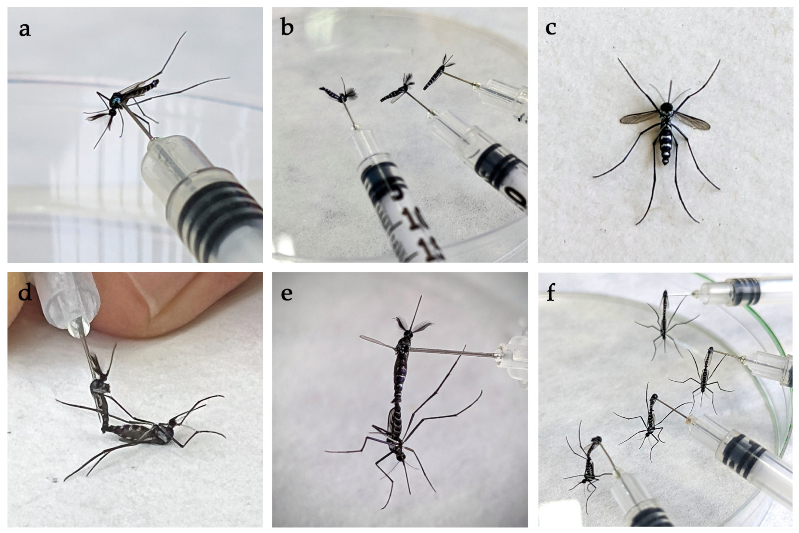
Male and non-blood fed female mosquitoes were anesthetized by exposure to ethyl acetate vapor as described in Section 2.2.3. Initially, 5–6 males were mounted laterally through the thorax on needles of 0.5 mL insulin syringes (Figure 2a). Mosquito legs were removed using soft entomological forceps (Figure 2b) but males were not decapitated at first, which is a common technique to induce mating [33,34]. However, when mosquito numbers increased, the heads of males were removed to speed up the copulation process. Around 2–3 anesthetized females were placed, with their ventral surface facing up (Figure 2c), on the lid of a white microcentrifuge box which could be rotated to allow easy manipulation. The male and female terminalia were introduced at an angle of slightly more than 90° (Figure 2d) (Video S1). If the mosquito pairs were still unresponsive after a short time, a different male was offered to a female. Male mosquitoes were reused once or twice if they remained viable after copulation. Once the terminalia were fully interlocked (Figure 2e), mating pairs were left to rest on the lid of a Petri dish until they separated (Figure 2f). The female was then quickly, but gently, aspirated and transferred to a new pre-prepared cage with distilled water and 10% sugar solution, and left to rest until the next day. Additional mosquitoes were prepared as needed.

2.3.3. Modifications to Blood Feeding and Oviposition

Only small numbers of F1 females blood fed initially and repeatedly separating blood fed from non-blood fed females resulted in high mortality. To counter this, all copulated F1 females, regardless of the strength of interaction with the male, were separated into two 20 × 18 cm plastic cages containing oviposition substrates. The substrates comprised 150 mL clear plastic containers lined with filter paper and a thick layer of cotton wool saturated with distilled water (Figure S3). An extra layer of filter paper was placed above the cotton wool to prevent mosquitoes becoming entangled. Mosquitoes were offered blood daily following the method described in Section 2.2.2, in the hope that some would refeed and survive through multiple gonotrophic cycles. Water and sugar solution were removed no more than two hours before feeding to reduce the risk of desiccation. Oviposition substrates were usually changed every seven days.

2.4. F2 Generation

Similarities with Previous Generations and Further Modifications

F2 generation eggs were immersed, hatched, and reared in a bamboo leaf infusion for up to nine cycles as described in Section 2.3.1. To overcome low egg yields, we reverted to separating blood fed from non-blood fed females using a slightly modified method, and only used blood fed mosquitoes for copulation. Adult males were separated into 15 × 15 cm cages by date of eclosion. Females were added to a single 15 × 15 cm modified feeding cage covered with fine mesh netting and with a lateral hole plugged with cotton wool. Female mosquitoes were offered blood daily as described in Section 2.2.2. However, the 45 °C water was not changed, and blood was only offered for 20–30 min. Mosquitoes that had visibly fed were aspirated via the lateral hole. Blood fed females were copulated and left to rest for at least five days before oviposition was attempted using individual mosquitoes placed in filter-paper lined Petri dishes (Section 2.2.3).

2.5. Dissections and Hind Leg Measurements

Once dead, a subsample of individually maintained F2 generation mosquitoes were dissected in a drop of saline solution using a Zeiss Stemi SV 6 stereomicroscope and fine needles. The presence/absence of blood and eggs in the abdomen, and the number of eggs retained, was recorded. Additionally, a 10 mm stage micrometer with 0.1 mm divisions (Ted Pella Inc., Redding, CA, USA) was used to measure the length of the hindtarsi of a subsample of field collected, F1, and F2 generation mosquitoes as a proxy for relative size.

2.6. Identifying Mosquitoes

Field collected mosquitoes were sampled at a site where >97% of anthropophilic Haemagogus were known to be Hg. janthinomys [36]. All individually maintained mosquitoes (field collected and F2 adult females) were morphologically identified using a stereomicroscope and relevant taxonomic keys [2,37,38,39].

2.7. Statistics

Wilcoxon rank sum tests for non-normally distributed data were used to compare egg to larva, larva to pupa, and pupa to adult development times between generations. A simple linear regression was used to investigate the effect of larval density on development time. A Fisher’s exact test was used to compare the number of individually maintained mosquitoes ovipositing between field collected and F2 generations. Wilcoxon rank sum tests were also used to compare the number of days until the first eggs were laid, and the number of eggs laid between respective generations. A Kruskal–Wallis test and pairwise post hoc Wilcoxon Each Pair comparisons were used to compare the length of hindtarsi of adult mosquitoes between generations. Analyses were performed using JMP 14 [40].

3. Results

3.1. Source Material

A total of 418 adult female Haemagogus spp. were collected at the Ducke reserve between February and April 2022, of which 226 (54.1%) blood fed at first attempt in the laboratory (Table 1). Eggs that were morphologically distinct from Hg. janthinomys were removed from egg papers before hatching was attempted. Small numbers of Hg. leucocelaenus adults were also removed from cages of F1 mosquitoes. The remaining individually maintained adults were morphologically identified as Hg. janthinomys.

3.2. Oviposition of Field Collected and F1 Generation Mosquitoes

The 226-blood fed, field collected adults produced 2486 F1 eggs (11 per mosquito), which were laid in Petri dishes and subsequently used for hatching. Since few F1 adults blood fed initially, and repeated manipulation resulted in mortality, we added 445 copulated F1 females to cages containing oviposition substrates as described in Section 2.3.3. These produced 944 F2 eggs (2.1 per mosquito) over 55 days (38.0% of the original 2486).

3.3. Installment Hatching of F1 and F2 Generation Eggs

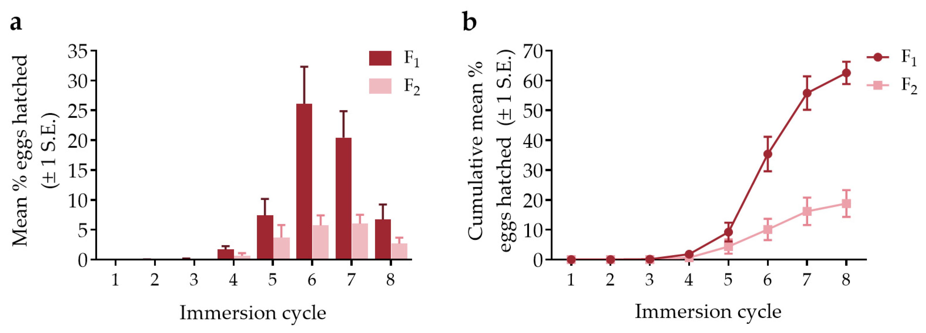
Almost no hatching occurred during the first three immersion cycles for both the F1 and F2 generations (Figure 3, Dataset S1). Eclosion began to increase during the fourth and fifth cycles and peaked during the sixth and seventh cycles for the F1 generation, before quickly declining. The F2 generation followed a similar pattern but there was no clear peak in egg hatching. The cumulative mean % eggs hatched after eight immersion cycles was considerably higher for the F1 generation (mean 62.5%) than the F2 generation (18.8%).

3.4. Development from Egg to Adult

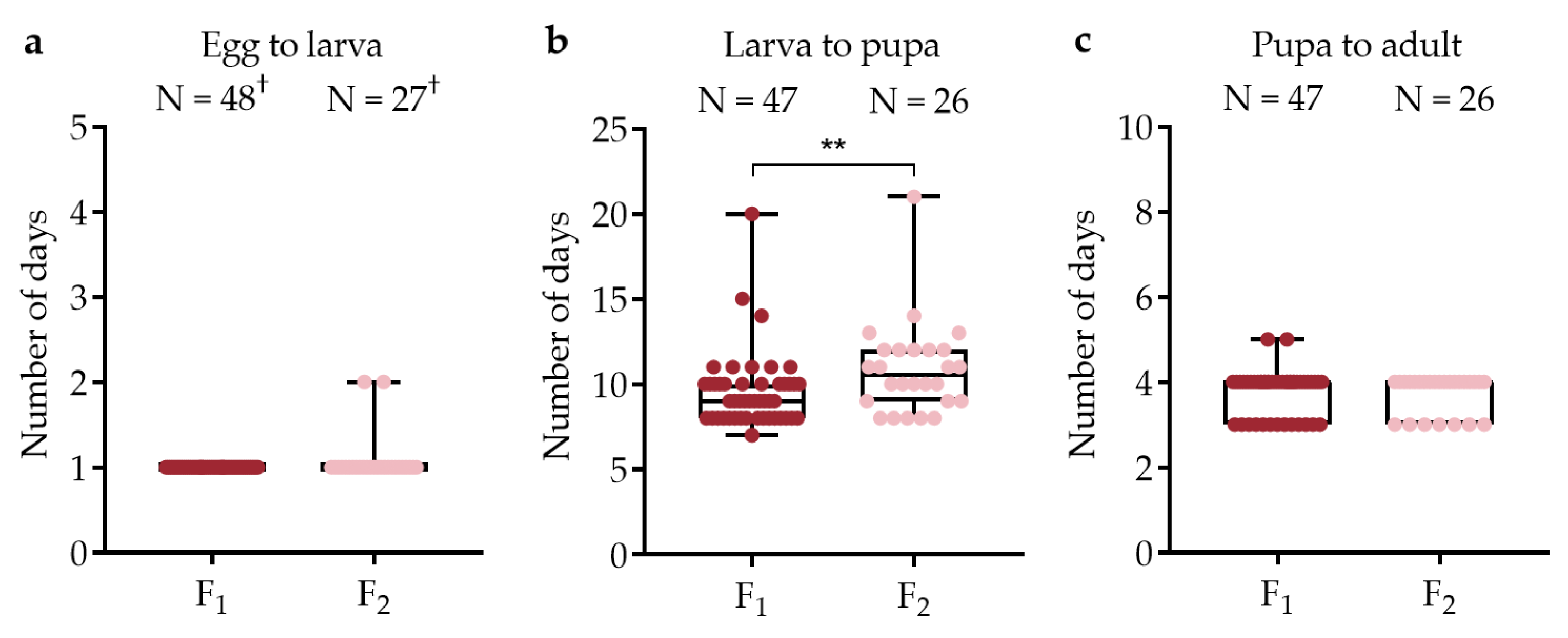
Rearing was relatively straightforward and successful once larvae had emerged with a total 86.1% (1345/1562) F1 and 93.7% (178/190) F2 mosquitoes surviving to adults. The first larvae usually hatched within 5–6 h of immersion, although results were recorded the following day. There was no significant difference in the number of days until the first larva emerged between generations (Wilcoxon test, DF = 1, χ2 = 3.6, p = 0.058), but there was a difference in development time from first larva until first pupa (DF = 1, χ2 = 7.38, p = 0.007), with the F2 generation developing more slowly (Figure 4). A simple linear regression revealed a negative relationship (DF = 1, F = 6.9, p = 0.01) between larval density (number of eggs hatched) per container and the larval to pupal development time for both generations combined. There was no difference in development time between generations from first pupa until first adult (Wilcoxon test, DF = 1, χ2 = 0.04, p = 0.83). The development time from egg to adult was completed within a mean 15 days (min = 12, max = 26). The male to female adult sex ratio was 1:1.1 for the F1 generation and 1:1.2 for the for the F2 generation.

3.5. Individually Maintained Mosquitoes

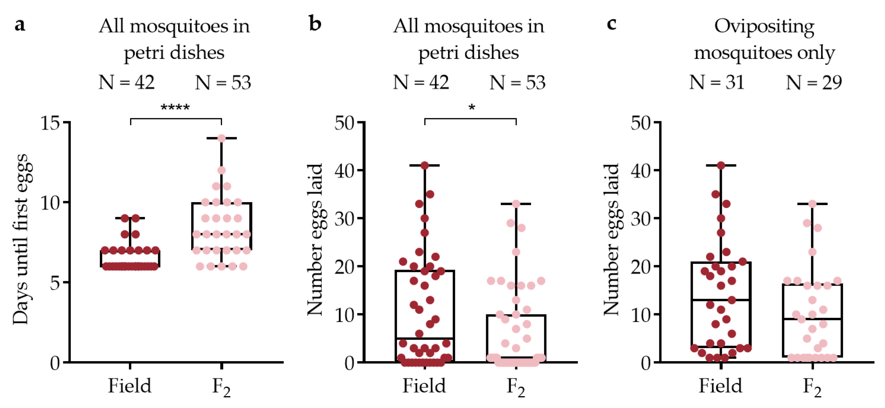
A subset of field collected and F2 generation mosquitoes were maintained individually in Petri dishes to obtain detailed data about egg development and oviposition. Oviposition rates were higher among field collected mosquitoes (31/42, 73.8% laid eggs) than the F2 generation (29/53, 54.7%), although this difference was not significant (Fisher’s Exact test, DF = 1, N = 95, p = 0.086). Field collected mosquitoes laid their first eggs more quickly than F2 generation mosquitoes (Figure 5a, Table 2) (Wilcoxon test, DF = 1, χ2 = 18.4, p < 0.0001). The median number of eggs laid by mosquitoes added to Petri dishes that had a chance to oviposit was higher for the field collected generation than for the F2 generation (DF = 1, χ2 = 6.18, p = 0.013) (Figure 5b). The same was also true when considering ovipositing mosquitoes only (Figure 5c), although the difference was not significant (DF = 1, χ2 = 2.70, p = 0.10). There was considerable egg retention among all dissected F2 mosquitoes (N = 46, mean eggs retained ±1 S.E. = 17.0 ± 1.74) and among those that laid at least one egg (N = 29, mean eggs retained ±1 S.E. = 13.5 ± 2.09).

3.6. Mosquito Size by Generation

A comparison of the size of female mosquitoes based on a subsample of 269 specimens (N = 102 field, 95 F1, and 72 F2 mosquitoes) revealed a significant difference in mean hindtarsus length between generations (Kruskal–Wallis test, DF = 2, χ2 = 44.0, p < 0.0001). Post hoc Wilcoxon Each Pair tests showed that both laboratory generations were significantly larger than the field collected generation (p < 0.0001 for both comparisons), while the F2 generation was also larger than the F1 generation (p = 0.0001).

4. Discussion

We reared Hg. janthinomys beyond an F1 generation for the first time. While our techniques could not sustain a laboratory colony, the study provides a foundation for improving maintenance methods. Crucially, our artificial blood feeding method will enable future in situ Hg. janthinomys vector competence studies to be performed using F1 adult females, which are relatively easy to rear in large numbers. These are necessary in a region affected by hyperendemic transmission of DENV, ZIKV, and CHIKV, and where the risk of sylvatic spillback is high.
We collected relatively small numbers of mosquitoes in the field over eight weeks, which was sufficient to perform this study but not ideal for establishing a colony. Sampling more intensively over a shorter time would yield higher numbers of F1 mosquitoes during each immersion cycle, allowing more control when blood feeding and copulating mosquitoes of a certain age. Shorter, more intensive collections have been successfully used to establish Anopheles colonies elsewhere [35,41], but are even more important for Hg. janthinomys given the additional difficulties associated with installment hatching.
The artificial feeding method was effective in delivering blood to each generation. We discarded unengorged field collected mosquitoes after the first feeding attempt since they did not appear to feed well thereafter, and we were unable to quantify feeding rates of F1 generation mosquitoes maintained in cages since they could not easily be counted. However, we were able to show that more than 85% of F2 females fed when presented with blood daily. Anecdotally, we observed blood feeding starting at around five days post emergence. However, we did not systematically record this since the small numbers of F2 adults available would have needed to be maintained and fed individually by day of emergence. If confirmed, our observation would be consistent with delayed feeding responses of arboreal Sabethes (Sabethoides) chloropterus (von Humboldt, 1819) which do not feed before five days post emergence [42], but slower than Hg. equinus [28] and the major Aedes vectors [43,44], which all begin feeding within three days post emergence. It was necessary to heat water used in the feeding system to 45 °C to achieve good rates of blood feeding, but it was not necessary to change the water as we did with earlier generations. Most mosquitoes that were going to feed did so within 20–30 min of being offered a blood meal.
Our forced mating technique differed slightly from the WHO method for Anopheles mosquitoes [33], which recommends introducing the terminalia at about 45 degrees. We found that an angle of slightly more than 90 degrees was appropriate for Hg. janthinomys, which is similar to the angle used when forced mating Ae. aegypti [45]. Other factors influencing forced mating outcome may include mosquito age and duration of copulation [46,47,48]. We attempted to use 3–5-day old adult males and 1–3-day old females. However, this was difficult since the effects of installment hatching meant that we often only had small numbers of mosquitoes to work with on a given day.
Oviposition rates were higher among field collected mosquitoes than the F2 generation, although the difference was not significant. The number of F2 generation mosquitoes that laid eggs (29) was also not consistent with the number copulated and added to Petri dishes (53), which may indicate that some mosquitoes were not inseminated despite coupling. This has been reported elsewhere [46,49] and it has been stated that at least six seconds of firm copulation are needed for insemination to occur in Ae. aegypti [47]. Our copulations lasted from a few seconds to several minutes, but the exact duration was not recorded on this occasion. The high number of eggs retained may also be indicative of unsuccessful insemination [50]. It was always likely that forced mating would be less effective than the natural process and it would be beneficial to compare insemination rates between generations through observation of spermathecae [51].
The mean and median number of eggs laid per mosquito was higher among the field collected generation than the laboratory generations (Table 1), but this could only be tested statistically with individually maintained specimens. Our respective mean (14.3) and median (13) number of eggs per ovipositing field mosquito (Table 2) was lower than reported by Bates [15] who produced 22 eggs per ovipositing female. Mondet [16] produced 32.2 eggs per mosquito, but that was based on a sample of 10 specimens which had completed a single gonotrophic cycle. It is, therefore, worth noting that 153 of our 301 F3 eggs were laid by just seven mosquitoes within the space of two days (Dataset S1). Cages of free laying females, capable of passing through several gonotrophic cycles, will probably be required to maintain a colony given the small number of eggs produced per cycle. Better oviposition substrates, possibly mimicking tree holes [52], may improve egg yields. Furthermore, testing the effects of additional larval nutrients on reproductive output (fertility and fecundity) may be worthwhile, since the quality of larval nutrition can affect such adult phenotypes [53].
A total of 62.8% F1 generation eggs hatched during ≤10 immersion cycles, decreasing to 20.1% in the F2 generation. It has been suggested that a period of conditioning (partial drying) is required before Hg. janthinomys eggs will hatch [25,54], but also that repeated immersions during embryogenesis influence Hg. leucocelaenus hatch rates [55]. Season may also affect rates of eclosion among Hg. janthinomys and Hg. leucocelaenus [22,25,56]. Immediately initiating immersion cycles may mimic rainy season conditions resulting in increased eclosion, while a period of conditioning may simulate dry season conditions, but data are needed to clearly show this. Our installment hatching results are consistent with Alencar et al. [23] who showed negligible effects of immersions beyond the ninth cycle on Hg. janthinomys eclosion. Given that our work extended from the rainy season into the early dry season, there may have been a temporal effect on the percentage of eggs hatched. However, it is also likely that egg fertilization rates were lower among the F2 generation than among eggs laid by field mosquitoes.
Once hatched, rearing was relatively straightforward with 93.7% of F2 larvae surviving to adults. Poor survival among the F1 generation was associated with biofilm growth, a common problem when rearing [57,58], or with overcrowding resulting in the production of small adults. These were either unable to escape the surface tension of water or were difficult to use for mating. The first adults usually emerged within 13–16 days, corresponding reasonably well with estimated natural development times of 11–13 days for Hg. janthinomys [11]. We also documented an increase in Hg. janthinomys occurrence associated with increasing 7-day cumulative rainfall at a lag of 1 week in the field (i.e., an increase in adults 14 days after the onset of particularly heavy rainfall) [59]. In the laboratory, we found significant differences in development time at the larval stage between generations. The ad hoc addition of yeast was potentially confounding, but since food resources were plentiful, we believe the negative density-development relationship was accurate. The number of days until the first eggs were laid by field mosquitoes was similar to the 6.2 to 7.2 days reported by Bates [15], although the F2 generation took slightly longer to oviposit.
We reared larger mosquitoes in the insectary than we collected in the field, mainly to increase chances of survival and for ease of manipulation. It is known that the number of eggs produced by a mosquito is related to its size [60], so rearing larger mosquitoes should facilitate colony maintenance. However, mosquito size may also affect vector competence [61], which should be taken into consideration when planning Hg. janthinomys transmission experiments.

5. Conclusions

Haemagogus janthinomys is the major vector of YFV and thought to be a prominent vector of MAYV in the neotropics, but it is not known whether it can transmit DENV, ZIKV, or CHIKV. Our pilot attempts at laboratory maintenance should form the basis of future experimental studies. Our artificial blood feeding method will permit future vector competence studies involving this species.

Supplementary Materials

The following supporting information can be downloaded at: https://www.mdpi.com/article/10.3390/v15010045/s1, Figure S1: Generic cages, Figure S2: Oviposition in Petri dishes, Figure S3: Oviposition in cages, Video S1: Forced mating, Dataset S1: Complete dataset.

Author Contributions

Conceptualization, A.H. and N.V.; Data curation, A.H.; Formal analysis, A.H. and E.H.-A.; Funding acquisition, K.A.H. and N.V.; Investigation, A.H., N.F.F., D.V., F.P.A. and J.T.A.J.; Methodology, A.H., N.F.F., D.V., B.A.C., L.F.A.d.S., R.A.G.S., A.d.C.P., M.M.M.S., C.A. and K.A.H.; Supervision, M.V.G.d.L. and N.V.; Visualization, A.H.; Writing—original draft, A.H.; Writing—review and editing, A.H., V.M.S., M.V.G.d.L., K.A.H. and N.V. All authors have read and agreed to the published version of the manuscript.

Funding

This research was funded by an International Collaborations in Infectious Disease Research (ICIDR) grant 1U01AI115577 and a Centers for Research in Emerging Infectious Diseases (CREID) 1U01AI151807 grant awarded to N.V. and K.A.H. by the National Institutes of Health (NIH). E.H.-A. is funded by a Consejo Nacional de Ciencia y Tecnología fellowship (709139) awarded by the Government of Mexico, M.V.G.L. is a CNPq fellow, and M.M.M.S is a CAPES fellow (proc. no. 88882.444402/2019-01).

Institutional Review Board Statement

The study was conducted according to the guidelines of the Declaration of Helsinki and approved by the Fundação de Medicina Tropical Doutor Heitor Vieira Dourado Ethics Review Board (CAAE: 53963521.0.0000.0005) on 11 February 2022.

Informed Consent Statement

Informed consent was obtained from all subjects involved in the study.

Data Availability Statement

The data presented in this study are available in the article and its supplementary material.

Acknowledgments

The authors wish to acknowledge Júlia Melissa da Rocha Albuquerque and Maurício Lima (FMT-HVD) for assistance developing the blood feeding protocol; Claudia Mendonça, Igor Pedrosa, and Taly Nayandra (FMT-HVD) for assistance maintaining mosquitoes in the insectary; Seth Irish (Swiss TPH) for assistance with the forced mating methods; Aleksey Lima (FMT-HVD) for administrative support; Raimundo Coelho (FMT-HVD) for driving and logistics, and the Instituto Nacional de Pesquisas da Amazônia (INPA) for permission to use the Adolpho Ducke Reserve to collect mosquitoes for this study.

Conflicts of Interest

The authors declare no conflict of interest. The funders had no role in the design of the study; in the collection, analyses, or interpretation of data; in the writing of the manuscript, or in the decision to publish the results.

References

  1. Shannon, R.C.; Whitman, L.; Franca, M. Yellow fever virus in jungle mosquitoes. Science 1938, 88, 110–111. [Google Scholar] [CrossRef]
  2. Arnell, J.H. Mosquito studies (Diptera, Culicidae) XXXII: A revision of the genus Haemagogus. Contrib. Am. Entomol. Inst. 1973, 10, 1–174. [Google Scholar]
  3. Pinheiro, G.G.; Rocha, M.N.; de Oliveira, M.A.; Moreira, L.A.; Andrade Filho, J.D. Detection of yellow fever virus in sylvatic mosquitoes during disease outbreaks of 2017–2018 in Minas Gerais State, Brazil. Insects 2019, 10, 136. [Google Scholar] [CrossRef] [Green Version]
  4. Abreu, F.V.S.; Ribeiro, I.P.; Ferreira-de-Brito, A.; Santos, A.; Miranda, R.M.; Bonelly, I.S.; Neves, M.; Bersot, M.I.; Santos, T.P.D.; Gomes, M.Q.; et al. Haemagogus leucocelaenus and Haemagogus janthinomys are the primary vectors in the major yellow fever outbreak in Brazil, 2016–2018. Emerg. Microbes Infect. 2019, 8, 218–231. [Google Scholar] [CrossRef] [Green Version]
  5. Pezzi, L.; Diallo, M.; Rosa-Freitas, M.G.; Vega-Rua, A.; Ng, L.F.P.; Boyer, S.; Drexler, J.F.; Vasilakis, N.; Lourenco-de-Oliveira, R.; Weaver, S.C.; et al. GloPID-R report on chikungunya, o’nyong-nyong and Mayaro virus, part 5: Entomological aspects. Antiviral Res. 2020, 174, 104670. [Google Scholar] [CrossRef]
  6. Hoch, A.L.; Peterson, N.E.; LeDuc, J.W.; Pinheiro, F.P. An outbreak of Mayaro virus disease in Belterra, Brazil. III. Entomological and ecological studies. Am. J. Trop. Med. Hyg. 1981, 30, 689–698. [Google Scholar] [CrossRef]
  7. Bates, M.; Roca-Garcia, M. The development of the virus of yellow fever in Haemagogus mosquitoes. Am. J. Trop. Med. Hyg. 1946, 26, 585–605. [Google Scholar] [CrossRef]
  8. Bates, M.; Roca-García, M. Experiments with various Colombian marsupials and primates in laboratory cycles of yellow fever. Am. J. Trop. Med. Hyg. 1946, S1–26, 437–453. [Google Scholar] [CrossRef]
  9. Galindo, P.; Rodaniche, E.d.; Trapido, H. Experimental transmission of yellow fever by Central American species of Haemagogus and Sabethes chloropterus. Am. J. Trop. Med. Hyg. 1956, 5, 1022–1031. [Google Scholar] [CrossRef]
  10. Bates, M.; Roca-Garcia, M. Laboratory studies of the Saimiri-Haemagogus cycle of jungle yellow fever. Am. J. Trop. Med. Hyg. 1945, S1–25, 203–216. [Google Scholar] [CrossRef]
  11. Degallier, N.; De Oliveira Monteiro, H.A.; Castro, F.C.; Da Silva, O.V.; Filho, G.C.S.; Elguero, E. An indirect estimation of the developmental time of Haemagogus janthinomys (Diptera: Culicidae), the main vector of yellow fever in South America. Stud. Neotrop. Fauna Environ. 2006, 41, 117–122. [Google Scholar] [CrossRef]
  12. Kumm, H.W.; Frobisher, M. Attempts to transmit yellow fever with certain Brazilian mosquitoes (Culicidae) and with bedbugs (Cimex hemipterus). Am. J. Trop. Med. Hyg. 1932, S1–12, 349–361. [Google Scholar] [CrossRef]
  13. Antunes, P.C.A.; Whitman, L. Studies on the capacity of mosquitoes of the genus Haemagogus to transmit yellow fever. Am. J. Trop. Med. Hyg. 1937, S1–17, 825–831. [Google Scholar] [CrossRef]
  14. Bates, M. The Saimiri monkey as an experimental host for the virus of yellow fever. Am. J. Trop. Med. Hyg. 1944, S1–24, 83–89. [Google Scholar] [CrossRef]
  15. Bates, M. The development and longevity of Haemagogus mosquitoes under laboratory conditions. Ann. Entomol. Soc. Am. 1947, 40, 1–12. [Google Scholar] [CrossRef]
  16. Mondet, B. Condições de sobrevivência em laboratório de Haemagogus janthinomys Dyar, 1921 (Diptera: Culicidae). Rev. Soc. Bras. Med. Trop. 1997, 30, 11–14. [Google Scholar] [CrossRef] [PubMed] [Green Version]
  17. Lowe, R.; Lee, S.; Martins Lana, R.; Torres Codeço, C.; Castro, M.C.; Pascual, M. Emerging arboviruses in the urbanized Amazon rainforest. BMJ 2020, 371, m4385. [Google Scholar] [CrossRef] [PubMed]
  18. Althouse, B.M.; Vasilakis, N.; Sall, A.A.; Diallo, M.; Weaver, S.C.; Hanley, K.A. Potential for Zika virus to establish a sylvatic transmission cycle in the Americas. PLoS Negl. Trop. Dis. 2016, 10, e0005055. [Google Scholar] [CrossRef]
  19. Lourenço-de-Oliveira, R.; Failloux, A.B. High risk for chikungunya virus to initiate an enzootic sylvatic cycle in the tropical Americas. PLoS Negl. Trop. Dis. 2017, 11, e0005698. [Google Scholar] [CrossRef] [Green Version]
  20. Alencar, J.; Gil-Santana, H.R.; de Oliveira, R.d.F.N.; Dégallier, N.; Guimarães, A.É. Natural breeding sites for Haemagogus mosquitoes (Diptera, Culicidae) in Brazil. Entomol. News 2010, 121, 393–396, 394. [Google Scholar] [CrossRef]
  21. Yanoviak, S.P.; Lounibos, L.P.; Weaver, S.C. Land use affects macroinvertebrate community composition in phytotelmata in the Peruvian Amazon. Ann. Entomol. Soc. Am. 2006, 99, 1172–1181. [Google Scholar] [CrossRef]
  22. Alencar, J.; Gleiser, R.M.; Morone, F.; Mello, C.F.; Silva Jdos, S.; Serra-Freire, N.M.; Guimarães, A.É. A comparative study of the effect of multiple immersions on Aedini (Diptera: Culicidae) mosquito eggs with emphasis on sylvan vectors of yellow fever virus. Mem. Inst. Oswaldo Cruz 2014, 109, 114–117. [Google Scholar] [CrossRef] [PubMed] [Green Version]
  23. Alencar, J.; de Almeida, H.M.; Marcondes, C.B.; Guimarães, A.É. Effect of multiple immersions on eggs and development of immature forms of Haemagogus janthinomys from south-eastern Brazil (Diptera: Culicidae). Entomol. News 2008, 119, 239–244. [Google Scholar] [CrossRef] [Green Version]
  24. Silva, S.O.F.; de Mello, C.F.; Gleiser, R.M.; Oliveira, A.A.; Maia, D.A.; Alencar, J. Evaluation of multiple immersion effects on eggs from Haemagogus leucocelaenus, Haemagogus janthinomys, and Aedes albopictus (Diptera: Culicidae) under experimental conditions. J. Med. Entomol. 2018, 55, 1093–1097. [Google Scholar] [CrossRef] [PubMed]
  25. Hovanitz, W. Comparisons of mating behavior, growth rate, and factors influencing egg-hatching in South American Haemagogus mosquitoes. Physiol. Zool. 1946, 19, 35–53. [Google Scholar] [CrossRef] [PubMed]
  26. Waddell, M.B.; Kumm, H.W. Haemagogus capricornii Lutz as a laboratory vector of yellow fever. Am. J. Trop. Med. Hyg. 1948, S1–28, 247–252. [Google Scholar] [CrossRef]
  27. Vasconcelos, P.F.; Rodrigues, S.G.; Degallier, N.; Moraes, M.A.; da Rosa, J.F.; da Rosa, E.S.; Mondet, B.; Barros, V.L.; da Rosa, A.P. An epidemic of sylvatic yellow fever in the southeast region of Maranhao State, Brazil, 1993-1994: Epidemiologic and entomologic findings. Am. J. Trop. Med. Hyg. 1997, 57, 132–137. [Google Scholar] [CrossRef] [Green Version]
  28. Dutary, B.E. Transovarial transmission of yellow fever virus by a sylvatic vector, Haemagogus equinus (Theobald). Ph.D. Thesis, The University of Texas Health Science Center at Houston School of Public Health, Houston, TX, USA, 1981. [Google Scholar]
  29. Osorno Mesa, E. Organización de una colonia de Haemagogus equinus Theobald. Caldasia 1944, 3, 39–45. [Google Scholar]
  30. Waddell, M.B. Comparative efficacy of certain South American Aëdes and Haemagogus mosquitoes as laboratory vectors of yellow fever. Am. J. Trop. Med. Hyg. 1949, S1–29, 567–575. [Google Scholar] [CrossRef]
  31. Bates, M.; Roca-Garcia, M. The douroucouli (Aotus) in laboratory cycles of yellow fever. Am. J. Trop. Med. Hyg. 1945, S1–25, 385–389. [Google Scholar] [CrossRef]
  32. McDaniel, I.N.; Horsfall, W.R. Induced copulation of Aedine mosquitoes. Science 1957, 125, 745. [Google Scholar] [CrossRef] [PubMed]
  33. World Health Organization. Manual on Practical Entomology in Malaria, Part II: Methods and Techniques; World Heath Organization: Geneva, Switzerland, 1975; p. 191. [Google Scholar]
  34. Bryan, J.H.; Southgate, B.A. Studies of forced mating techniques on anopheline mosquitoes. Mosq. News 1978, 38, 338–342. [Google Scholar]
  35. Zhu, G.; Zhou, H.; Li, J.; Tang, J.; Bai, L.; Wang, W.; Gu, Y.; Liu, Y.; Lu, F.; Cao, Y.; et al. The colonization of pyrethroid resistant strain from wild Anopheles sinensis, the major Asian malaria vector. Parasit. Vectors. 2014, 7, 582. [Google Scholar] [CrossRef] [PubMed]
  36. Hendy, A.; Valério, D.; Fé, N.F.; Hernandez-Acosta, E.; Mendonça, C.; Andrade, E.; Pedrosa, I.; Costa, E.R.; Júnior, J.T.A.; Assunção, F.P.; et al. Microclimate and the vertical stratification of potential bridge vectors of mosquito-borne viruses captured by nets and ovitraps in a central Amazonian forest bordering Manaus, Brazil. Sci. Rep. 2021, 11, 21129. [Google Scholar] [CrossRef] [PubMed]
  37. Forattini, O.P. Culicidologia Médica: Identificacao, Biologia e Epidemiologia; EDUSP: São Paulo, Brazil, 2002; Volume 2, p. 864. [Google Scholar]
  38. Lane, J. Neotropical Culicidae; University of São Paulo: São Paulo, Brazil, 1953; Volumes 1 and 2, p. 1112. [Google Scholar]
  39. Consoli, R.; Lourenço-de-Oliveira, R. Principais Mosquitos de Importância Sanitária no Brasil; Fundação Oswaldo Cruz (Fiocruz): Rio de Janeiro, Brazil, 1994; p. 228. [Google Scholar]
  40. SAS Institute Inc. JMP v14; SAS Institute Inc.: Cary, NC, USA, 2018. [Google Scholar]
  41. Villarreal-Treviño, C.; Vásquez, G.M.; López-Sifuentes, V.M.; Escobedo-Vargas, K.; Huayanay-Repetto, A.; Linton, Y.-M.; Flores-Mendoza, C.; Lescano, A.G.; Stell, F.M. Establishment of a free-mating, long-standing and highly productive laboratory colony of Anopheles darlingi from the Peruvian Amazon. Malar. J. 2015, 14, 227. [Google Scholar] [CrossRef] [PubMed] [Green Version]
  42. Galindo, P. Bionomics of Sabethes chloropterus Humboldt, a vector of sylvan yellow fever in Middle America. Am. J. Trop. Med. Hyg. 1958, 7, 429–440. [Google Scholar] [CrossRef]
  43. Alto, B.W.; Lounibos, L.P.; Juliano, S.A. Age-dependent bloodfeeding of Aedes aegypti and Aedes albopictus on artificial and living hosts. J. Am. Mosq. Control Assoc. 2003, 19, 347–352. [Google Scholar]
  44. Tallon, A.K.; Hill, S.R.; Ignell, R. Sex and age modulate antennal chemosensory-related genes linked to the onset of host seeking in the yellow-fever mosquito, Aedes aegypti. Sci. Rep. 2019, 9, 43. [Google Scholar] [CrossRef] [Green Version]
  45. Wheeler, R.E. A simple apparatus for forced copulation of mosquitoes. Mosq. News 1962, 22, 402–403. [Google Scholar]
  46. Gwadz, R.W.; Craig Jr, G.B. Sexual receptivity in female Aedes aegypti. Mosq. News 1968, 28, 586–593. [Google Scholar]
  47. Spielman, A.; Leahy, M.G.; Skaff, V. Failure of effective insemination of young female Aedes aegypti mosquitoes. J. Insect Physiol. 1969, 15, 1471–1479. [Google Scholar] [CrossRef] [PubMed]
  48. Jones, J.C.; Wheeler, R.E. Studies on spermathecal filling in Aedes aegypti (Linnaeus). I. Description. Biol. Bull. 1965, 129, 134–150. [Google Scholar] [CrossRef]
  49. Akey, D.H.; Jones, J.C. Sexual responses of adult male Aedes aegypti using the forced-copulation technique. Biol. Bull. 1968, 135, 445–453. [Google Scholar] [CrossRef] [PubMed]
  50. Villarreal, S.M.; Pitcher, S.; Helinski, M.E.H.; Johnson, L.; Wolfner, M.F.; Harrington, L.C. Male contributions during mating increase female survival in the disease vector mosquito Aedes aegypti. J. Insect Physiol. 2018, 108, 1–9. [Google Scholar] [CrossRef] [PubMed]
  51. Carrasquilla, M.C.; Lounibos, L.P. Detection of insemination status in live Aedes aegypti females. J. Insect Physiol. 2015, 75, 1–4. [Google Scholar] [CrossRef] [Green Version]
  52. Karna, A.K.; Azar, S.R.; Plante, J.A.; Yun, R.; Vasilakis, N.; Weaver, S.C.; Hansen, I.A.; Hanley, K.A. Colonized Sabethes cyaneus, a sylvatic New World mosquito species, shows a low vector competence for Zika virus relative to Aedes aegypti. Viruses 2018, 10, 434. [Google Scholar] [CrossRef]
  53. Yan, J.; Kibech, R.; Stone, C.M. Differential effects of larval and adult nutrition on female survival, fecundity, and size of the yellow fever mosquito, Aedes aegypti. Frontiers in Zoology 2021, 18, 10. [Google Scholar] [CrossRef]
  54. Galindo, P.; Carpenter, S.J.; Trapido, H. Ecological observations on forest mosquitoes of an endemic yellow fever area in Panama. Am. J. Trop. Med. Hyg. 1951, 31, 98–137. [Google Scholar] [CrossRef]
  55. Silva, S.O.F.; de Mello, C.F.; Figueiró, R.; Docile, T.; Serdeiro, M.; Fumian, F.F.; Alencar, J. Oviposition behavior of wild yellow fever vector mosquitoes (Diptera: Culicidae) in an Atlantic Forest fragment, Rio de Janeiro state, Brazil. Sci. Rep. 2021, 11, 6081. [Google Scholar] [CrossRef]
  56. Alencar, J.; de Mello, C.F.; Leite, P.J.; Bastos, A.Q.; Freitas Silva, S.O.; Serdeiro, M.; Dos Santos Silva, J.; Müller, G.A. Oviposition activity of Haemagogus leucocelaenus (Diptera: Culicidae) during the rainy and dry seasons, in areas with yellow fever virus circulation in the Atlantic Forest, Rio de Janeiro, Brazil. PLoS ONE 2021, 16, e0261283. [Google Scholar] [CrossRef]
  57. Hunt, C.M.; Collins, C.M.; Benedict, M.Q. Measuring and reducing biofilm in mosquito rearing containers. Parasit. Vectors. 2020, 13, 439. [Google Scholar] [CrossRef] [PubMed]
  58. Heymann, E.W.; Encarnación C, F.; Canaquin Y, J.E. Primates of the Río Curaray, northern Peruvian Amazon. Int. J. Primatol. 2002, 23, 191–201. [Google Scholar] [CrossRef]
  59. Hendy, A.; Hernandez-Acosta, E.; Valério, D.; Mendonça, C.; Costa, E.R.; Júnior, J.T.A.; Assunção, F.P.; Scarpassa, V.M.; Gordo, M.; Fé, N.F.; et al. The vertical stratification of potential bridge vectors of mosquito-borne viruses in a central Amazonian forest bordering Manaus, Brazil. Sci. Rep. 2020, 10, 18254. [Google Scholar] [CrossRef] [PubMed]
  60. Lyimo, E.O.; Takken, W. Effects of adult body size on fecundity and the pre-gravid rate of Anopheles gambiae females in Tanzania. Med. Vet. Entomol. 1993, 7, 328–332. [Google Scholar] [CrossRef]
  61. Alto, B.W.; Reiskind, M.H.; Lounibos, L.P. Size alters susceptibility of vectors to dengue virus infection and dissemination. Am. J. Trop. Med. Hyg. 2008, 79, 688–695. [Google Scholar] [CrossRef]
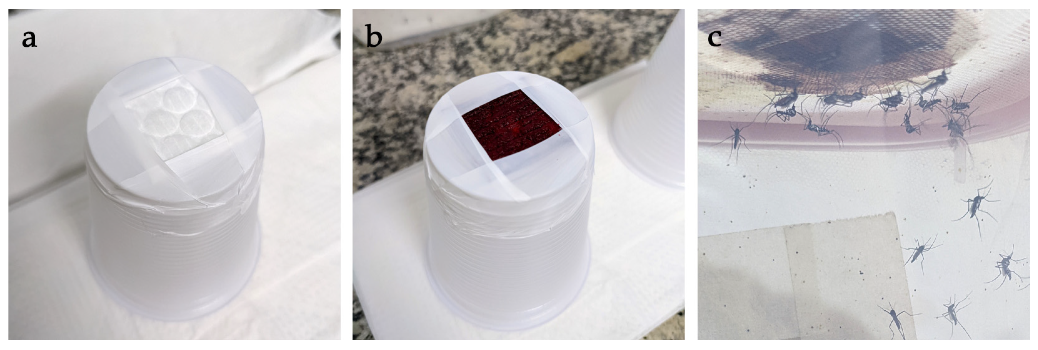
Figure 1. Artificial blood feeding. (a) cotton pad secured to the base of a plastic feeding cup with Teflon tape; (b) feeding cup after the application of blood; and (c) Hg. janthinomys feeding on a cup inverted over the top of a cage and partially filled with 45 °C water.
Viruses 15 00045 g001
Figure 2. Forced mating F1 generation Hg. janthinomys. (a) a male mosquito mounted laterally through the thorax on a 0.5 mL syringe; (b) mounted males with legs removed resting on a Petri dish; (c) a fully anesthetized female with legs relaxed; (d) introduction of mosquito terminalia at an angle of about 90°; (e) fully interlocked male and female; and (f) decapitated male and female mating pairs at rest on the lid of a Petri dish.
Viruses 15 00045 g002
Figure 3. Haemagogus janthinomys egg eclosion shown for the first eight immersion cycles for N = 10 F1 and N = 8 F2 egg papers (total 2486 F1 and 944 F2 eggs, respectively). Eggs were immersed as soon as possible once recovered from Petri dishes or cages. (a) mean percentage of eggs hatched ± 1 standard error (S.E.); and (b) the cumulative mean percentage of eggs hatched (±1 S.E.).
Viruses 15 00045 g003
Figure 4. Egg to adult development times for F1 and F2 Hg. janthinomys in trays where at least one egg hatched. Box plots show median values and interquartile range (IQR) for development times based on the number of days until the emergence of the first (a) larva, (b) pupa, and (c) adult in each rearing tray. Significant differences indicated by asterisks (** p < 0.01). ‘N =’ = number of trays where at least one egg hatched. All larvae died in one rearing tray in each generation.
Viruses 15 00045 g004
Figure 5. Egg development and oviposition for individually maintained field collected (Field) and F2 mosquitoes. Box plots show median values and IQR for (a) number of days until first eggs were laid; (b) number of eggs laid per mosquito for all mosquitoes in Petri dishes; and (c) number of eggs laid per mosquito for ovipositing mosquitoes in Petri dishes. Significant differences indicated by asterisks (* p < 0.05, **** p < 0.001). Number of individual mosquitoes (N) above bars.
Viruses 15 00045 g005
Table 1. Summary table showing progression from field collected mosquitoes through to F3 eggs for all mosquitoes included in the study.
StageN (%)Summary
Field collected mosquitoes418
Blood fed226 (54.1)Range 29.1–90.5%
F1 eggs laid 2486Mean 11 per ♀
F1 eggs hatched1562 (62.8)Range 51.4–87%
F1 reared to adult1345 (86.1)Range 19.2–100%
F1 ratio ♂:♀638:707 (47.4:52.6)
F1 ♀ copulated501
F1 ♀ in cages445
F2 eggs laid944Mean 2.1 per ♀
F2 eggs hatched190 (20.1)Range 3.1–41.4%
F2 reared to adult178 (93.7)Range 66.7–100%
F2 ratio ♂:♀80:98 (44.9:55.1)
F2 ♀ for blood feeding92
F2 ♀ blood fed79 (85.9)
F2 ♀ copulated59 (73.4 *)
F2 ♀ in Petri dishes53
F3 eggs301Mean 3.3 per ♀ **
* % blood fed mosquitoes copulated. ** 5.1 eggs per blood fed + copulated mosquito; 5.68 eggs per mosquito in Petri dish; 10.4 eggs per ovipositing mosquito.
Table 2. Summary table showing mean, median, and IQR for comparisons shown in Figure 5. Mean values are included to facilitate comparisons with previously published work [15,16]. ‘Gen.’ = Generation, ‘N =’ = number of individual mosquitoes.
ComparisonGen.N =MeanMedianIQRMin.Max.
Days until first eggs laidField426.66169
F2538.483614
Eggs laid per mosquito for all individual mosquitoes in Petri dishesField4210.6519.25041
F2535.7110033
Eggs laid per mosquito for ovipositing mosquitoes onlyField3114.31318041
F22910.4915.5033
Disclaimer/Publisher’s Note: The statements, opinions and data contained in all publications are solely those of the individual author(s) and contributor(s) and not of MDPI and/or the editor(s). MDPI and/or the editor(s) disclaim responsibility for any injury to people or property resulting from any ideas, methods, instructions or products referred to in the content.