Article

Effective Treatment of Staphylococcus aureus Intramammary Infection in a Murine Model Using the Bacteriophage Cocktail StaphLyse™

1
Département de Biologie, Faculté des Sciences, Université de Sherbrooke, Sherbrooke, QC J1K 2R1, Canada
2
Techniques de Santé Animale, Cégep de Sherbrooke, Sherbrooke, QC J1E 4K1, Canada
3
Sherbrooke Research and Development Centre, Agriculture and Agri-Food Canada, Sherbrooke, QC J1M 0C8, Canada
4
Phileo by Lesaffre North America Office, 7475 West Main Street, Milwaukee, WI 53214, USA
5
Intralytix, Inc., Columbia, MD 21046, USA
*
Author to whom correspondence should be addressed.
Viruses 2023, 15(4), 887; https://doi.org/10.3390/v15040887
Submission received: 9 February 2023 / Revised: 21 March 2023 / Accepted: 28 March 2023 / Published: 30 March 2023
(This article belongs to the Section Bacterial Viruses)

Abstract

:
Staphylococcus aureus causes intramammary infections (IMIs), which are refractory to antibiotic treatment and frequently result in chronic mastitis. IMIs are the leading cause of conventional antibiotic use in dairy farms. Phage therapy represents an alternative to antibiotics to help better manage mastitis in cows, reducing the global spread of resistance. A mouse mastitis model of S. aureus IMI was used to study the efficacy of a new cocktail of five lytic S. aureus-specific phages (StaphLyse™), administered either via the intramammary (IMAM) route or intravenously (IV). The StaphLyse™ phage cocktail was stable in milk for up to one day at 37 °C and up to one week at 4 °C. The phage cocktail was bactericidal in vitro against S. aureus in a dose-dependent manner. A single IMAM injection of this cocktail given 8 h after infection reduced the bacterial load in the mammary glands of lactating mice infected with S. aureus, and as expected, a two-dose regimen was more effective. Prophylactic use (4 h pre-challenge) of the phage cocktail was also effective, reducing S. aureus levels by 4 log10 CFU per gram of mammary gland. These results suggest that phage therapy may be a viable alternative to traditional antibiotics for the control of S. aureus IMIs.

1. Introduction

Intramammary infections (IMIs) and subsequent mastitis cause a decrease in milk production and degrade milk quality. Mastitis management in herds, based on hygiene and antibiotic therapy, involves the administration of intramammary or parenteral antibiotics to treat clinical cases during lactation, and to treat or prevent subclinical cases at dry-off. Antibiotic dry cow therapy (DCT) was introduced in the dairy industry in the 1970s and involves systematically treating all cows with intramammary antibiotics. DCT is still being used today to remove pre-existing IMI at dry-off and to prevent new IMI during the nonlactating period [1,2,3]. The cost associated with antibiotic treatment, the economic loss caused by having to discard milk due to the risk of antibiotic residues in milk, as well as early animal culling due to persistent mastitis, cause significant economic losses to the dairy industry worldwide [4,5].
One of the most common etiological agents of IMIs in dairy cattle is Staphylococcus aureus. This pathogen can cause both clinical mastitis, which is characterized by macroscopic signs of inflammation, and subclinical mastitis, which is suspected by an elevated count of somatic cells in milk and can be confirmed via bacteriological analysis of milk samples [6,7]. While most pathogens can usually be eradicated from infected quarters using antibiotics, S. aureus is frequently refractory to treatment and often establishes chronic IMIs [8]. Chronic S. aureus IMIs may last for several weeks during lactation because pathogen clearance by the host immune system is difficult [9]. Furthermore, virulence factors promoting cell invasion and biofilm formation render antibiotic therapy ineffective [10,11,12,13]. On top of it all, while some S. aureus strains are frequently refractory to antibiotic treatments, bovine mastitis induced by S. aureus is frequently difficult to cure despite the absence of antibiotic resistance [14]. The widespread use and mismanagement of antibiotic treatment in bovine mastitis contribute to the global spread of antibiotic resistance in S. aureus, with resistance to antimicrobials used against this pathogen steadily increasing since 2009 [15,16,17]. To better control S. aureus mastitis in the dairy industry and reduce the spread of resistance to antibiotics, a One Health management approach must be implemented. More effective dynamic farm management, the prudent use of antibiotics that are critically important for human medicine, and the development of alternatives to antibiotics for the control of S. aureus mastitis are required [15,18,19].
There is growing interest in developing bacteriophage-based products for the treatment of infections in food animals. Using bacteriophages instead of antibiotics may have several advantages, including their specific mode of action (i.e., phages will kill only the targeted pathogenic bacteria, with no deleterious impact on the normal, and often beneficial, microflora) and their excellent safety profile; phages can also be effective against bacteria that have developed resistance to all commonly used antibiotics, and the use of phages is not expected to promote the emergence of antibiotic-resistant bacteria. Bacteria can develop resistance to phages; however, resistance development may be reduced by using cocktails composed of several phages [19]. Bacteriophage (phage) therapy is still uncommon because antibiotics have been the preferred treatment for bacterial infections since the 1940s [20,21]. Lytic phages act as bactericidal agents by replicating inside the bacterial cells and killing them. Phages have been used to treat human infections in the Eastern world for decades [20], and the treatment has been found to be safe and, when phage preparations were properly selected, effective [22]. However, more phage therapy research and clinical trials are needed to determine the optimal conditions to achieve a successful treatment [23,24]. Animal studies on S. aureus-targeted phage therapy have shown the potential of this approach in contaminated catheters [25], subcutaneous abscesses [26], septicemia [27], bacteremia [28], sinusitis [29], and recently, in IMIs in mice [30,31,32,33,34].
Lytic phages have been isolated from cows with S. aureus IMI, suggesting that the conditions encountered in the mammary gland may allow phage replication [35,36,37,38]. Several factors can affect phage efficacy, including the multiplicity of infection, the dose and timing of administration, the presence of neutralizing antibodies, and the relative spectrum or specificity of the phage [39], and as a result, treatment conditions must be optimized for the treatment to be effective.
In this report, we investigated the efficacy of StaphLyse™, a cocktail of five bacteriophages lytic for S. aureus, in a well-characterized S. aureus mouse mastitis model that we have previously used to study the efficacy of novel antibiotics and vaccines for the control of S. aureus IMIs [40,41,42,43,44,45,46,47]. We also evaluated the effect of single and multiple phage doses, as well as the timing and route of administration on treatment outcomes.

2. Materials and Methods

2.1. S. aureus Strains

A collection of 709 S. aureus strains was used to determine the lytic range of the phages. This collection includes 89 methicillin-susceptible S. aureus (MSSA), 396 methicillin-resistant S. aureus (MRSA), and 2 vancomycin-intermediate S. aureus (VISA) strains. They were obtained from a variety of providers, including ATCC, BEI Resources, dairy farms, hospitals, and various universities. The isolates were obtained from different infections, such as wounds, mastitis, and body fluids.
S. aureus ATCC 49775 and ATCC 29740 were used in the subsequent experiments. S. aureus Rosenbach ATCC 49775 was originally isolated from a case of human furunculosis. S. aureus Rosenbach ATCC 29740, also known as S. aureus Newbould 305 [48], was originally isolated in 1958 from a cow with bovine mastitis. S. aureus ATCC 29740 is used as a model strain to reproducibly induce chronic mastitis in cows and is commonly used in murine models of IMIs.

2.2. StaphLyse™ Phage Cocktail

The StaphLyse™ phage cocktail is composed of approximately equal concentrations of five bacteriophages with lytic potency against S. aureus (Table 1). All five phages included in StaphLyse™ have been fully sequenced and characterized (Figure 1). These five bacteriophages belong to the Herelleviridae family, in the class Caudoviricetes; their sizes range from 137,842 bp (SAML-229) to 140,609 bp (SAML-150). These five bacteriophages meet the 40 CFR § 725.421 safety criteria established by the FDA for biological products contemplated for human therapeutic applications in terms of the absence of undesirable genes, such as genes coding for antibiotic resistance and virulence factors in their genomes [49].

2.3. Lytic Range of the StaphLyse™

The lytic range of the StaphLyse™ cocktail was evaluated using susceptibility testing against a collection of 709 S. aureus strains. Susceptibility testing was performed using the classical agar layer (Luria Bertani (LB) agar)/spot test assay essentially as described previously [51]. A force-directed graph depicting the host-killing range and the interaction network of the 5 monophages included in the StaphLyse™ cocktail was generated using the proprietary PhageSelector™ program (Intralytix, Inc., Columbia, MD, USA).

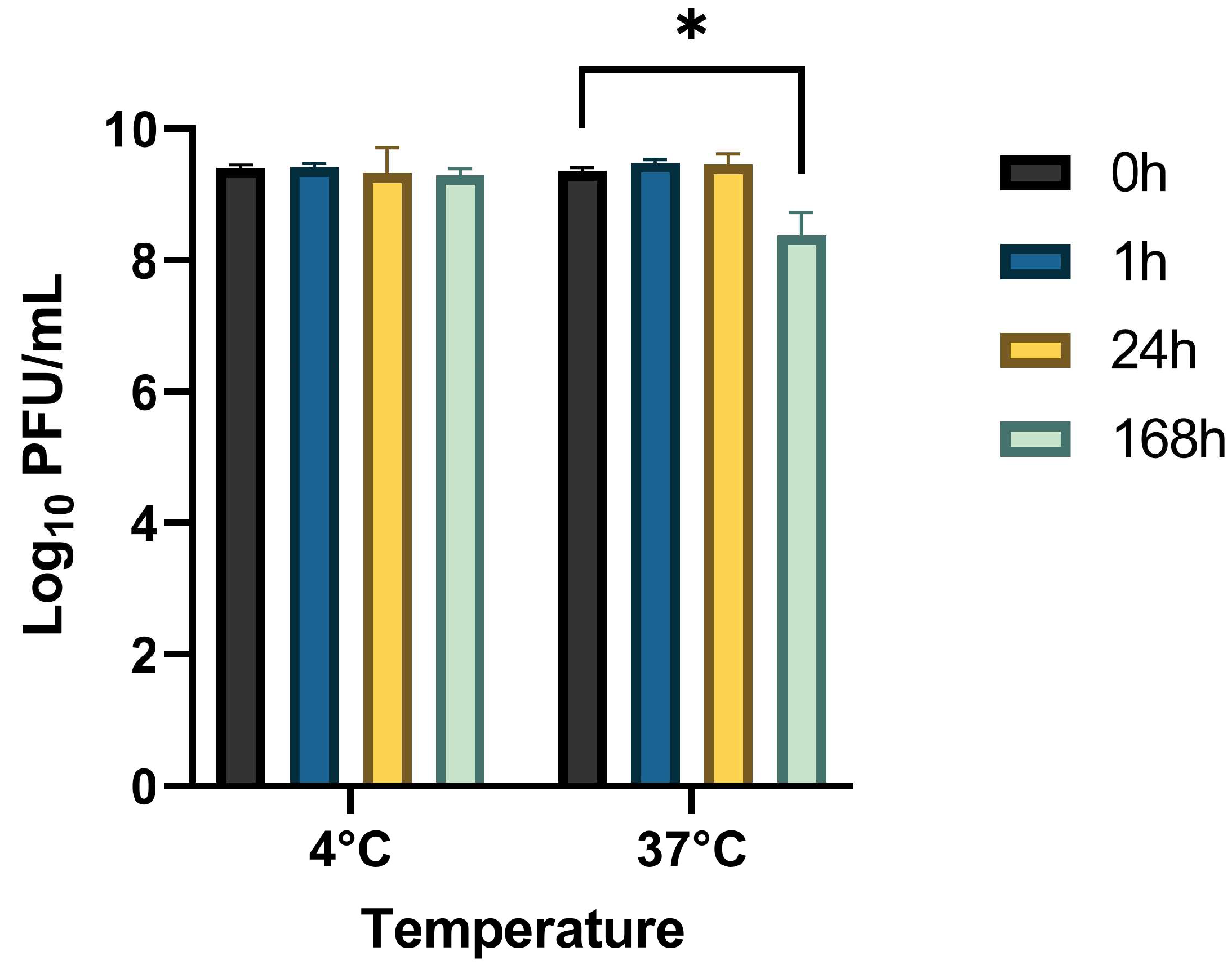
2.4. Stability Study

The stability of StaphLyse™ in whole milk was evaluated for 0, 1, 24 (1 day), and 168 (7 days) h. Aliquots (2.5 mL) of milk were pre-equilibrated to 37 °C or 4 °C, and 250 µL of StaphLyse™ was added to triplicate milk samples. To determine the initial phage titer, an aliquot of each sample was immediately serially ten-fold diluted in SM buffer (50 mM Tris-HCl pH7.5, 100 mM NaCl, 8 mM MgSO4) through the 10−6 dilution. Then, 10 µL triplicate aliquots of the 10−5 and 10−6 dilutions were spotted onto lawns of the titer host strain, S. aureus ATCC 49775. The spot test plates (LB agar) were incubated overnight at 30 °C, and plaques were counted the next day to obtain titers. The titer method was repeated at 1 h, 24 h, and 168 h (7 days).

2.5. In Vitro Killing of S. aureus in Whole Milk

The ability of StaphLyse™ to kill S. aureus ATCC 29740 in whole milk was analyzed at 0 and 1 h. The challenge S. aureus ATCC 29740 was grown to an OD of 0.2, and 375 µL of the bacterial suspension (~5 × 10⁷ CFU/mL) was added to each of three tubes containing 3 mL pre-warmed milk (37 °C). The control tube received 375 µL of SM buffer, one tube received 375 µL full concentration StaphLyse™ (~1 × 10¹⁰ PFU/mL), and 375 µL of a 1:10 dilution of StaphLyse™ (~1 × 10⁹ PFU/mL) was added to the third tube. The tubes were incubated at 37 °C for 1 h at 200 rpm. To determine the concentration of bacteria, an aliquot of each sample was serially diluted ten-fold in SM buffer through the 10−6 dilution method at 0 h and at 1 h. Bacterial cell counts were determined by plating 10 µL of each of the dilutions in triplicate on LB agar. The plates were incubated overnight at 37 °C, and colonies were counted the next day to obtain bacterial concentrations. The experiment was repeated on four separate days.

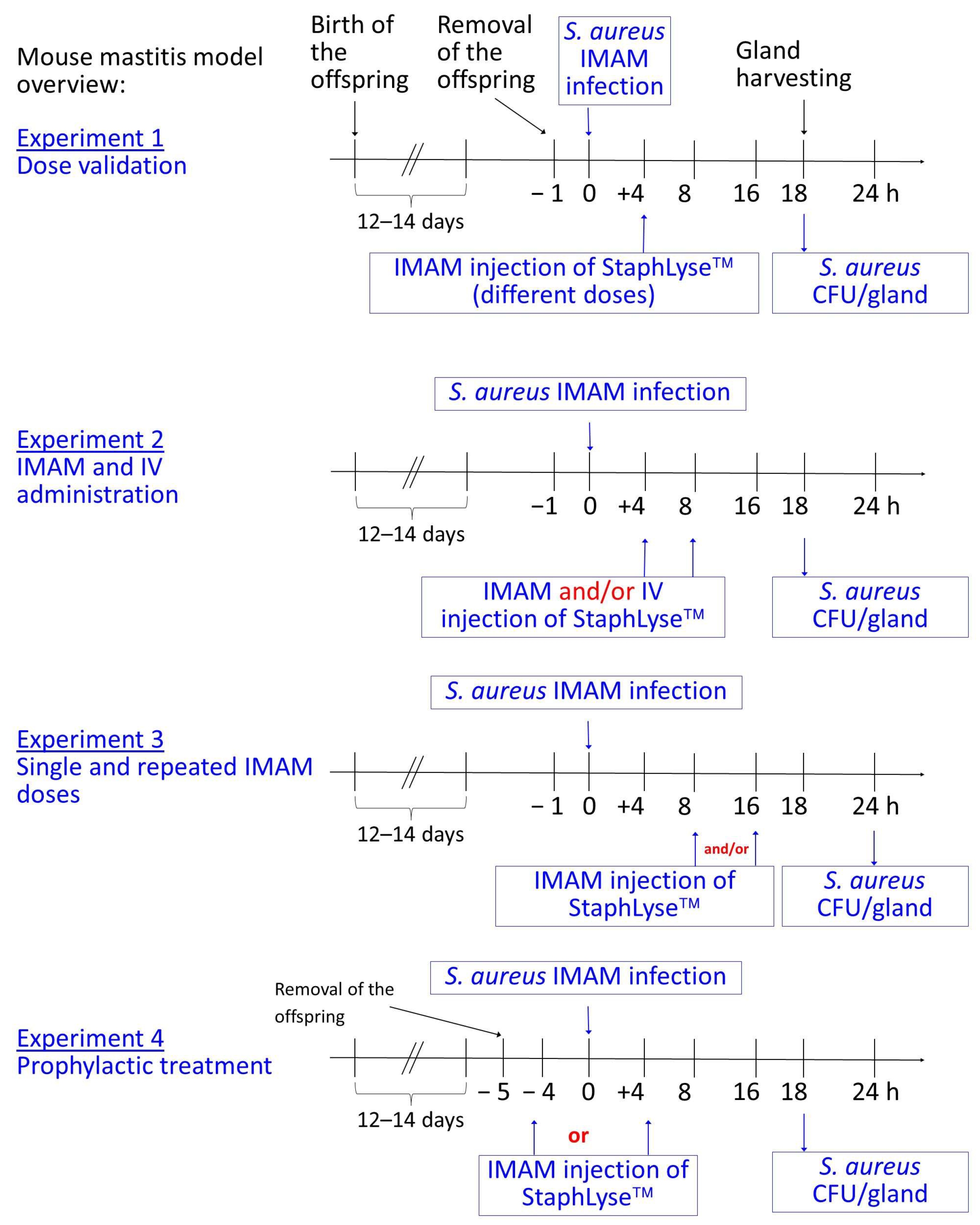
2.6. Mouse Mastitis Model

A well-characterized and validated mouse mastitis model [44] was used. Briefly, CD-1 lactating mice (Charles River Laboratories, Sherbrooke, QC, Canada) were separated from their 12–14-day-old pups, anesthetized with a mixture of ketamine/xylazine (87 and 13 mg/kg of body weight, respectively), and the teats of the fourth pair of mammary glands located from head to tail were disinfected with 70% ethanol prior to the intramammary injection of S. aureus ATCC 29740 (~100 CFU in 100 µL for each gland). All the injections into the mammary glands were carried out under a binocular at the experimental time points specified in the figure legends, using 1 mL tuberculin syringes and 31-gauge blunt needles. For all the experiments, each group contained four mice, with each mouse providing up to two glands (the fourth, i.e., the biggest ones).
Figure 2 shows the general outline of the mastitis model as well as the different experimental designs that were used in this study. Four experimental designs were used. First of all, in experiment 1, the effect of different doses of StaphLyse™ was evaluated to determine the best dose to use in the following experiments. In this experiment, separate doses of StaphLyse™ (0, 106, 107, or 108 PFU delivered in 100 µL) were administered as a single intramammary (IMAM) dose 4 h after infection. In experiment 2, we studied the effect of administration routes. StaphLyse™ or PBS were given either IMAM 4 h after infection, IMAM 8 h after infection, or IMAM 4 h after infection followed by IV (intra veinous) 4 h later (at 8 h post infection). In this experiment, each dose of StaphLyse™ contains 108 PFU in a 100 µL volume. In experiment 3, we used IMAM administration, which was found to be more effective in experiment 2, and we studied the effect of administering a single dose versus two repeated doses of phage cocktail. StaphLyse™ was injected via the IMAM route at 8 and/or at 16 h after S. aureus infection. In experiment 4, the model was used to evaluate the efficacy of prophylactic treatment to evaluate the possibility to use StaphLyse™ as a preventive treatment for the nonlactating period in cows (DCT, as defined in the introduction section). StaphLyse™ was injected via the IMAM route either 4 h before the S. aureus infection (−4) or 4 h after the infection (+4).
At the end of each experiment, mice were humanely euthanized; mammary glands were harvested, weighed, and homogenized in phosphate-buffered saline (PBS); and CFUs were determined by plating serial dilutions of homogenates on tryptic soy agar (TSA). Colonies were counted after 20 h of incubation at 37 °C. In some experiments, phage titers in some of the glands were also determined using a double-layer method [52]. In brief, samples were diluted in 5 mL of molten TSA (0.7% agar) at 50 °C containing 100 µL of an overnight culture of S. aureus ATCC 29740. The molten agar mixture was poured onto TSA plates and allowed to set, and plates were incubated at 37 °C for 24 h. Phage plaques were counted to determine PFU/mL.

2.7. Statistical Analysis

Statistical analysis was performed using GraphPad Prism for Windows (version 9.3.1; GraphPad Software; San Diego, CA, USA; www.graphpad.com). For the stability of StaphLyse™ in whole milk, the data were analyzed using a two-way analysis of variance (two-way ANOVA). Sidak’s multiple-comparison test was performed to compare the stability of StaphLyse™ in milk at each temperature between 0 h and the other time points (1 h, 24 h, and 168 h). Tukey’s multiple-comparison test was performed to compare the effect of StaphLyse™ on S. aureus at 0 h and 1 h treatment times. In the mouse experiments, statistical significance was determined using the Kruskal–Wallis comparison test followed by Dunn’s multiple-comparison test. PFU and CFU were log10-transformed prior to statistical analysis. A p value < 0.05 was considered statistically significant.

3. Results

3.1. Lytic Range of the StaphLyse™ In Vitro

StaphLyse™ demonstrated lytic activity against a large collection of well-characterized S. aureus strains including MSSA (including S. aureus ATCC 29740 that was used in the mouse experiments), MRSA, and VISA strains. The cocktail lysed 92.7% of the strains tested at a titer of 2 × 10⁴ PFU/mL and 100% of these same strains when tested at a higher titer of 1 × 10⁹ PFU/mL (Table 2).
Figure 3 presents the host-killing range and interaction network of the five monophages included in the StaphLyse™ cocktail. It shows that a large number of strains are being lysed by multiple phages (i.e., redundant lytic spectrum coverage).

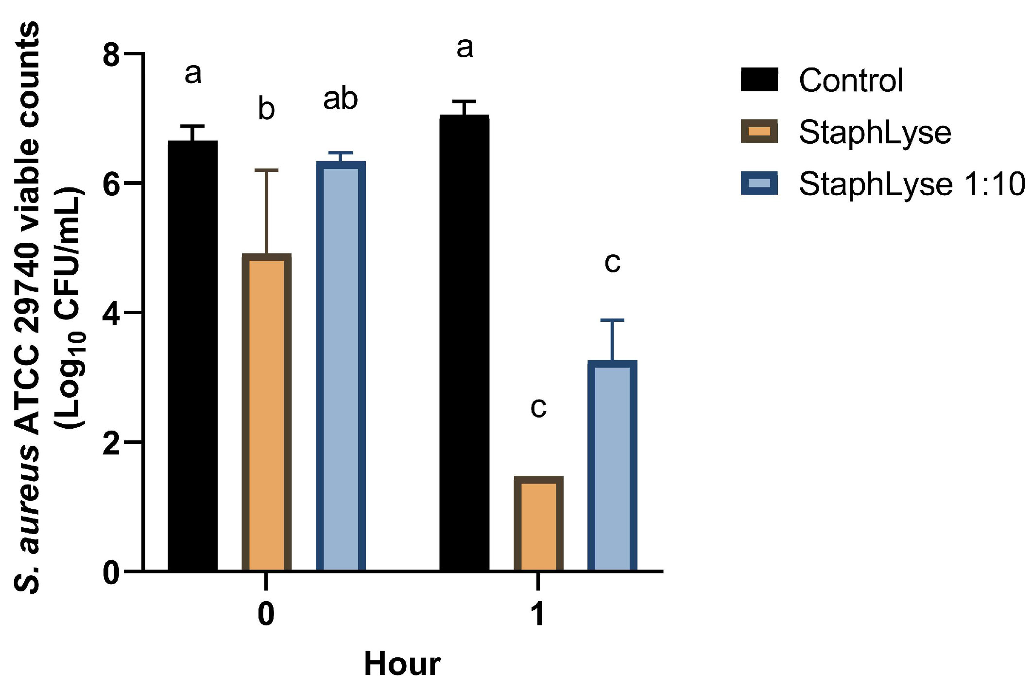
3.2. Stability of StaphLyse™ and In Vitro Killing of S. aureus in Whole Milk

The stability of StaphLyse™ was evaluated after 1 h, 24 h (1 day), and 168 h (7 days) at 4 and 37 °C in milk using an initial phage concentration of ~2.5 × 10⁹ PFU/mL. Total PFU/mL counts were maintained at 4 and 37 °C for up to one day and for up to 7 days at 4 °C, while after a period of 7 days at 37 °C, a reduction of ~1.0 log10 PFU/mL was observed (Figure 4, p < 0.05).
To study the in vitro killing of S. aureus in whole milk, two concentrations of StaphLyse™ were used: fully concentrated StaphLyse™ ~1010 PFU/mL and a diluted StaphLyse™ at ~109 PFU/mL (StaphLyse™ 1:10). After 1 h in milk, StaphLyse™ killed 5.8 and 3.2 log10 CFU/mL of the initial S. aureus ATCC 29740 inoculum (6.7 log10 CFU/mL). The killing occurred quickly, as by the end of the processing at the first time point (t = 0), we already observed a reduction of 0.35 and 1.0 log10 CFU/mL at phage concentrations of 109 and 1010 PFU/mL, respectively (Figure 5).

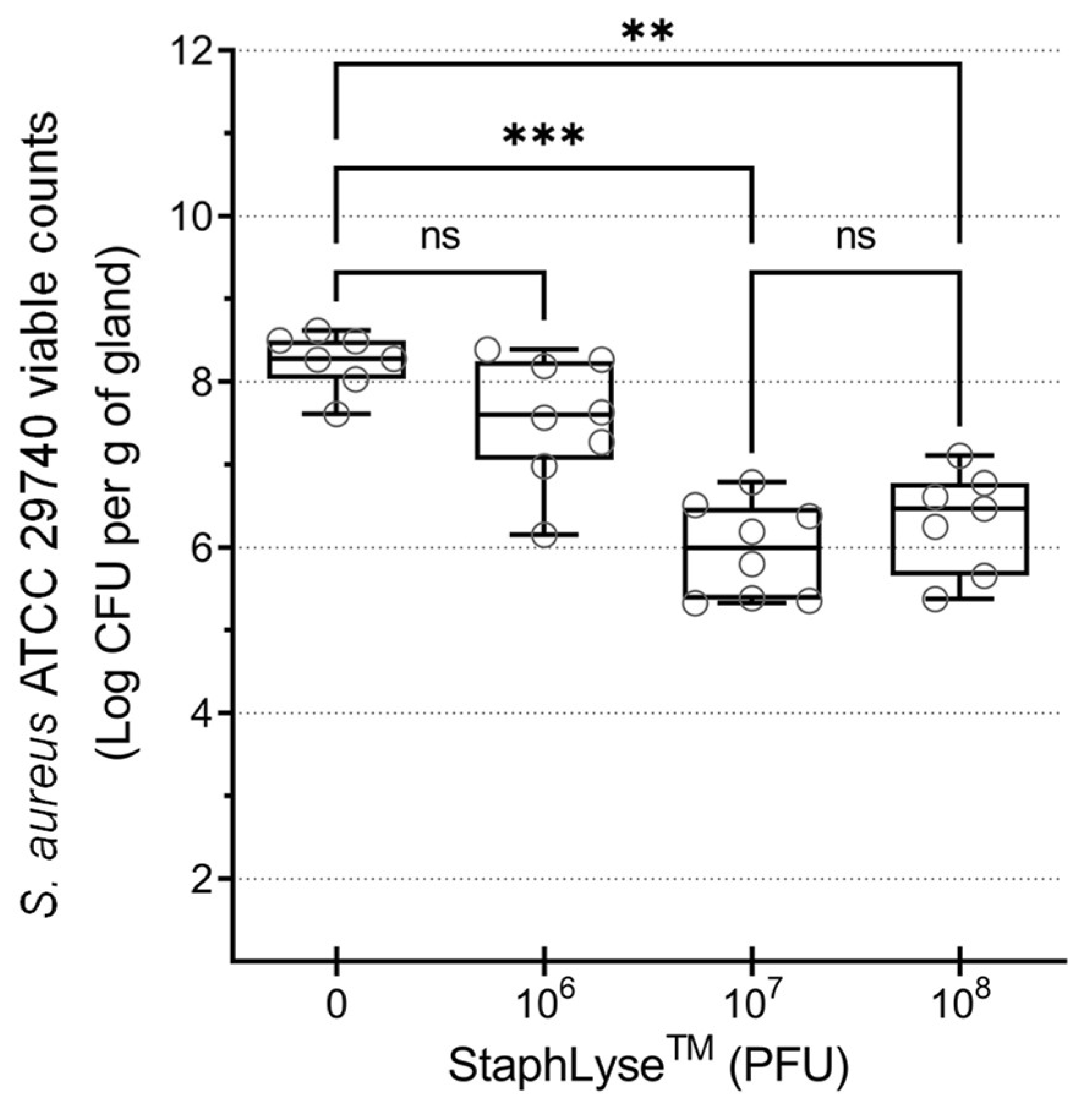
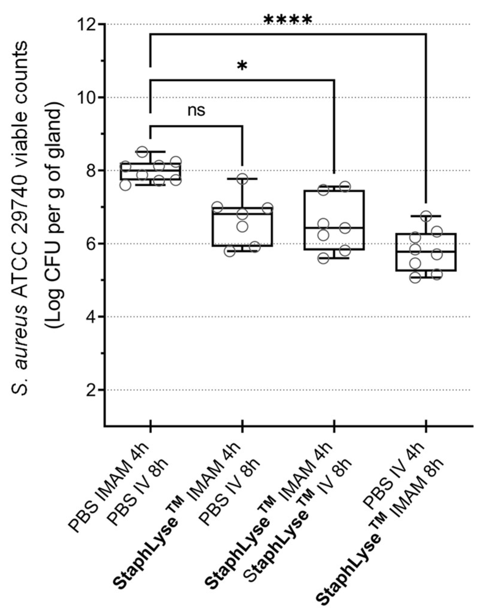
3.3. StaphLyse™ Efficacy in a Mouse Model to Treat S. aureus Intramammary Infection

The efficacy of therapy with the StaphLyse™ phage cocktail was studied using a well-established mouse model of S. aureus IMI infection. In this model, StaphLyse™ dose ranging revealed that a single dose at phage titers of 107 or 108 PFU, injected via the IMAM route 4 h after infection with S. aureus ATCC 29740, was effective, resulting in a significant decrease in the bacterial load in the glands of infected animals, whereas a low phage titer of 106 PFU was insufficient to significantly reduce the bacterial load present after 18 h of infection (Figure 6). The dose of 108 PFU was selected for further experiments.
StaphLyse™ was then administered to mice via the IMAM and/or IV routes at a dose of 108 PFU once or twice at 4 and/or 8 h after IMAM S. aureus infection, and mammary glands were harvested 18 h after infection to determine the remaining bacterial load. A single IMAM dose of phage cocktail given 8 h after infection significantly reduced the bacterial load in the mammary glands, whereas the same dose given 4 h after infection did not generate a significant response. Furthermore, after an initial IMAM treatment 4 h after infection, an additional IV dosing of the phage cocktail 8 h later did not improve response, indicating that the IV route of administration and/or the multiplicity of infection (MOI) may not be suitable for reaching the site of infection and eliciting a response in this model (Figure 7).
We further studied the effect of single and repeated doses of StaphLyse™, using IMAM administration. We found that two IMAM doses administered at 8 and 16 h after infection was the most effective treatment regimen, significantly reducing bacterial load by 2.82 log10 CFU per gram of gland. In this experiment, amoxicillin was used as a positive control. It was administered IMAM at 8 h after the infection at a concentration of 75 µg per gland or approximately 75 µg/mL based on the volume of the gland, which is approximately 150 times higher than the MIC of amoxicillin for S. aureus ATCC 29740 (MIC = 0.5 µg/mL). As expected, this amoxicillin treatment resulted in a significant reduction of 4.45 log10 CFU per gram of gland (Figure 8). As an indicative measure, the number of phages in some glands was also estimated at the end of the experiment (24 h). Each dose of StaphLyse™ contains a total of 1.25 × 10⁸ PFU in a volume of 100 µL. There was an increase of 0.44 and 0.76 log10 PFU at 24 h in the sampled glands of the two groups that had only received one dose of the phage cocktail at either 8 h or 16 h, suggesting that phages remained stable in the gland or may have replicated. We recovered an equivalent amount of PFU in the gland as was initially inoculated in the sampled glands of the group that received two doses of the phage cocktail at 8 and 16 h after infection. The first phage cocktail injection may have reduced the bacterial population, and the second dose of phages may have further reduced the number of live bacteria in the gland, resulting in less phage replication. More systematic gland sampling for phage quantitation would be needed to confirm such a hypothesis.

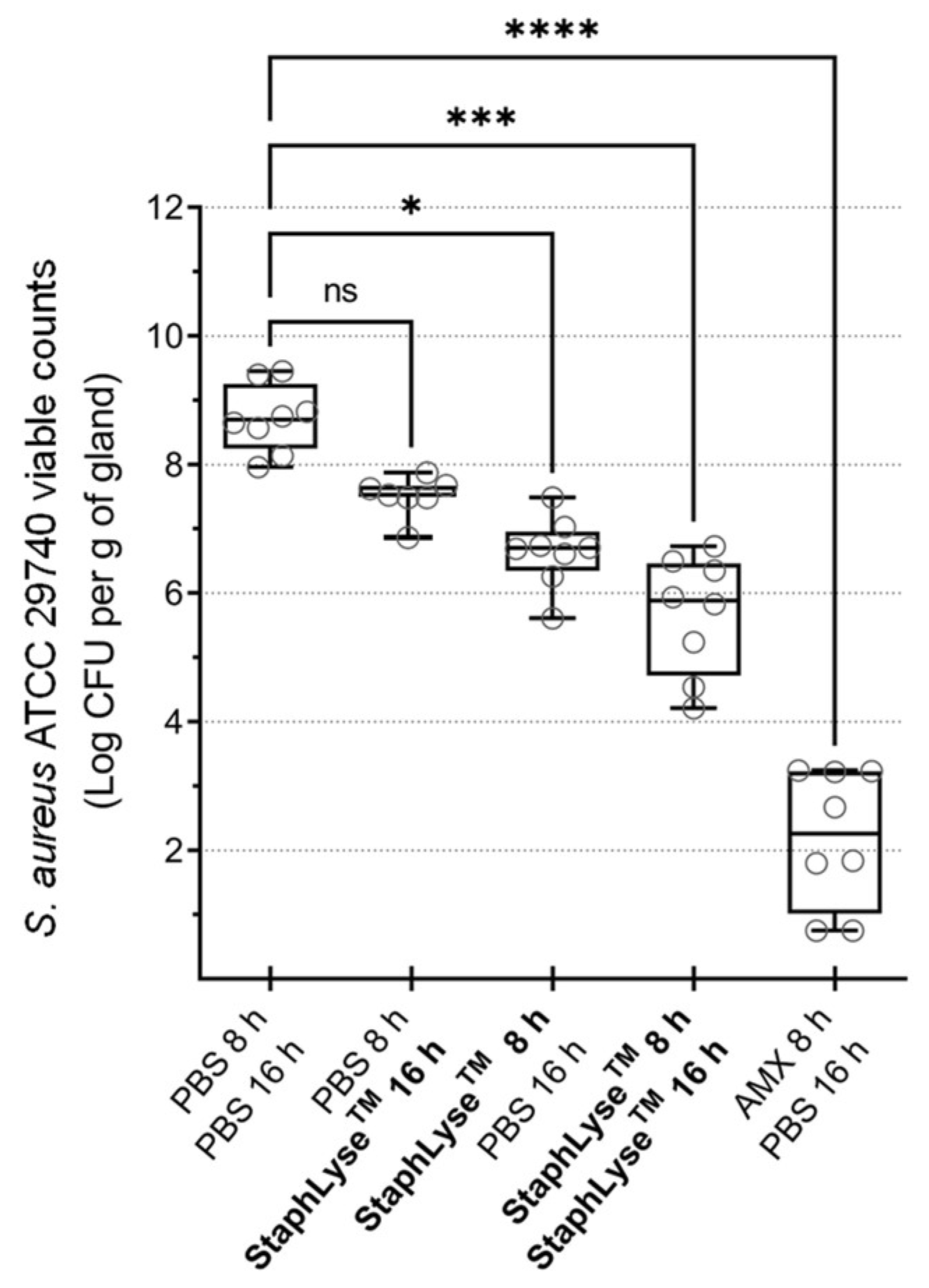
3.4. Prophylactic Treatment of S. aureus Intramammary Infection Using StaphLyse™

In the dairy sector, antibiotic treatment during the cow’s nonlactating period (DCT) is performed to prevent new infection of the mammary gland when lactation resumes after calving. To test the potential of StaphLyse™ as a DCT, prophylactic treatment was evaluated in the murine model. The administration of StaphLyse™ 4 h prior to infection as well as 4 h after infection both significantly reduced the bacterial loads by 4.03 and 2.81 log10 CFU per gram of gland, respectively. Moreover, there was no significant difference in bacterial load in the glands between the two treated groups, whether the phage cocktail was administered before or after infection (Figure 9A). When the experiment was performed with amoxicillin, similar results were observed (Figure 9B). The administration of amoxicillin 4 h prior to infection as well as 4 h after infection both significantly reduced the bacterial loads by 4.07 and 4.85 log10 CFU per gram of gland, respectively. As for StaphLyse™, there was no significant difference in bacterial load in the glands between the two treated groups, whether the phage cocktail was administered before or after infection. The StaphLyse™ and amoxicillin used in prophylaxis remained active in the mammary gland for at least 4 h.

4. Discussion

Antibiotic resistance is currently on the rise, and it is predicted that by 2050, it will be responsible for 10 million human deaths per year, as well as USD 1 trillion loss to the global economy [53]. The overuse and misuse of antibiotics for the treatment of bovine mastitis by the dairy industry are recognized as important factors contributing to the global spread of antibiotic resistance [15,16,17]. Without available alternatives, the outright prohibition of antibiotic use in the dairy industry would result in a significant increase in production costs, due to the increased incidence of infection, and would threaten the sustainability of dairy farming [17,54]. Thus, there is an urgent need to develop alternatives to antibiotics for the treatment of bovine mastitis.
Lytic bacteriophages act as bactericidal agents and specifically infect and kill targeted bacteria. Phage therapy thus has the potential to become part of the arsenal to tackle S. aureus IMIs and significantly reduce the use of antibiotics by the dairy industry [55]. Phage-based products are currently used in the food safety and processing industry; however, none are yet available for administration to food animals during production [56].
In this study, we investigated the efficacy of a new phage cocktail named StaphLyse™ for the control and prevention of S. aureus mastitis. This phage cocktail is composed of five fully sequenced and characterized lytic phages specifically targeting S. aureus. We showed that the in vitro lytic activity of the cocktail was dose-dependent and that it lysed 92.7% and 100% of the strains tested (n = 709) at a titer of 2 × 10⁴ and 1 × 10⁹ PFU/mL, respectively (Table 2 and Figure 3). The combination of the five phages present in StaphLyse™ appears to cover a much broader range of S. aureus strains than that previously reported by Geng et al. for a phage cocktail composed of only two phages, one from the Myoviridae family (vBSM-A1) and the other from the Podoviridae family (vBSP-A2), with vBSM-A1 lysing 85% and vBSP-A2 lysing 59% of 23 S. aureus strains tested [31]. This suggests that combining more than two phages in a cocktail may be desirable to ensure efficacy against a broader spectrum of S. aureus strains and perhaps avoid treatment failure; however, competitive interference between different phages has been shown in E. coli and must be kept in mind when developing such cocktails [57]. Combining multiple phages in a therapeutic cocktail may also reduce the likelihood of resistance development [19].
A mouse mastitis model was used to study the in vivo efficacy of the phage cocktail against S. aureus IMI. This model is currently used to evaluate the efficacy of novel treatments for mastitis, and we have previously characterized clinical symptoms, pathological conditions, and levels of cytokines and shown that they are similar to that of a naturally occurring infection in mice [44]. Using mouse mastitis models allows researchers to test new alternatives to antibiotics in well-controlled settings and at a scale that is appropriate for the evaluation of emerging therapies. Without a doubt, mice cannot completely replace cows for all research purposes. There are differences between mouse and bovine mammary glands, and caution is needed when extrapolating mouse data to cows. However, this model clearly allows for an appreciation of the potential of novel therapies and provides further justification for the significant investment required to move forward and test such therapies in infected cows, which can only be carried out in a biosafety level 2 barn and with larger quantities of phage cocktail.
In this study, we demonstrated that the phage cocktail StaphLyse™ was effective in controlling S. aureus IMI in mice. A single IMAM injection of this phage cocktail given 8 h after infection via the IMAM route reduced the bacterial load in mammary glands of lactating mice infected with S. aureus (reduction of 2 to 2.2 log10 CFU per gram of gland, as observed in Figure 7 and Figure 8). As expected, a double-dose regimen was more effective, with a reduction of 2.82 log10 CFU per gram of gland (Figure 8). We also showed that the IV administration of phages, following an initial IMAM administration, did not improve the effectiveness of the initial IMAM treatment (Figure 7).
The pharmacodynamics and pharmacokinetics of a phage cocktail are undoubtedly different from that of traditional small-molecule antibiotics. Bacteriophages can not only kill bacteria, but they can also multiply in the environment. As a result, their pharmacology is influenced by both their direct interactions with bacterial cells as well as their capacity to penetrate tissues and reach infection sites [58]. It was shown that the tight junctions between the epithelial cells constituting the inner lining of the alveolar structure of the mammary gland are affected by neutrophil diapedesis occurring in response to inflammation caused by infection [59]. During an IMI in a cow, neutrophils migrate from the blood to the infection site within 2 to 4 h, reaching a peak in the milk between 10 and 16 h, as cytokines are released [60]. Phages injected directly into the blood may gain access to the alveolar lumen as the inflammatory response increases over time. It is also known that the inflammatory response allows blood components to enter the milk (such as serum albumin and antibodies) and vice versa (lactose from the milk leaking into the blood) as the permeability of the mammary epithelium increases [61]. This inflammatory state is typical of IMI-induced bovine mastitis, and it may have an effect on the efficacy of phage therapy administered in cows via IV injections. However, using the mouse mastitis model, Iwano et al. showed that only 1% of phages reached mammary glands and were rapidly eliminated when administered via the IV route [32].
The IMAM administration of a phage cocktail may be more practical to implement for the treatment of cows because phages can be selectively injected into the infected mammary gland quarter, directly at the site of infection. Furthermore, contrary to what was previously found for other phages in the presence of raw milk or milk whey proteins in in vitro assays [62,63], our in vitro results showed that milk did not interfere with or neutralize the lytic effect of the StaphLyse™ phage cocktail (Figure 5), suggesting that IMAM administration may be appropriate.
Finally, our study showed that a cocktail of lytic phages can be used prophylactically to control S. aureus mastitis in mice (Figure 9). This is an exciting finding because, if proven similarly effective in cows, phage prophylactic therapy could replace prophylactic DCT, and significantly reduce the number of antibiotics used in dairy farms.

5. Conclusions

The present study suggests that a cocktail of lytic phages could be a safe and effective alternative to antibiotics for the treatment or prevention of IMIs caused by S. aureus. The StaphLyse™ phage cocktail was stable in milk at 37 °C for up to one day and for up to one week at 4 °C. The phage cocktail was bactericidal in vitro in a dose-dependent manner against S. aureus. A single IMAM injection of this phage cocktail given 8 h after infection via the IMAM route reduced the bacterial load in the mammary glands of lactating mice infected with S. aureus, and as expected, a two-dose regimen was more effective. The prophylactic use of the phage cocktail was also effective. These results support further research to determine the efficacy of the phage cocktail StaphLyse™ in dairy cows as an alternative to traditional antibiotics therapy to control S. aureus IMIs.

6. Patents

The phages contained in StaphLyse™ and the StaphLyse™ formulation are the subject of several issued and pending patent applications, including US patent #7,745,194.

Author Contributions

Conceptualization, F.M., S.C., J.W. and T.K.; methodology, E.B., G.M., J.-P.R., G.M. and L.H.; investigation, validation, and formal analysis, J.W., L.H., S.H., E.B., G.M., C.S., S.C. and F.M.; resources, F.M.; writing—original draft preparation, E.B. and S.C.; writing—review and editing, C.S., J.W., G.M., T.K. and F.M.; supervision, F.M.; project administration, F.M. and T.K.; funding acquisition, F.M. All authors have read and agreed to the published version of the manuscript.

Funding

Intralytix developed and manufactured StaphLyse™ and performed in vitro tests and phage characterization. The mastitis mouse model and therapy tests were funded by a research contract (no. 37574) between Université de Sherbrooke and Phileo-Lesaffre Inc. (Société Industrielle Lesaffre SA). The authors also acknowledge logistical support to J.-P.R. and F.M. laboratories from Op+lait, the Regroupement pour un lait de qualité optimale supported by the Fonds de Recherche du Québec-Nature et Technologie (FRQ-NT) (Université de Montréal, St-Hyacinthe, QC, Canada).

Institutional Review Board Statement

The animal studies were performed in compliance with the guidelines of the Canadian Council on Animal Care and this research project was approved by the institutional ethics committee on animal experimentation of the Faculté des Sciences of Université de Sherbrooke (protocol code 2017-1966, FM2017-01B and date of approval 9 February 2021).

Data Availability Statement

All data, generated or analyzed, and materials during this study are included in this published article.

Acknowledgments

We are grateful to Jennifer Schwartz, Bradley Anderson, and Alexander Sulakvelidze for their helpful comments and assistance with writing this manuscript.

Conflicts of Interest

The corresponding author declares no conflict of interest. Phileo-Lesaffre Inc. had no direct involvement in the collection and interpretation of the data related to intramammary infections and treatment tests conducted at Université de Sherbrooke.

References

  1. Huijps, K.; Hogeveen, H. Stochastic modeling to determine the economic effects of blanket, selective, and no dry cow therapy. J. Dairy Sci. 2007, 90, 1225–1234. [Google Scholar] [CrossRef] [PubMed]
  2. Kabera, F.; Dufour, S.; Keefe, G.; Cameron, M.; Roy, J.P. Evaluation of quarter-based selective dry cow therapy using Petrifilm on-farm milk culture: A randomized controlled trial. J. Dairy Sci. 2020, 103, 7276–7287. [Google Scholar] [CrossRef] [PubMed]
  3. Oliver, S.P.; Murinda, S.E. Antimicrobial resistance of mastitis pathogens. Vet. Clin. N. Am. Food Anim. Pract. 2012, 28, 165–185. [Google Scholar] [CrossRef]
  4. Aghamohammadi, M.; Haine, D.; Kelton, D.F.; Barkema, H.W.; Hogeveen, H.; Keefe, G.P.; Dufour, S. Herd-level mastitis-associated costs on Canadian dairy farms. Front. Vet. Sci. 2018, 5, 100. [Google Scholar] [CrossRef] [PubMed]
  5. Krömker, V.; Leimbach, S. Mastitis treatment—Reduction in antibiotic usage in dairy cows. Reprod. Domest. Anim. 2017, 52, 21–29. [Google Scholar] [CrossRef] [PubMed] [Green Version]
  6. Reyher, K.K.; Dufour, S.; Barkema, H.W.; Des Côteaux, L.; DeVries, T.J.; Dohoo, I.R.; Keefe, G.P.; Roy, J.P.; Scholl, D.T. The National Cohort of Dairy Farms—A data collection platform for mastitis research in Canada. J. Dairy Sci. 2011, 94, 1616–1626. [Google Scholar] [CrossRef] [Green Version]
  7. Wellnitz, O.; Bruckmaier, R.M. The innate immune response of the bovine mammary gland to bacterial infection. Vet. J. 2012, 192, 148–152. [Google Scholar] [CrossRef]
  8. Peton, V.; Le Loir, Y. Staphylococcus aureus in veterinary medicine. Infect. Genet. Evol. 2014, 21, 602–615. [Google Scholar] [CrossRef] [PubMed]
  9. Pichette-Jolette, S.; Millette, G.; Demontier, E.; Bran-Barrera, D.; Cyrenne, M.; Ster, C.; Haine, D.; Keefe, G.; Malouin, F.; Roy, J.P. Partial prediction of the duration and the clinical status of Staphylococcus aureus bovine intramammary infections based on the phenotypic and genotypic analysis of isolates. Vet. Microbiol. 2019, 228, 188–195. [Google Scholar] [CrossRef]
  10. Jacques, M.; Aragon, V.; Tremblay, Y.D. Biofilm formation in bacterial pathogens of veterinary importance. Anim. Health Res. Rev. Conf. Res. Work Anim. Dis. 2010, 11, 97–121. [Google Scholar] [CrossRef]
  11. Melchior, M.B.; Fink-Gremmels, J.; Gaastra, W. Extended antimicrobial susceptibility assay for Staphylococcus aureus isolates from bovine mastitis growing in biofilms. Vet. Microbiol. 2007, 125, 141–149. [Google Scholar] [CrossRef] [PubMed]
  12. Proctor, R.A.; von Eiff, C.; Kahl, B.C.; Becker, K.; McNamara, P.; Herrmann, M.; Peters, G. Small colony variants: A pathogenic form of bacteria that facilitates persistent and recurrent infections. Nat. Rev. Microbiol. 2006, 4, 295–305. [Google Scholar] [CrossRef] [PubMed]
  13. Ster, C.; Lebeau, V.; Leclerc, J.; Fugere, A.; Veh, K.A.; Roy, J.P.; Malouin, F. In vitro antibiotic susceptibility and biofilm production of Staphylococcus aureus isolates recovered from bovine intramammary infections that persisted or not following extended therapies with cephapirin, pirlimycin or ceftiofur. Vet. Res. 2017, 48, 56. [Google Scholar] [CrossRef] [Green Version]
  14. Tong, S.Y.C.; Davis, J.S.; Eichenberger, E.; Holland, T.L.; Fowler, V.G. Staphylococcus aureus infections: Epidemiology, pathophysiology, clinical manifestations, and management. Clin. Microbiol. Rev. 2015, 28, 603–661. [Google Scholar] [CrossRef] [PubMed] [Green Version]
  15. McEwen, S.A.; Collignon, P.J. Antimicrobial resistance: A one health perspective. Microbiol. Spectr. 2018, 6, ARBA-0009-2017. [Google Scholar] [CrossRef] [PubMed] [Green Version]
  16. Molineri, A.I.; Camussone, C.; Zbrun, M.V.; Suárez Archilla, G.; Cristiani, M.; Neder, V.; Calvinho, L.; Signorini, M. Antimicrobial resistance of Staphylococcus aureus isolated from bovine mastitis: Systematic review and meta-analysis. Prev. Vet. Med. 2021, 188, 105261. [Google Scholar] [CrossRef] [PubMed]
  17. Park, S.; Ronholm, J. Staphylococcus aureus in agriculture: Lessons in evolution from a multispecies pathogen. Clin. Microbiol. Rev. 2021, 34, 1–27. [Google Scholar] [CrossRef] [PubMed]
  18. Garcia, S.N.; Osburn, B.I.; Cullor, J.S. A one health perspective on dairy production and dairy food safety. One Health 2019, 7, 100086. [Google Scholar] [CrossRef]
  19. Kahn, L.H.; Bergeron, G.; Bourassa, M.W.; De Vegt, B.; Gill, J.; Gomes, F.; Malouin, F.; Opengart, K.; Ritter, G.D.; Singer, R.S.; et al. From farm management to bacteriophage therapy: Strategies to reduce antibiotic use in animal agriculture. Ann. N. Y. Acad. Sci. 2019, 1441, 31–39. [Google Scholar] [CrossRef]
  20. Wittebole, X.; De Roock, S.; Opal, S.M. A historical overview of bacteriophage therapy as an alternative to antibiotics for the treatment of bacterial pathogens. Virulence 2014, 5, 226–235. [Google Scholar] [CrossRef]
  21. Henein, A. What are the limitations on the wider therapeutic use of phage? Bacteriophage 2013, 3, e24872. [Google Scholar] [CrossRef] [PubMed] [Green Version]
  22. Speck, P.; Smithyman, A. Safety and efficacy of phage therapy via the intravenous route. FEMS Microbiol. Lett. 2015, 363, 1–5. [Google Scholar] [CrossRef] [PubMed]
  23. Kutter, E.M.; Kuhl, S.J.; Abedon, S.T. Re-establishing a place for phage therapy in western medicine. Future Microbiol. 2015, 10, 685–688. [Google Scholar] [CrossRef] [Green Version]
  24. Zduńczyk, S.; Janowski, T. Bacteriophages and associated endolysins in therapy and prevention of mastitis and metritis in cows: Current knowledge. Anim. Reprod. Sci. 2020, 218, 106504. [Google Scholar] [CrossRef] [PubMed]
  25. Lungren, M.P.; Donlan, R.M.; Kankotia, R.; Paxton, B.E.; Falk, I.; Christensen, D.; Kim, C.Y. Bacteriophage K antimicrobial-lock technique for treatment of Staphylococcus aureus central venous catheter-related infection: A leporine model efficacy analysis. J. Vasc. Interv. Radiol. 2014, 25, 1627–1632. [Google Scholar] [CrossRef] [PubMed]
  26. Wills, Q.F.; Kerrigan, C.; Soothill, J.S. Experimental bacteriophage protection against Staphylococcus aureus abscesses in a rabbit model. Antimicrob. Agents Chemother. 2005, 49, 1220–1221. [Google Scholar] [CrossRef] [Green Version]
  27. Takemura-Uchiyama, I.; Uchiyama, J.; Osanai, M.; Morimoto, N.; Asagiri, T.; Ujihara, T.; Daibata, M.; Sugiura, T.; Matsuzaki, S. Experimental phage therapy against lethal lung-derived septicemia caused by Staphylococcus aureus in mice. Microbes Infect. 2014, 16, 512–517. [Google Scholar] [CrossRef]
  28. Sunagar, R.; Patil, S.A.; Chandrakanth, R.K. Bacteriophage therapy for Staphylococcus aureus bacteremia in streptozotocin-induced diabetic mice. Res. Microbiol. 2010, 161, 854–860. [Google Scholar] [CrossRef]
  29. Drilling, A.; Morales, S.; Boase, S.; Jervis-Bardy, J.; James, C.; Jardeleza, C.; Tan, N.C.W.; Cleland, E.; Speck, P.; Vreugde, S.; et al. Safety and efficacy of topical bacteriophage and ethylenediaminetetraacetic acid treatment of Staphylococcus aureus infection in a sheep model of sinusitis. Int. Forum Allergy Rhinol. 2014, 4, 176–186. [Google Scholar] [CrossRef]
  30. Breyne, K.; Honaker, R.W.; Hobbs, Z.; Richter, M.; Zaczek, M.; Spangler, T.; Steenbrugge, J.; Lu, R.; Kinkhabwala, A.; Marchon, B.; et al. Efficacy and safety of a bovine-associated Staphylococcus aureus phage cocktail in a murine model of mastitis. Front. Microbiol. 2017, 8, 2348. [Google Scholar] [CrossRef]
  31. Geng, H.; Zou, W.; Zhang, M.; Xu, L.; Liu, F.; Li, X.; Wang, L.; Xu, Y. Evaluation of phage therapy in the treatment of Staphylococcus aureus-induced mastitis in mice. Folia Microbiol. 2020, 65, 339–351. [Google Scholar] [CrossRef]
  32. Iwano, H.; Inoue, Y.; Takasago, T.; Kobayashi, H.; Furusawa, T.; Taniguchi, K.; Fujiki, J.; Yokota, H.; Usui, M.; Tanji, Y.; et al. Bacteriophage ΦSA012 has a broad host range against Staphylococcus aureus and effective lytic capacity in a mouse mastitis model. Biology 2018, 7, 8. [Google Scholar] [CrossRef] [PubMed] [Green Version]
  33. Schmelcher, M.; Powell, A.M.; Becker, S.C.; Camp, M.J.; Donovan, D.M. Chimeric phage lysins act synergistically with lysostaphin to kill mastitis-causing Staphylococcus aureus in murine mammary glands. Appl. Environ. Microbiol. 2012, 78, 2297–2305. [Google Scholar] [CrossRef] [PubMed] [Green Version]
  34. Teng, F.; Xiong, X.; Zhang, S.; Li, G.; Wang, R.; Zhang, L.; Wang, X.; Zhou, H.; Li, J.; Li, Y.; et al. Efficacy assessment of phage therapy in treating Staphylococcus aureus-induced mastitis in mice. Viruses 2022, 14, 620. [Google Scholar] [CrossRef] [PubMed]
  35. Li, L.; Zhang, Z. Isolation and characterization of a virulent bacteriophage SPW specific for Staphylococcus aureus isolated from bovine mastitis of lactating dairy cattle. Mol. Biol. Rep. 2014, 41, 5829–5838. [Google Scholar] [CrossRef]
  36. O’Flaherty, S.; Ross, R.P.; Flynn, J.; Meaney, W.J.; Fitzgerald, G.F.; Coffey, A. Isolation and characterization of two anti-staphylococcal bacteriophages specific for pathogenic Staphylococcus aureus associated with bovine infections. Lett. Appl. Microbiol. 2005, 41, 482–486. [Google Scholar] [CrossRef]
  37. Kwiatek, M.; Parasion, S.; Mizak, L.; Gryko, R.; Bartoszcze, M.; Kocik, J. Characterization of a bacteriophage, isolated from a cow with mastitis, that is lytic against Staphylococcus aureus strains. Arch. Virol. 2012, 157, 225–234. [Google Scholar] [CrossRef]
  38. Han, J.E.; Kim, J.H.; Hwang, S.Y.; Choresca, C.H.; Shin, S.P.; Jun, J.W.; Chai, J.Y.; Park, Y.H.; Park, S.C. Isolation and characterization of a Myoviridae bacteriophage against Staphylococcus aureus isolated from dairy cows with mastitis. Res. Vet. Sci. 2013, 95, 758–763. [Google Scholar] [CrossRef]
  39. Ly-Chatain, M.H. The factors affecting effectiveness of treatment in phages therapy. Front. Microbiol. 2014, 5, 51. [Google Scholar] [CrossRef] [Green Version]
  40. Boulanger, D.; Brouillette, E.; Jaspar, F.; Malouin, F.; Mainil, J.; Bureau, F.; Lekeux, P. Helenalin reduces Staphylococcus aureus infection in vitro and in vivo. Vet. Microbiol. 2007, 119, 330–338. [Google Scholar] [CrossRef]
  41. Brouillette, E.; Hyodo, M.; Hayakawa, Y.; Karaolis, D.K.R.; Malouin, F. 3′,5′-cyclic diguanylic acid reduces the virulence of biofilm-forming Staphylococcus aureus strains in a mouse model of mastitis infection. Antimicrob. Agents Chemother. 2005, 49, 3109–3113. [Google Scholar] [CrossRef] [PubMed] [Green Version]
  42. Brouillette, E.; Grondin, G.; Talbot, B.G.; Malouin, F. Inflammatory cell infiltration as an indicator of Staphylococcus aureus infection and therapeutic efficacy in experimental mouse mastitis. Vet. Immunol. Immunopathol. 2005, 104, 163–169. [Google Scholar] [CrossRef] [PubMed]
  43. Brouillette, E.; Grondin, G.; Lefebvre, C.; Talbot, B.G.; Malouin, F. Mouse mastitis model of infection for antimicrobial compound efficacy studies against intracellular and extracellular forms of Staphylococcus aureus. Vet. Microbiol. 2004, 101, 253–262. [Google Scholar] [CrossRef]
  44. Brouillette, E.; Malouin, F. The pathogenesis and control of Staphylococcus aureus-induced mastitis: Study models in the mouse. Microbes Infect. 2005, 7, 560–568. [Google Scholar] [CrossRef] [PubMed]
  45. Côté-Gravel, J.; Brouillette, E.; Obradović, N.; Ster, C.; Talbot, B.G.; Malouin, F. Characterization of a vraG mutant in a genetically stable Staphylococcus aureus small-colony variant and preliminary assessment for use as a live-attenuated vaccine against intrammamary infections. PLoS ONE 2016, 11, e0166621. [Google Scholar] [CrossRef] [PubMed] [Green Version]
  46. Mulhbacher, J.; Brouillette, E.; Allard, M.; Fortier, L.C.; Malouin, F.; Lafontaine, D.A. Novel riboswitch ligand analogs as selective inhibitors of guanine-related metabolic pathways. PLoS Pathog. 2010, 6, e1000865. [Google Scholar] [CrossRef] [PubMed] [Green Version]
  47. Diarra, M.S.; Block, G.; Rempel, H.; Oomah, B.D.; Harrison, J.; McCallum, J.; Boulanger, S.; Brouillette, É.; Gattuso, M.; Malouin, F. In vitro and in vivo antibacterial activities of cranberry press cake extracts alone or in combination with β-lactams against Staphylococcus aureus. BMC Complement. Altern. Med. 2013, 13, 90. [Google Scholar] [CrossRef] [Green Version]
  48. Prasad, L.B.; Newbould, F.H. Inoculation of the bovine teat duct with Staph. aureus: The relationship of teat duct length, milk yield and milking rate to development of intramammary infection. Can. Vet. J. 1968, 9, 107–114. [Google Scholar]
  49. FDA. T.F.a.D.A. Bacteriophage Therapy: Scientific and Regulatory Issues Public Workshop. Available online: https://www.federalregister.gov/documents/2017/06/08/2017-11862/bacteriophage-therapy-scientific-and-regulatory-issues-public-workshop (accessed on 2 September 2021).
  50. Darling, A.E.; Mau, B.; Perna, N.T. Progressivemauve: Multiple genome alignment with gene gain, loss and rearrangement. PLoS ONE 2010, 5, e11147. [Google Scholar] [CrossRef] [PubMed] [Green Version]
  51. Adams, M.H. Methods of study bacterial viruses. In Bacteriophages; Interscience Publishers, Ltd.: London, UK, 1959; pp. 443–522. [Google Scholar]
  52. Kropinski, A.M.; Mazzocco, A.; Waddell, T.E.; Lingohr, E.; Johnson, R.P. Enumeration of bacteriophages by double agar overlay plaque assay. Methods Mol. Biol. 2009, 501, 69–76. [Google Scholar] [CrossRef]
  53. Sugden, R.; Kelly, R.; Davies, S. Combatting antimicrobial resistance globally. Nat. Microbiol. 2016, 1, 16187. [Google Scholar] [CrossRef] [PubMed]
  54. Lhermie, G.; Tauer, L.W.; Gröhn, Y.T. The farm cost of decreasing antimicrobial use in dairy production. PLoS ONE 2018, 13, e0194832. [Google Scholar] [CrossRef] [PubMed] [Green Version]
  55. Lin, D.M.; Koskella, B.; Lin, H.C. Phage therapy: An alternative to antibiotics in the age of multi-drug resistance. World J. Gastrointest. Pharmacol. Ther. 2017, 8, 162–173. [Google Scholar] [CrossRef] [PubMed]
  56. Moye, Z.D.; Woolston, J.; Sulakvelidze, A. Bacteriophage applications for food production and processing. Viruses 2018, 10, 205. [Google Scholar] [CrossRef] [Green Version]
  57. Liu, D.; Van Belleghem, J.D.; de Vries, C.R.; Burgener, E.; Chen, Q.; Manasherob, R.; Aronson, J.R.; Amanatullah, D.F.; Tamma, P.D.; Suh, G.A. The safety and toxicity of phage therapy: A review of animal and clinical studies. Viruses 2021, 13, 1268. [Google Scholar] [CrossRef]
  58. Danis-Wlodarczyk, K.; Dąbrowska, K.; Abedon, S.T. Phage therapy: The pharmacology of antibacterial viruses. Curr. Issues Mol. Biol. 2020, 40, 81–163. [Google Scholar] [CrossRef]
  59. Lin, Y.; Xia, L.; Turner, J.D.; Zhao, X. Morphologic observation of neutrophil diapedesis across bovine mammary gland epithelium in vitro. Am. J. Vet. Res. 1995, 56, 203–207. [Google Scholar]
  60. Burton, J.L.; Erskine, R.J. Immunity and mastitis: Some new ideas for an old disease. Vet. Clin. North Am. Food Anim. Pract. 2003, 19, 1–45. [Google Scholar] [CrossRef]
  61. Harmon, R.J. Physiology of mastitis and factors affecting somatic cell counts. J. Dairy Sci. 1994, 77, 2103–2112. [Google Scholar] [CrossRef]
  62. Gill, J.J.; Pacan, J.C.; Carson, M.E.; Leslie, K.E.; Griffiths, M.W.; Sabour, P.M. Efficacy and pharmacokinetics of bacteriophage therapy in treatment of subclinical Staphylococcus aureus mastitis in lactating dairy cattle. Antimicrob. Agents Chemother. 2006, 50, 2912–2918. [Google Scholar] [CrossRef] [Green Version]
  63. O’Flaherty, S.; Coffey, A.; Meaney, W.J.; Fitzgerald, G.F.; Ross, R.P. Inhibition of bacteriophage K proliferation on Staphylococcus aureus in raw bovine milk. Lett. Appl. Microbiol. 2005, 41, 274–279. [Google Scholar] [CrossRef] [PubMed]
Figure 1. Mauve comparative genomic sequence analysis of the five S. aureus phages included in StaphLyse™. This method aligns conserved regions in two or more genomes. The genomes of the five StaphLyse™ phages are lined up parallel to each other. The red vertical lines indicate the genome boundaries, which were determined using the genome assembly tool (defined by long terminal repeats, which are typical of the Herelleviridae family). Colored similarity plots are shown for each genome. Different colors represent different conserved regions within the genome. These regions are referred to as locally collinear blocks (LCBs). Similar colors with connecting lines show which regions are similar between the genomes. Shading and spacing within the LCB depict variation. In this figure, there are three LCBs, shown as blue, yellow, and green, shared among the five phage genomes [50].
Viruses 15 00887 g001
Figure 2. General outline of the mouse mastitis model and experimental designs. Four experimental designs were used in this study. In experiment 1, various doses of StaphLyse™ were given intramammary (IMAM). In experiment 2, StaphLyse™ was given either IMAM 4 h after S. aureus infection, IMAM 8 h after infection, or IMAM 4 h after infection followed by intra veinous (IV) 4 h later (at 8 h). In experiment 3, StaphLyse™ was injected via the IMAM route at 8 and/or at 16 h after S. aureus infection. In experiment 4, the model was used to evaluate the efficacy of prophylactic treatment. StaphLyse™ was injected via the IMAM route either 4 h prior to (−4) or 4 h after (+4) S. aureus infection. The results of these experiments are shown in Figures 6–9.
Viruses 15 00887 g002
Figure 3. Visualization of the lytic spectrum of StaphLyse™ at two concentrations. Shown is a force-directed graph depicting the host-killing range and interaction network of the 5 monophages included in StaphLyse™ generated by the proprietary PhageSelector™ program (Intralytix, Inc., Columbia, MD, USA). Monophages are represented by green nodes, which are sized based on the total strains each kills. Blue nodes are collections of S. aureus strains. Lines connecting green nodes to blue nodes indicate a monophage that is capable of lysing the strains from a node at a given concentration. Blue nodes that are larger in size and are clustered closer to the center of the figure indicate that a greater number of strains are being lysed by multiple phages (i.e., redundant lytic spectrum coverage), whereas smaller nodes that are clustered closer to the edges of the figure indicate that a smaller number of strains are susceptible to fewer monophages.
Viruses 15 00887 g003
Figure 4. Stability of StaphLyse™ in milk at two temperatures. The average of triplicate samples is presented, and error bars represent SEM. Asterisks indicate significant differences (p < 0.05) for each time point compared to the respective time of 0 h for that temperature.
Viruses 15 00887 g004
Figure 5. StaphLyse™ in vitro killing of S. aureus ATCC 29740 in whole milk. The average of four experiments is presented, and error bars represent SEM. Bars with different letters are significantly different (p < 0.05) from one another.
Viruses 15 00887 g005
Figure 6. Dose-finding experiment in the mouse mastitis model of S. aureus infection. S. aureus ATCC 29740 was inoculated IMAM, at a dose of 100 CFU in a volume of 100 µL, in the 2 fourth glands in lactating CD-1 mice separated from pups 1 h prior to infection. Different doses of StaphLyse™ (in a volume of 100 µL) were administered as a single IMAM dose 4 h after infection. Mammary glands were harvested 18 h after infection, and bacterial loads into the glands were determined (CFU per gram of gland). Each group included 4 mice, with each mouse providing up to 2 glands. Each dot on the graph represents the bacterial load for each gland. The horizontal bars within the boxes indicate the median value for each group. The whiskers show the minimum and maximum values. The results were compared with the results of the placebo control group, which received PBS. Statistical significance was determined using the Kruskal–Wallis comparison test followed by Dunn’s multiple-comparison test (ns: not significant, ** p ˂ 0.01, *** p ˂ 0.001).
Viruses 15 00887 g006
Figure 7. Efficacy of single and repeated doses of StaphLyse™ and effect of route of administration in the S. aureus mouse mastitis model. StaphLyse™ was administered via the IMAM and/or IV routes, once or twice at 4 and/or 8 h after IMAM S. aureus infection in lactating CD-1 mice separated from pups 1 h prior to infection. Each dose of StaphLyse™ contains 108 PFU in a 100 µL volume. Each group contained 4 mice, with each mouse providing 2 glands. Glands were harvested 18 h after infection. The bacterial load for each gland is represented by a dot on the graph. The horizontal bars within the boxes indicate the median value for each group. The whiskers show the minimum and maximum values. Statistical significance was determined using the Kruskal–Wallis comparison test followed by Dunn’s multiple-comparison test (ns: not significant, * p = 0.02, **** p ˂ 0.0001).
Viruses 15 00887 g007
Figure 8. Efficacy of single and repeated doses of StaphLyse™ administered IMAM in the S. aureus mouse mastitis model. S. aureus ATCC 29740 was inoculated IMAM, at a concentration of 100 CFU in a volume of 100 µL in the 2 fourth glands, in lactating CD-1 mice separated from pups 1 h prior to infection. StaphLyse™ (108 PFU in 100 µL) was administered IMAM 8 h and/or 16 h after the infection. Glands were harvested (2 per mouse) 24 h after infection. Each dot on the graph represents the bacterial load for each gland. Amoxicillin was used as a comparator and was administered IMAM at a concentration of 75 µg per gland. The horizontal bars within the boxes indicate the median value for each group. The whiskers show the minimum and maximum values. Statistical significance was determined using the Kruskal–Wallis comparison test followed by Dunn’s multiple-comparison tests (ns: not significant, * p = 0.02, *** p ˂ 0.001, **** p ˂ 0.0001).
Viruses 15 00887 g008
Figure 9. Effect of prophylactic administration of StaphLyse™ (A) or amoxicillin (B) in the S. aureus mouse mastitis model. S. aureus ATCC 29740 was inoculated IMAM, at a concentration of 100 CFU in a volume of 100 µL, in the 2 fourth glands in lactating CD-1 mice separated from pups 1 h prior to infection. StaphLyse™ (108 PFU per gland) or amoxicillin (AMX, 75 µg per gland) was administered via the IMAM route at either 4 h before infection (−4 h) or 4 h after infection (+4 h). Mammary glands were harvested 18 h after infection for determination of the bacterial load (CFU per gram of gland). Each dot on the graph represents the bacterial load for each gland, whereas whiskers show the minimum and maximum values. The horizontal bars within the boxes indicate the median value for each group. Log CFU differences between controls and tests are shown in italic. Statistical significance was evaluated by the Kruskal–Wallis comparison test followed by Dunn’s multiple-comparison tests (ns: not significant, ** p ˂ 0.01, *** p ˂ 0.001).
Viruses 15 00887 g009
Table 1. Lytic spectrum of individual S. aureus monophages and cumulative lytic spectrum of the StaphLyse™ phage cocktail at two concentrations.
Monophage and CocktailGenBank NumberLysis of Staphylococcus aureus Strains 1
2 × 10⁴ PFU/mL1 × 109 PFU/mL
n%Unique Kill 2n%Unique Kill 2
SAML-4OP35212155277.9570098.70
SAML-12OP35212250170.71460885.8 30
SAML-150OP35212342359.71568396.31
SAML-229OP35212458081.81270098.70
SATA-8505OQ59477449569.78463189.0 30
StaphLyse™-65792.7-709100-
1 The S. aureus strain collection (709 strains) used in this study includes both methicillin-sensitive and -resistant strains. 2 The column “unique kill” presents the number of strains that are only lysed by the monophage. 3 A subset of strains (i.e., those not susceptible at 2 × 10⁴ PFU/mL) were tested for SAML-12 and SATA-8505 at the higher dose (1 × 10⁹ PFU/mL); the percent kill assumes that isolates susceptible at the lower dose are also susceptible at 1 × 10⁹ PFU/mL.
Table 2. Lytic range of StaphLyse™ against S. aureus.
Phenotype 1Number of Strains TestedLysis by StaphLyse™
2 × 10⁴ PFU/mL1 × 109 PFU/mL
n%n%
MSSA 2897684.489100
MRSA39637694.9396100
VISA 32150.02100
Others22220491.9222100
Total70965792.7709100
1 MSSA, methicillin-susceptible S. aureus; MRSA, methicillin-resistant S. aureus; VISA, vancomycin-intermediate S. aureus; Others, isolates for which methicillin or vancomycin susceptibility was not determined. 2 StaphLyse™ was effective against S. aureus ATCC 29740, which was used in the mouse experiments. 3 VISA strains were also resistant to methicillin.
Disclaimer/Publisher’s Note: The statements, opinions and data contained in all publications are solely those of the individual author(s) and contributor(s) and not of MDPI and/or the editor(s). MDPI and/or the editor(s) disclaim responsibility for any injury to people or property resulting from any ideas, methods, instructions or products referred to in the content.

