Article

Gut Bacterial Diversity of Field and Laboratory-Reared Aedes albopictus Populations of Rio de Janeiro, Brazil

by
João M. C. Baltar
1,
Márcio G. Pavan
1,
Jessica Corrêa-Antônio
1,
Dinair Couto-Lima
1,
Rafael Maciel-de-Freitas
1,2 and
Mariana R. David
1,*
1
Laboratório de Mosquitos Transmissores de Hematozoários, Oswaldo Cruz Institute, Oswaldo Cruz Foundation, Rio de Janeiro 21040-360, RJ, Brazil
2
Department of Arbovirology, Bernhard Nocht Institute of Tropical Medicine, 20359 Hamburg, Germany
*
Author to whom correspondence should be addressed.
Viruses 2023, 15(6), 1309; https://doi.org/10.3390/v15061309
Submission received: 3 April 2023 / Revised: 25 May 2023 / Accepted: 30 May 2023 / Published: 31 May 2023
(This article belongs to the Special Issue Mosquito-Borne Virus Ecology 2.0)

Abstract

:
Background: The mosquito microbiota impacts different parameters in host biology, such as development, metabolism, immune response and vector competence to pathogens. As the environment is an important source of acquisition of host associate microbes, we described the microbiota and the vector competence to Zika virus (ZIKV) of Aedes albopictus from three areas with distinct landscapes. Methods: Adult females were collected during two different seasons, while eggs were used to rear F1 colonies. Midgut bacterial communities were described in field and F1 mosquitoes as well as in insects from a laboratory colony (>30 generations, LAB) using 16S rRNA gene sequencing. F1 mosquitoes were infected with ZIKV to determine virus infection rates (IRs) and dissemination rates (DRs). Collection season significantly affected the bacterial microbiota diversity and composition, e.g., diversity levels decreased from the wet to the dry season. Field-collected and LAB mosquitoes’ microbiota had similar diversity levels, which were higher compared to F1 mosquitoes. However, the gut microbiota composition of field mosquitoes was distinct from that of laboratory-reared mosquitoes (LAB and F1), regardless of the collection season and location. A possible negative correlation was detected between Acetobacteraceae and Wolbachia, with the former dominating the gut microbiota of F1 Ae. albopictus, while the latter was absent/undetectable. Furthermore, we detected significant differences in infection and dissemination rates (but not in the viral load) between the mosquito populations, but it does not seem to be related to gut microbiota composition, as it was similar between F1 mosquitoes regardless of their population. Conclusions: Our results indicate that the environment and the collection season play a significant role in shaping mosquitoes’ bacterial microbiota.

1. Introduction

Aedes mosquitoes are vectors of paramount relevance due to arbovirus transmission to humans [1,2]. Aedes albopictus has been implicated in dengue and chikungunya outbreaks in Europe, Africa and Asia [3,4,5,6]. It is also able to transmit at least 17 arboviruses under laboratory conditions, including Zika virus (ZIKV) and yellow fever virus (YFV) [7,8,9]. The successful establishment of this species is due to its ecological plasticity, which allows its survival in sylvatic and urban areas, where this mosquito blood-feeds on several vertebrate hosts and resists temperate climate; its larvae breed in natural and artificial water containers [7,8,10,11,12].
Aedes albopictus was first detected in Brazil in 1986, and since then, its local role as an arbovirus vector has been discussed [9]. Brazilian Ae. albopictus populations have low–moderate competence to ZIKV under laboratory conditions [13,14], and natural infections have been sporadically detected in field mosquitoes through the country [15,16]. Moreover, since it is a forest edge mosquito, it can potentially act as a bridge vector, carrying zoonotic pathogens from sylvatic animals to humans and vice versa [7,9,11]. This scenario raises the need to improve the knowledge about the ecology of Ae. albopictus, especially regarding vector competence to local arbovirus strains.
Among vector competence determinants, the host-associated microbiota has been pointed out as a modulator of arbovirus infection and transmission [17]. Some of the mechanisms by which the microbiota affects pathogen replication include the activation of vector immunity, competition with pathogens for resources and cell receptors in the midgut and the production of antiviral metabolites [18,19,20,21,22,23]. Thus, differences in microbiota composition can potentially explain the variations in vector competence usually observed among mosquito populations [24,25,26]. The microbiota can also influence mosquito development [27], blood digestion [28], nutrient acquisition [29] and peritrophic matrix synthesis [30].
The environment is largely indicated as a key factor shaping mosquito microbiota composition [31,32,33,34], as mosquitoes exhibit a similar microbiota when reared in the same breeding site or when collected in the same place [32,35,36]. Moreover, although some bacteria from the larvae microbiota can resist the metamorphosis to pupae and adult stages, the adult emerges with very few bacteria in the gut. Thus, its microbiota is mainly acquired from the environment by ingesting water from the breeding site [37] and exploring different food sources, such as floral nectar and blood meals on vertebrate hosts [38,39,40].
Therefore, we hypothesized that gut microbiota of natural Ae. albopictus populations vary according to the collection area, as well as to laboratory rearing/colonization. Thus, we investigated the gut bacterial diversity in field-collected and laboratory-reared (F1 and >F30 generations) adult females from three localities with different landscapes in Rio de Janeiro, Brazil, using Next-Generation Sequencing (NGS) of the 16S rRNA gene. Moreover, since gut microbiota can impact vector competence, we also orally infected mosquitoes with ZIKV and related body and head (i.e., dissemination) infection rates to potential variations in gut bacteria composition.

2. Materials and Methods

2.1. Study Areas

Aedes albopictus adult females and eggs were collected in three sites in Rio de Janeiro, Brazil [41,42]. Those localities were chosen due to their different landscapes: (1) Represa dos Ciganos (RPC), Rio de Janeiro, RJ (22°55′ S, 43°18′ O)—a forest area; (2) Jurujuba (JU), Niterói, RJ (22°55′ S, 43°06′ O)—a typical Brazilian slum—and (3) Jardim Guanabara (JDG), Rio de Janeiro, RJ (22°48′ S, 43°12′ O)—an urban neighborhood. The linear distance between these localities ranged from 16.5 to 20 km (Figure 1). Field collections were conducted in two different seasons in 2019: February (summer, wet season: 27.3 °C and 127.6 mm in Rio de Janeiro/109.8 mm in Niterói) and July to August (winter, dry season: 20.3 °C and 112 mm in Rio de Janeiro/71.6 mm in Niterói) [43].

2.2. Adult Collection and Identification

Adult mosquitoes were collected using backpack aspirators and transported alive to the laboratory in small cages. In JU and JDG, collections were conducted in the peridomestic area of 5–10 randomly selected premises in an area at least ~500 m long. All of them are close to vegetated areas, where it was possible to find Ae. albopictus. In RPC, collections were conducted along a trail in three fixed points: the beginning of the trail (forest edge), the middle (~500 m to 1 km from the edge) and at the end of the trail (~1.5–2 km from the edge). Adult mosquitoes were anesthetized on ice and taxonomically identified according to Consoli and Oliveira [44].

2.3. Egg Collection and Mosquito Rearing

Twenty ovitraps were installed in each collection point during the dry season of 2019. Their wooden paddles were replaced weekly until we obtained a minimum of 500 Ae. albopictus per sampled area. In the insectary (27 ± 2 °C and 70 ± 10% of relative humidity), paddles were submerged in 3 L of tap water in plastic trays containing 0.26 g of dry yeast. Larvae were fed daily with 0.45 g of Tetramin fish food (Tetra, Melle, Germany). Pupae were transferred to cages according to collection area and taxonomically identified using keys [44]. Aedes albopictus were fed with a solution of 10% glucose ad libitum and on anesthetized mice weekly for egg production (approved by the Fiocruz Ethical Committee for Animal Use—CEUA: L028/2018). Microbiota investigation and ZIKV infections proceeded with F1 mosquitoes (6–7 day-old) from each area reared and maintained following the same aforementioned protocol.

2.4. Gut Processing and DNA Extraction

Microbiota diversity was individually investigated in field-caught adult Ae. albopictus collected during the wet and dry seasons of 2019 in each area or reared in the laboratory for one generation (F1). We also determined gut bacterial diversity of Ae. albopictus from a colony maintained for more than ten years in the laboratory (F > 30, LAB) to address the long-term effects of colonization on microbiota composition (Table 1).
Female mosquitoes were anesthetized on ice, surface disinfected with 70% ethanol for one minute and rinsed four times in sterile 1x phosphate-buffered saline solution (PBS). All dissection steps were performed using sterilized tweezers and a sterilized workbench. As a sterilization control for this process, we plated 100 µL from the last rinse in Luria–Bertani (LB) agar medium. The LB plates were incubated at room temperature for 48h to check for bacterial growth from the body surface of dissected mosquitoes, which did not occur. The guts were dissected under a Stereo Microscope (ZEISS), where mosquitoes were placed in a drop of 1x sterile PBS (approximately 8 µL) on a microscope slide. The midguts were removed and individually stored at −20 °C in 200 µL of PBS. Only insects with no apparent blood in the midgut were submitted to microbiota analyses.
Genomic DNA was extracted from individual midguts using the DNeasy Blood and Tissue Kit (Qiagen, Hilden, Germany) in a Biosafety cabinet (Trox®, Offenbach am Main, Germany) following the manufacturer’s instructions for Gram-positive bacteria. Blank samples were utilized as a negative control of the DNA extraction, which consisted of all reagents without Ae. albopictus midguts. In order to avoid any “batch effect”, samples had the DNA extracted in three different rounds of extraction, randomizing field, F1 and LAB gut samples [45].

2.5. Sequencing of the V3–V4 Region of the 16s rRNA Gene

Polymerase chain reactions (PCRs) amplified the V3-V4 hyper-variable region of the 16S rRNA gene using 341F (CCTACGGGNGGCWGCAG) and 805R (GACTACHVGGGTATCTAATC) primers [46] according to the 16S Metagenomic Sequencing Library Preparation protocol from Illumina [47]. Once individualized midguts had low microbial biomass [45], we increased the template DNA from 5 µL to 10.5 µL (1–3 ng of DNA), added 1 µL (10 mM) of forward and reverse primers (instead of 5 µL each) and performed 40 cycles of amplification. In addition to these modifications, we used 12.5 µL 2 × KAPA HiFi HotStart PCR mix (Roche, Switzerland) according to the manufacturer’s protocol [47]. Ultrapure distilled water (Gibco, Grand Island, NY, USA) with all PCR reagents and primers was used as negative control. In order to avoid any “batch effect”, samples had the 16S rDNA amplified in two different rounds, randomizing FIELD, F1 and LAB samples [45].
Samples (i.e., individual midguts) were purified using AMPure XP Beads and 16S libraries were prepared according to [47]. Library quantification was conducted using a Qubit® 4 Fluorometer (Thermo Fisher Scientific, USA) and quality assessment was conducted using the Agilent TapeStation 4200. Paired-end (2 × 250 bp) sequencing was performed with an Illumina Miseq using kit MiSeq®v2 Reagent 500 cycles (Illumina Inc, San Diego, CA, USA). In total, 96 samples consisting of 94 individual midgut samples (detailed in Table S1) and 2 negative controls (1 pool of 3 extraction blanks and a pool of 2 PCR negative controls) were sent for 16s rRNA gene sequencing.

2.6. Bioinformatic Analysis

Illumina paired-end reads were demultiplexed in the Illumina BaseSpace Sequence Hub [48]. The QIIME2 (v. 2021.4) software [49] was used to process reads, infer the taxonomic affiliation and perform diversity and abundance analysis. The removal of sequence errors, index and adapters was performed with the cutadapt plugin [50], whereas quality control, chimera removal and dereplication were performed with the DADA2 plugin [51]. Taxonomic affiliation of amplicon sequence variants (ASVs) was inferred using the Greengenes 13_8 99% classifier [52,53]. Specifically for the reads assigned to the Acetobacteraceae family, we additionally used the BLASTN [54] and RDP classifier [55] for taxonomic identification. The dataset was submitted to a decontamination step using the microDecon package [56] in R (v. 4.1.1) to remove sequences considered to be cross-contamination (between biological samples) and contaminants from the laboratory environment, human manipulation [45], DNA extraction kit and PCR reagents (“kitome”) [57].

2.7. Diversity Analysis

Rarefaction curves were constructed to check the sampling depth and describe richness (number of ASVs) as functions of the sequences generated. Rarefaction was also used to equalize the number of sequences per sample, avoiding any bias due to variations in sequencing depth. The rarefaction threshold was established as 3800 sequences per sample. This value was chosen after the observation that the majority of samples reached a plateau in rarefaction curves, suggesting that the majority of bacterial taxa was detected (Figure S1). Four mosquito samples, one from groups RPC-F1 and JDG and two from JU-F1, with <3800 sequences were excluded from the dataset.
The analysis of microbial diversity was conducted using rarefied data through quantitative diversity indexes calculated from richness and abundance of ASVs (α diversity) and through a comparative matrix of microbiota composition between samples (β diversity) in QIIME2 [49]. Four α diversity metrics comprising estimates of richness, ecological diversity and equitability were calculated: observed ASVs, Shannon–Weaver index, Faith’s phylogenetic diversity and Pielou evenness. The results were visualized using boxplots generated using QIIME2 and the package qiime2R in R (v. 4.1.1) [58]. Differences in α diversity according to collection season, area, origin and population (detailed in Table 2) were investigated using the non-parametric Kruskal–Wallis test followed by pairwise comparisons, if necessary. The p-value was adjusted by the Benjamini–Hochberg multiple hypothesis false discovery rate (FDR) corrections for multiple comparisons (q-value) [59].
The β diversity was described through Bray–Curtis dissimilarity and Weighted Unifrac matrixes. Dissimilarities in microbiota composition between the aforementioned variables (Table 2) were visualized using Principal Co-ordinates Analysis (PCoA) plots generated using QIIME2 [49]. The β diversity indices were compared between groups using PERMANOVA (999 permutations) with a p-value < 0.05 considered statistically significant. In addition, we conducted pairwise PERMANOVA tests with p-values adjusted by the FDR (q-value) to detect significant differences in microbiota composition between the distinct mosquito groups.

2.8. Taxonomic Composition, Differential Abundance Analysis and Core Microbiota

The relative abundance of bacteria was visualized through stacked barplots of the top 12 bacterial taxa at the lowest taxonomic level possible using qiime2R packages in R (v.4.1.1) [58]. Analysis of Composition of Microbiomes (ANCOM) [60] was employed to detect the differentially abundant bacterial taxa between mosquito groups (Table 2). As this method is sensitive to rare ASVs, ASVs with less than ten sequences and singletons (ASVs that occurred in only one sample from a group) were removed. Taxa were considered differentially abundant according to the W statistic, which indicates the number of times the null hypothesis was rejected for each taxon. The threshold for the W value is automatically determined by the qiime2 plugin according to dataset characteristics (e.g., the number of taxa and groups compared) [60].
The core microbiota was described as the ASVs (with at least ten reads) detected in at least 90% of host mosquitoes from a specific group [61]. The core was described considering all mosquitoes analyzed, sample origin and mosquito population (Table 2).

2.9. ZIKV Infection

Aedes albopictus oral infection was performed using F1 mosquitoes from the three populations with artificial feeders [62] using 2 mL of defibrinated rabbit blood, 1 mL of Leibovitz medium (L-15) and 1 mL of ~106 PFU of a ZIKV strain isolated in Pernambuco, Brazil, during the 2015 outbreak of the infection (BRPE243/2015, Asian lineage) [63]. Aedes aegypti from the Urca neighborhood, Rio de Janeiro (AEG), was used as a positive control of virus infectivity for mosquitoes, since this vector population is known to be highly susceptible to ZIKV [64]. As a negative control, mosquitoes were fed with blood but with virus-free L-15 medium. Only visually blood-engorged females were selected for viral detection at 14 and 21 days post-infection (dpi) by RT-qPCR [65]. Viral copy number was determined in the mosquito’s body and head by absolute quantification using a standard curve with a seven-point serial dilution (101–106 copies) of an in vitro transcribed viral RNA [66]. Only samples with at least one thousand copies of ZIKV were considered positive. Infection rates (IRs) and dissemination rates (DRs) were calculated as the proportion of females with infected bodies among the total tested and the proportion of females with infected heads among those with infected bodies, respectively. The influence of Ae. albopictus population and dpi on IR and DR was estimated using logistic regression models and expressed as relative risks (RRs) and 95% confidence intervals (CIs). Differences in viral loads were compared using the Kruskal–Wallis test followed by the paired Dunn test, when necessary, applying the Bonferroni correction for multiple tests. The data from Ae. aegypti were not considered for the statistical analysis.

3. Results

3.1. Sequencing Data

Illumina Miseq sequencing generated 4,169,369 raw reads, with an average of 43,431 reads per sample. Due to low Phred scores (<25) in most of the reverse sequences, we proceeded with the analysis with only the forward sequences. After the filtering and denoising steps, 1,871,448 sequences and 1619 ASVs were retained. The frequencies of filtered reads are described in Table S2. Decontamination retained 1,245,552 sequences, and seven ASVs were excluded as they were considered contaminants (Table S3). A dataset with 1612 ASVs was used for comparative analysis of gut bacterial diversity and composition.

3.2. Microbiota Diversity

3.2.1. Alpha Diversity

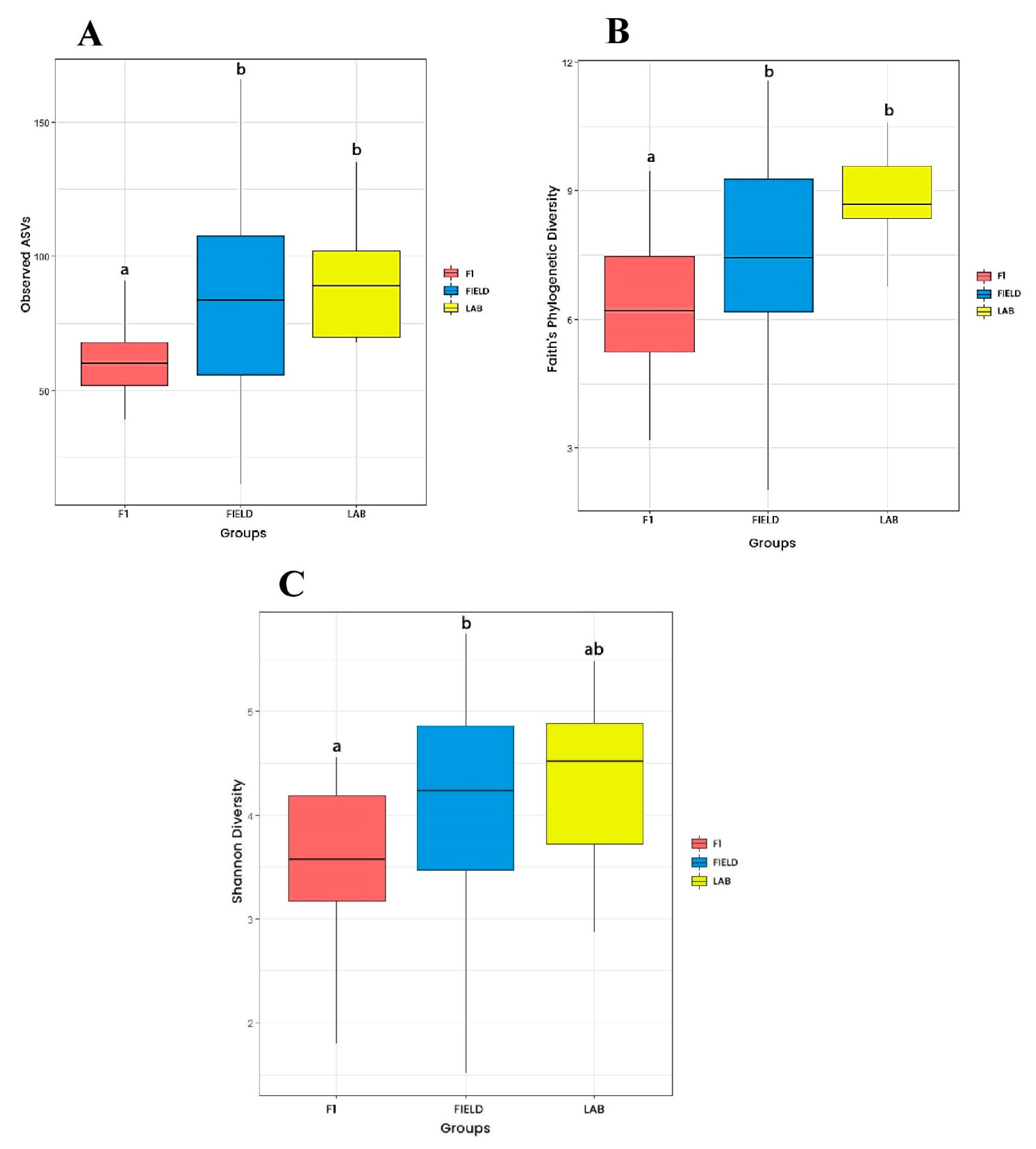
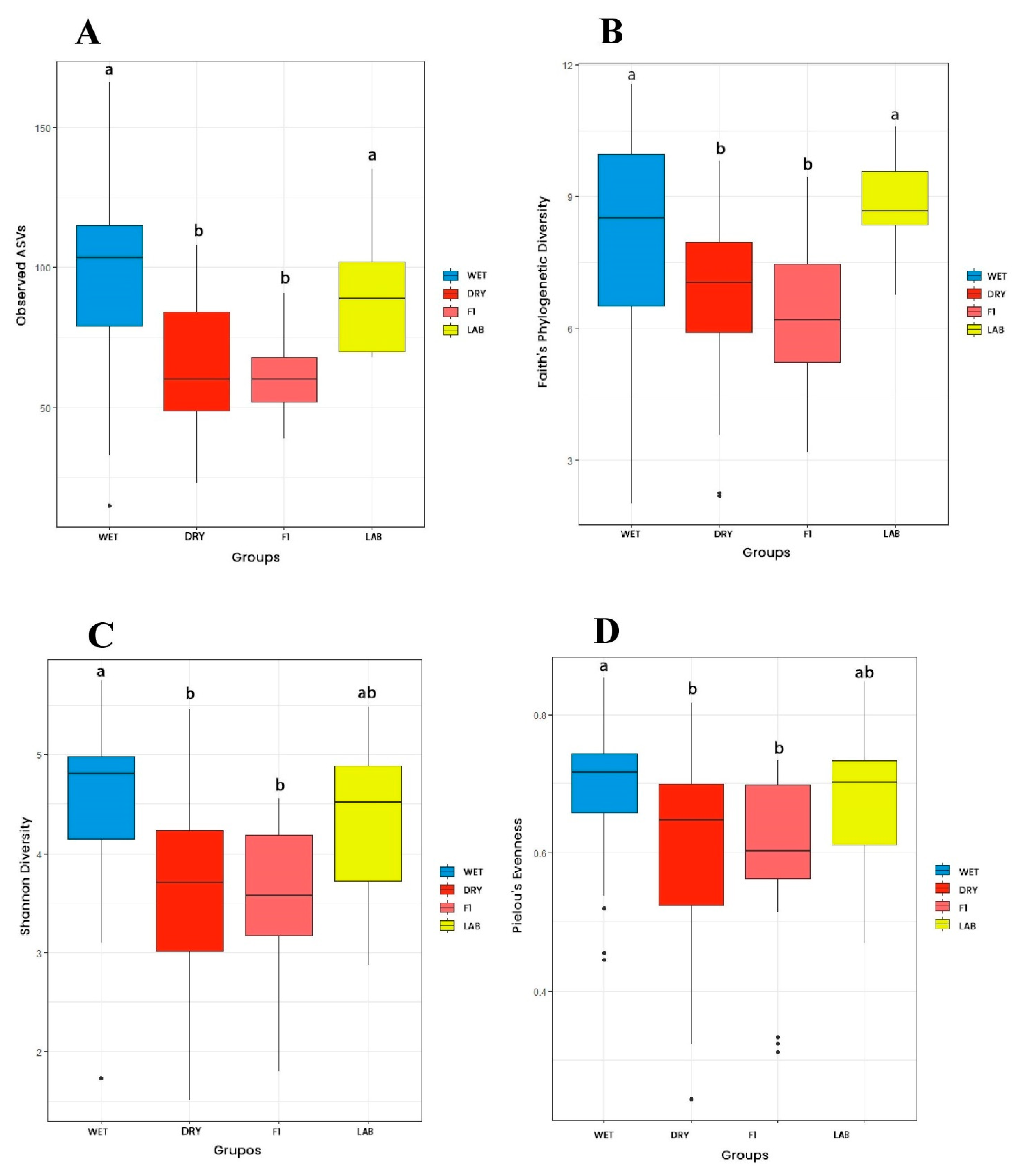
Regarding origin, Faith’s index and observed ASVs were higher for FIELD and LAB groups when compared to the F1 group (Figure 2A,B). The Shannon index was higher for FIELD in comparison to F1 samples, whereas LAB did not differ from both groups (Figure 2C). Considering collection season, all four indices were higher for the microbiota of Ae. albopictus from WET in comparison to those from the DRY season (Figure 3). Observed ASVs and Faith’s index were higher for LAB in relation to F1 mosquitoes (Figure 3A,B). In general, microbiota diversity did not vary significantly considering the collection area nor the population of Ae. albopictus. Significant differences were noticed only for the Faith’s index, with lower phylogenetic diversity of JDG-F1 when compared to LAB and RPC. Regarding population, a reduction in phylogenetic diversity was detected in JDG and JU mosquito microbiota in relation to the LAB group. Statistics can be found in figure legends, whereas pairwise comparisons for the four indices can be found in Table S4.

3.2.2. Beta Diversity

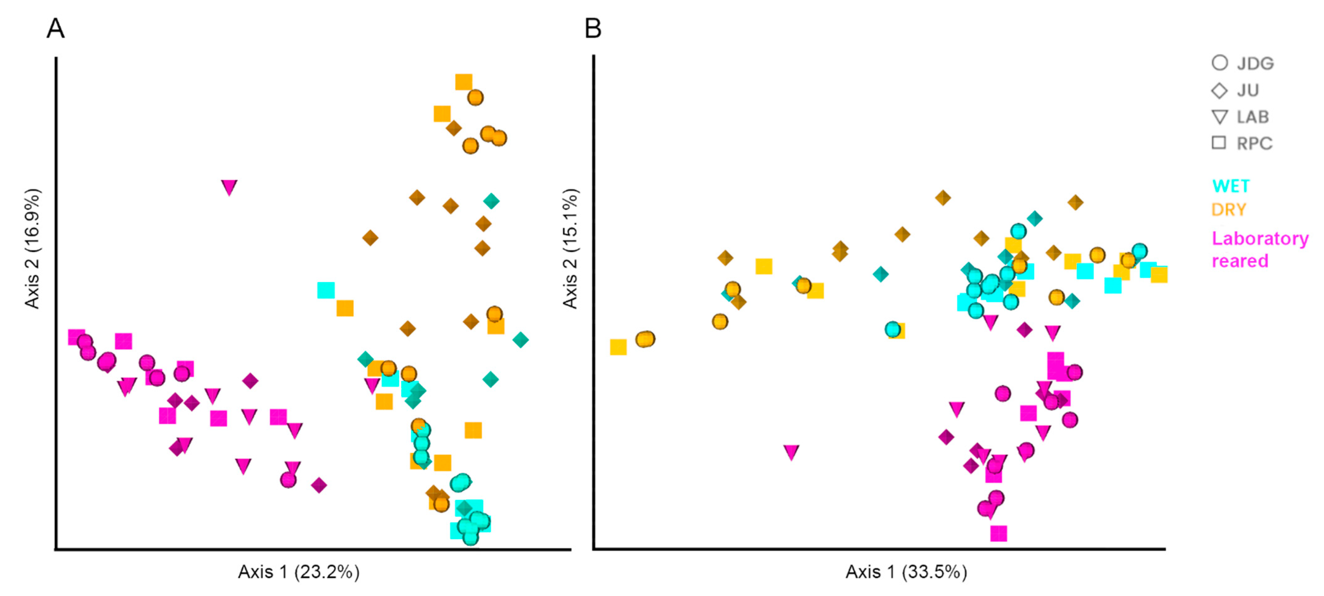
The PERMANOVA analysis suggested a significant influence of all variables in gut microbiota composition, but pairwise comparisons only detected significant variations according to mosquito origin and collection season (Table S5), with a distinction between field vs. laboratory samples (F1 vs. FIELD Bray–Curtis PERMANOVA: pseudo-F = 20, q-value = 0.001; LAB vs. FIELD Bray–Curtis PERMANOVA: pseudo-F = 8.3, q-value = 0.001) and wet x dry season samples (WET vs. DRY Weighted Unifrac PERMANOVA: pseudo-F = 6.1, q-value = 0.001). The PCoA corroborates the pairwise test from PERMANOVA showing clustering between mosquito gut samples from the laboratory (F1 from the three field populations and LAB) versus those from field mosquitoes, regardless of collection area. Moreover, it is also possible to notice a cluster of samples from the WET season, whereas the DRY samples are more dispersed in the plot, suggesting a higher heterogeneity in the microbiota composition of mosquitoes from this group (Figure 4).

3.3. Aedes albopictus Microbiota Taxonomic Composition

In total, more than 100 bacterial genera were detected through 16S rDNA sequencing. However, the top 10 most abundant phylum and top 12 most abundant family/genera usually comprised >75% of sequences (Figure 5 and Figures S2 and S3). Proteobacteria was the predominant phylum in the microbiota of almost all Ae. albopictus females, with a mean relative abundance per group ranging from 66 to 88%, followed by Firmicutes, Actinobacteria and Bacteroidetes (Figure S2). Acetobacteraceae, Vibrio, Wolbachia, Sphingomonas and Enterobacteriaceae were, in general, the most abundant taxa in the midgut of Ae. albopictus (Figure 5).
Acetobacteraceae was abundant only in laboratory mosquitoes (LAB and F1 of all populations), with an average abundance of 48, 45, 33 and 27% in JDG-F1, RPC-F1, LAB and JU-F1, respectively, and showed a much lower average abundance (<0.2%) in field-collected mosquitoes (JDG, JU and RPC) (Figure 5). In contrast, Wolbachia was detected in most field mosquitoes, regardless of the collection season, whereas it was only detected in three of ten samples from the LAB group (Figure 5). The presence of Wolbachia varied among field mosquitoes, but when detected, it was often one of the most abundant taxa. JDG had 6/19 samples with Wolbachia, with abundance ranging from 0.2% to 77.6% and an average abundance of 13.6% considering all gut samples. Wolbachia was detected in 14/19 of JU samples, varying from 0.8% to 55% of sequences (17.2% in average). The RPC group had the lowest Wolbachia average abundance (6.7%), with the widest variation in relative abundance between positive samples: 0.7% to 84.5%. This symbiont was detected in 7/20 of RPC samples. Wolbachia was more abundant in mosquitoes collected during the DRY season than those captured during the WET season (Figure 5).
Based on PERMANOVA results, we ran ANCOM according to sample origin and collection season. The ANCOM for mosquito origin corroborated the presence of Acetobacteraceae, as well as Rahnella and an unclassified Alphaproteobacteria, as characteristic of F1 and LAB mosquitoes. Twelve of thirteen bacteria were considered differentially abundant in LAB samples, six of them exclusive of this group (Table 3). Regarding collection season, Acetobacteraceae was also differentially abundant taxa in F1, LAB and DRY samples (Table 4). Wolbachia was considered a signature of DRY, WET and LAB samples, whereas it was less abundant in the F1 group, reinforcing the results from Figure 5A (Table 4). Furthermore, Ralstonia and Methylobacterium were pointed out as typical microbiota from mosquitoes collected during the wet season (Table 4).

3.4. Aedes albopictus Core Microbiota

Aedes albopictus core microbiota was composed of four bacterial families (Enterobacteriaceae, Moraxellaceae, Pseudomonadaceae and Halomonadaceae) and one order (Actinomycetales) (Table S6). The core microbiota of field mosquitoes (JDG, JU and RPC from both seasons) was composed of Bacillaceae, Propionibacteriaceae, Sphingomonadaceae, Streptococcaceae and Vibrionaceae, whereas the core microbiota of laboratory Ae. albopictus (F1 from all areas and LAB) was only composed of Acetobacteraceae. The JDG and JU core had Vibrionaceae as the most dominant bacterial taxa; the JU core had also Xanthomonadaceae, whereas the RPC core was composed only of Bacillaceae (Table S6).

3.5. Vector Competence to ZIKV

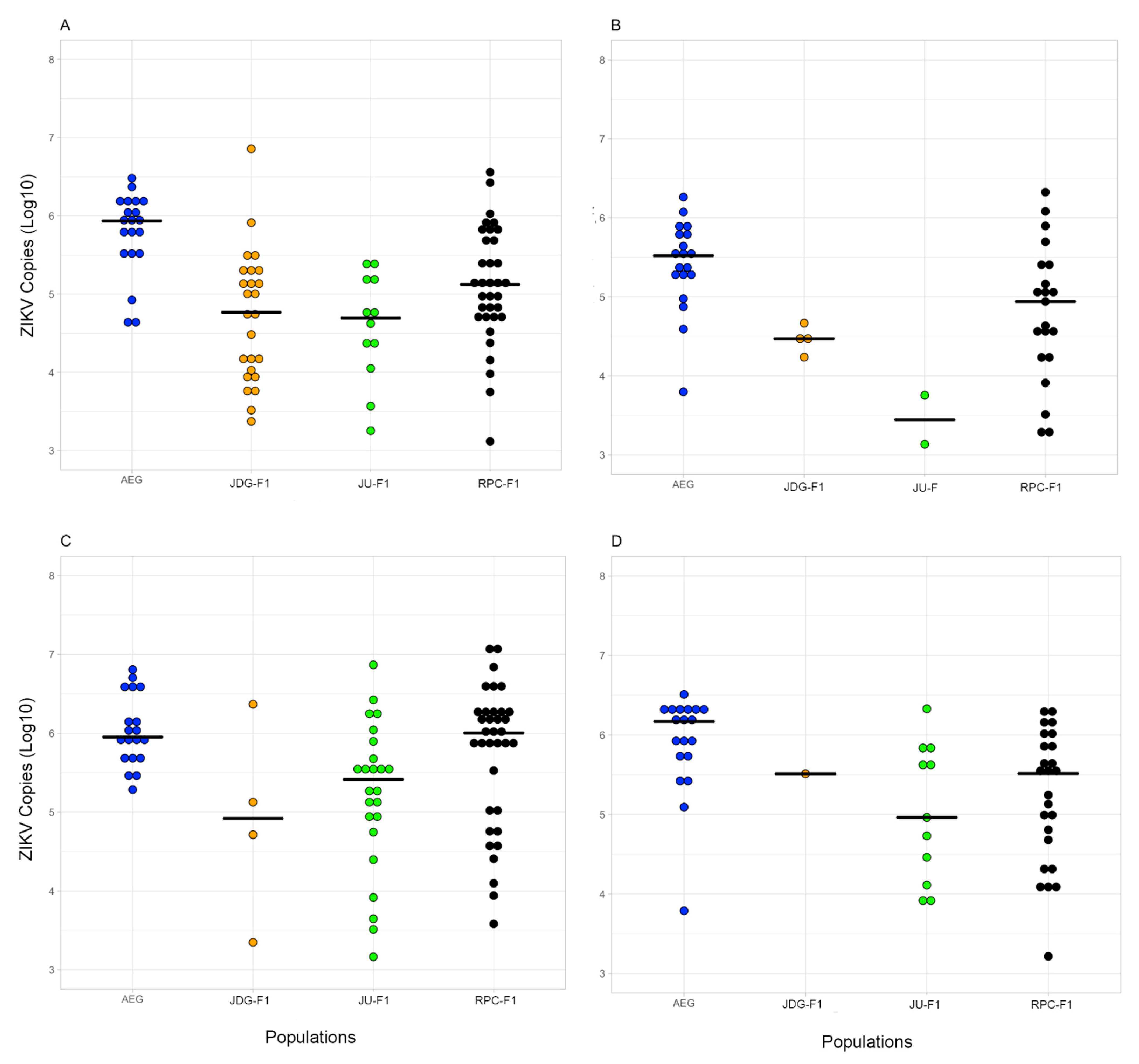
Approximately 600 Ae. albopictus females from each population and 200 from Ae. aegypti were exposed to ZIKV in two independent experimental infections. The infection rates ranged from 30 to 85.4%, while the dissemination rates ranged from 16 to 71.4% in Ae. albopictus (Table 5). Considering the IR, a significant population effect was observed on the proportion of infected insects (RR for JU: 0.42, RR 95% CI: 0.20–0.89, p-value = 0.02; RR for RPC: 3.97, RR 95% CI: 1.70–9.29, p-value = 0.001), whereas there was no difference between 14 and 21 dpi (RR: 1.02, RR 95% CI: 0.93–1.12, p-value = 0.61). Similarly, populations varied in DR, with markedly higher values for RPC at both dpi (RR for JU: 1.88, RR 95% CI: 0.53–6.63, p-value = 0.33; RR for RPC: 8.09, RR 95% CI: 2.65–24.74, p-value < 0.001). There was no significant difference between 14 and 21 dpi (RR: 1.09, RR 95% CI: 0.97–1.23, p-value = 0.13). In general, the number ZIKV copies was highly heterogeneous in the analyzed mosquitoes irrespective of their localities, with variations in up to three orders of magnitude. However, considering 14 and 21 dpi, there was no significant difference in viral load in the body and head of females between the three populations of Ae. albopictus tested (Kruskal–Wallis p-value > 0.05) (Figure 6A–D).

4. Discussion

Aedes albopictus gut microbiota diversity and composition was determined according to collection area and season, origin and population. Our results evidenced that bacterial diversity levels decreased when mosquitoes were reared for one generation in the laboratory (F1) in comparison to field-collected specimens, irrespective of Ae. albopictus population. Diversity was also lower in mosquitoes collected during the dry season in comparison to those from the wet season. On the other hand, diversity levels were similar between field Ae. albopictus and those from a lab colony with >30 generations in the insectary (LAB). Altogether, our data suggest that mosquito microbiota was mainly influenced by the environment, and the ZIKV infection and dissemination rates were not related to gut microbiota composition.
Investigations on the diversity of the microbiota of Ae. albopictus have been executed worldwide, especially in Asia, Europe and the United States [67,68,69,70,71]. However, few were conducted in Latin America, including Brazil. At the phylum level, Proteobacteria, Firmicutes, Actinobacteria and Bacteroidetes were the most dominant phyla, as they often are for different mosquito species [27,35,36,72,73,74], including in adults and larvae of Ae. albopictus [36,75,76,77]. Proteobacteria was the most abundant phylum, regardless of whether the mosquitoes were from the field or laboratory, which is in line with what has been described for Chinese and Brazilian Ae. albopictus populations [68,76,78].
The variability in mosquito gut microbiota has been linked to larval breeding site and/or habitat factors [77]. Despite this, microbiota diversity and composition were not significantly different here between female Ae. albopictus collected in areas with different landscapes. Although geographical variations in Ae. albopictus microbiota composition have been described in Hawaii and China [68,79], other studies pointed to a weak or no contribution of collection area to diversity levels or microbiota composition of these insects [77,80,81]. For example, there was no correlation between microbiota diversity and the level of urbanization for populations from the island of O’ahu (Hawaii) [70]. On the other hand, the microbiota of An. coluzzi and Ae. aegypti were more diverse in mosquitoes collected in urban areas than in those collected in rural areas [82,83]. Such contrasting findings could be explained both by ecological and/or geographic factors. Aedes albopictus exhibits high ecological plasticity, colonizing a variety of natural and artificial breeding sites [7,8]. It is frequently found in urban environments and artificial containers in China [84], while it is considered a forest edge mosquito in Brazil [11]. Thus, the similarity between the microbiota of Ae. albopictus seen here could be due to similar breeding sites commonly available in vegetated areas from the three sampled sites. Moreover, the absence of a geographic signature in the microbiota could be also explained by the proximity of collection sites in this study (up to 20 km). In contrast, the gut microbiota of six different mosquito species, including Ae. albopictus, collected across eight areas separated from each other by at least 47 km in China, was correlated with geographic origin.
We observe a higher richness (observed ASVs), diversity (Faith, Shannon), evenness (Pielou) and heterogeneity in the microbiota of mosquitoes collected during the wet season than in those sampled during the dry season. This indicates a temporal instability of the mosquito microbiota, possibly by changes in climatic conditions and/or environmental microbes that can colonize the mosquito’s midgut [34,85]. Our experimental design cannot confirm these hypotheses, but laboratory experiments have indicated an increase in heat-tolerant taxa in the microbiota of mosquitoes submitted to higher environmental temperatures [86,87]. Monitoring the temperature and the microbiota of wild mosquitoes over time would help our understanding of the influence of this variable in mosquito-associated microbe diversity.
We noticed significant differences in the richness, diversity levels and composition of gut microbiota according to mosquito origin (field, F1 and LAB). This lower diversity and richness in the microbiota after laboratory colonization are in line with previous studies with Ae. albopictus [88,89,90]. The sudden change of environment from the field to the laboratory may have caused a remodeling in gut microbiota composition, as F1 and LAB specimens had a more similar gut microbiota composition to each other than to insects from the field (Figure 4). This microbiota shift might also have resulted from the loss of genetic variability after mosquito colonization [91] and/or standardized diet (10% glucose) and rearing conditions in the insectary. Even though wild mosquitoes were processed without visible blood in the midgut, we had no control over their diet in the field. Diet and blood sources affect gut microbiota diversity [92]: field-collected Ae. aegypti fed on human blood showed a greater diversity and richness in their microbiota compared to those that fed on non-human blood sources or were non-blood fed [93]. Moreover, we do not know the age of the wild mosquitoes, while the laboratory-reared specimens were processed at 6–7 days old. Age impacts the microbiota diversity of Ae. albopictus, favoring specific taxa according to the physiological state of the mosquito (newly emerged, young, older) [94].
Microbiota diversity levels were similar between wild Ae. albopictus and those from a colony with >30 generations of laboratory rearing (LAB) in terms of number of ASVs. Over generations, the insect gut tends to be colonized by bacterial taxa adapted to the type of food, rearing water and controlled temperature and humidity conditions of an insectary [94]. Thus, after some time, microbiota diversity would reach levels similar to field mosquitoes (but with a distinct composition). Studies comparing field populations with their respective laboratory colonies over several generations are needed to test this hypothesis and determine whether and when microbiota diversity levels would be restored.
Changes in microbiota after mosquito colonization may have implications on vector competence studies. As microbiota can influence vector susceptibility to pathogens, the infection pattern of laboratory-reared mosquitoes might not reflect that of field mosquitoes. It is laborious to carry out vector competence experiments with wild-caught adult Aedes due to the large number of insects required [95], unknown mosquito age and physiological stage and low survival and blood-fed success caused by capture physical stress. An alternative way to produce mosquitoes with a more similar microbiota than their wild counterparts would be their rearing in the laboratory with natural breeding site water. This methodology allowed the conservation of ~50% of the bacterial families found in field mosquitoes in the microbiota of An. gambiae (F10) from Ghana [96].
There was a significant effect of Ae. albopictus population on the IR and DR of ZIKV, with markedly higher values for RPC at both 14 and 21 dpi. We have no evidence that this variation is related to the microbiota, since gut bacterial composition was similar between F1 mosquitoes from the three populations (Figure 4). However, we must consider that the microbiota was investigated in a sample of mosquitoes from the same batch of those exposed to experimental infections, i.e., we do not have ZIKV and microbiota information for the same specimens. Thus, we cannot exclude the possibility that the individual heterogeneity seen in gut bacterial composition may have impacted virus establishment in the vector. As we intended to identify possible gut symbionts related to virus establishment and dissemination in the mosquito, we opted to characterize the microbiota in mosquitoes before virus exposure, otherwise it would be elusive whether potential differences in gut bacterial diversity and composition between F1 mosquitoes from the three areas are a cause or consequence of ZIKV infection [97].
The interaction between microorganisms can modulate the microbiota composition of mosquitoes [88,98]. The Acetobacteraceae family of bacteria was more prevalent and relatively abundant in LAB and F1 mosquitoes, while Wolbachia was more prevalent and relatively abundant in FIELD mosquitoes, suggesting a possible co-exclusion interaction between these bacterial taxa. Acetobacteraceae are acetic acid bacteria (AAB), Gram-negative aerobic bacteria that carry out the oxidation of ethanol into acetic acid [99]. AAB are often found in insects with diets rich in sugar, colonizing different organs and tissues, including the midgut. They grow in acidic pH and produce an extracellular matrix of polysaccharides, which allow their contact with the intestinal epithelium without activating the host immune system [100]. Our laboratory colonies were fed with sugar solutions which were probably the source of acquisition and transmission of AAB between mosquitoes [101].
Wolbachia is an obligate intracellular bacterium that naturally colonizes >40% of insect species [102]. Aedes albopictus naturally harbors two Wolbachia strains, wAlbA and wAlbB [103], that are in higher density in the gonads than in the mosquito’s midgut [104]. A negative correlation between Asaia and Wolbachia was previously described in the reproductive tissues of Ae. albopictus, Ae. aegypti, An. gambiae and Cx. quinquefasciatus [105,106]. In our work, we used laboratory-reared females fed only with 10% glucose solution and processed at 6–7 days old. In this scenario, the molecular mechanisms by which these bacteria interact still need to be elucidated.
Despite the possible co-exclusion interaction between Acetobacteraceae and Wolbachia, some LAB specimens exhibited a considerable abundance of both bacteria. This may be due to the longer period of colonization in the laboratory, which might have facilitated the establishment of new bacterial taxa from this environment in the gut and/or changes in the interactions between microorganisms, favoring the reestablishment of Wolbachia levels even in the presence of Acetobacteraceae. This hypothesis is supported by the higher diversity levels of LAB Ae. albopictus in relation to the F1, in which the increase in Acetobacteraceae possibly favored the decrease in Wolbachia. Nevertheless, the individual variation in Wolbachia relative abundance in mosquitoes from the same group could also be explained by host and/or environmental characteristics, as well as technical issues. We worked with the mosquitoes’ midguts, where Wolbachia density is usually low, which could explain why it was undetectable in some samples. In addition, the ages of field-collected mosquitoes could have influenced Wolbachia density, as older Ae. albopictus have lower densities of both wAlbA and B strains [107,108]. Although these Wolbachia strains are resistant to temperature increase [109] and there was no differential abundance of Wolbachia between the two collection seasons, environmental stresses from the field could also affect the endosymbiont density. Finally, even if we avoided blood-fed mosquitoes in which the microbiota multiplies up to a thousand times [110], fluctuations in gut microbiota load may have hindered the detection of Wolbachia in samples whose symbiont density was already low.
The core microbiota of adult Ae. albopictus from all groups was formed by the bacterial families Enterobacteriaceae, Halomonadaceae, Moraxellaceae and Pseudomonadaceae and the order Actinomycetales. Enterobacteriaceae comprises genera often associated with Ae. Albopictus, such as Enterobacter, Klebsiella and Pantoea [79,85,94,111]. Halomonadaceae was mainly from the Halomonas genus, a halotolerant bacterium (i.e., that survives in saline environments [112]) previously detected in the gut microbiota of field Ae. albopictus from the United States [88]. Pseudomonadaceae and Moraxellaceae families were mainly from Pseudomonas and Acinetobacter genera, respectively, which are also commonly found in the microbiota core of different mosquito vector species, such as Ae. aegypti, Anopheles spp. and Ae. albopictus [72,73,75,113,114,115]. Once acquired, Pseudomonas colonizes the intestine of mosquitoes and increases in abundance after blood-feeding [92,94], as it resists the oxidative stress caused by the heme from red blood cells [72]. It can be found in breeding water and in all stages of mosquito development, with evidence of transtadial transmission, since it was detected in the Malpighian tubules [116]. Acinetobacter is not only found in the gut of mosquitoes but also in breeding sites and food sources such as vertebrate host skin and plants. It may be involved with important functions for the physiology of Ae. albopictus, such as blood digestion and the assimilation of sugars from nectar [117]. Lastly, bacteria from the Actinomycetales order are found in soils and aquatic habitats [118,119,120], including domestic water storage containers holding Ae. aegypti larvae [121]. Actinomycetes can have antimicrobial properties and larvicidal activity, e.g., the genus Streptomyces has recently been indicated as a potential biolarvicide against Ae. albopictus [122].
The core microbiota analysis corroborates the influence of the environment on the microbiota of mosquitoes discussed above: the microbiota core of laboratory mosquitoes (F1 + LAB) only had Acetobacteraceae, while the core of field mosquitoes had Bacillaceae, Propionibacteriaceae, Sphingomonadaceae, Streptococcaceae and Vibrionaceae families. These bacterial taxa are commonly found in the environment, associated with the breeding site water or soil (e.g., Bacillaceae, Sphingomonadaceae and Vibrionaceae) [123,124,125]. Some taxa are associated with the human skin and mucosa [126], such as Propionibacterium (Propionibacteriaceae) [127] and Streptococcaceae, and could possibly be acquired by Ae. albopictus during blood-feeding or contacting materials manipulated by humans.

5. Conclusions

This study provides insights into mosquito–microbiota interactions. We explored the variations in Ae. albopictus gut bacterial diversity and composition according to collection area and season, origin and population. Our results indicated that mosquitoes collected during the wet season had a more diverse microbiota than those collected during the dry season, and field mosquitoes (irrespective of the collection area) presented a different microbiota composition than those reared in the laboratory (F1 and LAB). Richness and diversity of the gut bacterial communities were greater in field mosquitoes compared with mosquitoes reared for only one generation in the laboratory (F1). We detected significant differences in ZIKV virus infection and dissemination rates between the three different F1 populations of Ae. albopictus, which is probably not related to microbiota composition as it was similar between them. A possible co-exclusion interaction between Wolbachia and Acetobacteraceae was detected, with the former being absent or undetectable in F1 mosquitoes, while the latter was frequently the most dominant taxa. Future studies concerning mosquito microbe–microbe interactions at the molecular level are necessary to disentangle the network of bacterial interactions that shapes mosquito microbiota.

Supplementary Materials

The following supporting information can be downloaded at: https://www.mdpi.com/article/10.3390/v15061309/s1, Figure S1: Rarefaction curves; Figure S2: Relative abundance of bacterial taxa per sample at phylum level, Figure S3: Relative abundance of bacterial taxa per sample at family level, Table S1: Midgut samples sent for 16S rDNA sequencing; Table S2: Sequencing parameters; Table S3: ASVs removed after decontamination; Table S4: Pairwise comparisons for the four alpha diversity indices; Table S5: Pairwise comparisons for the two beta diversity indices; Table S6: Core microbiota of Aedes albopictus.

Author Contributions

Conceptualization, J.M.C.B. and M.R.D.; methodology: J.M.C.B., M.G.P., J.C.-A., D.C.-L. and M.R.D.; formal analysis, J.M.C.B. and M.R.D.; resources, M.R.D., M.G.P. and R.M.-d.-F.; writing—original draft preparation, J.M.C.B. and M.R.D.; writing—review and editing, J.M.C.B., M.G.P., J.C.-A., D.C.-L., R.M.-d.-F. and M.R.D.; funding acquisition, M.R.D. All authors have read and agreed to the published version of the manuscript.

Funding

This work was carried out with the support of the Coordenação de Aperfeiçoamento Pessoal de Nível Superior (CAPES), Brazil (Financing Code 001); Edital Universal CNPq, 2018 (429625/2018-9); and Inova Fiocruz/Fundação Oswaldo Cruz, Edital Geração de Conhecimento—Novos Talentos 2018 (VPPCB-008-FIO-18-2-36).

Institutional Review Board Statement

Not applicable.

Informed Consent Statement

Not applicable.

Data Availability Statement

Raw read sequences are stored in Sequence Read Archive (SRA) in fastq format (accession: PRJNA951432).

Acknowledgments

We thank Mauro Menezes, Marcelo Celestino and Renato Carvalho† for contributions to the fieldwork. We are grateful to the Plataforma de Sequenciamento de Alto Desempenho from Instituto Oswaldo Cruz (IOC–FIOCRUZ) for all the support in the library construction and sequencing. We also thank the moderator’s team from the Qiime2 forum (https://forum.qiime2.org/ accessed on 24 May 2022) for insightful comments and suggestions in bioinformatic analyses. In memory of Renato Carvalho.

Conflicts of Interest

The authors declare no conflict of interest.

References

  1. Harbach, R.E. Mosquito Taxonomic Inventory. Available online: https://mosquito-taxonomic-inventory.myspecies.info (accessed on 6 September 2022).
  2. Weaver, S.C.; Reisen, W.K. Present and Future Arboviral Threats. Antivir. Res. 2010, 85, 328–345. [Google Scholar] [CrossRef] [PubMed]
  3. Wu, J.-Y.; Lun, Z.-R.; James, A.A.; Chen, X.-G. Dengue Fever in Mainland China. Am. J. Trop. Med. Hyg. 2010, 83, 664. [Google Scholar] [CrossRef]
  4. Lazzarini, L.; Barzon, L.; Foglia, F.; Manfrin, V.; Pacenti, M.; Pavan, G.; Rassu, M.; Capelli, G.; Montarsi, F.; Martini, S.; et al. First Autochthonous Dengue Outbreak in Italy, August 2020. Eurosurveillance 2020, 25, 2001606. [Google Scholar] [CrossRef] [PubMed]
  5. Monge, S.; García-Ortúzar, V.; López Hernández, B.; Lopaz Pérez, M.Á.; Delacour-Estrella, S.; Sánchez-Seco, M.P.P.; Fernández Martinez, B.; García San Miguel, L.; García-Fulgueiras, A.; Sierra Moros, M.J.J. Characterization of the First Autochthonous Dengue Outbreak in Spain (August–September 2018). Acta Trop. 2020, 205, 105402. [Google Scholar] [CrossRef]
  6. Pagès, F.; Peyrefitte, C.N.; Mve, M.T.; Jarjaval, F.; Brisse, S.; Iteman, I.; Gravier, P.; Nkoghe, D.; Grandadam, M. Aedes albopictus Mosquito: The Main Vector of the 2007 Chikungunya Outbreak in Gabon. PLoS ONE 2009, 4, e4691. [Google Scholar] [CrossRef]
  7. Pereira-Dos-Santos, T.; Roiz, D.; Lourenço-De-Oliveira, R.; Paupy, C. A Systematic Review: Is Aedes albopictus an Efficient Bridge Vector for Zoonotic Arboviruses? Pathogens 2020, 9, 266. [Google Scholar] [CrossRef]
  8. Bonizzoni, M.; Gasperi, G.; Chen, X.; James, A.A. The Invasive Mosquito Species Aedes albopictus: Current Knowledge and Future Perspectives. Trends Parasitol. 2013, 29, 460–468. [Google Scholar] [CrossRef] [PubMed]
  9. Ferreira-de-Lima, V.H.; Câmara, D.C.P.; Honório, N.A.; Lima-Camara, T.N. The Asian Tiger Mosquito in Brazil: Observations on Biology and Ecological Interactions since Its First Detection in 1986. Acta Trop. 2020, 205, 105386. [Google Scholar] [CrossRef]
  10. Kamgang, B.; Nchoutpouen, E.; Simard, F.; Paupy, C. Notes on the Blood-Feeding Behavior of Aedes albopictus (Diptera: Culicidae) in Cameroon. Parasites Vectors 2012, 5, 57. [Google Scholar] [CrossRef]
  11. Pereira dos Santos, T.; Roiz, D.; Santos de Abreu, F.V.; Luz, S.L.B.; Santalucia, M.; Jiolle, D.; Santos Neves, M.S.A.; Simard, F.; Lourenço-de-Oliveira, R.; Paupy, C. Potential of Aedes albopictus as a Bridge Vector for Enzootic Pathogens at the Urban-Forest Interface in Brazil. Emerg. Microbes Infect. 2018, 7, 1–8. [Google Scholar] [CrossRef]
  12. Medley, K.A.; Westby, K.M.; Jenkins, D.G. Rapid Local Adaptation to Northern Winters in the Invasive Asian Tiger Mosquito Aedes albopictus: A Moving Target. J. Appl. Ecol. 2019, 56, 2518–2527. [Google Scholar] [CrossRef]
  13. Lambrechts, L.; Scott, T.W.; Gubler, D.J. Consequences of the Expanding Global Distribution of Aedes albopictus for Dengue Virus Transmission. PLoS Negl. Trop. Dis. 2010, 4, e646. [Google Scholar] [CrossRef] [PubMed]
  14. Fernandes, R.S.; O’connor, O.; Bersot, M.I.L.; Girault, D.; Dokunengo, M.R.; Pocquet, N.; Dupont-Rouzeyrol, M.; Lourenço-De-oliveira, R. Vector Competence of Aedes aegypti, Aedes albopictus and Culex quinquefasciatus from Brazil and New Caledonia for Three Zika Virus Lineages. Pathogens 2020, 9, 575. [Google Scholar] [CrossRef] [PubMed]
  15. Bezerra, J.M.T.; Araújo, R.G.P.; Melo, F.F.; Gonçalves, C.M.; Chaves, B.A.; Silva, B.M.; Silva, L.D.; Brandão, S.T.; Secundino, N.F.C.; Norris, D.E.; et al. Aedes (Stegomyia) Albopictus’ Dynamics Influenced by Spatiotemporal Characteristics in a Brazilian Dengue-Endemic Risk City. Acta Trop. 2016, 164, 431–437. [Google Scholar] [CrossRef]
  16. Ferreira-De-Lima, V.H.; Lima-Camara, T.N. Natural Vertical Transmission of Dengue Virus in Aedes aegypti and Aedes albopictus: A Systematic Review. Parasit. Vectors 2018, 11, 77. [Google Scholar] [CrossRef]
  17. Cansado-Utrilla, C.; Zhao, S.Y.; Mccall, P.J.; Coon, K.L.; Hughes, G.L. The Microbiome and Mosquito Vectorial Capacity: Rich Potential for Discovery and Translation. Microbiome 2021, 9, 111. [Google Scholar] [CrossRef]
  18. Xi, Z.; Ramirez, J.L.; Dimopoulos, G. The Aedes aegypti Toll Pathway Controls Dengue Virus Infection. PLoS Pathog. 2008, 4, e1000098. [Google Scholar] [CrossRef]
  19. Dong, Y.; Manfredini, F.; Dimopoulos, G. Implication of the Mosquito Midgut Microbiota in the Defense against Malaria Parasites. PLoS Pathog. 2009, 5, e1000423. [Google Scholar] [CrossRef]
  20. Cirimotich, C.M.; Dong, Y.; Clayton, A.M.; Sandiford, S.L.; Souza-Neto, J.A.; Mulenga, M.; Dimopoulos, G. Natural Microbe-Mediated Refractoriness to Plasmodium Infection in Anopheles gambiae. Science 2011, 332, 855–858. [Google Scholar] [CrossRef] [PubMed]
  21. Apte-Deshpande, A.D.; Paingankar, M.S.; Gokhale, M.D.; Deobagkar, D.N. Serratia Odorifera Mediated Enhancement in Susceptibility of Aedes aegypti for Chikungunya Virus. Indian J. Med. Res. 2014, 139, 762–768. [Google Scholar] [PubMed]
  22. Caragata, E.P.; Tikhe, C.V.; Dimopoulos, G. Curious Entanglements: Interactions between Mosquitoes, Their Microbiota, and Arboviruses. Curr. Opin. Virol. 2019, 37, 26–36. [Google Scholar] [CrossRef]
  23. Saraiva, R.G.; Dimopoulos, G. Bacterial Natural Products in the Fight against Mosquito-Transmitted Tropical Diseases. Nat. Prod. Rep. 2020, 37, 338–354. [Google Scholar] [CrossRef]
  24. Charan, S.S.; Pawar, K.D. Comparative Analysis of Midgut Bacterial Communities of Aedes aegypti Mosquito Strains Varying in Vector Competence to Dengue Virus. Parasitol. Res. 2013, 112, 2627–2637. [Google Scholar] [CrossRef] [PubMed]
  25. Charan, S.S.; Pawar, K.D.; Gavhale, S.D.; Tikhe, C.V.; Charan, N.S.; Angel, B.; Joshi, V.; Patole, M.S.; Shouche, Y.S. Comparative Analysis of Midgut Bacterial Communities in Three Aedine Mosquito Species from Dengue-Endemic and Non-Endemic Areas of Rajasthan, India. Med. Vet. Entomol. 2016, 30, 264–277. [Google Scholar] [CrossRef] [PubMed]
  26. Arévalo-Cortés, A.; Damania, A.; Granada, Y.; Zuluaga, S.; Mejia, R.; Triana-Chavez, O. Association of Midgut Bacteria and Their Metabolic Pathways with Zika Infection and Insecticide Resistance in Colombian Aedes aegypti Populations. Viruses 2022, 14, 2197. [Google Scholar] [CrossRef]
  27. Coon, K.L.; Vogel, K.J.; Brown, M.R.; Strand, M.R. Mosquitoes Rely on Their Gut Microbiota for Development. Mol. Ecol. 2014, 23, 2727–2739. [Google Scholar] [CrossRef]
  28. Gaio, A.D.O.; Gusmão, D.S.; Santos, A.V.; Berbert-Molina, M.A.; Pimenta, P.F.P.; Lemos, F.J.A. Contribution of Midgut Bacteria to Blood Digestion and Egg Production in Aedes aegypti (Diptera: Culicidae) (L.). Parasit. Vectors 2011, 4, 105. [Google Scholar] [CrossRef]
  29. Guégan, M.; Tran Van, V.; Martin, E.; Minard, G.; Tran, F.H.; Fel, B.; Hay, A.E.; Simon, L.; Barakat, M.; Potier, P.; et al. Who Is Eating Fructose within the Aedes albopictus Gut Microbiota? Environ. Microbiol. 2020, 22, 1193–1206. [Google Scholar] [CrossRef] [PubMed]
  30. Rodgers, F.H.; Gendrin, M.; Wyer, C.A.S.; Christophides, G.K. Microbiota-Induced Peritrophic Matrix Regulates Midgut Homeostasis and Prevents Systemic Infection of Malaria Vector Mosquitoes. PLoS Pathog. 2017, 13, e1006391. [Google Scholar] [CrossRef]
  31. Bascuñán, P.; Niño-Garcia, J.P.; Galeano-Castañeda, Y.; Serre, D.; Correa, M.M. Factors Shaping the Gut Bacterial Community Assembly in Two Main Colombian Malaria Vectors. Microbiome 2018, 6, 146. [Google Scholar] [CrossRef]
  32. Muturi, E.J.; Lagos-Kutz, D.; Dunlap, C.; Ramirez, J.L.; Rooney, A.P.; Hartman, G.L.; Fields, C.J.; Rendon, G.; Kim, C.H. Mosquito Microbiota Cluster by Host Sampling Location. Parasites Vectors 2018, 11, 468. [Google Scholar] [CrossRef]
  33. Bogale, H.N.; Cannon, M.V.; Keita, K.; Camara, D.; Barry, Y.; Keita, M.; Coulibaly, D.; Kone, A.K.; Doumbo, O.K.; Thera, M.A.; et al. Relative Contributions of Various Endogenous and Exogenous Factors to the Mosquito Microbiota. Parasites Vectors 2020, 13, 619. [Google Scholar] [CrossRef] [PubMed]
  34. Pérez-Ramos, D.W.; Ramos, M.M.; Payne, K.C.; Giordano, B.V.; Caragata, E.P. Collection Time, Location, and Mosquito Species Have Distinct Impacts on the Mosquito Microbiota. Front. Trop. Dis. 2022, 3, 896289. [Google Scholar] [CrossRef]
  35. Buck, M.; Nilsson, L.K.J.; Brunius, C.; Dabiré, R.K.; Hopkins, R.; Terenius, O. Bacterial Associations Reveal Spatial Population Dynamics in Anopheles gambiae Mosquitoes. Sci. Rep. 2016, 6, 22806. [Google Scholar] [CrossRef] [PubMed]
  36. Coon, K.L.; Brown, M.R.; Strand, M.R. Mosquitoes Host Communities of Bacteria That Are Essential for Development but Vary Greatly between Local Habitats. Mol. Ecol. 2016, 25, 5806–5826. [Google Scholar] [CrossRef]
  37. Lindh, J.M.; Borg-Karlson, A.-K.; Faye, I. Transstadial and Horizontal Transfer of Bacteria within a Colony of Anopheles gambiae (Diptera: Culicidae) and Oviposition Response to Bacteria-Containing Water. Acta Trop. 2008, 107, 242–250. [Google Scholar] [CrossRef]
  38. Dillon, R.J.; Dillon, V.M. The Gut Bacteria of Insects: Nonpathogenic Interactions. Annu. Rev. Entomol. 2004, 49, 71–92. [Google Scholar] [CrossRef]
  39. Fridman, S.; Izhaki, I.; Gerchman, Y.; Halpern, M. Bacterial Communities in Floral Nectar. Environ. Microbiol. Rep. 2012, 4, 97–104. [Google Scholar] [CrossRef]
  40. Bassene, H.; Hadji, E.; Niang, A.; Fenollar, F.; Doucoure, S.; Faye, O.; Raoult, D.; Sokhna, C.; Mediannikov, O. Role of Plants in the Transmission of Asaia sp., which Potentially Inhibit the Plasmodium Sporogenic Cycle in Anopheles Mosquitoes. Sci. Rep. 2020, 10, 7144. [Google Scholar] [CrossRef]
  41. Honório, N.A.; Silva, W.D.C.; Leite, P.J.; Gonçalves, J.M.; Lounibos, L.P.; Lourenço-de-Oliveira, R. Dispersal of Aedes aegypti and Aedes albopictus (Diptera: Culicidae) in an Urban Endemic Dengue Area in the State of Rio de Janeiro, Brazil. Mem. Inst. Oswaldo Cruz 2003, 98, 191–198. [Google Scholar] [CrossRef]
  42. De Oliveira, S.; Villela, D.A.M.; Dias, F.B.S.; Moreira, L.A.; Maciel de Freitas, R. How Does Competition among Wild Type Mosquitoes Influence the Performance of Aedes aegypti and Dissemination of Wolbachia Pipientis? PLoS Negl. Trop. Dis. 2017, 11, e0005947. [Google Scholar] [CrossRef] [PubMed]
  43. INMET. Instituto Nacional de Meteorologia. Tabela de Dados Das Estações. Available online: https://tempo.inmet.gov.br/CondicoesRegistradas (accessed on 1 September 2019).
  44. Consoli, R.A.; Oliveira, R.L.D. Principais Mosquitos de Importância Sanitária No Brasil; Editora Fiocruz: Rio de Janeiro, RJ, Brazil, 1994. [Google Scholar]
  45. Eisenhofer, R.; Minich, J.J.; Marotz, C.; Cooper, A.; Knight, R.; Weyrich, L.S. Contamination in Low Microbial Biomass Microbiome Studies: Issues and Recommendations. Trends Microbiol. 2019, 27, 105–117. [Google Scholar] [CrossRef] [PubMed]
  46. Klindworth, A.; Pruesse, E.; Schweer, T.; Peplies, J.; Quast, C.; Horn, M.; Glöckner, F.O. Evaluation of General 16S Ribosomal RNA Gene PCR Primers for Classical and Next-Generation Sequencing-Based Diversity Studies. Nucleic Acids Res. 2013, 41, e1. [Google Scholar] [CrossRef]
  47. Illumina. 16S Metagenomic Sequencing Library; Illumina: San Diego, CA, USA, 2013; pp. 1–28. [Google Scholar]
  48. Illumina. BaseSpace Sequence Hub; Illumina: San Diego, CA, USA, 2017; pp. 1–6. [Google Scholar]
  49. Bolyen, E.; Rideout, J.R.; Dillon, M.R.; Bokulich, N.A.; Abnet, C.C.; Al-Ghalith, G.A.; Alexander, H.; Alm, E.J.; Arumugam, M.; Asnicar, F.; et al. Reproducible, Interactive, Scalable and Extensible Microbiome Data Science Using QIIME 2. Nat. Biotechnol. 2019, 37, 852–857. [Google Scholar] [CrossRef] [PubMed]
  50. Martin, M. Cutadapt Removes Adapter Sequences from High-Throughput Sequencing Reads. EMBnet. J. 2011, 17, 10–12. [Google Scholar] [CrossRef]
  51. Callahan, B.J.; McMurdie, P.J.; Rosen, M.J.; Han, A.W.; Johnson, A.J.A.; Holmes, S.P. DADA2: High-Resolution Sample Inference from Illumina Amplicon Data. Nat. Methods 2016, 13, 581–583. [Google Scholar] [CrossRef]
  52. McDonald, D.; Price, M.N.; Goodrich, J.; Nawrocki, E.P.; Desantis, T.Z.; Probst, A.; Andersen, G.L.; Knight, R.; Hugenholtz, P. An Improved Greengenes Taxonomy with Explicit Ranks for Ecological and Evolutionary Analyses of Bacteria and Archaea. ISME J. 2012, 6, 610–618. [Google Scholar] [CrossRef]
  53. Bokulich, N.A.; Kaehler, B.D.; Rideout, J.R.; Dillon, M.; Bolyen, E.; Knight, R.; Huttley, G.A.; Gregory Caporaso, J. Optimizing Taxonomic Classification of Marker-Gene Amplicon Sequences with QIIME 2’s Q2-Feature-Classifier Plugin. Microbiome 2018, 6, 90. [Google Scholar] [CrossRef]
  54. Camacho, C.; Coulouris, G.; Avagyan, V.; Ma, N.; Papadopoulos, J.; Bealer, K.; Madden, T.L. BLAST+: Architecture and Applications. BMC Bioinform. 2009, 10, 421. [Google Scholar] [CrossRef]
  55. Wang, Q.; Garrity, G.M.; Tiedje, J.M.; Cole, J.R. Naïve Bayesian Classifier for Rapid Assignment of RRNA Sequences into the New Bacterial Taxonomy. Appl. Environ. Microbiol. 2007, 73, 5261–5267. [Google Scholar] [CrossRef]
  56. McKnight, D.T.; Huerlimann, R.; Bower, D.S.; Schwarzkopf, L.; Alford, R.A.; Zenger, K.R. MicroDecon: A Highly Accurate Read-Subtraction Tool for the Post-Sequencing Removal of Contamination in Metabarcoding Studies. Environ. DNA 2019, 1, 14–25. [Google Scholar] [CrossRef]
  57. Salter, S.J.; Cox, M.J.; Turek, E.M.; Calus, S.T.; Cookson, W.O.; Moffatt, M.F.; Turner, P.; Parkhill, J.; Loman, N.J.; Walker, A.W. Reagent and Laboratory Contamination Can Critically Impact Sequence-Based Microbiome Analyses. BMC Biol. 2014, 12, 87. [Google Scholar] [CrossRef] [PubMed]
  58. Bisanz, J.E. Qiime2R: Importing QIIME2 Artifacts and Associated Data into R Sessions. Available online: https://github.com/jbisanz/qiime2R (accessed on 1 July 2021).
  59. Benjamini, Y.; Hochberg, Y. Controlling the False Discovery Rate: A Practical and Powerful Approach to Multiple Testing. J. R. Stat. Soc. Ser. B 1995, 57, 289–300. [Google Scholar] [CrossRef]
  60. Mandal, S.; Van Treuren, W.; White, R.A.; Eggesbø, M.; Knight, R.; Peddada, S.D. Analysis of Composition of Microbiomes: A Novel Method for Studying Microbial Composition. Microb. Ecol. Heal. Dis. 2015, 26, 27663. [Google Scholar] [CrossRef]
  61. Guégan, M.; Zouache, K.; Démichel, C.; Minard, G.; Tran Van, V.; Potier, P.; Mavingui, P.; Valiente Moro, C. The Mosquito Holobiont: Fresh Insight into Mosquito-Microbiota Interactions. Microbiome 2018, 6, 49. [Google Scholar] [CrossRef]
  62. Rutledge, L.C.; Ward, R.A.; Gould, D.J. Studies on the Feeding Response of Mosquitoes to Nutritive Solutions in a New Membrane Feeder. Mosq. News 1964, 24, 407–419. [Google Scholar]
  63. Faria, N.R.; Azevedo, R.d.S.d.S.; Kraemer, M.U.G.; Souza, R.; Cunha, M.S.; Hill, S.C.; Thézé, J.; Bonsall, M.B.; Bowden, T.A.; Rissanen, I.; et al. Zika Virus in the Americas: Early Epidemiological and Genetic Findings. Science 2016, 352, 345–349. [Google Scholar] [CrossRef]
  64. Fernandes, R.S.; Campos, S.S.; Ferreira-de-Brito, A.; Miranda, R.M.D.; Barbosa da Silva, K.A.; de Castro, M.G.; Raphael, L.M.S.; Brasil, P.; Failloux, A.-B.; Bonaldo, M.C.; et al. Culex quinquefasciatus from Rio de Janeiro Is Not Competent to Transmit the Local Zika Virus. PLoS Negl. Trop. Dis. 2016, 10, e0004993. [Google Scholar] [CrossRef]
  65. Ferreira-De-Brito, A.; Ribeiro, I.P.; De Miranda, R.M.; Fernandes, R.S.; Campos, S.S.; Da Silva, K.A.B.; De Castro, M.G.; Bonaldo, M.C.; Brasil, P.; Lourenço-De-Oliveira, R. First Detection of Natural Infection of Aedes aegypti with Zika Virus in Brazil and throughout South America. Mem. Inst. Oswaldo Cruz 2016, 111, 655–658. [Google Scholar] [CrossRef]
  66. Bonaldo, M.C.; Ribeiro, I.P.; Lima, N.S.; dos Santos, A.A.C.; Menezes, L.S.R.; da Cruz, S.O.D.; de Mello, I.S.; Furtado, N.D.; de Moura, E.E.; Damasceno, L.; et al. Isolation of Infective Zika Virus from Urine and Saliva of Patients in Brazil. PLoS Negl. Trop. Dis. 2016, 10, e0004816. [Google Scholar] [CrossRef]
  67. Minard, G.; Tran, F.H.; Dubost, A.; Tran-Van, V.; Mavingui, P.; Valiente Moro, C. Pyrosequencing 16S RRNA Genes of Bacteria Associated with Wild Tiger Mosquito Aedes albopictus: A Pilot Study. Front. Cell. Infect. Microbiol. 2014, 4, 59. [Google Scholar] [CrossRef] [PubMed]
  68. Kang, X.; Wang, Y.; Li, S.; Sun, X.; Lu, X.; Rajaofera, M.J.N.; Lu, Y.; Kang, L.; Zheng, A.; Zou, Z.; et al. Comparative Analysis of the Gut Microbiota of Adult Mosquitoes from Eight Locations in Hainan, China. Front. Cell. Infect. Microbiol. 2020, 10, 596750. [Google Scholar] [CrossRef] [PubMed]
  69. Lee, J.M.; Yek, S.H.; Wilson, R.F.; Rahman, S. Characterization of the Aedes albopictus (Diptera: Culicidae) Holobiome: Bacterial Composition across Land Use Type and Mosquito Sex in Malaysia. Acta Trop. 2020, 212, 105683. [Google Scholar] [CrossRef] [PubMed]
  70. Medeiros, M.C.I.; Seabourn, P.S.; Rollins, R.L.; Yoneishi, N.M. Mosquito Microbiome Diversity Varies Along a Landscape–Scale Moisture Gradient. Microb. Ecol. 2021, 84, 893–900. [Google Scholar] [CrossRef] [PubMed]
  71. Tawidian, P.; Coon, K.L.; Jumpponen, A.; Cohnstaedt, L.W.; Michel, K. Host-Environment Interplay Shapes Fungal Diversity in Mosquitoes. Msphere 2021, 6, e00646-21. [Google Scholar] [CrossRef]
  72. Wang, Y.; Gilbreath, T.M.; Kukutla, P.; Yan, G.; Xu, J. Dynamic Gut Microbiome across Life History of the Malaria Mosquito Anopheles gambiae in Kenya. PLoS ONE 2011, 6, e24767. [Google Scholar] [CrossRef]
  73. Minard, G.; Mavingui, P.; Moro, C.V. Diversity and Function of Bacterial Microbiota in the Mosquito Holobiont. Parasites Vectors 2013, 6, 146. [Google Scholar] [CrossRef]
  74. Scolari, F.; Casiraghi, M.; Bonizzoni, M. Aedes spp. and Their Microbiota: A Review. Front. Microbiol. 2019, 10, 2036. [Google Scholar] [CrossRef]
  75. Yadav, K.K.; Datta, S.; Naglot, A.; Bora, A.; Hmuaka, V.; Bhagyawant, S.; Gogoi, H.K.; Veer, V.; Raju, P.S. Diversity of Cultivable Midgut Microbiota at Different Stages of the Asian Tiger Mosquito, Aedes albopictus from Tezpur, India. PLoS ONE 2016, 11, e0167409. [Google Scholar] [CrossRef]
  76. Lin, D.; Zheng, X.; Sanogo, B.; Ding, T.; Sun, X.; Wu, Z. Bacterial Composition of Midgut and Entire Body of Laboratory Colonies of Aedes aegypti and Aedes albopictus from Southern China. Parasites Vectors 2021, 14, 586. [Google Scholar] [CrossRef]
  77. Bennett, K.L.; Gómez-Martínez, C.; Chin, Y.; Saltonstall, K.; McMillan, W.O.; Rovira, J.R.; Loaiza, J.R. Dynamics and Diversity of Bacteria Associated with the Disease Vectors Aedes aegypti and Aedes albopictus. Sci. Rep. 2019, 9, 12160. [Google Scholar] [CrossRef] [PubMed]
  78. Pereira, T.N.; Carvalho, F.D.; Rugani, J.N.; de Carvalho, V.R.; Jarusevicius, J.; Souza-Neto, J.A.; Moreira, L.A. Mayaro Virus: The Potential Role of Microbiota and Wolbachia. Pathogens 2021, 10, 525. [Google Scholar] [CrossRef] [PubMed]
  79. Seabourn, P.; Spafford, H.; Yoneishi, N.; Medeirosid, M. The Aedes albopictus (Diptera: Culicidae) Microbiome Varies Spatially and with Ascogregarine Infection. PLoS Negl. Trop. Dis. 2020, 14, e0008615. [Google Scholar] [CrossRef] [PubMed]
  80. Novakova, E.; Woodhams, D.C.; Rodríguez-Ruano, S.M.; Brucker, R.M.; Leff, J.W.; Maharaj, A.; Amir, A.; Knight, R.; Scott, J. Mosquito Microbiome Dynamics, a Background for Prevalence and Seasonality of West Nile Virus. Front. Microbiol. 2017, 8, 526. [Google Scholar] [CrossRef]
  81. Arévalo-Cortés, A.; Mejia-Jaramillo, A.M.; Granada, Y.; Coatsworth, H.; Lowenberger, C.; Triana-Chavez, O. The Midgut Microbiota of Colombian Aedes aegypti Populations with Different Levels of Resistance to the Insecticide Lambda-Cyhalothrin. Insects 2020, 11, 584. [Google Scholar] [CrossRef]
  82. Akorli, J.; Gendrin, M.; Pels, N.A.P.; Yeboah-manu, D. Seasonality and Locality Affect the Diversity of Anopheles gambiae and Anopheles coluzzii Midgut Microbiota from Ghana. PLoS ONE 2016, 11, e0157529. [Google Scholar] [CrossRef]
  83. Thongsripong, P.; Chandler, J.A.; Green, A.B.; Kittayapong, P.; Wilcox, B.A.; Kapan, D.D.; Bennett, S.N. Mosquito Vector-Associated Microbiota: Metabarcoding Bacteria and Eukaryotic Symbionts across Habitat Types in Thailand Endemic for Dengue and Other Arthropod-Borne Diseases. Ecol. Evol. 2017, 8, 1352–1368. [Google Scholar] [CrossRef]
  84. Li, Y.; Kamara, F.; Zhou, G.; Puthiyakunnon, S.; Li, C.; Liu, Y.; Zhou, Y.; Yao, L.; Yan, G.; Chen, X.-G. Urbanization Increases Aedes albopictus Larval Habitats and Accelerates Mosquito Development and Survivorship. PLoS Negl. Trop. Dis. 2014, 8, e3301. [Google Scholar] [CrossRef]
  85. Wang, X.; Liu, T.; Wu, Y.; Zhong, D.; Zhou, G.; Su, X.; Xu, J.; Sotero, C.F.; Sadruddin, A.A.; Wu, K.; et al. Bacterial Microbiota Assemblage in Aedes albopictus Mosquitoes and Its Impacts on Larval Development. Mol. Ecol. 2018, 27, 2972–2985. [Google Scholar] [CrossRef]
  86. Onyango, G.M.; Bialosuknia, M.S.; Payne, F.A.; Mathias, N.; Ciota, T.A.; Kramer, D.L. Increase in Temperature Enriches Heat Tolerant Taxa in Aedes aegypti Midguts. Sci. Rep. 2020, 10, 19135. [Google Scholar] [CrossRef]
  87. Onyango, M.G.; Lange, R.; Bialosuknia, S.; Payne, A.; Mathias, N.; Kuo, L.; Vigneron, A.; Nag, D.; Kramer, L.D.; Ciota, A.T. Zika Virus and Temperature Modulate Elizabethkingia Anophelis in Aedes albopictus. Parasites Vectors 2021, 14, 573. [Google Scholar] [CrossRef] [PubMed]
  88. Hegde, S.; Khanipov, K.; Albayrak, L.; Golovko, G.; Pimenova, M.; Saldaña, M.A.; Rojas, M.M.; Hornett, E.A.; Motl, G.C.; Fredregill, C.L.; et al. Microbiome Interaction Networks and Community Structure from Laboratory-Reared and Field-Collected Aedes aegypti, Aedes albopictus, and Culex quinquefasciatus Mosquito Vectors. Front. Microbiol. 2018, 9, 2160. [Google Scholar] [CrossRef] [PubMed]
  89. Ranasinghe, K.; Gunathilaka, N.; Amarasinghe, D.; Rodrigo, W.; Udayanga, L. Diversity of Midgut Bacteria in Larvae and Females of Aedes aegypti and Aedes albopictus from Gampaha District, Sri Lanka. Parasites Vectors 2021, 14, 433. [Google Scholar] [CrossRef] [PubMed]
  90. Tuanudom, R.; Yurayart, N.; Rodkhum, C.; Tiawsirisup, S. Diversity of Midgut Microbiota in Laboratory-Colonized and Field-Collected Aedes albopictus (Diptera: Culicidae): A Preliminary Study. Heliyon 2021, 7, e08259. [Google Scholar] [CrossRef]
  91. Conn, J.E.; Lainhart, W.; Rios, C.T.; Vinetz, J.M.; Bickersmith, S.A.; Moreno, M. Changes in Genetic Diversity from Field to Laboratory During Colonization of Anopheles darlingi Root (Diptera: Culicidae). Am. J. Trop. Med. Hyg. 2015, 93, 998–1001. [Google Scholar] [CrossRef]
  92. Muturi, E.J.; Njoroge, T.M.; Dunlap, C.; Cáceres, C.E. Blood Meal Source and Mixed Blood-Feeding Influence Gut Bacterial Community Composition in Aedes aegypti. Parasites Vectors 2021, 14, 83. [Google Scholar] [CrossRef]
  93. Sarma, D.K.; Kumar, M.; Dhurve, J.; Pal, N.; Sharma, P.; James, M.M.; Das, D.; Mishra, S.; Shubham, S.; Kumawat, M.; et al. Influence of Host Blood Meal Source on Gut Microbiota of Wild Caught Aedes aegypti, A Dominant Arboviral Disease Vector. Microorganisms 2022, 10, 332. [Google Scholar] [CrossRef]
  94. Chen, S.; Zhang, D.; Augustinos, A.; Doudoumis, V.; Bel Mokhtar, N.; Maiga, H.; Tsiamis, G.; Bourtzis, K. Multiple Factors Determine the Structure of Bacterial Communities Associated with Aedes albopictus Under Artificial Rearing Conditions. Front. Microbiol. 2020, 11, 605. [Google Scholar] [CrossRef]
  95. Kain, M.P.; Skinner, E.B.; Athni, T.S.; Ramirez, A.L.; Mordecai, E.A.; van den Hurk, A.F. Not All Mosquitoes Are Created Equal: A Synthesis of Vector Competence Experiments Reinforces Virus Associations of Australian Mosquitoes. PLoS Negl. Trop. Dis. 2022, 16, e0010768. [Google Scholar] [CrossRef]
  96. Akorli, J.; Namaali, P.A.; Ametsi, G.W.; Egyirifa, R.K.; Pels, N.A.P. Generational Conservation of Composition and Diversity of Field-Acquired Midgut Microbiota in Anopheles gambiae (Sensu Lato) during Colonization in the Laboratory. Parasites Vectors 2019, 12, 27. [Google Scholar] [CrossRef]
  97. Boissière, A.; Tchioffo, M.T.; Bachar, D.; Abate, L.; Marie, A.; Nsango, S.E.; Shahbazkia, H.R.; Awono-Ambene, P.H.; Levashina, E.A.; Christen, R.; et al. Midgut Microbiota of the Malaria Mosquito Vector Anopheles gambiae and Interactions with Plasmodium Falciparum Infection. PLoS Pathog. 2012, 8, e1002742. [Google Scholar] [CrossRef] [PubMed]
  98. Kozlova, E.V.; Hegde, S.; Roundy, C.M.; Golovko, G.; Saldaña, M.A.; Hart, C.E.; Anderson, E.R.; Hornett, E.A.; Khanipov, K.; Popov, V.L.; et al. Microbial Interactions in the Mosquito Gut Determine Serratia Colonization and Blood-Feeding Propensity. ISME J. 2021, 15, 93–108. [Google Scholar] [CrossRef] [PubMed]
  99. Kersters, K.; Lisdiyanti, P.; Komagata, K.; Swings, J. The Family Acetobacteraceae: The Genera Acetobacter, Acidomonas, Asaia, Gluconacetobacter, Gluconobacter, and Kozakia. In The Prokaryotes; Springer: New York, NY, USA, 2006; pp. 163–200. [Google Scholar]
  100. Crotti, E.; Rizzi, A.; Chouaia, B.; Ricci, I.; Favia, G.; Alma, A.; Sacchi, L.; Bourtzis, K.; Mandrioli, M.; Cherif, A.; et al. Acetic Acid Bacteria, Newly Emerging Symbionts of Insects. Appl. Environ. Microbiol. 2010, 76, 6963–6970. [Google Scholar] [CrossRef] [PubMed]
  101. Saab, S.A.; Dohna, H.Z.; Nilsson, L.K.; Onorati, P.; Nakhleh, J.; Terenius, O.; Osta, M.A. The Environment and Species Affect Gut Bacteria Composition in Laboratory Co-Cultured Anopheles gambiae and Aedes albopictus Mosquitoes. Sci. Rep. 2020, 10, 3352. [Google Scholar] [CrossRef] [PubMed]
  102. Zug, R.; Hammerstein, P. Still a Host of Hosts for Wolbachia: Analysis of Recent Data Suggests That 40% of Terrestrial Arthropod Species Are Infected. PLoS ONE 2012, 7, 7–9. [Google Scholar] [CrossRef]
  103. Sinkins, S.P.; Braig, H.R.; Oneill, S.L. Wolbachia Pipientis: Bacterial Density and Unidirectional Cytoplasmic Incompatibility between Infected Populations of Aedes albopictus. Exp. Parasitol. 1995, 81, 284–291. [Google Scholar] [CrossRef]
  104. Zouache, K.; Voronin, D.; Tran-Van, V.; Mousson, L.; Failloux, A.B.; Mavingui, P. Persistent Wolbachia and Cultivable Bacteria Infection in the Reproductive and Somatic Tissues of the Mosquito Vector Aedes albopictus. PLoS ONE 2009, 4, e6388. [Google Scholar] [CrossRef]
  105. Hughes, G.L.; Dodson, B.L.; Johnson, R.M.; Murdock, C.C.; Tsujimoto, H.; Suzuki, Y.; Patt, A.A.; Cui, L.; Nossa, C.W.; Barry, R.M.; et al. Native Microbiome Impedes Vertical Transmission of Wolbachia in Anopheles Mosquitoes. Proc. Natl. Acad. Sci. USA 2014, 111, 12498–12503. [Google Scholar] [CrossRef]
  106. Rossi, P.; Ricci, I.; Cappelli, A.; Damiani, C.; Ulissi, U.; Mancini, M.V.; Valzano, M.; Capone, A.; Epis, S.; Crotti, E.; et al. Mutual Exclusion of Asaia and Wolbachia in the Reproductive Organs of Mosquito Vectors. Parasites Vectors 2015, 8, 278. [Google Scholar] [CrossRef]
  107. Tortosa, P.; Charlat, S.; Labbé, P.; Dehecq, J.S.; Barré, H.; Weill, M. Wolbachia Age-Sex-Specific Density in Aedes albopictus: A Host Evolutionary Response to Cytoplasmic Incompatibility? PLoS ONE 2010, 5, e9700. [Google Scholar] [CrossRef]
  108. Calvitti, M.; Marini, F.; Desiderio, A.; Puggioli, A.; Moretti, R. Wolbachia Density and Cytoplasmic Incompatibility in Aedes albopictus: Concerns with Using Artificial Wolbachia Infection as a Vector Suppression Tool. PLoS ONE 2015, 10, e0121813. [Google Scholar] [CrossRef] [PubMed]
  109. Mancini, M.V.; Herd, C.S.; Ant, T.H.; Murdochy, S.M.; Sinkins, S.P. Wolbachia Strain WAu Efficiently Blocks Arbovirus Transmission in Aedes albopictus. PLoS Negl. Trop. Dis. 2020, 14, e0007926. [Google Scholar] [CrossRef] [PubMed]
  110. Oliveira, J.H.M.; Gonçalves, R.L.S.; Lara, F.A.; Dias, F.A.; Gandara, A.C.P.; Menna-Barreto, R.F.S.; Edwards, M.C.; Laurindo, F.R.M.; Silva-Neto, M.A.C.; Sorgine, M.H.F.; et al. Blood Meal-Derived Heme Decreases ROS Levels in the Midgut of Aedes aegypti and Allows Proliferation of Intestinal Microbiota. PLoS Pathog. 2011, 7, e1001320. [Google Scholar] [CrossRef] [PubMed]
  111. Valiente Moro, C.; Tran, F.H.; Nantenaina Raharimalala, F.; Ravelonandro, P.; Mavingui, P. Diversity of Culturable Bacteria Including Pantoea in Wild Mosquito Aedes albopictus. BMC Microbiol. 2013, 13, 70. [Google Scholar] [CrossRef]
  112. Chen, Y.-H.; Lu, C.-W.; Shyu, Y.-T.; Lin, S.-S. Revealing the Saline Adaptation Strategies of the Halophilic Bacterium Halomonas Beimenensis through High-Throughput Omics and Transposon Mutagenesis Approaches. Sci. Rep. 2017, 7, 13037. [Google Scholar] [CrossRef]
  113. Rani, A.; Sharma, A.; Rajagopal, R.; Adak, T.; Bhatnagar, R.K. Bacterial Diversity Analysis of Larvae and Adult Midgut Microflora Using Culture-Dependent and Culture-Independent Methods in Lab-Reared and Field-Collected Anopheles stephensi-an Asian Malarial Vector. BMC Microbiol. 2009, 9, 96. [Google Scholar] [CrossRef]
  114. David, M.R.; Maria, L.; Carolina, A.; Vicente, P.; Maciel-de-freitas, R. Effects of Environment, Dietary Regime and Ageing on the Dengue Vector Microbiota: Evidence of a Core Microbiota throughout Aedes aegypti Lifespan. Memórias Do Inst. Oswaldo Cruz 2016, 111, 577–587. [Google Scholar] [CrossRef]
  115. Rosso, F.; Tagliapietra, V.; Albanese, D.; Pindo, M.; Baldacchino, F.; Arnoldi, D.; Donati, C.; Rizzoli, A. Reduced Diversity of Gut Microbiota in Two Aedes Mosquitoes Species in Areas of Recent Invasion. Sci. Rep. 2018, 8, 16091. [Google Scholar] [CrossRef]
  116. Chavshin, A.R.; Oshaghi, M.A.; Vatandoost, H.; Yakhchali, B.; Zarenejad, F. Malpighian Tubules Are Important Determinants of Pseudomonas Transstadial Transmission and Longtime Persistence in Anopheles stephensi. Parasites Vectors 2015, 8, 585. [Google Scholar] [CrossRef]
  117. Minard, G.; Tran, F.H.; Raharimalala, F.N.; Hellard, E.; Ravelonandro, P.; Mavingui, P.; Valiente Moro, C. Prevalence, Genomic and Metabolic Profiles of Acinetobacter and Asaia Associated with Field-Caught Aedes albopictus from Madagascar. FEMS Microbiol. Ecol. 2013, 83, 63–73. [Google Scholar] [CrossRef]
  118. Colquhoun, J.A.; Heald, S.C.; Li, L.; Tamaoka, J.; Kato, C.; Horikoshi, K.; Bull, A.T. Taxonomy and Biotransformation Activities of Some Deep-Sea Actinomycetes. Extremophiles 1998, 2, 269–277. [Google Scholar] [CrossRef] [PubMed]
  119. George, M.; Anjumol, A.; George, G.; Mohamed Hata, A.A. Distribution and Bioactive Potential of Soil Actinomycetes from Different Ecological Habitats. Afr. J. Microbiol. Res. 2012, 6, 2265–2271. [Google Scholar] [CrossRef]
  120. Chaudhary, H.S.; Soni, B.; Shrivastava, A.R.; Shrivastava, S. Diversity and Versatility of Actinomycetes and Its Role in Antibiotic Production. J. Appl. Pharm. Sci. 2013, 3, S83–S94. [Google Scholar] [CrossRef]
  121. Dada, N.; Jumas-Bilak, E.; Manguin, S.; Seidu, R.; Stenström, T.A.; Overgaard, H.J. Comparative Assessment of the Bacterial Communities Associated with Aedes aegypti Larvae and Water from Domestic Water Storage Containers. Parasites Vectors 2014, 7, 391. [Google Scholar] [CrossRef]
  122. Dahmana, H.; Sambou, M.; Raoult, D.; Fenollar, F.; Mediannikov, O. Biological Control of Aedes albopictus: Obtained from the New Bacterial Candidates with Insecticidal Activity. Insects 2020, 11, 403. [Google Scholar] [CrossRef]
  123. Lindh, J.M.; Kännaste, A.; Knols, B.G.J.; Faye, I.; Borg-Karlson, A.K. Oviposition Responses of Anopheles gambiae s.s. (Diptera: Culicidae) and Identification of Volatiles from Bacteria-Containing Solutions. J. Med. Entomol. 2008, 45, 1039–1049. [Google Scholar] [CrossRef]
  124. Vaz-Moreira, I.; Nunes, O.C.; Manaia, C.M. Diversity and Antibiotic Resistance Patterns of Sphingomonadaceae Isolates from Drinking Water. Appl. Environ. Microbiol. 2011, 77, 5697–5706. [Google Scholar] [CrossRef]
  125. Mandic-Mulec, I.; Stefanic, P.; van Elsas, J.D. Ecology of Bacillaceae. Microbiol. Spectr. 2015, 3, 59–85. [Google Scholar] [CrossRef]
  126. Grice, E.A.; Segre, J.A. The Skin Microbiome. Nat. Rev. Microbiol. 2011, 9, 244–253. [Google Scholar] [CrossRef]
  127. Van Wyk, J.; Morkel, R.A.; Dolley, L. Metabolites of Propionibacterium: Techno- and Biofunctional Ingredients. Altern. Replace. Foods 2018, 17, 205–260. [Google Scholar] [CrossRef]
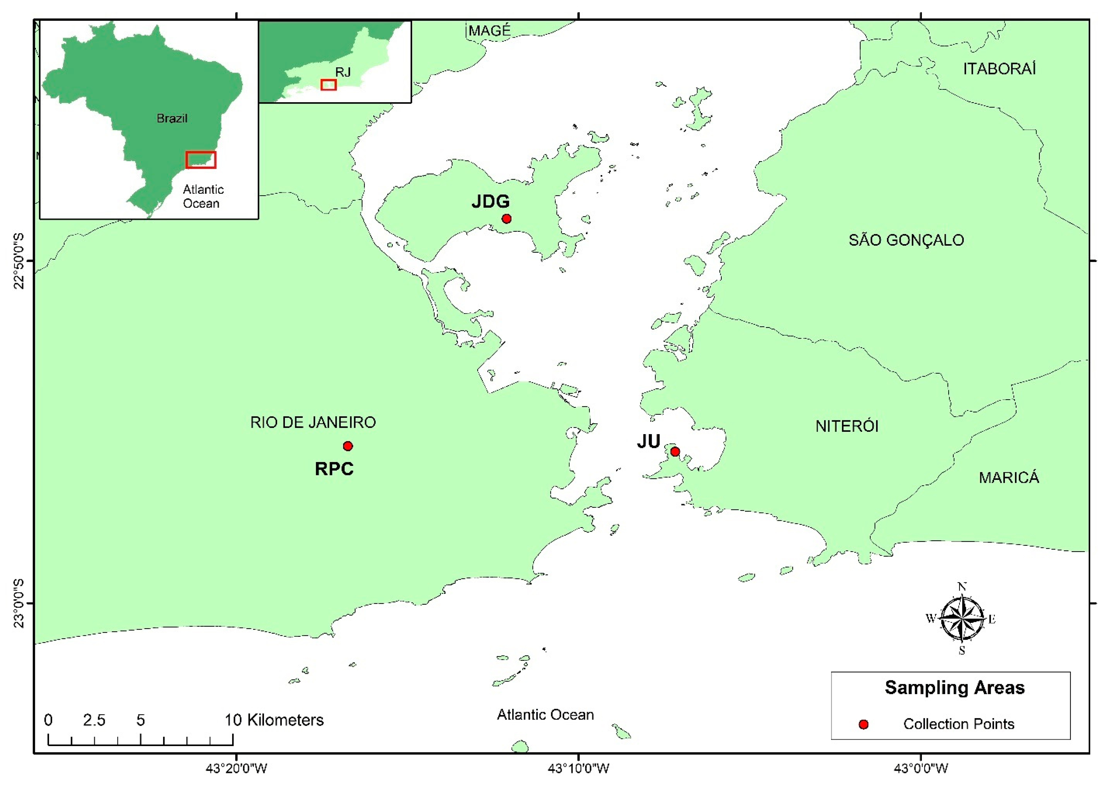
Figure 1. Mosquito collection areas. Map built using ArcGIS, sampling areas with different landscapes in Rio de Janeiro and Niterói cities. RPC—Represa dos Ciganos; JDG—Jardim Guanabara; JU—Jurujuba.
Viruses 15 01309 g001
Figure 2. Alpha diversity of Aedes albopictus gut microbiota according to origin. (A) Observed ASVs (Kruskal–Wallis: H = 9.73; p-value < 0.001); (B) Faith (Kruskal–Wallis: H = 11.44; p-value < 0.05); (C) Shannon–Weaver (Kruskal–Wallis: H = 7.38; p-value < 0.05). Black lines indicate medians, and black dots indicate outliers. Groups indicated with the same letters are not significantly different (paired Kruskal–Wallis q-value > 0.05) (Table S4).
Viruses 15 01309 g002
Figure 3. Alpha diversity of Aedes albopictus gut microbiota according to collection season. (A) Observed ASVs (Kruskal–Wallis: H = 29.60, p-value < 0.001); (B) Faith (Kruskal–Wallis: H = 17.58, p-value < 0.001); (C) Shannon–Weaver (Kruskal–Wallis: H = 21.94, p-value < 0.001); (D) Pielou (Kruskal–Wallis: H = 11.95, p-value < 0.05). Black lines indicate medians, and black dots indicate outliers. Groups indicated with the same letters are not significantly different (paired Kruskal–Wallis q-value > 0.05) (Table S4).
Viruses 15 01309 g003
Figure 4. Principal component analysis (PCoA) of the gut microbiota of Aedes albopictus: (A) based on the Bray–Curtis dissimilarity matrix and (B) based on the Weighted Unifrac matrix. Shapes indicate the different populations, while colors show whether mosquitoes were collected during the wet or dry season or were laboratory reared (F1 and LAB). The variation explained by the PCoA axes is shown in parentheses.
Viruses 15 01309 g004
Figure 5. Relative abundance of ASVs, classified at the lowest taxonomic level possible according to (A) collection season, (B) origin and (C) collection area, detected in the midgut of mosquitoes from the groups described in Table 2. Legends at the upper right: the remainder belong to bacterial taxa with <3% of abundance (C).
Viruses 15 01309 g005
Figure 6. Quantification of ZIKV copies (log10) by RT-qPCR in the body (A for 14 dpi and C for 21 dpi) and head (B for 14 dpi and D for 21 dpi) of Ae. albopictus. The black horizontal line represents the median number of viral copies.
Viruses 15 01309 g006
Table 1. Aedes albopictus samples classified according to their collection area, season and generation.
Sample ClassificationCollection AreaCollection SeasonOriginGeneration
JDG-WJardim GuanabaraWetFieldF0
JU-WJurujubaWetFieldF0
RPC-WRepresa dos CiganosWetFieldF0
JDG-DJardim GuanabaraDryFieldF0
JU-DJurujubaDryFieldF0
RPC-DRepresa dos CiganosDryFieldF0
JDG-F1Jardim GuanabaraLAB *LaboratoryF1
JU-F1JurujubaLAB *LaboratoryF1
RPC-F1Represa dos CiganosLAB *LaboratoryF1
LABLaboratoryLAB *Laboratory>F30
* Reared under standardized laboratory conditions.
Table 2. Variables Used in Diversity Analyses.
VariableDescriptionGroups (In Bold)
AreaSamples classified according to collection area (regardless of the collection season) and its respective F1.JDG (JDG-W + JDG-D),
JU (JU-W + JU-D),
RPC (RPC-W+RPC-D),
JDG-F1, JU-F1, RPC-F1 vs. LAB
Collection seasonPeriod in which insects were collected (regardless of collection area)WET (JDG-W + JU-W + RPC-W) vs. DRY (JDG-D + JU-D + RPC-D);
F1 (JDG-F1 + JU-F1 + RPC-F1) vs. LAB
OriginPlace of mosquito rearing (field or laboratory, regardless collection area and season)FIELD (JDG-W + JU-W + RPC-W + JDG-D + JU-D + RPC-D),
F1 (JDG-F1 + JU-F1 + RPC-F1) vs. LAB
PopulationMosquito population (regardless of
collection area, season or rearing place)
JDG (JDG-W + JDG-D + JDG-F1), JU (JU-W + JU-D + JU-F1),
RPC (RPC-W + RPC-D + RPC-F1) vs.
LAB
Table 3. Differentially abundant taxa according to Aedes albopictus origin.
Significantly Different TaxaW *F1FIELDLAB
Propionibacterium202Viruses 15 01309 i001Viruses 15 01309 i001
Acetobacteraceae_unclassified genus202Viruses 15 01309 i001 Viruses 15 01309 i001
Alphaproteobacteria_unclassified order200Viruses 15 01309 i001 Viruses 15 01309 i001
Peptostreptococcaceae_unclassified genus196 Viruses 15 01309 i001Viruses 15 01309 i001
Rahnella188Viruses 15 01309 i001 Viruses 15 01309 i001
Cupriavidus187 Viruses 15 01309 i001
Moraxellaceae_unclassified genus186 Viruses 15 01309 i001
Pedobacter185 Viruses 15 01309 i001
Reyranella182 Viruses 15 01309 i001
MLE1-12_unclassified family182 Viruses 15 01309 i001
Rhodospirillaceae_unclassified genus176 Viruses 15 01309 i001Viruses 15 01309 i001
Hydrocarboniphaga175 Viruses 15 01309 i001
Sphingobacteriales_unclassified family172 Viruses 15 01309 i001Viruses 15 01309 i001
* Considering mosquito origin, a given taxa was considered differentially abundant when W ≥ 172.
Table 4. Differentially abundant taxa according to Aedes albopictus collection season.
Significantly Different TaxaW *WETDRYF1LAB
Propionibacterium202Viruses 15 01309 i001Viruses 15 01309 i001Viruses 15 01309 i001
Acetobacteraceae_unclassified genus202 Viruses 15 01309 i001Viruses 15 01309 i001Viruses 15 01309 i001
Wolbachia202Viruses 15 01309 i001Viruses 15 01309 i001 Viruses 15 01309 i001
Methylobacterium202Viruses 15 01309 i001
Peptostreptococcaceae_unclassified genus201Viruses 15 01309 i001Viruses 15 01309 i001 Viruses 15 01309 i001
Alphaproteobacteria_unclassified order199Viruses 15 01309 i001 Viruses 15 01309 i001Viruses 15 01309 i001
Methylobacteriaceae_unclassified genus193Viruses 15 01309 i001Viruses 15 01309 i001
Cupriavidus190 Viruses 15 01309 i001
Moraxellaceae_unclassified genus187 Viruses 15 01309 i001
Pedobacter183 Viruses 15 01309 i001
Reyranella183 Viruses 15 01309 i001
MLE1-12_unclassified family183 Viruses 15 01309 i001
Rahnella177 Viruses 15 01309 i001Viruses 15 01309 i001
Rhizobiales_unclassified family177Viruses 15 01309 i001Viruses 15 01309 i001 Viruses 15 01309 i001
Hydrocarboniphaga177 Viruses 15 01309 i001
Rhodospirillaceae_unclassified genus174 Viruses 15 01309 i001Viruses 15 01309 i001
Sphingomonadaceae_unclassified genus172Viruses 15 01309 i001Viruses 15 01309 i001Viruses 15 01309 i001
Sphingobacteriales_unclassified family171Viruses 15 01309 i001 Viruses 15 01309 i001
Bdellovibrio171 Viruses 15 01309 i001
Ralstonia171Viruses 15 01309 i001 Viruses 15 01309 i001Viruses 15 01309 i001
Chitinophagaceae_unclassified genus169 Viruses 15 01309 i001
* Considering collection season, a given taxa was considered differentially abundant when W ≥ 169.
Table 5. Infection rates (IRs) and dissemination rates (DRs) of ZIKV at 14 and 21 days post-infection (dpi) of Ae. albopictus from natural populations collected in locations with different landscapes in Rio de Janeiro.
PopulationIR—14dpi
(%)
IR—21dpi
(%)
DR—14dpi
(%)
DR—21dpi
(%)
JDG-F162.5 (25/40)40 (4/10)16 (4/25)25 (1/4)
JU-F130 (12/40)44.4 (24/54)16.7 (2/12)45.8 (11/24)
RPC-F185 (34/40)85.4 (35/41)64.7 (22/34)71.4 (25/35)
Ae. aegypti (control)100 (20/20)95 (19/20)95 (19/20)100 (19/19)
The numbers in parentheses indicate ZIKV-positive samples/tested samples.
Disclaimer/Publisher’s Note: The statements, opinions and data contained in all publications are solely those of the individual author(s) and contributor(s) and not of MDPI and/or the editor(s). MDPI and/or the editor(s) disclaim responsibility for any injury to people or property resulting from any ideas, methods, instructions or products referred to in the content.