Article

Emergence and Prevalence of an African Swine Fever Virus Variant in Wild Boar Populations in South Korea from 2019 to 2022

Wildlife Disease Response Team, National Institute of Wildlife Disease Control and Prevention (NIWDC), 1 Songam-gil, Gwangsan-gu, Gwangju 62407, Republic of Korea
*
Author to whom correspondence should be addressed.
Viruses 2023, 15(8), 1667; https://doi.org/10.3390/v15081667
Submission received: 9 June 2023 / Revised: 26 July 2023 / Accepted: 27 July 2023 / Published: 31 July 2023
(This article belongs to the Section Animal Viruses)

Abstract

:
African swine fever (ASF), a viral disease caused by the African swine fever virus (ASFV), is associated with high mortality rates in domestic pigs and wild boars. ASF has been spreading since its discovery in wild boars in Korea in October 2019. Genomic analyses have provided insights into the genetic diversity of the ASFV isolated from various regions, enabling a better understanding of the virus origin and transmission patterns. We conducted a genome analysis to evaluate the diversity and mutations of ASFV spreading among wild boars in Korea during 2019–2022. We compared the genomes of ASFV strains isolated from Korean wild boars and publicly available ASFV genomes. Genomic analysis revealed several single-nucleotide polymorphisms within multigene families (MGFs) 360-1La and 360-4L in Korean ASFV. MGF 360-1La and 360-4L variations were not observed in other ASFV strains, including those of genotype II. Finally, we partially analyzed MGFs 360-1La and 360-4L in ASFV-positive samples between 2019 and 2022, confirming the geographical distribution of the variants. Our findings can help identify new genetic markers for epidemiological ASFV analysis and provide essential information for effective disease management.

1. Introduction

African swine fever (ASF), a highly contagious hemorrhagic disease caused by the African swine fever virus (ASFV), is a major cause of death in domestic pigs and wild boars [1]. ASF transmission can occur through direct contact with infected animals or indirect contact with contaminated materials such as equipment, clothing, feed, and ticks [2]. ASF was first identified in Kenya in 1921, which later spread to Africa, Europe, and Asia. Furthermore, ASF was also reported in the Dominican Republic and Haiti [3,4,5,6]. In Korea, ASF has spread nationwide since the first case in wild boars in October 2019 [7]. Therefore, the Korean government has been implementing measures, including fence cutting, population control, carcass searching, and safe disposal, to control the spread of ASF in domestic wild boars [8,9]. Despite these efforts, new cases of ASF occurring in wild boars are still being reported in Korea.
ASFV is a member of the Asfarviridae family and is a large icosahedral DNA virus [10]. The ASFV 170–190 kb genome encodes 150–170 open reading frames (ORFs) [11]. Most ORFs encode structural and nonstructural viral proteins involved in viral replication, transcription, translation, and virulence [12]. Demand for the complete ASFV genome sequence has surged owing to the rapid spread of the pathogen [13]. Moreover, the virus is an emerging threat to the swine industry; hence, detailed genetic information is urgently required to understand its biology [14]. Therefore, studies are being conducted to better understand the genetic diversity, evolution, and virulence of different ASFV strains [15].
The ASFV genomic diversity has been explored using pairwise sequence similarity and ORF distribution comparisons [16]. The nucleotide sequences of ASFV genomes have been compared to determine their similarity, and the genetic variations in ASFV and factors influencing its evolution have been revealed [11]. ASFV strains are classified into 24 genotypes based on the nucleotide sequence analysis of the B646L (p72) variable region, with all gene variants associated with the disease [17,18]. Over 140 ASFV genome sequences are available from various sources, exhibiting varying virulence degrees from different geographical locations. Sequence analysis of variable genomic regions is extensively used in molecular ASFV epidemiological studies [16]. X64R, EP152R, EP153R, EP402R, EP364R, and CP2475L have been identified in regions characterized by a high genetic ASFV diversity [19]. High variability is observed within the 35 kb at the 3′ end and the 15 kb at the 5′ end of the genome [20,21]. These two regions contain the multigene families (MGFs); ASFV has five MGFs, namely MGF-100, -110, -300, -360, and -505 [22]. The ASFV genome MGF region is highly variable and characterized by frequent mutations and genetic rearrangements [23]. Genes within the ASFV MGF-360 and MGF-505 regions reportedly inhibit host interferon production and are associated with host specificity, innate immune mechanisms, and virulence [24].
Analyzing ASFV isolates from Poland using whole-genome sequencing has revealed single-nucleotide polymorphisms (SNPs). These SNPs were identified in the K145R and MGF 505-5R genes, distinguishing the Polish isolates from the reference Georgia 2007/1 strain [25]. During the initial ASF outbreak in India, complete genome sequencing revealed several distinct nonsynonymous mutations in MGF 369-11L and 505-4R [26]. In addition, recent epidemiological simulations of the spread of the ASFV variant and its genetics have demonstrated the emergence of probabilistic and geographically distinct variant clusters in nonselective populations of wild boars. Furthermore, specific mutation sites have been identified as genetic markers, enabling genome dynamics analysis of various ASFV outbreak variants in Germany [27]. Therefore, identifying intriguing genetic variations in Korean wild boar ASFV is essential, as it will help distinguish isolates from Korea and those from other regions, and provide valuable information for specific diagnosis and treatment opportunities.
In this study, we aimed to analyze the ASFV genome isolated from ASFV-infected wild boars in Korea during 2019–2022. We identified characteristic mutations in the MGF 360-1La and MGF 360-4L regions for the first time. These mutations exhibited geographically distinguishable cluster patterns. The findings of this study, which reveal the genetic diversity of ASFV in domestic wild boars, have major implications. The mutations can serve as valuable genetic markers for conducting epidemiological analyses and developing preventive measures. Furthermore, the comprehensive analysis of ASFV diversity based on its distinct characteristics within the country can provide essential data for the development of effective vaccines.

2. Materials and Methods

2.1. Analysis of ASF Outbreak Distribution

We obtained data on ASF diagnosis in Korean wild boars, and these results were deposited in the World Organization for Animal Health. We analyzed ASF occurrences during 2019–2022 and used open-source Geographic Information System software version 3.24 (www.qgis.org, accessed on 25 July 2023) to investigate the geographical distribution of these outbreaks. A cartographic analysis was conducted based on the geographical coordinates of each ASF outbreak.

2.2. DNA Extraction and ASFV Detection in Wild Boar Samples

The Maxwell RSC Viral Total Nucleic Purification Kit (Promega, Madison, WI, USA) was used to extract DNA from the blood of wild boars, according to the manufacturer’s instructions. Polymerase chain reaction (PCR) was performed to detect the presence of ASFV DNA, using the ASFV diagnostic primers PPA1 (5′-AGTTATGGGAAACCCGACCC-3′), PPA2 (5′-CCCTGAATCGGAGCATCCT-3′) [28], P72D (5′-GTACTGTAACGCAGCACAG-3′), and P72U (5′-GGCACAAGTTCGGACATGT-3′), which partially amplified B646L (p72) [17]. The MGF 360-1La and 360-4L regions of ASFV were detected using the primers MGF 360-1La (F: CCGATTAATGTCAGCCCCCA, R: TGCAGACATCAGCTTTGGGT) and MGF 360-4L (F: CTCTAAGGCACGGTCAAGGT, R: TTCCTCATTTAGATCTTTGGCGT). The cycling protocol comprised an initial step at 95 °C for 2 min, followed by 45 denaturation cycles at 95 °C for 20 s, annealing at 58 °C for 40 s, and extension at 72 °C for 40 s. After the cycling steps, a final extension was performed at 72 °C for 5 min. Electrophoresis was performed using 1.5% agarose gels to visualize the amplification products. The gels were stained with Ecodye (BIOFACT, Seoul, Republic of Korea). Sanger sequencing was performed by Macrogen Inc., an outsourcing service specializing in Sanger sequencing, to determine the nucleotide sequence of the PCR products.

2.3. Complete ASFV Genome Sequencing

The Maxwell Viral Total Nucleic Purification Kit (Promega) was used to extract total DNA from 200 μL whole blood from wild boars, following the manufacturer’s instructions. As outlined in the OIE manual, the samples were amplified through PCR using the PPA1/PPA2 and P72 primers to detect ASFV. Library preparation was performed using an enzymatic preparation kit (Celemics, Seoul, Republic of Korea) after shearing genomic DNA (gDNA). Subsequently, the gDNA library and capture probes were hybridized using a target enrichment kit (Celemics), which includes chemically synthesized capture probes hybridized to the target regions. Post-PCR amplification was performed to enrich the captured regions. The target-captured library was sequenced against a next-generation sequencing library that was generated using the Illumina NextSeq 550 platform (Illumina, San Diego, CA, USA), with a target capture 2 × 150 bp read layout. Adaptor sequences and low-quality bases were trimmed using the Fastx Toolkit 0.0.14 (http://hannolab.cshl.edu/fastx_toolkit/, accessed on 25 July 2023), and AdapterRemoval (version 2.2.2) was used to trim the sequences. The reads were mapped onto a reference ASFV genome (accession number: FR682468) using Burrows–Wheeler Aligner software version 0.7.10. SNPs, indels, and structural variation were detected using Genome Analysis TK 4.0.4.0. The read alignment quality was assessed using the SAMtools software (samtools 1.1) and Python software package (numpy 1.11.0).

2.4. ASFV Complete-Genome Annotation

We used Genome Annotation Transfer Utility (GATU) software available at the Viral Bioinformatics Resource Center to annotate the newly constructed genome. We used the Georgia2007 strain as a reference for annotation.

2.5. Genetic ASFV Characterization and B646L (p72) Phylogenetic Analysis

We selected 16 closely related viruses and constructed a maximum likelihood (ML) phylogenetic tree of their whole-genome sequences to explore the phylogenetic relationships among different ASFV strains. We used Randomized Axelerated Maximum Likelihood version 8.0 with default parameters and a general time-reversible model, accounting for the gamma-distributed rate variation among sites. The genome sequences were aligned using multiple alignment within a fast Fourier transform algorithm in Geneious Prime version 2023.0.4 (https:www.geneious.com/prime, accessed on 25 July 2023). We constructed an ML phylogenetic tree of p72 using Molecular Evolutionary Genetics Analysis (MEGA) X to assess support for our phylogenetic tree. We performed 1000 replicates of ML bootstrapping to evaluate the robustness of the tree topologies. In addition, we aligned the nucleotide sequence of B646L (p72) of the Korean ASFV strains with those of other ASFV strains belonging to the same B646L (p72) genotype using the ClustalW algorithm in MEGA X. The evolutionary history was inferred using the maximum composite likelihood model method. We used the neighbor-joining method with 1000 bootstrap replicates to construct the phylogenetic tree.

3. Results

3.1. Geographical Distribution of ASF Outbreaks in South Korea during 2019–2022

The ASF outbreaks in South Korea during 2019–2022 are shown in Figure 1. The first outbreak occurred in Yeoncheon (Gyeonggi-do) in October 2019, followed by Yeoncheon (Paju) and Cheorwon (Gangwon-do). In 2020, the ASF epidemic spread from the western to the eastern regions. Of the total ASF cases occurring during 2019–2020, 49% and 51% occurred in Gyeonggi-do and Gangwon-do, respectively (Figure 1a,d). In 2021, the ASF cases reported in Gyeonggi-do, Gangwon-do, and Chungcheongbuk-do accounted for 21%, 73%, and 5% of the total cases, respectively. Compared with that during 2019–2020, the ASF incidence rate in Gyeonggi-do decreased in 2021 (Figure 1b,d). While the incidence rate in Gangwon-do decreased in 2022 compared with that in 2021, it remained the highest between 2019 and 2022, particularly in the southern region. In Gyeonggi-do, which had an incidence rate similar to that in Gangwon-do during 2019–2020, the incidence decreased between 2021 and 2022 to 2.3% of the total incidence rate. After the first occurrence in 2021 in the Chungcheongbuk-do region, the incidence rate of ASF substantially increased 4.7 times in 2022, and a new outbreak occurred in Gyeongsangbuk-do (Figure 1c,d). These results show that the occurrence diverged southward from the northernmost region of Korea.

3.2. Geographical Representation of the ASFV Genome Isolated from a Wild Boar with ASF in South Korea

We generated the whole-genome sequences of ASFV isolated from wild boars with ASF in Hwacheon and Inje in 2020 during the active spread of the outbreaks (Figure 2a). The complete genome sequences of the ASFV Korea/HC224/2020 and Korea/IJ702/2020 strains, with total lengths of 188,645 bp and 188,598 bp, respectively, were determined; the GC content was 38.4%. Korea/HC224/2020 and Korea/IJ702/2020 strain sequence annotations were created using the GATU software [29]. The genome length of Georgia 2007/1 is 190,854 bp, and sequence differences are present in the 5′- and 3′-ITR regions [30], resulting in an approximate 2000 bp difference compared to that in the YC1/2019, HC224/2020, and IJ702/2020 strains (Figure 2b).
In the Korea/HC224/2020 and Korea/IJ702/2020 strains, 46 MGF genes were identified, namely, MGF-100 (three members), -110 (11 members), -300 (three members), -360 (19 members), and -505 (10 members). The Korea/HC224/2020 strain genome sequences have been deposited in GenBank under accession number OP628183.

3.3. Phylogenetic Analysis of Complete ASFV Strain Genomes

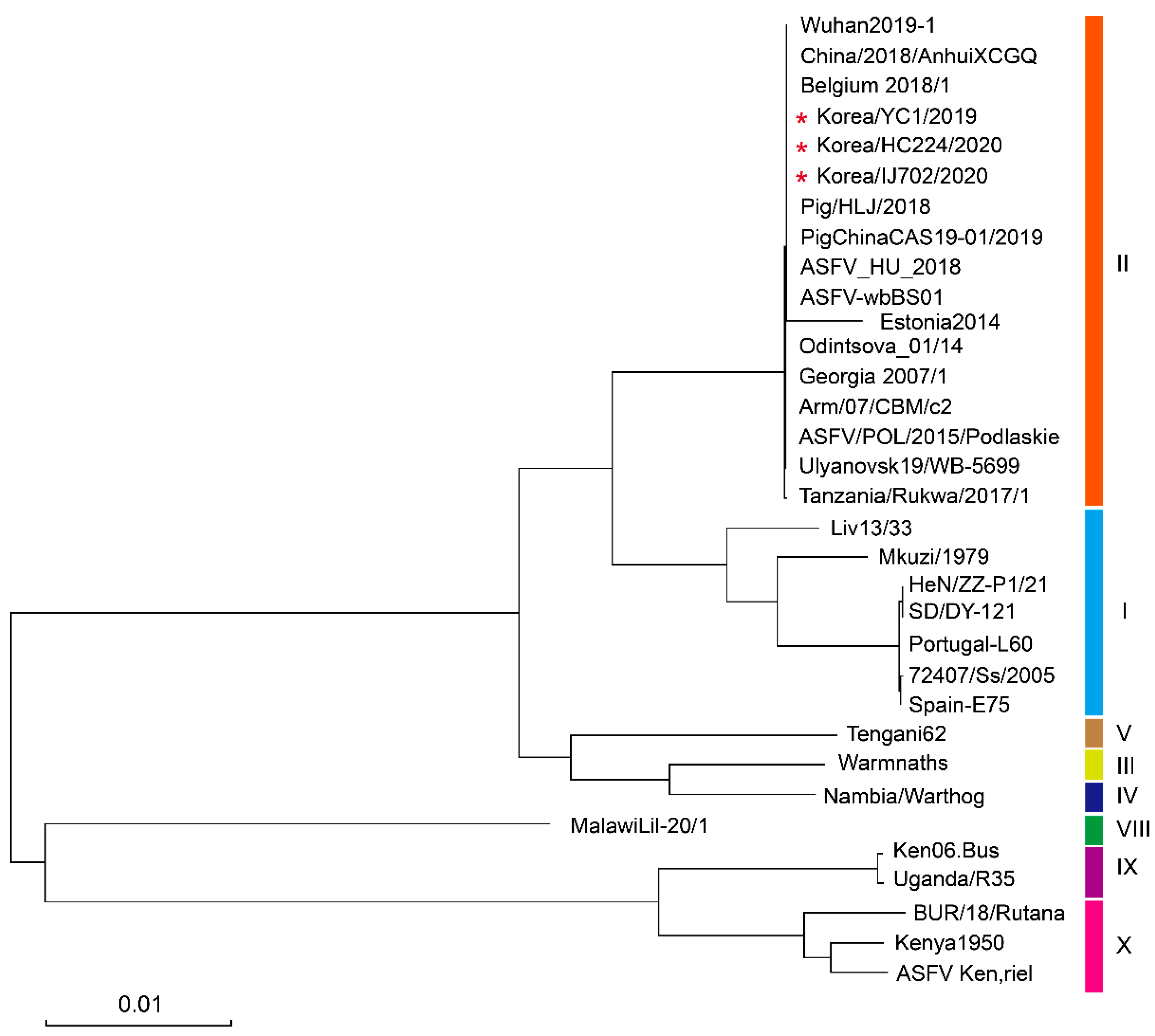
A phylogenetic tree of ASFV p72 was constructed to determine the genetic relationship between HC224/2020 and IJ702/2020 strains and the other isolated ASFV strains. An ML phylogenetic ASFV dendrogram was constructed by aligning genome sequences. A phylogenetic analysis of the ASFV genome showed that the strains clustered according to genotype. The HC224/2020 and IJ702/2020 strains belonged to p72 genotype II (Figure 3).

3.4. ASFV Genome Comparison of Korean Strains with Reported ASFV Strains

We compared pairwise sequence similarities to investigate the genome-wide gene content of 30 ASFV strains. Table 1 shows the strain information, and the ML phylogenetic tree of the ASFV genome sequences is shown on the left (Figure 4).
The overall whole-genome sequence similarity varied between 75.37% and 99.9%. The sequence divergence observed within genotype II was wide, varying between 91.42% and 99.99%. Similarly, the pairwise sequence similarities observed among Korean ASFV strains were highly divergent within genotype I, with a range of 86.5%–90.8%. The highest inter-genotype pairwise similarity was observed for genotype II (Georgia 2007/1, ASFV_HU_2018, China/2018/AnhuiXCGQ, Belgium 2018/1) at 99.9%. In contrast, the lowest inter-genotype pairwise similarity was found between genotypes X (Kenya1950, Ken.rie1 and BUR/18/Rutana) and VIII (MalawiLil-20/1), at 80.54% and 85.39%, respectively. Although the Korea/HC224/2020 and Korea/YC1/2019 strains showed 100% sequence similarity, the Korea/IJ702/2020 strain exhibited 99.97% similarity to the Korea/HC224/2020 and Korea/YC1/2019 strains.

3.5. Amino Acid Sequence Alignment in the MGF 360-1La and 360-4L Regions

We analyzed similarities in the genomes of Korea/HC224/2020, Korea/YC1/2019, and Korea/IJ702/2020 strains, and found three novel mutation sites (MGFs 360-1La, 4L, and 360-10L) when comparing the Korea/HC224/2020 and Korea/IJ702/2020 strains with the Korea/YC1/2019 strain. Two synonymous mutations (MGFs 360-1La and 360-4L) and one nonsynonymous mutation (MGF 360-10L) were observed (Table 2).
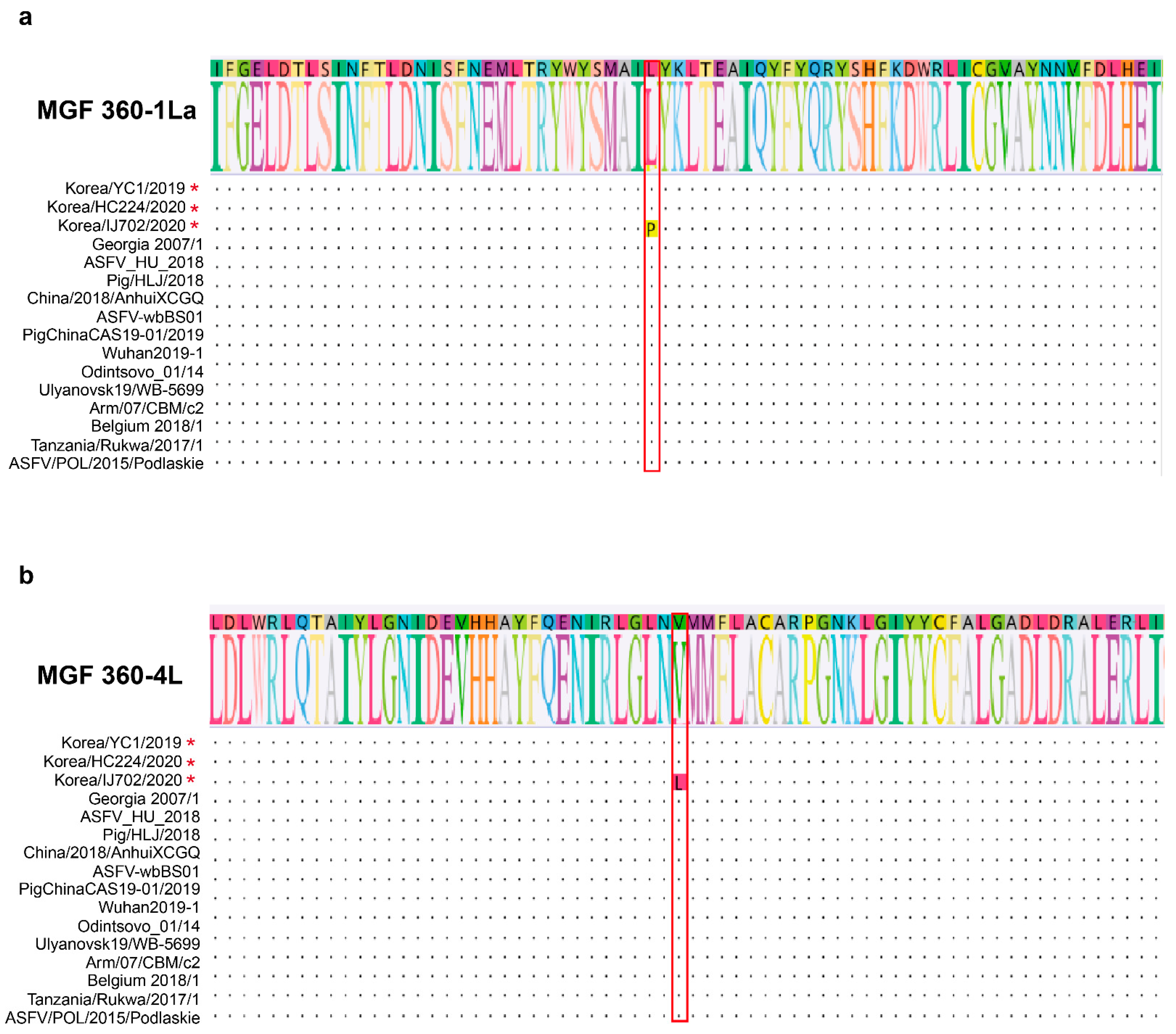
MGFs 360-1La and 360-4L are encoded in the reverse strand sequence of the left variable region of the ASF genome. MGF 360-1L is upstream of MGF 360-2L, and MGF 360-4L is between MGFs 110-13L and 360-6L. MGFs 360-1La and 360-4L encode proteins comprising 277 and 387 amino acids, respectively. The sequence analysis revealed an SNP within MGFs 360-1La and 360-4L. The amino acid at MGF 360-1La position 106, leucine (L), is replaced with proline (P). In MGF 360-4L, the amino acid at position 243, valine (V), is replaced with leucine (L) (Figure 5a,b). SNPs were identified using the data from aligning the sequence to the Georgia 2007/1 strain.

3.6. Geographic Distribution of Variant ASFV in Korea during 2019–2022

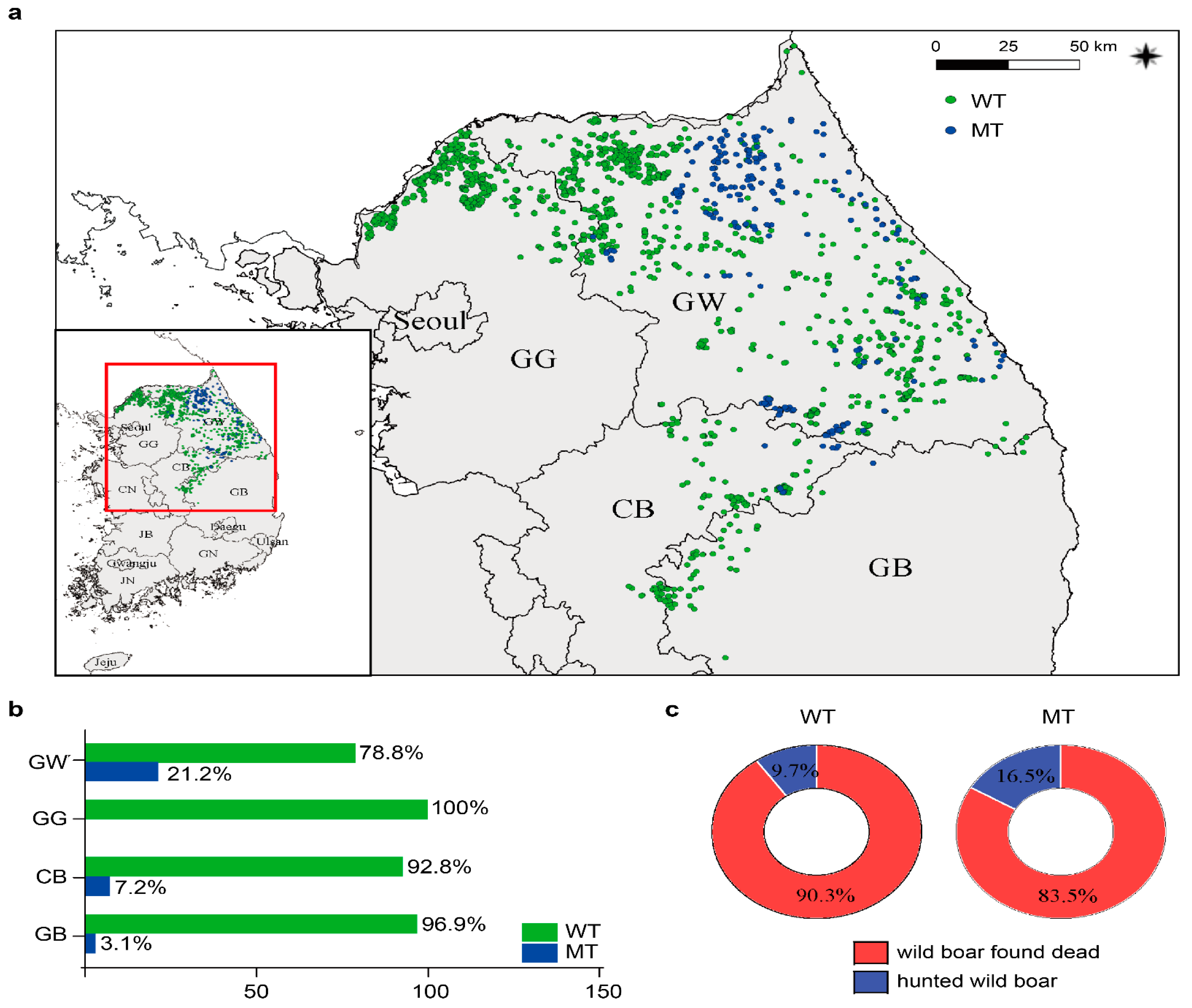
We confirmed the mutation sites in the MGF360-1La and MGF360-4L regions of the Korea/IJ702/2020 strain (Figure 5 and Table 2). We then analyzed ASFV samples from 2019 to 2022 to investigate whether the identified mutations also exist in other ASFV strains. Therefore, we performed partial PCR and sequencing analysis targeting the MGF360-1La and MGF360-4L regions. The mutant virus first discovered in Inje spread to Gangwon-do and moved south to Chungcheongbuk-do in November 2021. The mutant type was most frequently identified in Gangwon-do, followed by Chungcheongbuk-do and Gyeongsangbuk-do, adjacent to Gangwon-do. In contrast, no mutant viruses were identified in Gyeonggi-do (Figure 6a,b). The wild type spread more widely without being confined to specific regions. In contrast, the mutant virus showed a pattern of sporadic occurrence in the western regions.
Furthermore, we compared the occurrence rate of genetic mutations between hunted wild boars and carcasses. The wild type was identified in 90.3% of carcasses and 9.7% of hunted boars. The mutant types were identified in 83.5% of carcasses and 16.5% of hunted boars (Figure 6c). Therefore, approximately 5% more mutant forms were identified in the carcasses compared to the wild type. However, further research is needed to determine if this phenomenon is characteristic of the mutant virus or influenced by other factors.

4. Discussion

Partial nucleotide sequencing of specific ASFV regions is traditionally used to diagnose ASF and determine ASFV genotypes [51]. Analyzing ASFV genome information is essential as it broadens our understanding of the circulating ASFV variant characteristics, genetic diversity, and evolutionary pathways [52]. ASF first occurred in Yeoncheon in 2019 and disseminated toward the east in 2020, before progressing southward in 2021 and 2022. We analyzed the ASFV genome to investigate the characteristics of the ongoing wild boar ASF outbreaks. ASFV detected in Korean wild boars belonged to genotype II, remarkably similar to other strains in the genotype II group, particularly the Georgia 2007/1, ASFV-wbBS01, China/2018/AnhuiXCGQ, Pig/HLJ/2018, CAS19-01/2019, and Wuhan2019-1 strains. ASFV genome similarity analysis confirmed that the genetic nucleotide sequences of the Yeoncheon and Hwacheon strains were identical, whereas the sequence of the Inje strain differed slightly from the sequences of these two strains. We also identified SNPs within MGFs 360-1La and 360-4L. These SNPs distinguished the Inje strain from the Yeoncheon, Hwacheon, and Georgia 2007/1 strains. Furthermore, no differences were observed between the MGF 360-1La and 360-4L genes in the ASFV isolates from domestic farms in Korea. These results indicate that certain viral strains, such as the Inje strain, occur only in wild boars. The identification of new variants holds the potential for the strategic management and control of ASF in both wild boar and domestic pig populations. Because it allows for the adjustment of response measures to effectively contain ASFV spread and prevent further outbreaks, continuous efforts in identifying new ASFV variants via genome analysis are emphasized.
ASFV, the only member of the Asfvirus genus in the Asfarviridae family, has a high molecular weight and linear double-stranded DNA, with a genome size of 170–193 kb, containing 150–167 ORFs, one-third of which have unknown functions [53,54]. ASFV has a relatively low mutation frequency, attributed to the proofreading activity of DNA polymerase and repair mechanisms of the virus [55]. The primary factor responsible for genome length and gene number differences among viruses is ORF gain or loss in the MGFs [12]. ASFV genomes typically contain a conserved central and variable region at both ends, including five MGFs, and most genome variations are attributed to the varied number of MGF genes in the left and right variable regions [56]. ASFV has five MGFs, namely, MGF-100, -110, -300, -360, and -505, which can modulate the immune response of the host and exhibit host specificity [57]. However, the functions and mechanisms of these MGFs have only been partially studied. According to our results, mutant-type MGF-360 1La and -360 4L were present in 16.5% of hunted wild boars, which is 1.7-fold higher than seen in wild types. More than 80% of the mutant types were found in wild boar carcasses, suggesting that the mutant type does not affect virulence. However, additional molecular studies are needed to understand the basis for the observed 1.7-fold increase in the survival of the mutant types compared with that of the wild type. We obtained limited information from genetic analysis and could not determine whether the identified mutations increased the severity of the disease. In addition, further research is necessary to evaluate the effect of the mutations identified in MGFs 360-1La and 360-4L on the protein structure and the signaling of the host cell immune response. In particular, the emergence of new variants may necessitate the development of new vaccines or updating of existing ones. Understanding the genetic characteristics of these variants can aid in the development of effective vaccines targeted to combat specific mutations.
This study identified a new ASFV variant form for the first time in the Inje region. However, the actual origin of the mutation is unknown and several possibilities are being considered. First, owing to the pattern of domestic ASF outbreaks spreading from the western to the eastern regions, the mutation possibly occurred naturally during this spread or a new type of virus from an external source rather than the domestic virus was introduced. However, extensive research is necessary to elucidate the fundamental origin of the emergence of domestically mutated forms during ASF outbreaks. The transboundary spread of ASF raises concerns regarding variant transmission to diverse regions. The data on identified ASF variants obtained from this study can facilitate in preventing its dissemination via international cooperation and exchange of information.
Genetic epidemiology is essential for determining the origin of a virus, creating maps for prevention, and observing its spread. Therefore, the newly identified mutation in Germany can be used as a genetic marker to investigate the genetic epidemiology of various ASFV occurrences [27]. The mutant type was detected most frequently in Chungcheongbuk-do and Gyeongsangbuk-do after its initial detection in Gangwon-do. During the same period, ASF persisted in Gyeonggi-do; however, the mutant type was not detected. In this study, we were able to confirm the geographically clustered occurrence patterns of ASFV variants based on the genetic characteristics discovered. The newly identified mutation sites can be used as genetic markers to track the transmission pathways of ASFV outbreaks in South Korea. With this information, we can elucidate the transmission pathway of ASFV between wild boars and domestic pigs, and utilize the identified markers to differentiate the spatial and temporal variations in ASFV variants.
In conclusion, during the cyclic domestic occurrence of ASF in 2019–2022, we identified characteristic mutations in the ASFV genome through genetic ASFV analysis in wild boar, specifically in the MGF 360-1La and MGF 360-4L regions. These mutations exhibited a clustered pattern of ASF incidence in the local area. The results can improve our understanding of the spread of ASF through genetic ASFV variant diversity. Without a vaccine against ASFV, the best approach to control and prevent ASFV spread is through monitoring and using the identified characteristic mutations as genetic markers for genetic epidemiological analysis. Furthermore, we will continue to analyze the characteristics of ASFV strains in wild boar populations through genomic analysis to control ASFV infection and aid the development of effective drugs.

Author Contributions

G.K.; methodology, W.-J.K.; software, J.-C.K.; validation, G.K. and S.-J.K.; formal analysis, S.-H.L.; investigation, J.-H.K.; resources, S.-G.L.; data curation, E.-S.K.; writing—original draft preparation, G.K.; writing—review and editing, G.K.; visualization, S.-J.K.; supervision, W.-H.J.; project administration. All authors have read and agreed to the published version of the manuscript.

Funding

This research was funded by the National Institute of Wildlife Disease Control and Prevention (NIWDC) and the Ministry of Environment, Republic of Korea, grant number 2022-001.

Institutional Review Board Statement

Not applicable.

Informed Consent Statement

Not applicable.

Data Availability Statement

Not applicable.

Acknowledgments

The authors would like to thank their colleagues who worked on ASF diagnosis and response tasks.

Conflicts of Interest

The authors declare no conflict of interest.

References

  1. Salas, M.L. African Swine Fever Virus (Asfarviridae). In Encyclopedia of Virology, 2nd ed.; Elsevier: Amsterdam, The Netherlands, 1999; pp. 30–38. [Google Scholar] [CrossRef]
  2. Costard, S.; Wieland, B.; De Glanville, W.; Jori, F.; Rowlands, R.; Vosloo, W.; Roger, F.; Pfeiffer, D.U.; Dixon, L.K. African swine fever: How can global spread be prevented? Philos. Trans. R. Soc. B Biol. Sci. 2009, 364, 2683–2696. [Google Scholar] [CrossRef] [Green Version]
  3. Sánchez-Vizcaíno, J.M.; Mur, L.; Gomez-Villamandos, J.C.; Carrasco, L. An Update on the Epidemiology and Pathology of African Swine Fever. J. Comp. Pathol. 2015, 152, 9–21. [Google Scholar] [CrossRef]
  4. Penrith, M.-L. History of’swine fever in Southern Africa: Historical article. J. S. Afr. Vet. Assoc. 2013, 84, 1–6. [Google Scholar] [CrossRef] [Green Version]
  5. Khomenko, S.; Beltrán-Alcrudo, D.; Rozstanlnyy, A.; Gogin, A.; Kolbasov, D.; Pinto, J.; Lubroth, J.; Martin, V. African Swine Fever in the Russian Federation: Risk Factors for Europe and Beyond. EMPRES Watch 2013, 28, 1–14. Available online: https://www.fao.org/publications/card/fr/c/456c5e1d-f450-524f-a664-/ (accessed on 25 July 2023).
  6. Gonzales, W.; Moreno, C.; Duran, U.; Henao, N.; Bencosme, M.; Lora, P.; Reyes, R.; Núñez, R.; De Gracia, A.; Perez, A.M. African swine fever in the Dominican Republic. Transbound. Emerg. Dis. 2021, 68, 3018–3019. [Google Scholar] [CrossRef]
  7. Kim, H.J.; Cho, K.H.; Lee, S.K.; Kim, D.Y.; Nah, J.J.; Kim, H.J.; Kim, H.J.; Hwang, J.Y.; Sohn, H.J.; Choi, J.G.; et al. Outbreak of African swine fever in South Korea, 2019. Transbound. Emerg. Dis. 2020, 67, 473–475. [Google Scholar] [CrossRef]
  8. Yoo, D.; Kim, H.; Lee, J.Y.; Yoo, H.S. African swine fever: Etiology, epidemiological status in Korea, and perspective on control. J. Vet. Sci. 2020, 21, e38. [Google Scholar] [CrossRef] [Green Version]
  9. Lim, J.-S.; Andraud, M.; Kim, E.; Vergne, T. Three Years of African Swine Fever in South Korea (2019–2021): A Scoping Review of Epidemiological Un-derstanding. Transbound. Emerg. Dis. 2023, 2023, 4686980. [Google Scholar] [CrossRef]
  10. Karger, A.; Pérez-Núñez, D.; Urquiza, J.; Hinojar, P.; Alonso, C.; Freitas, F.B.; Revilla, Y.; Le Potier, M.-F.; Montoya, M. An Update on African Swine Fever Virology. Viruses 2019, 11, 864. [Google Scholar] [CrossRef] [Green Version]
  11. Yáñez, R.J.; Rodriguez, J.M.; Nogal, M.L.; Yuste, L.; Enriquez, C.; Rodriguez, J.F.; Viñuela, E. Analysis of the Complete Nucleotide Sequence of African Swine Fever Virus. Virology 1995, 208, 249–278. [Google Scholar] [CrossRef] [Green Version]
  12. Dixon, L.K.; Chapman, D.A.; Netherton, C.L.; Upton, C. African swine fever virus replication and genomics. Virus Res. 2013, 173, 3–14. [Google Scholar] [CrossRef]
  13. Wen, X.; He, X.; Zhang, X.; Zhang, X.; Liu, L.; Guan, Y.; Zhang, Y.; Bu, Z. Genome sequences derived from pig and dried blood pig feed samples provide important insights into the transmission of African swine fever virus in China in 2018. Emerg. Microbes Infect. 2019, 8, 303–306. [Google Scholar] [CrossRef]
  14. Martins, C.; Boinas, F.S.; Iacolina, L.; Ruiz-Fons, F.; Gavier-Widén, D. African swine fever (ASF), the pig health challenge of the century. In Understanding and Combatting African Swine Fever: A European Perspective; Wageningen Academic Publishers: Wageningen, The Netherlands, 2021; pp. 149–154. [Google Scholar]
  15. Alonso, C.; Borca, M.; Dixon, L.; Revilla, Y.; Rodriguez, F.; Escribano, J.M.; ICTV Report Consortium. ICTV Virus Taxonomy Profile Asfarviridae. J. Gen. Virol. 2018, 99, 613–614. [Google Scholar] [CrossRef]
  16. Bao, J.; Zhang, Y.; Shi, C.; Wang, Q.; Wang, S.; Wu, X.; Cao, S.; Xu, F.; Wang, Z. Genome-Wide Diversity Analysis of African Swine Fever Virus Based on a Curated Dataset. Animals 2022, 12, 2446. [Google Scholar] [CrossRef]
  17. Bastos, A.D.S.; Penrith, M.-L.; Crucière, C.; Edrich, J.L.; Hutchings, G.; Roger, F.; Couacy-Hymann, E.; Thomson, G.R. Genotyping field strains of African swine fever virus by partial p72 gene characterisation. Arch. Virol. 2003, 148, 693–706. [Google Scholar] [CrossRef]
  18. Achenbach, J.E.; Gallardo, C.; Nieto-Pelegrín, E.; Rivera-Arroyo, B.; Degefa-Negi, T.; Arias, M.; Jenberie, S.; Mulisa, D.D.; Gizaw, D.; Gelaye, E.; et al. Identification of a New Genotype of African Swine Fever Virus in Domestic Pigs from Ethiopia. Transbound. Emerg. Dis. 2017, 64, 1393–1404. [Google Scholar] [CrossRef]
  19. Li, X.; Xiao, K.; Zhang, Z.; Yang, J.; Wang, R.; Shen, X.; Pan, J.; Irwin, D.M.; Chen, R.-A.; Shen, Y. The recombination hot spots and genetic diversity of the genomes of African swine fever viruses. J. Infect. 2020, 80, 121–142. [Google Scholar] [CrossRef]
  20. Qu, H.; Ge, S.; Zhang, Y.; Wu, X.; Wang, Z. A systematic review of genotypes and serogroups of African swine fever virus. Virus Genes 2022, 58, 77–87. [Google Scholar] [CrossRef]
  21. Cackett, G.; Matelska, D.; Sýkora, M.; Portugal, R.; Malecki, M.; Bähler, J.; Dixon, L.; Werner, F. The African swine fever virus transcriptome. J. Virol. 2020, 94, 10–1128. [Google Scholar] [CrossRef] [Green Version]
  22. González, A.; Calvo, V.; Almazán, F.; Almendral, J.M.; Ramírez, J.C.; de la Vega, I.; Blasco, R.; Viñuela, E. Multigene families in African swine fever virus: Family 360. J. Virol. 1990, 64, 2073–2081. [Google Scholar] [CrossRef] [Green Version]
  23. Farlow, J.; Donduashvili, M.; Kokhreidze, M.; Kotorashvili, A.; Vepkhvadze, N.G.; Kotaria, N.; Gulbani, A. Intra-epidemic genome variation in highly pathogenic African swine fever virus (ASFV) from the country of Georgia. Virol. J. 2018, 15, 190. [Google Scholar] [CrossRef] [PubMed] [Green Version]
  24. Afonso, C.L.; Piccone, M.E.; Zaffuto, K.M.; Neilan, J.; Kutish, G.F.; Lu, Z.; Balinsky, C.A.; Gibb, T.R.; Bean, T.J.; Zsak, L.; et al. African Swine Fever Virus Multigene Family 360 and 530 Genes Affect Host Interferon Response. J. Virol. 2004, 78, 1858–1864. [Google Scholar] [CrossRef] [PubMed] [Green Version]
  25. Mazur-Panasiuk, N.; Woźniakowski, G.; Niemczuk, K. The first complete genomic sequences of African swine fever virus isolated in Poland. Sci. Rep. 2019, 9, 4556. [Google Scholar] [CrossRef] [Green Version]
  26. Senthilkumar, D.; Rajukumar, K.; Venkatesh, G.; Singh, F.; Tosh, C.; Kombiah, S.; Dubey, C.K.; Chakravarty, A.; Barman, N.N.; Singh, V.P. Complete genome analysis of African swine fever virus isolated from domestic pigs during the first ASF outbreaks in India. Transbound. Emerg. Dis. 2022, 69, e2020–e2027. [Google Scholar] [CrossRef] [PubMed]
  27. Forth, J.H.; Calvelage, S.; Fischer, M.; Hellert, J.; Sehl-Ewert, J.; Roszyk, H.; Deutschmann, P.; Reichold, A.; Lange, M.; Thulke, H.H.; et al. African swine fever virus–variants on the rise. Emerg. Microbes Infect. 2023, 12, 2146537. [Google Scholar] [CrossRef]
  28. Aguero, M.; Fernandez, J.; Romero, L.; Mascaraque, C.S.; Arias, M.; Sanchez-Vizcaino, J.M. Highly Sensitive PCR Assay for Routine Diagnosis of African Swine Fever Virus in Clinical Samples. J. Clin. Microbiol. 2003, 41, 4431–4434. [Google Scholar] [CrossRef] [Green Version]
  29. Tcherepanov, V.; Ehlers, A.; Upton, C. Genome Annotation Transfer Utility (GATU): Rapid annotation of viral genomes using a closely related reference genome. BMC Genom. 2006, 7, 150. [Google Scholar] [CrossRef]
  30. Forth, J.H.; Forth, L.F.; King, J.; Groza, O.; Hübner, A.; Olesen, A.S.; Höper, D.; Dixon, L.K.; Netherton, C.L.; Rasmussen, T.B.; et al. A Deep-Sequencing Workflow for the Fast and Efficient Generation of High-Quality African Swine Fever Virus Whole-Genome Sequences. Viruses 2019, 11, 846. [Google Scholar] [CrossRef] [Green Version]
  31. Kim, G.; Park, J.-E.; Kim, S.-J.; Kim, Y.; Kim, W.; Kim, Y.-K.; Jheong, W. Complete genome analysis of the African swine fever virus isolated from a wild boar responsible for the first viral outbreak in Korea, 2019. Front. Veter. Sci. 2022, 9, 1080397. [Google Scholar] [CrossRef]
  32. Chapman, D.A.; Darby, A.C.; Da Silva, M.; Upton, C.; Radford, A.D.; Dixon, L.K. Genomic analysis of highly virulent Georgia 2007/1 isolate of African swine fever virus. Emerg. Infect. Dis. 2011, 17, 599. [Google Scholar] [CrossRef]
  33. Jia, L.; Jiang, M.; Wu, K.; Hu, J.; Wang, Y.; Quan, W.; Hao, M.; Liu, H.; Wei, H.; Fan, W.; et al. Nanopore sequencing of African swine fever virus. Sci. China Life Sci. 2020, 63, 160–164. [Google Scholar] [CrossRef]
  34. Pikalo, J.; Schoder, M.E.; Sehl, J.; Breithaupt, A.; Tignon, M.; Cay, A.B.; Gager, A.M.; Fischer, M.; Beer, M.; Blome, S. The African swine fever virus isolate Belgium 2018/1 shows high virulence in European wild boar. Transbound. Emerg. Dis. 2020, 67, 1654–1659. [Google Scholar] [CrossRef] [PubMed] [Green Version]
  35. Pérez-Núñez, D.; Castillo-Rosa, E.; Vigara-Astillero, G.; García-Belmonte, R.; Gallardo, C.; Revilla, Y. Identification and Isolation of Two Different Subpopulations Within African Swine Fever Virus Arm/07 Stock. Vaccines 2020, 8, 625. [Google Scholar] [CrossRef] [PubMed]
  36. Njau, E.P.; Entfellner, J.-B.D.; Machuka, E.M.; Bochere, E.N.; Cleaveland, S.; Shirima, G.M.; Kusiluka, L.J.; Upton, C.; Bishop, R.P.; Pelle, R.; et al. The first genotype II African swine fever virus isolated in Africa provides insight into the current Eurasian pandemic. Sci. Rep. 2021, 11, 13081. [Google Scholar] [CrossRef]
  37. Bao, J.; Wang, Q.; Lin, P.; Liu, C.; Li, L.; Wu, X.; Chi, T.; Xu, T.; Ge, S.; Liu, Y.; et al. Genome comparison of African swine fever virus China/2018/Anhui XCGQ strain and related European p72 Genotype II strains. Transbound. Emerg. Dis. 2019, 66, 1167–1176. [Google Scholar] [CrossRef] [PubMed]
  38. Pershin, A.; Shevchenko, I.; Igolkin, A.; Zhukov, I.; Mazloum, A.; Aronova, E.; Vlasova, N.; Shevtsov, A. A Long-Term Study of the Biological Properties of ASF Virus Isolates Originating from Various Regions of the Russian Federation in 2013–2018. Veter. Sci. 2019, 6, 99. [Google Scholar] [CrossRef] [Green Version]
  39. Zani, L.; Forth, J.H.; Forth, L.; Nurmoja, I.; Leidenberger, S.; Henke, J.; Carlson, J.; Breidenstein, C.; Viltrop, A.; Höper, D.; et al. Deletion at the 5′-end of Estonian ASFV strains associated with an attenuated phenotype. Sci. Rep. 2018, 8, 65. [Google Scholar] [CrossRef] [Green Version]
  40. Olasz, F.; Mészáros, I.; Marton, S.; Kaján, G.L.; Tamás, V.; Locsmándi, G.; Magyar, T.; Bálint, A.; Bányai, K.; Zádori, Z. A Simple Method for Sample Preparation to Facilitate Efficient Whole-Genome Sequencing of African Swine Fever Virus. Viruses 2019, 11, 1129. [Google Scholar] [CrossRef] [Green Version]
  41. Mazloum, A.; van Schalkwyk, A.; Shotin, A.; Igolkin, A.; Shevchenko, I.; Gruzdev, K.N.; Vlasova, N. Comparative Analysis of Full Genome Sequences of African Swine Fever Virus Isolates Taken from Wild Boars in Russia in 2019. Pathogens 2021, 10, 521. [Google Scholar] [CrossRef]
  42. Shi, K.; Liu, H.; Yin, Y.; Si, H.; Long, F.; Feng, S. Molecular Characterization of African Swine Fever Virus From 2019-2020 Outbreaks in Guangxi Province, Southern China. Front. Veter. Sci. 2022, 9, 912224. [Google Scholar] [CrossRef]
  43. de Villiers, E.P.; Gallardo, C.; Arias, M.; da Silva, M.; Upton, C.; Martin, R.; Bishop, R.P. Phylogenomic analysis of 11 complete African swine fever virus genome sequences. Virology 2010, 400, 128–136. [Google Scholar] [CrossRef] [Green Version]
  44. Portugal, R.; Coelho, J.; Hoeper, D.; Little, N.S.; Smithson, C.; Upton, C.; Martins, C.; Leitão, A.; Keil, G.M. Related strains of African swine fever virus with different virulence: Genome comparison and analysis. J. Gen. Virol. 2015, 96, 408–419. [Google Scholar] [CrossRef]
  45. Sun, E.; Huang, L.; Zhang, X.; Zhang, J.; Shen, D.; Zhang, Z.; Wang, Z.; Huo, H.; Wang, W.; Huangfu, H.; et al. Genotype I African swine fever viruses emerged in domestic pigs in China and caused chronic infection. Emerg. Microbes Infect. 2021, 10, 2183–2193. [Google Scholar] [CrossRef]
  46. Chastagner, A.; Pereira De Oliveira, R.; Hutet, E.; Le Dimna, M.; Paboeuf, F.; Lucas, P.; Blanchard, Y.; Dixon, L.; Vial, L.; Le Potier, M.F. Coding-complete genome sequence of an African swine fever virus strain Liv13/33 isolate from ex-perimental transmission between pigs and Ornithodoros moubata ticks. Microbiol. Resour. Announc. 2020, 9, e00185-20. [Google Scholar] [CrossRef]
  47. Zsak, L.; Borca, M.V.; Risatti, G.R.; Zsak, A.; French, R.A.; Lu, Z.; Kutish, G.F.; Neilan, J.G.; Callahan, J.D.; Nelson, W.M.; et al. Preclinical Diagnosis of African Swine Fever in Contact-Exposed Swine by a Real-Time PCR Assay. J. Clin. Microbiol. 2005, 43, 112–119. [Google Scholar] [CrossRef] [Green Version]
  48. Rodriguez, F.; Alcaraz, C.; Eiras, A.; Yáñez, R.J.; Rodriguez, J.M.; Alonso, C.; Escribano, J.M. Characterization and molecular basis of heterogeneity of the African swine fever virus envelope protein p54. J. Virol. 1994, 68, 7244–7252. [Google Scholar] [CrossRef] [Green Version]
  49. Njau, E.P.; Machuka, E.M.; Cleaveland, S.; Shirima, G.M.; Kusiluka, L.J.; Okoth, E.A.; Pelle, R. African Swine Fever Virus (ASFV): Biology, Genomics and Genotypes Circulating in Sub-Saharan Africa. Viruses 2021, 13, 2285. [Google Scholar] [CrossRef] [PubMed]
  50. Ge, S.; Li, J.; Fan, X.; Liu, F.; Li, L.; Wang, Q.; Ren, W.; Bao, J.; Liu, C.; Wang, H.; et al. Molecular Characterization of African Swine Fever Virus, China, 2018. Emerg. Infect. Dis. 2018, 24, 2131–2133. [Google Scholar] [CrossRef] [PubMed] [Green Version]
  51. Sánchez-Vizcaíno, J.M.; Laddomada, A.; Arias, M.L. African swine fever virus. In Diseases of Swine, 11th ed.; John Wiley & Sons, Inc.: Hoboken, NJ, USA, 2019; pp. 443–452. [Google Scholar]
  52. Wang, L.; Luo, Y.; Zhao, Y.; Gao, G.F.; Bi, Y.; Qiu, H.J. Comparative genomic analysis reveals an ‘open’pan-genome of African swine fever virus. Transbound. Emerg. Dis. 2020, 67, 1553–1562. [Google Scholar] [CrossRef] [PubMed]
  53. Blasco, R.; de la Vega, I.; Almazan, F.; Aguero, M.; Viñuela, E. Genetic variation of african swine fever virus: Variable regions near the ends of the viral DNA. Virology 1989, 173, 251–257. [Google Scholar] [CrossRef] [PubMed]
  54. Galindo, I.; Alonso, C. African Swine Fever Virus: A Review. Viruses 2017, 9, 103. [Google Scholar] [CrossRef] [PubMed] [Green Version]
  55. Forth, J.H.; Tignon, M.; Cay, A.B.; Forth, L.F.; Höper, D.; Blome, S.; Beer, M. Comparative analysis of whole-genome sequence of African swine fever virus Belgium 2018/1. Emerg. Infect. Dis. 2019, 25, 1249. [Google Scholar] [CrossRef] [PubMed]
  56. Blasco, R.; Agüero, M.; Almendral, J.; Viñuela, E. Variable and constant regions in african swine fever virus DNA. Virology 1989, 168, 330–338. [Google Scholar] [CrossRef] [PubMed]
  57. He, W.-R.; Yuan, J.; Ma, Y.-H.; Zhao, C.-Y.; Yang, Z.-Y.; Zhang, Y.; Han, S.; Wan, B.; Zhang, G.-P. Modulation of Host Antiviral Innate Immunity by African Swine Fever Virus: A Review. Animals 2022, 12, 2935. [Google Scholar] [CrossRef]
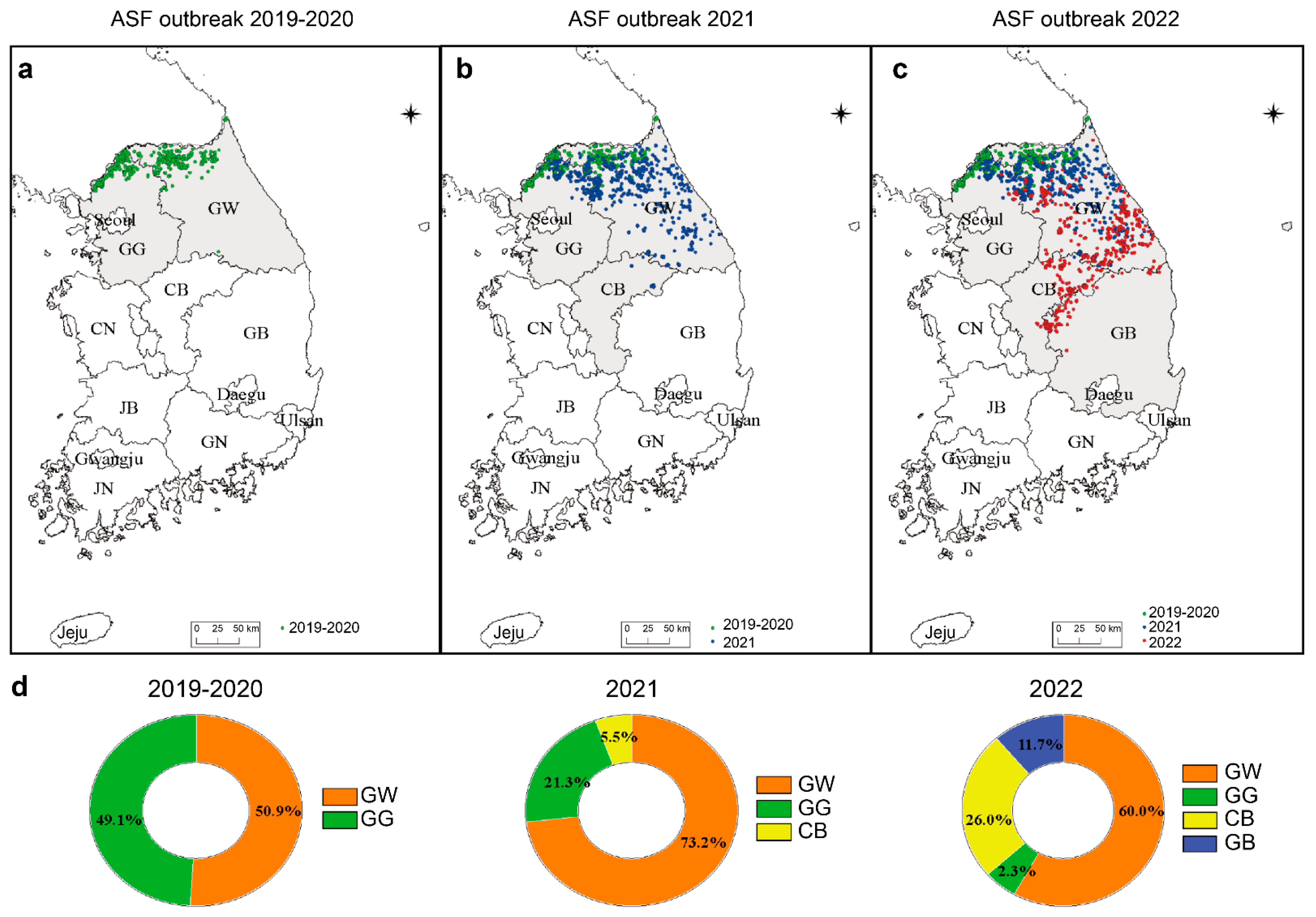
Figure 1. Analyzing the territorial distribution of African swine fever (ASF) throughout Korea during 2019–2022. The wild boar ASF outbreak is shown on the map by year, with (a) green dots representing 2019–2020, (b) blue dots for 2021, and (c) red dots for 2022. (d) ASF regional outbreaks between 2019 and 2022: orange, Gangwon-do; green, Gyeonggi-do; yellow, Chungcheongbuk-do; blue, Gyeongsangbuk-do. GW, Gangwon-do; GG, Gyeonggi-do; CB, Chungcheongbuk-do; CN, Chungcheongnam-do; GB, Gyeongsangbuk-do; GN, Gyeongsangnam-do; JB, Jeollabuk-do; JN, Jeollanam-do.
Viruses 15 01667 g001
Figure 2. The genome of the African swine fever virus (ASFV) obtained from a wild boar in South Korea was mapped geographically. The red dots on the map indicate the occurrence of ASF in wild boars from 2019 to 2022 (a) From left to right, the asterisks indicate the areas of Yeoncheon, Hwacheon, and Inje from where the ASFV genome has been registered in NCBI. (b) Visualization of the open reading frames (ORFs) of ASFV in Korea/YC1/2019, Korea/HCC224/2020, and Korea/IJ702/2020.
Viruses 15 01667 g002
Figure 3. Maximum likelihood phylogenetic tree constructed from ASFV genome sequences. The scale bar represents the number of substitutions per site, and the node values are shown as bootstrap values (1000 replications). The red asterisk represents the ASFV strain that occurred in wild boars in South Korea.
Viruses 15 01667 g003
Figure 4. The sequence similarity matrix plot displays pairwise comparisons of ASFV genome sequences in the curated dataset. The similarity degree between genome sequence pairs is represented by different colors, with dark red indicating 100% identity and blue indicating lower identity. The red asterisk represents the ASFV strain that occurred in wild boars in South Korea.
Viruses 15 01667 g004
Figure 5. Comparison of amino acid sequences in the MGFs 360-1La and 360-4L regions between the Korean wild boar ASFV strain and other ASFV strains. The partial amino acid sequence of the MGF-360 1La (a) and MGF-360 4L (b) regions of ASFV was aligned. Each character represents a single amino acid according to the international nomenclature. During amino acid alignment, identical amino acids were marked with dots, and isolates highlighted in different colors belong to different groups based on sequence variations caused by mutations. The red asterisk represents the ASFV strain that occurred in wild boars in South Korea.
Viruses 15 01667 g005
Figure 6. Spatial spread of different ASF strains in Korea from 2019 to 2022. (a) The green dots represent wild-type viruses, and the blue ones represent mutant viruses. (b) The graph shows the incidence rates of wild and mutant strains by region. (c) The wild type is on the left; the mutant-type virus is on the right; the hunted boar is in blue, and the carcass is in red. GW, Gangwon-do; GG, Gyeonggi-do; CB, Chungcheongbuk-d; GB, Gyeongsangbuk-do.
Viruses 15 01667 g006
Table 1. ASFV genome information used in this study.
StrainAccession NumberCountryYearHostLength (bp)GenotypeRef.
Korea/YC1/2019ON075797Korea2019Wild boar188,950II[31]
Korea/HC224/2020OP618183Korea2020Wild boar188,645IIUnpublished
Korea/IJ702/2020-Korea2020Wild boar188,598IIUnpublished
Georgia 2007/1FR682468.2Georgia2007Pig190,584II[32]
ASFV/pigChina/CAS19-01/2019MN172368China2019Pig189,405II[33]
Belgium 2018/1LR536725Belgium2018Wild boar189,404II[34]
ASFV-wbBS01MK645909China2018Wild boar189,394IIUnpublished
Arm/07/CBM/c2LR812933Armenia2007Pig190,145II[35]
Wuhan2019-1MN393477China2019Pig190,576IIUnpublished
Tanzania/Rukwa/2017/1LR813622Tanzania2007Pig183,186II[36]
China/2018/AnhuiXCGQMK128995China2018Pig189,393II[37]
ASFV/POL/2015/PodlaskieMH681419Poland2015Pig189,394II[25]
Odintsovo_02/14KP843857Russia2014Wild boar189,333II[38]
Estonia 2014LS478113Estonia2014Wild boar182,446II[39]
ASFV_HU_2018MN715134China2018Pig190,601II[40]
ASFV/Ulyanovsk 19/WB-5699MW306192Russia2019 189,263II[41]
Pig/HLJ/2018MK333180China2018Pig189,404II[42]
Spain-E75NC_044958.1Spain 181,187I[43]
Portugal-L60NC_044941Portugal1960 182,362I[44]
HeN/ZZ-P1/21MZ945536China2021Pig171,235I[45]
SD/DY-1-21MZ945537China2021Pig172,025I[45]
72407/Ss/2005MN270978Italy2005Sus scrofa181,699IUnpublished
Mkuzi/1979AY261362South Africa1979 192,714IUnpublished
Liv13/33MN913970Zambia2017Ornithodoros moubata188,277I[46]
WarmbathsAY261365South Africa1987Tick190,773IIIUnpublished
Namibia/WarthogAY261366Namibia1980Warthog186,5258IV[47]
Ken06.BusKM111295Kenya2006Pig184,368IXUnpublished
Uganda/R35MH025920Uganda2015Pig188,629IXUnpublished
Tengani62AY261364Malawi1962Pig185,689VUnpublished
MalawiLil-20/1AY261361Malawi1983Tick187,162VIII[48]
ASFV Ken.rie1LR899131Kenya2019Tick190,592XUnpublished
Kenya1950AY261360Kenya1950Pig193,886X[49]
BUR/18/RutanaMW856067Burundi2018Pig176,564X[50]
Table 2. Genetic differences in Korean ASFV strains compared with other ASFV strains.
NameAccession NumberNonsynonymousSynonymous
MGF 360-1LaMGF360-4LMGF 360-10L
Korea/YC1/2019ON075797ACG
Korea/HC224/2020OP618183ACG
Korea/IJ702/2020-GGC
Georgia2007FR682468ACC
HU2018MN715134ACC
Pig/HLJMK333180ACC
AnhuiXCGQ/2018MK128995ACC
wbBS01MK645909ACC
Pig/china2019MN172368ACC
Wuhan2019MN393477ACC
OdintsovoKP843857ACC
ulyanovsk19MW306192.1ACC
Arm/07/CBMLR812933ACC
Belgium2018LR536725ACC
Tanzania2017LR813622ACC
Podlaskie2015MH681419ACC
Disclaimer/Publisher’s Note: The statements, opinions and data contained in all publications are solely those of the individual author(s) and contributor(s) and not of MDPI and/or the editor(s). MDPI and/or the editor(s) disclaim responsibility for any injury to people or property resulting from any ideas, methods, instructions or products referred to in the content.