Communication

Genomes of Alphanucleorhabdovirus Physostegiae Isolates from Two Different Cultivar Groups of Solanum melongena

by
Nikita Gryzunov
1,2,†,
Sergey Yu. Morozov
3,†,
Tatiana Suprunova
4,
Viktoriya Samarskaya
1,
Nadezhda Spechenkova
1,
Sofiya Yakunina
1,
Natalia O. Kalinina
1,3 and
Michael Taliansky
1,5,*
1
Shemyakin-Ovchinnikov Institute of Bioorganic Chemistry of the Russian Academy of Sciences, 117997 Moscow, Russia
2
Faculty of Bioengineering and Bioinformatics, Lomonosov Moscow State University, 119234 Moscow, Russia
3
Belozersky Institute of Physico-Chemical Biology, Lomonosov Moscow State University, 119991 Moscow, Russia
4
Doka-Gene Technologies Ltd., 141880 Rogachevo, Russia
5
The James Hutton Institute, Invergowrie, Dundee DD2 5DA, UK
*
Author to whom correspondence should be addressed.
These authors contributed equally to this work.
Viruses 2024, 16(10), 1538; https://doi.org/10.3390/v16101538
Submission received: 26 August 2024 / Revised: 13 September 2024 / Accepted: 27 September 2024 / Published: 28 September 2024
(This article belongs to the Section Viruses of Plants, Fungi and Protozoa)

Abstract

:
Plant rhabdoviruses cause considerable economic losses and are a threat to the agriculture of Solanaceae plants. Two novel virus isolates belonging to the family Rhabdoviridae are identified by high-throughput sequencing (HTS) in Russian eggplant cultivars grown in the Volga river delta region for the first time. The phylogenetic inference of L protein (polymerase) shows that these virus isolates belong to Alphanucleorhabdovirus physostegia (Physostegia chlorotic mottle virus—PhCMoV), and their minus-sense RNA genomes have the typical gene order 3′-nucleocapsid (N)—X protein (X)—phosphoprotein (P)—Y protein (Y)—matrix protein (M)—glycoprotein (G)—polymerase (L)-5′ observed in some plant-infecting alphanucleorhabdoviruses. One of the PhCMoV isolates from the eggplant cultivar Almaz is genetically very similar to the Russian PhCMoV isolate from tomato and grouped in a subclade together with four isolates from Belgium, Germany, the Netherlands, and France. However, another eggplant-infecting isolate from the Russian cultivar Boggart is the most divergent compared with the other 45 virus genomes of European PhCMoV isolates. Thus, our comparative analysis reveals that two virus isolates from Russia may either share a close evolutionary relationship with European isolates or significantly diverge from all known virus isolates. The potential to use the protein sequence comparative analysis of accessory polypeptides, along with the early developed strategy of the nucleotide sequence comparison of the RNA genomes, is shown.

1. Introduction

Plant RNA viruses have an outstanding ability to achieve a high evolution rate due to fast replication and rapid accumulation of mutations since their RNA replicases lack proofreading activity [1,2]. The genetic variations produced by mutation and recombination processes are known to be under pressure of natural selection resulting from the competition between genetic variants influencing important aspects of the virus life cycle [3,4]. As a result, a limited number of variants with high reproductive fitness will pass to the next generations. In limited virus populations, genetic drift also influences the frequency of genetic variants by random chance [2,3,4]. Gene flow favors genetic uniformity between virus populations due to the selection process, whereas the restricted migration of genetic variants because of geographic/biological factors and genetic drift leads to enhanced genetic differentiation among populations. In general, the characterization of the genetic variability among virus populations is important for developing accurate detection and diagnostic tools and implementing efficient disease control strategies [4,5].
Eggplant (Solanum melongena L.) is an economically important vegetable crop worldwide. Eggplant ranks as the third most significant crop within the Solanaceae family after potatoes and tomatoes [6,7]. It is known that eggplant varieties can be infected by fungi, bacteria, and viruses that may cause important diseases with a negative impact on production and fruit quality [8,9,10]. In particular, there are two nucleorhabdoviruses named Alphanucleorhabdovirus melongenae [Eggplant mottled dwarf virus (EMDV)] and Alphanucleorhabdovirus physostegiae [Physostegia chlorotic mottle virus (PhCMoV)] that have negative effects on crop production [11,12,13]. These viruses cause deformed leaves of varying intensity, which have chlorotic to yellow discolorations of the veins. Foliar symptoms may be accompanied by mild-to-severe plant stunting and a lack of fruit. The fruits, when present, are small and often deformed [11,12,13]. These two virus species positioned in the genus Alphanucleorhabdovirus (family Rhabdoviridae, order Mononegavirales) encode five conserved proteins in the negative-sense RNA genome 3′-to-5′ direction [nucleocapsid (N), phosphoprotein (P), matrix (M), glycoprotein (G), and the polymerase (L)], like other rhabdoviruses. Furthermore, both EMDV and PhCMoV and three other nucleorhabdoviruses contain an accessory Y gene inserted between P and M and encoding the virus movement protein, as well as an additional auxiliary one (X gene) located between N and P, giving rise to an acidic protein with a currently unknown function [11,14,15,16,17,18]. All nucleorhabdovirus proteins are at least partly localized in the nuclei of host cells [19]. In the minus-sense rhabdovirus genomic RNA, these seven genes are separated by intergenic untranslated regions (UTRs), which, along with the 3′ leader and 5′ trailer sequences, are essential for the replication and transcription of the viral RNA. The intergenic UTRs contain termination and promoter signals, which are required during transcription to produce monocistronic mRNAs [11,13,15,16,17,18].
RNA viruses, such as nucleorhabdoviruses, exist as quasi-species, with the emergence of new strains and species being a continuous process [20]. EMDV and PhCMoV strain populations are widespread among crops (including eggplant) in the Mediterranean basin and have also been detected in many other European countries [11,12,13,21]. In 2022, to allow for efficient comparisons of epidemiologically important PhCMoV lineages, researchers from eight European laboratories proposed that “the current situation requires rapid characterization and a common response from European countries” to detect PhCMoV in Europe over a wide range of economically important plant crops [11]. However, currently, there is only a single report showing the presence of PhCMoV in the tomato sample from Russia [11]. Thus, data on the possible infection of eggplants by nucleorhabdoviruses in Russia are practically absent.
In the last few decades, in addition to well-established serological methods (mainly enzyme-linked immunosorbent assay, ELISA), molecular techniques (hybridization and DNA amplification, particularly PCR) have been developed for the detection of plant viruses [5,22]. High-throughput sequencing (HTS) technologies have resulted in enormous progress in plant virus diagnosis and identification [16,23,24]. HTS is not dependent on previous knowledge of viral sequences in a plant probe and results in the rapid diagnosis of all viruses (even unknown) present in plants (both symptomatic and asymptomatic), although often it does not show a direct association between the symptomatic disease and the detected viruses [25,26]. In this paper, we used HTS to detect nucleorhabdoviruses infecting eggplants in Russia (the Volga river delta region). The first report on the plant rhabdoviruses (represented by two isolates of PhCMoV) in Russian cultivars of Solanum melongena is presented.

2. Materials and Methods
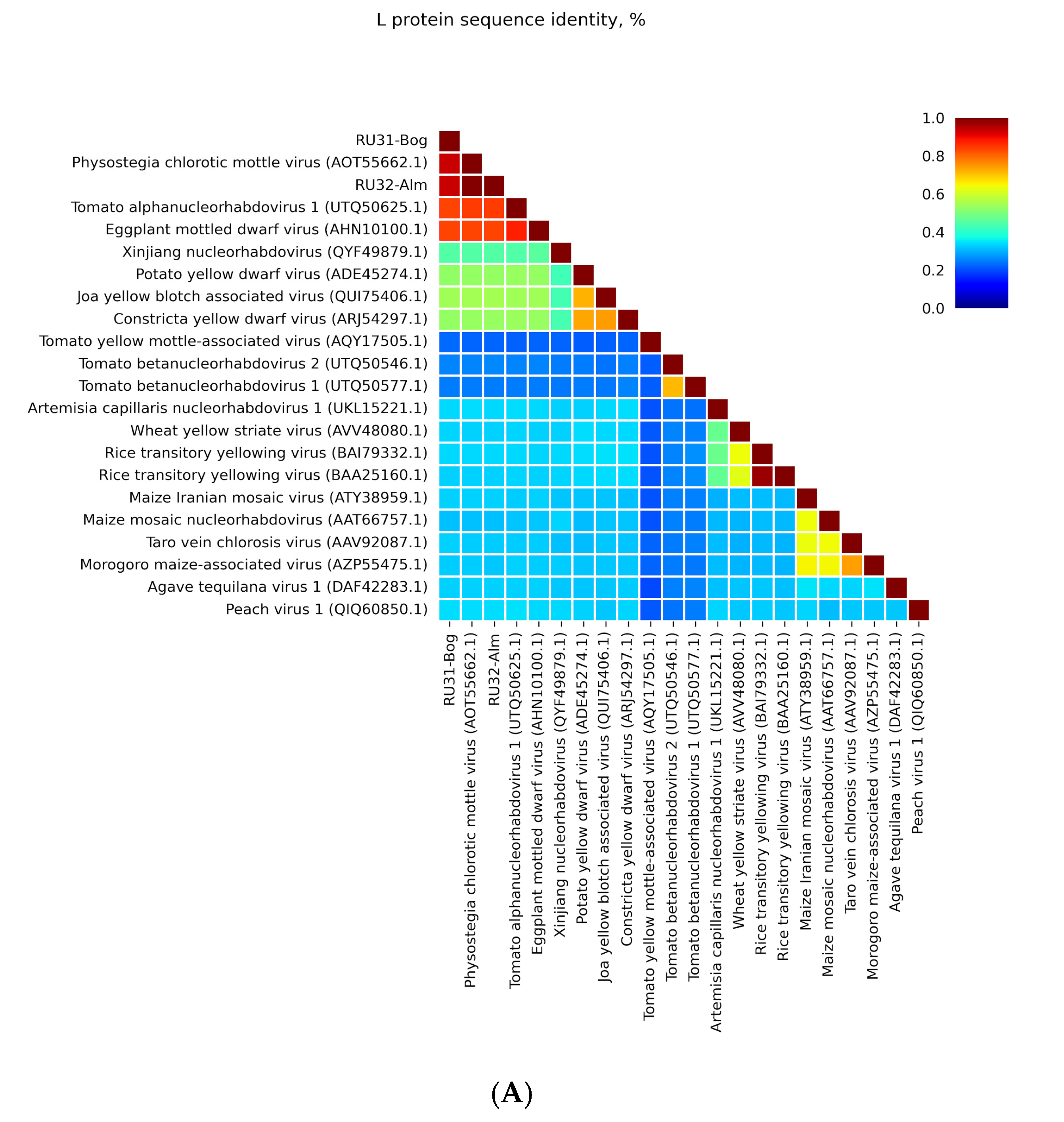
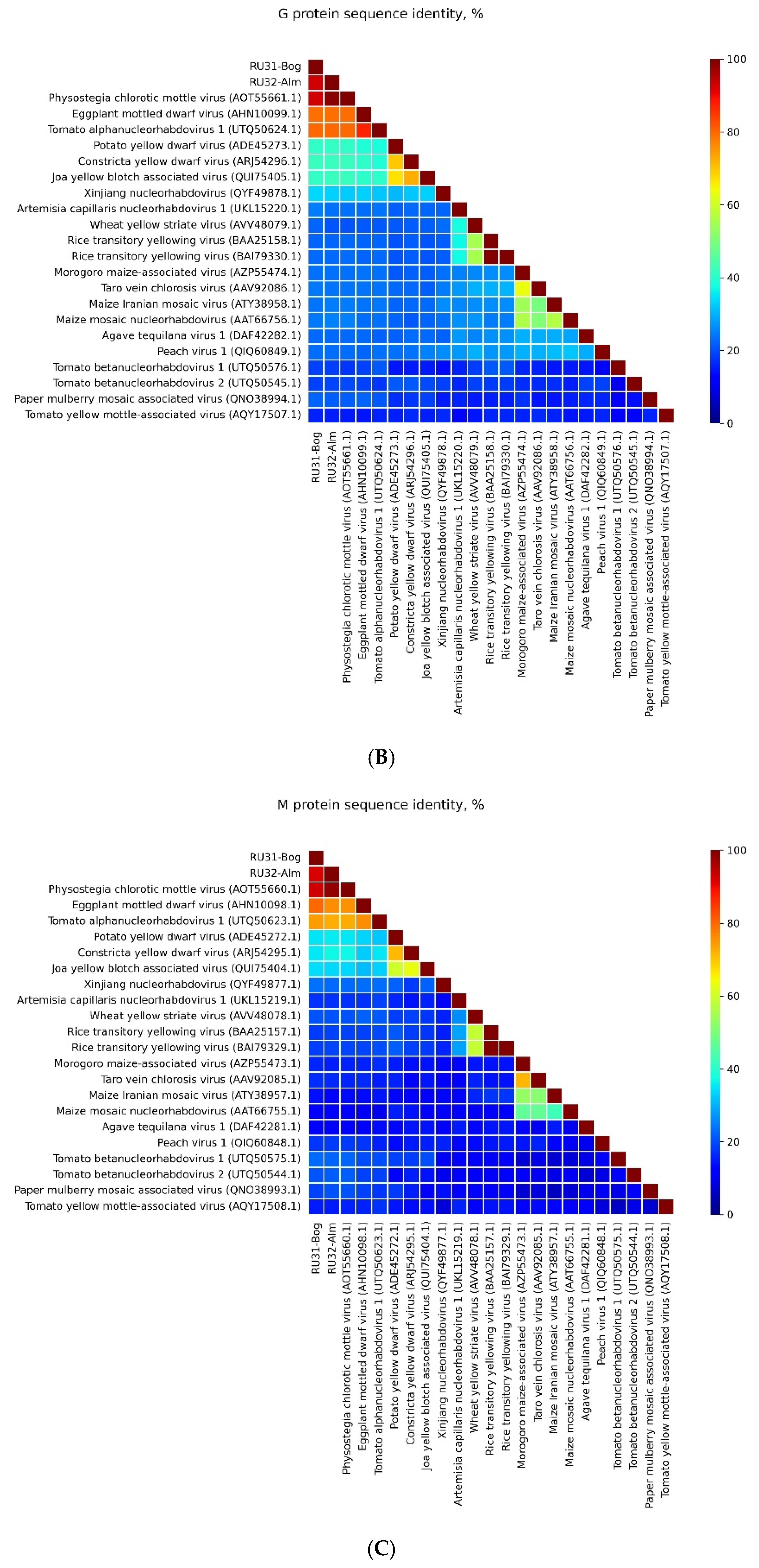
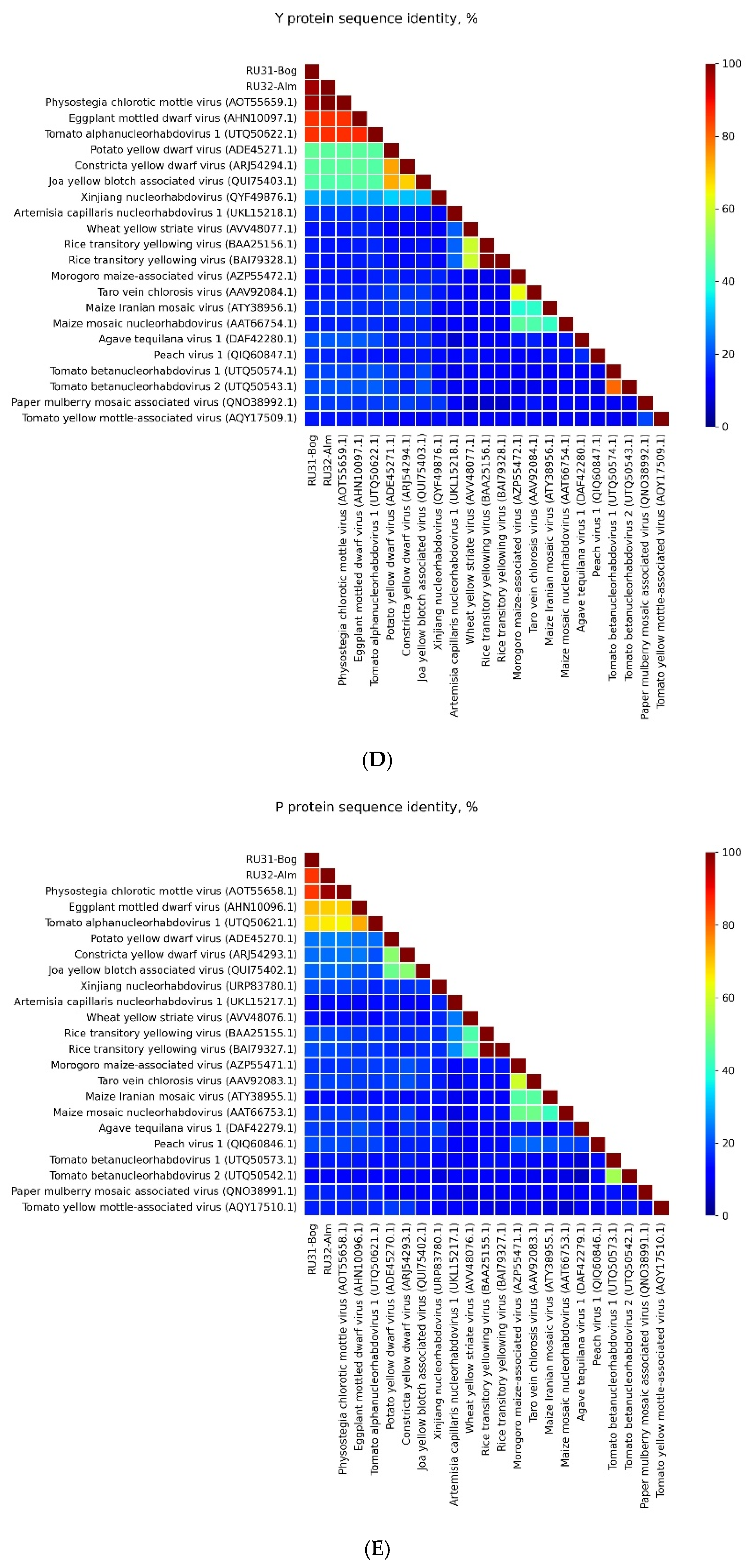
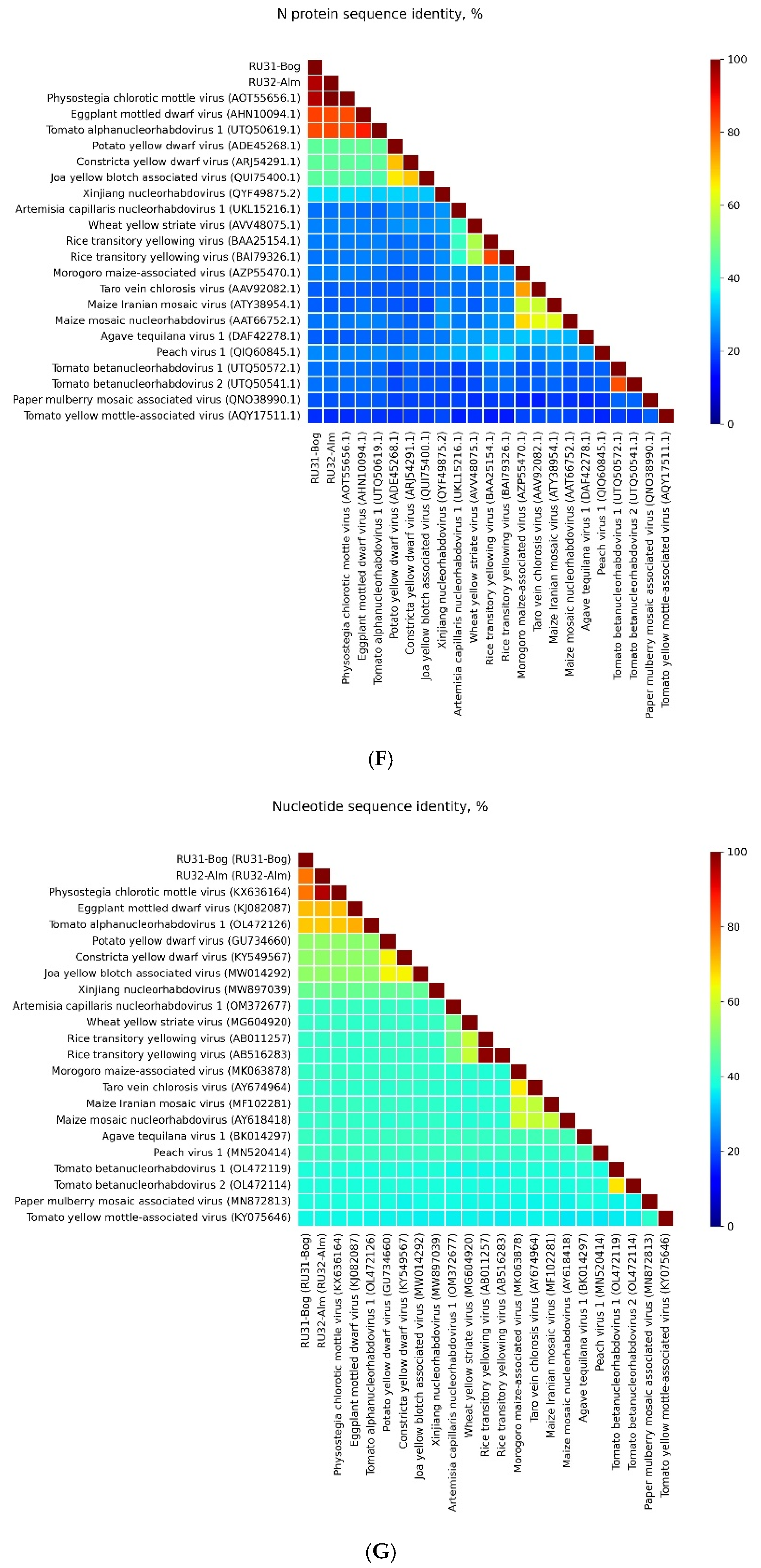
Samples of leaf material from two eggplant cultivar groups, Boggart (Bog) and Almaz (Alm), were obtained on 3 August 2023 from asymptomatic field-grown plants in the Astrakhan region near the Volga river delta in Russia from true leaves at stages 4–5. Leaves were collected in a fixative buffer (Fixator IntactRNA (Evrogen, No. BC031, Moscow, Russia), 20–22 plants per cultivar. The isolation of total RNA was performed using TRIzol, yielding two tubes of RNA preparations per sample, which were dissolved in nuclease-free water (NFW, Thermo Fisher Scientific, Waltham, MA, USA). RNA samples were treated with DNase I (RNase-free, 50 U/µL, Thermo #EN0523, a new batch from BioInnLabs, Rostov-on-Don, Russia) at a ratio of 1 unit per 1 µg of RNA. Moreover, 1 μL of RiboCare RNase Inhibitor was added to each sample (40 e.a./μL, EK005S, Eurogen, Moscow, Russia). The mixture was incubated at 37 °C for 30 min before being resuspended with TRIzol-chloroform without temperature termination. The RNA quantity and quality were determined using gel electrophoresis and a NanoDrop spectrophotometer (Thermo Fisher Scientific, Waltham, MA, USA). Total RNA libraries were constructed using TruSeq Stranded Total RNA with a Ribo-Zero Plant kit (Illumina, San Diego, CA, USA), which enables rRNA depletion. Libraries were sequenced on Illumina NovaSeq 6000 (2 × 101 bp). Reads were demultiplexed using Illumina bcl2fastq v2.20. Adapter removal was achieved through the use of Skewer v0.2.2 [27]. Sequencing, demultiplexing, and adapter removal were performed by CeGaT GmbH (Tuebingen, Germany). The RNA-sequencing libraries without adapters were deposited in the Sequence Read Archive (SRA) under BioProject accession number PRJNA1144656.
Read quality was evaluated with the FastQC v0.11.9 (https://www.bioinformatics.babraham.ac.uk/projects/fastqc/, accessed on 8 June 2024) and SeqKit v2.3.0 [28] tools. A total of 34,178,077 and 26,858,940 paired-end 101 nt reads were obtained for the RU31-Boggart and RU32-Almaz libraries, respectively, via HTS cDNA library construction. These two libraries demonstrated good sequencing quality, with Q30 values ranging from 88.32 to 94.09% across both libraries, and reads from the libraries were normalized using bbnorm.sh v39.01 (BBMap—Bushnell B.—https://sourceforge.net/projects/bbmap/, accessed on 8 June 2024) and then mapped to the Solanum melongena genome assembly GCA_000787875.1 using hisat2 v2.1.0 to remove host reads. Then, de novo assembly by Trinity v2.14.0 (standard k-mer length, 25 nt) [29] produced contig sets of 10,836 and 8956 sequences, respectively. Contig taxonomy was determined using BLASTn v2.14.0+ [30], and the ref_viruses_rep_genomes BLAST database (NCBI, Bethesda, MD, USA) was used for annotation. Several contigs were identified as associated with plant viruses. Two contigs (RU31-Bog and RU32-Alm) >13 kb in size revealed similarities to nucleorhabdoviruses (see the Section 3). The remaining contigs showed nearly 99% homology to cucumber mosaic virus RNA1, RNA2, and RNA3. RU31-Bog and RU32-Alm contigs were validated through independent assemblies using rnaSPAdes v3.15.4 [31] and rnaviralSPAdes v3.15.4 [32]. To further validate contigs, the original libraries were mapped to contigs using hisat2 v2.1.0 (https://daehwankimlab.github.io/hisat2/, accessed on 8 June 2024), followed by the removal of reads with mismatches. Both contigs had coverage depths of at least 100 reads throughout most of their length. These two contigs were submitted to GenBank with the accession numbers PQ206275 and PQ206276.
Detailed sequence comparisons were also carried out using the BLAST algorithm (BLASTn and BLASTp) at NCBI. Open reading frames (ORFs) were identified using the ORF Finder programs (https://www.ncbi.nlm.nih.gov/orffinder/, accessed on 8 June 2024). Conserved motif searches were conducted in CDD (http://www.ncbi.nlm.nih.gov/Structure/cdd/wrpsb.cgi/, accessed on 8 June 2024) databases. Phylogenetic analysis was performed with “Phylogeny.fr” (a free, simple-to-use web service dedicated to the reconstruction and analysis of phylogenetic relationships between molecular sequences) by constructing maximum likelihood phylogenetic trees (http://www.phylogeny.fr/simple_phylogeny.cgi/, accessed on 8 June 2024). Bootstrap percentages received from 1000 replications were used.

3. Results

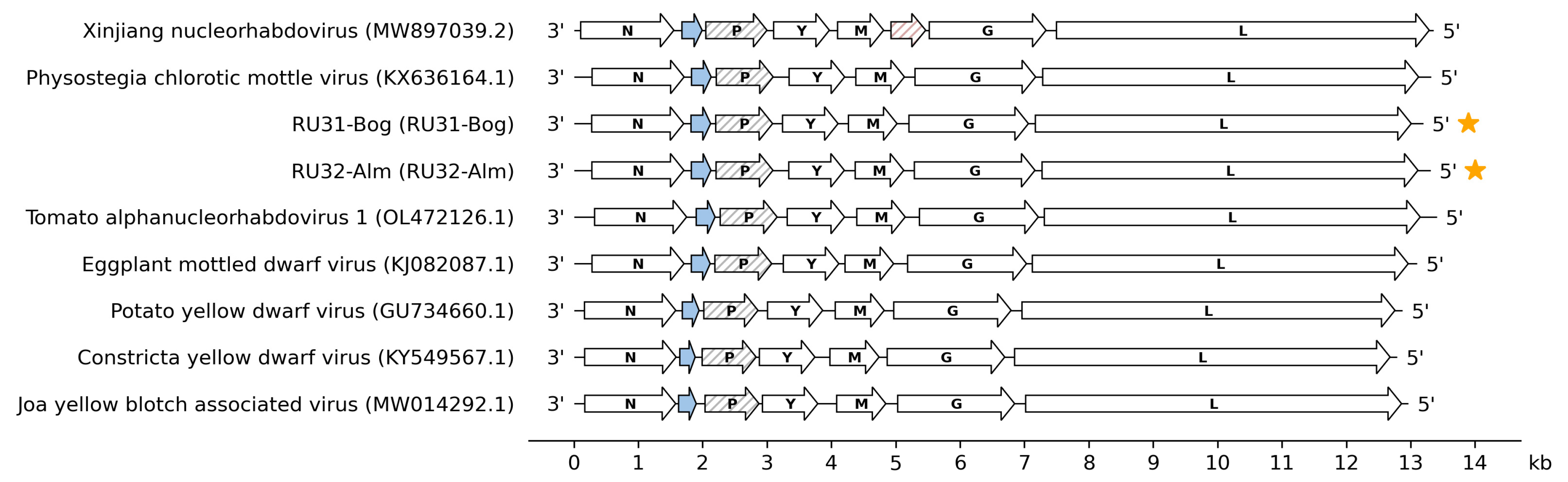
3.1. Genome Sequences of the Revealed Eggplant Rhabdoviruses

Our HTS studies of RNA samples isolated from two Russian eggplant cultivars, Boggart (Bog) and Almaz (Alm), have revealed, in each cultivar, long contigs resembling the size of plant rhabdovirus negative-sense RNA genomes. We have named these probable virus genomes RU31-Bog (size 13,197 nts) and RU32-Alm (size 13,307 nts). NCBI BLASTx analysis has unambiguously shown that both contigs encode polymerase proteins highly similar to those of virus species belonging to the genus Alphanucleorhabdovirus. Pairwise sequence comparisons at NCBI BLASTp have revealed that proteins G, M, Y, P, and N encoded by both found eggplant viruses are most similar to the corresponding proteins of PhCMoV (identity 92–94%), whereas RNA polymerases (protein L) have smaller similarity to other alphanucleorhabdoviruses, EMDV, and Tomato alphanucleorhabdovirus 1 (identity near 84%) (Figure 1A–F). The genomic nucleotide sequences of these sequenced isolates have 79% and 82% of their identity matched to most of the other PhCMoV isolates (Figure 1G).
These data strongly suggest that the sequenced eggplant viruses represent isolates (strains) of PhCMoV because one of the criteria used to differentiate species within the genus Alphanucleorhabdovirus is that the nucleotide sequence identities of entire genomes must be less than 75% [13]. Sequences of RU31-Bog and RU32-Alm have been translated into amino acid sequences of open reading frames (ORFs) via NCBI ORF Finder. Both genomes encode seven genes in the antigenomic sense (Figure 2). These genomic RNAs contain, in the 3′-to-5′ direction, coding regions for the proteins N, X, P, Y, M, G, and L. Their genome organization is similar to those of EMDV, Potato yellow dwarf virus (PYDV), Constricta yellow dwarf virus (CYDV), and Joa yellow blotch-associated virus (JYBaV) but differs from those of other alphanucleorhabdoviruses (Figure 2).
All genes of RU31-Bog and RU32-Alm are separated by conserved gene junction regions with the consensus sequence 3′-AAAUUAUUUUU GGG UUG-5′, which represents the polyadenylation signal of the upstream gene, untranscribed G-rich intergenic region (in bold letters), and transcription initiation site for the downstream gene and is similar to those of other alphanucleorhabdoviruses [14,16,18] (Table 1). The alphanucleorhabdovirus genome region containing seven genes is flanked by the so-called 3′ leader and 5′ trailer [17,18].

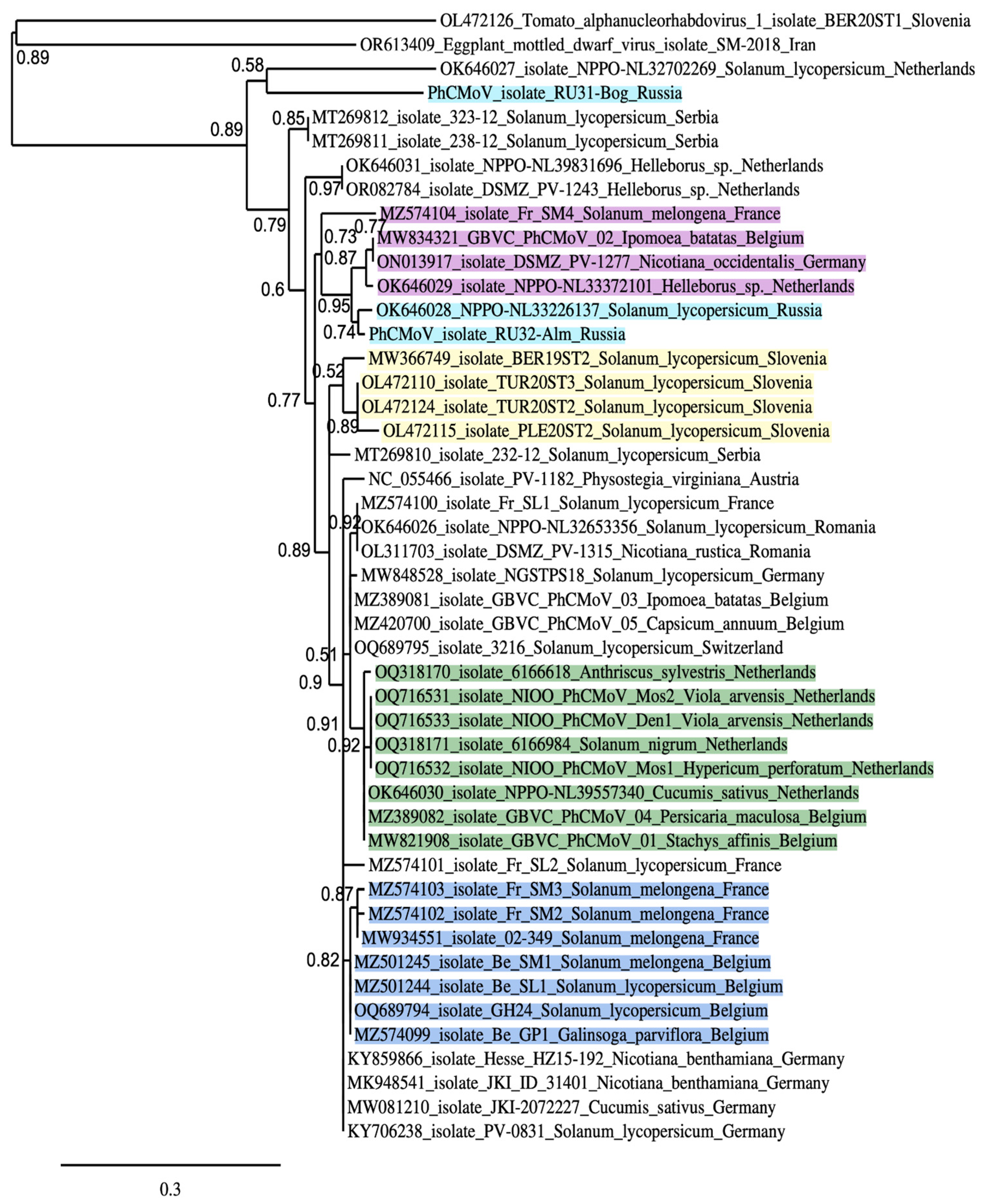
3.2. Placement of RU31-Bog and RU32-Alm in the Phylogenies of the Previously Sequenced PhCMoV Isolates

PhCMoV was first identified in Austria by the HTS method in Physostegia virginiana plants collected in 2014 [33]. Later, using HTS allowed for the detection of novel PhCMoV strains in Germany and Serbia on tomatoes [34,35]. Comprehensive comparative population analyses of more than 30 virus strains infecting plant species around Europe have previously been published [11,12]. PhCMoV has also been found to naturally infect 21 plant species across 15 plant families and is transmitted by a polyphagous leafhopper (genus Anaceratagallia) representing a natural vector [12]. It was previously revealed that relatively low genetic variability was observed for the genomic sequences (>93% nucleotide identity) in 28 out of 29 isolates [11]. Alternatively, to understand the evolutionary relationships among the 45 PhCMoV isolates in our study, amino acid identities were calculated for X proteins (Figure 3), which are the most divergent among plant nucleorhabdoviruses [11,17,21].
These phylogenetic data show that RU31-Bog is significantly diverged from other PhCMoV strains (Figure 3), like the strain NPPO-NL32702269 isolated from Solanum lycopersicum in the Netherlands [11]. Indeed genomic nucleotide sequences of these isolates have 79% and 82% common identity with most other isolates, which in turn show a common identity of 95–98% with each other (Table 2 and Table 3) [11]. RU31-Bog and NPPO-NL32702269 show 80% common identity in pairwise nucleotide sequence comparison. The phylogenetic tree of PhCMoV X proteins contains several well-defined basal clusters (Figure 3). In particular, two Russian isolates, RU32-Alm from eggplant (this paper) and NPPO-NL33226137 from tomato [11], are included in a common cluster with four other strains isolated in France (eggplant), Belgium (Ipomea batatas), Germany (Nicotiana occidentalis), and the Netherlands (Helleborus sp.) (Figure 3). Importantly, the members of this cluster also have highest common nucleotide sequence identity with RU32-Alm among the PhCMoV strains (Table 2), thus showing clear sequence similarity correlation between X protein amino acid sequences and genome nucleotide sequences. One more phylogenetic cluster has been found to contain four Slovenian isolates (Figure 3). The members of this cluster also show the highest common genomic nucleotide sequence identities among PhCMoV strains (Table 3). Since the need to organize PhCMoV strains into different genetic groups or clusters was first recognized in a recent paper when isolates were primarily divided into genogroups based on full-length genome sequences [11], our data further suggest that we should designate genogroups based on the complete X protein amino acid sequences together with the data on genome nucleotide sequences. Importantly, the use of well-conserved amino acid sequences of RNA polymerase (L protein) or nucleocapsid protein, which are universally used as phylogenetic anchors, provides only poorly informative dendrograms with negligible amounts of distinct sequence clusters (Figure S1) due to very high sequence similarity (92–98%) between PhCMoV isolates.

4. Discussion

To better understand the genetic diversity and evolution of PhCMoV in Europe, the accumulation of genomic sequences from many diverse isolates is required [11]. Recent data highlight the importance of the geographical location on the virus population evolution of PhCMoV strains [11]. Moreover, this is in line with observations on other plant rhabdoviruses, where the phylogenetic position among strains correlates with geographical localization but not with the host plant [21,36,37]. These observations suggest that certain genetic variations may have occurred during the virus’ geographic spread and then fixed to promote the virus’ viability in specific areas.
Our study of two Russian isolates adds some data on the occurrence and genomic diversity of PhCMoV isolates from a novel geographical location near the border between Europe and Asia. Importantly, Russian isolates (RU31-Bog and RU32-Alm) have been identified in the same relatively small region (the Astrakhan region near the Volga river delta). Nevertheless, when the amino acid sequence identities of the different isolates of PhCMoV were compared in phylogenetic and pairwise analyses, RU31-Bog was the most divergent isolate, having an isolated position in the phylogenetic tree of X proteins (Figure 3) and showing 79% nucleotide sequence identity compared with the other 45 strain genomes. Another most divergent isolate, NPPO-NL32702269, with 81–82% identity has been found in the Netherlands [11]. Both these isolates are quite distant from each other and evidently belong to PhCMoV strains, because one criterion used to differentiate species within the genus Alphanucleorhabdovirus is that the nucleotide sequence identity of entire genomes must be less than 75% [13].
Interestingly, RU32-Alm and the Russian isolate NPPO-NL33226137 from tomato are clearly clustered with four other strains isolated in Western Europe (France, Belgium, Germany, and the Netherlands). These data are not correlated with the high impact of the geographical dimension on the genetic evolution of PhCMoV. However, obvious clustering of four PhCMoV strains from Slovenia (Figure 3, Table 3) is in accordance with the proposed significance of geographical location as a determinant of genetic diversity. Revealing another sub-cluster of eight isolates from Anthriscus sylvestris, Viola arvensis, Solanum nigrum, Hypericum perforatum, Cucumis sativus, Persicaria maculosa and Stachys affinis found in the neighboring countries (The Netherlands and Belgium) (Figure 3) also supports the significance of a common geographical location for the existence of a close genetic relationship.
Taken together with a previous study [11], our data confirm that our current understanding of the determinants of genetic diversity in virus populations remains partly incomplete, and finding more viral strain genomes in samples of different origins (new location and new host) is important for gaining a better understanding of the genetic diversity and evolution of PhCMoV.

Supplementary Materials

The following supporting information can be downloaded at https://www.mdpi.com/article/10.3390/v16101538/s1. Figure S1: The phylogeny of the PhCMoV strains based on N (nucleocapsid) protein sequences.

Author Contributions

Conceptualization, S.Y.M., N.O.K. and M.T.; methodology, N.G., V.S. and N.S.; formal analysis, N.G., S.Y.M. and V.S.; investigation, T.S., N.G., N.S. and S.Y.; resources, M.T.; writing—original draft preparation, N.G. and S.Y.M.; writing—review and editing, S.Y.M., N.O.K. and M.T.; project administration, N.O.K.; funding acquisition, M.T. All authors have read and agreed to the published version of the manuscript.

Funding

This research was supported by the Russian Science Foundation, grant number 23-74-30003.

Institutional Review Board Statement

Not applicable.

Informed Consent Statement

Not applicable.

Data Availability Statement

The data supporting the findings of this study are available within the article and in the NCBI database, BioProject accession number PRJNA1144656, GenBank accession numbers PQ206275 and PQ206276.

Conflicts of Interest

The authors declare no conflict of interests, including commercial interests. The funders had no role in the design of this study; the collection, analyses, or interpretation of data; the writing of the manuscript; or the decision to publish the results.

References

  1. Roossinck, M.J. Mechanisms of plant virus evolution. Annu. Rev. Phytopathol. 1997, 35, 191–209. [Google Scholar] [CrossRef] [PubMed]
  2. Elena, S.F.; Fraile, A.; Garcıa-Arenal, F. Evolution and emergence of plant viruses. Adv. Virus Res. 2014, 88, 161–191. [Google Scholar] [CrossRef] [PubMed]
  3. Moya, A.; Holmes, E.C.; González-Candelas, F. The population genetics and evolutionary epidemiology of RNA viruses. Nat. Rev. Microbiol. 2004, 2, 279–288. [Google Scholar] [CrossRef] [PubMed]
  4. McLeish, M.J.; Fraile, A.; García-Arenal, F. Population Genomics of Plant Viruses: The Ecology and Evolution of Virus Emergence. Phytopathology 2021, 111, 32–39. [Google Scholar] [CrossRef]
  5. Rubio, L.; Galipienso, L.; Ferriol, I. Detection of plant viruses and disease management: Relevance of genetic diversity and evolution. Front. Plant Sci. 2020, 11, 1092. [Google Scholar] [CrossRef]
  6. Yang, X.; Cheng, Y.-F.; Deng, C.; Ma, Y.; Wang, Z.W.; Chen, X.H.; Xue, L.B. Comparative transcriptome analysis of eggplant (Solanum melongena L.) and Turkey berry (Solanum torvum Sw.): Phylogenomics and disease resistance analysis. BMC Genom. 2014, 15, 412. [Google Scholar] [CrossRef]
  7. Page, A.; Gibson, J.; Meyer, R.S.; Chapman, M.A. Eggplant domestication: Pervasive gene flow, feralisation and transcriptomic divergence. Mol. Biol. Evol. 2019, 36, 1359–1372. [Google Scholar] [CrossRef]
  8. Garcia-Estrada, R.S.; Cruz-Lachica, I.; Osuna-Garcia, L.A.; Marquez, I. First Report of Eggplant Fruit Rot Caused by Phytophthora nicotianae Breda de Haan in Mexico. Plant Dis. 2021, 105, 513. [Google Scholar] [CrossRef] [PubMed]
  9. Scholthof, K.-B.G. The Past Is Present: Coevolution of Viruses and Host Resistance within Geographic Centers of Plant Diversity. Annu. Rev. Phytopathol. 2023, 61, 119–136. [Google Scholar] [CrossRef] [PubMed]
  10. Zhao, M.; Gitaitis, R.; Dutta, B. Characterization of Pseudomonas capsici strains from pepper and tomato. Front. Microbiol. 2023, 14, 1267395. [Google Scholar] [CrossRef]
  11. Temple, C.; Blouin, A.G.; De Jonghe, K.; Foucart, Y.; Botermans, M.; Westenberg, M.; Schoen, R.; Gentit, P.; Visage, M.; Verdin, E.; et al. Biological and Genetic Characterization of Physostegia Chlorotic Mottle Virus in Europe Based on Host Range, Location, and Time. Plant Dis. 2022, 106, 2797–2807. [Google Scholar] [CrossRef] [PubMed]
  12. Temple, C.; Blouin, A.G.; Boezen, D.; Botermans, M.; Durant, L.; De Jonghe, K.; de Koning, P.; Goedefroit, T.; Minet, L.; Steyer, S.; et al. Biological Characterization of Physostegia Chlorotic Mottle Virus, an Emergent Virus Infecting Vegetables in Diversified Production Systems. Phytopathology 2024, 114, 1680–1688. [Google Scholar] [CrossRef] [PubMed]
  13. Alfaro-Fernández, A.; Taengua, R.; Font-San-Ambrosio, I.; Sanahuja-Edo, E.; Peiró, R.; Galipienso, L.; Rubio, L. Genetic Variation and Evolutionary Analysis of Eggplant Mottled Dwarf Virus Isolates from Spain. Plants 2024, 13, 250. [Google Scholar] [CrossRef] [PubMed]
  14. Pappi, P.G.; Dovas, C.I.; Efthimiou, K.E.; Maliogka, V.I.; Katis, N.I. A novel strategy for the determination of a rhabdovirus genome and its application to sequencing of Eggplant mottled dwarf virus. Virus Genes 2013, 47, 105–113. [Google Scholar] [CrossRef] [PubMed]
  15. Walker, P.J.; Dietzgen, R.G.; Joubert, D.A.; Blasdell, K.R. Rhabdovirus accessory genes. Virus Res. 2011, 162, 110–125. [Google Scholar] [CrossRef] [PubMed]
  16. Bejerman, N.; Dietzgen, R.G.; Debat, H. Illuminating the Plant Rhabdovirus Landscape through Metatranscriptomics Data. Viruses 2021, 13, 1304. [Google Scholar] [CrossRef]
  17. Dietzgen, R.G.; Bejerman, N.E.; Goodin, M.M.; Higgins, C.M.; Huot, O.B.; Kondo, H.; Martin, K.M.; Whitfield, A.E. Diversity and epidemiology of plant rhabdoviruses. Virus Res. 2020, 281, 197942. [Google Scholar] [CrossRef]
  18. Dietzgen, R.G.; Bejerman, N.E.; Mei, Y.; Jee, C.L.J.; Chabi-Jesus, C.; Freitas-Astúa, J.; Veras, S.M.; Kitajima, E.W. Joá yellow blotch-associated virus, a new alphanucleorhabdovirus from a wild solanaceous plant in Brazil. Arch. Virol. 2021, 166, 1615–1622. [Google Scholar] [CrossRef]
  19. Jang, C.; Wang, R.; Wells, J.; Leon, F.; Farman, M.; Hammond, J.; Goodin, M.M. Genome sequence variation in the constricta strain dramatically alters the protein interaction and localization map of Potato yellow dwarf virus. J. Gen. Virol. 2017, 98, 1526–1536. [Google Scholar] [CrossRef]
  20. Domingo, E.; Perales, C. Viral quasispecies. PLoS Genet. 2019, 15, e1008271. [Google Scholar] [CrossRef]
  21. Pappi, P.G.; Maliogka, V.I.; Amoutzias, G.D.; Katis, N.I. Genetic variation of eggplant mottled dwarf virus from annual and perennial plant hosts. Arch. Virol. 2016, 161, 631–639. [Google Scholar] [CrossRef] [PubMed]
  22. Boonham, N.; Kreuze, J.; Winter, S.; van der Vlugt, R.; Bergervoet, J.; Tomlinson, J.; Mumford, R. Methods in virus diagnostics: From ELISA to next generation sequencing. Virus Res. 2014, 186, 20–31. [Google Scholar] [CrossRef] [PubMed]
  23. Bejerman, N.; Debat, H.; Dietzgen, R.G. The Plant Negative-Sense RNA Virosphere: Virus Discovery Through New Eyes. Front. Microbiol. 2020, 11, 588427. [Google Scholar] [CrossRef] [PubMed]
  24. German, T.L.; Lorenzen, M.D.; Grubbs, N.; Whitfield, A.E. New Technologies for Studying Negative-Strand RNA Viruses in Plant and Arthropod Hosts. Mol. Plant-Microbe Interact. 2020, 33, 382–393. [Google Scholar] [CrossRef]
  25. Massart, S.; Olmos, A.; Jijakli, H.; Candresse, T. Current impact and future directions of high throughput sequencing in plant virus diagnostics. Virus Res. 2014, 188, 90–96. [Google Scholar] [CrossRef]
  26. Roossinck, M.J. Deep sequencing for discovery and evolutionary analysis of plant viruses. Virus Res. 2017, 239, 82–86. [Google Scholar] [CrossRef]
  27. Jiang, H.; Lei, R.; Ding, S.W.; Zhu, S. Skewer: A fast and accurate adapter trimmer for next-generation sequencing paired-end reads. BMC Bioinform. 2014, 15, 182. [Google Scholar] [CrossRef]
  28. Shen, W.; Le, S.; Li, Y.; Hu, F. SeqKit: A Cross-Platform and Ultrafast Toolkit for FASTA/Q File Manipulation. PLoS ONE 2016, 11, e0163962. [Google Scholar] [CrossRef] [PubMed]
  29. Grabherr, M.G.; Haas, B.J.; Yassour, M.; Levin, J.Z.; Thompson, D.A.; Amit, I.; Adiconis, X.; Fan, L.; Raychowdhury, R.; Zeng, Q.; et al. Full-length transcriptome assembly from RNA-Seq data without a reference genome. Nat. Biotechnol. 2011, 29, 644–652. [Google Scholar] [CrossRef]
  30. Camacho, C.; Coulouris, G.; Avagyan, V.; Ma, N.; Papadopoulos, J.; Bealer, K.; Madden, T.L. BLAST+: Architecture and applications. BMC Bioinform. 2009, 10, 421. [Google Scholar] [CrossRef]
  31. Bushmanova, E.; Antipov, D.; Lapidus, A.; Prjibelski, A.D. rnaSPAdes: A de novo transcriptome assembler and its application to RNA-Seq data. GigaScience 2019, 8, giz100. [Google Scholar] [CrossRef] [PubMed]
  32. Meleshko, D.; Hajirasouliha, I.; Korobeynikov, A. coronaSPAdes: From biosynthetic gene clusters to RNA viral assemblies. Bioinformatics 2021, 38, 1–8. [Google Scholar] [CrossRef] [PubMed]
  33. Menzel, W.; Richert-Poggeler, K.; Winter, S.; Knierim, D. Characterization of a nucleorhabdovirus from Physostegia. Acta Hortic. 2018, 1193, 29–38. [Google Scholar] [CrossRef]
  34. Vučurović, A.; Kutnjak, D.; Mehle, N.; Stanković, I.; Pecman, A.; Bulajić, A.; Krstić, B.; Ravnikar, M. Detection of Four New Tomato Viruses in Serbia Using Post Hoc High-Throughput Sequencing Analysis of Samples from a Large-Scale Field Survey. Plant Dis. 2021, 105, 2325–2332. [Google Scholar] [CrossRef] [PubMed]
  35. Gaafar, Y.Z.A.; Abdelgalil, M.A.M.; Knierim, D.; Richert-Poggeler, K.R.; Menzel, W.; Winter, S.; Ziebell, H. First report of physostegia chlorotic mottle virus on tomato (Solanum lycopersicum) in Germany. Plant Dis. 2018, 102, 255. [Google Scholar] [CrossRef]
  36. Parrella, G.; Greco, B. Sequence variation of block III segment identifies three distinct lineages within Eggplant mottled dwarf virus isolates from Italy, Spain and Greece. Acta Virol. 2016, 60, 100–105. [Google Scholar] [CrossRef]
  37. Yang, X.; Chen, B.; Zhang, T.; Li, Z.; Xu, C.; Zhou, G. Geographic distribution and genetic diversity of rice stripe mosaic virus in Southern China. Front. Microbiol. 2018, 9, 3068. [Google Scholar] [CrossRef]
Figure 1. Pairwise identity matrix of the amino acid (AF) and nucleotide (G) sequences in nucleorhabdovirus proteins and genomes: (A) RNA polymerase (L protein); (B) G protein; (C) M protein; (D) Y protein; (E) P protein; (F) nucleocapsid protein; (G) RNA genome sequences.
Viruses 16 01538 g001aViruses 16 01538 g001bViruses 16 01538 g001cViruses 16 01538 g001d
Figure 2. Genome organization in nucleorhabdovirus species. Accessory genes are colored blue (X protein). Hatches: P protein. Two novel isolates (RU31-Bog and RU32-Alm) are marked with orange stars.
Viruses 16 01538 g002
Figure 3. The phylogenies of the 45 PhCMoV strains based on X protein sequences. Accession numbers, host species, and countries of isolation are shown. Russian isolates are shown in blue. The cluster of Slovenian strains is shown in yellow. Strains related to RU32-Alm are shown in pink. The scale bar indicates the number of substitutions per site. The green and light blue colors indicate sequence clusters including isolates from Western Europe.
Viruses 16 01538 g003
Table 1. The genome positions of intergenic junction sequences in PhCMoV isolates sequenced in this paper.
Location of GGG Sequence in Leader and Intercistronic Regions of Genomic RNANucleotide Position of Central G Residue in RU31-Bog *Nucleotide Position of Central G Residue in RU32-Alm **
Leader/N gene junction13,00113,109
N gene/X gene11,38811,493
X gene/P gene11,01711,122
P gene/Y (MP) gene99689981
Y (MP) gene/M gene89438948
M gene/G gene80298037
G gene/L gene60386043
* Total genome size—13,187 nt; ** Total genome size—13,307 nt.
Table 2. Pairwise nucleotide sequence comparisons of RU32-Alm genomic RNA with some other related PhCMoV strains.
PhCMoV Strains *E-ValuePercentage of IdentityNCBI Accession
isolate GBVC_PhCMoV_02_IB0.097.90%MW834321
isolate DSMZ PV-12770.097.86%ON013917
isolate NPPO-NL333721010.097.89%OK646029
isolate NPPO-NL332261370.096.71%OK646028
isolate Fr_SM40.096.22%MZ574104
isolate PV-11820.095.42%NC055466
isolate Be_GP10.095.40%MZ574099
isolate 32160.095.40%OQ689795
isolate 232–120.095.36%MT269810
* Isolates forming a cluster of X proteins with RU32-Alm are in bold and underlined.
Table 3. Pairwise nucleotide sequence comparisons of genomic RNAs from Slovenian isolates with some other related PhCMoV strains.
PhCMoV Strains *E-ValuePercentage of IdentityNCBI Accession
isolate BER19ST20.0100.00%MW366749
isolate TUR20ST20.097.67%OL472124
isolate TUR20ST30.097.67%OL472110
isolate PLE20ST20.097.63%OL472115
isolate Be_GP10.096.41%MZ574099
isolate GH240.096.43%OQ689794
isolate 32160.095.40%OQ689795
* Isolates forming a cluster of X proteins (Slovenian strains) are shown in bold and underlined.
Disclaimer/Publisher’s Note: The statements, opinions and data contained in all publications are solely those of the individual author(s) and contributor(s) and not of MDPI and/or the editor(s). MDPI and/or the editor(s) disclaim responsibility for any injury to people or property resulting from any ideas, methods, instructions or products referred to in the content.

