Article

Molecular Characterization and Genomic Surveillance of SARS-CoV-2 Lineages in Central India

1
ICMR-National Institute of Research in Tribal Health, Jabalpur 482003, Madhya Pradesh, India
2
Department of Microbiology and Biotechnology Centre, Faculty of Science, The Maharaja Sayajirao University of Baroda, Vadodara 390002, Gujarat, India
3
Academy of Scientific and Innovative Research (AcSIR), Ghaziabad 201002, Uttar Pradesh, India
*
Authors to whom correspondence should be addressed.
Joint first author.
Joint second authors.
Viruses 2024, 16(10), 1608; https://doi.org/10.3390/v16101608
Submission received: 7 June 2024 / Revised: 3 July 2024 / Accepted: 9 July 2024 / Published: 14 October 2024
(This article belongs to the Special Issue Molecular Epidemiology of SARS-CoV-2, 3rd Edition)

Abstract

:
Since the first reported case of COVID-19 in December 2019, several SARS-CoV-2 variants have evolved, and some of them have shown higher transmissibility, becoming the prevalent strains. Genomic epidemiological investigations into strains from different time points, including the early stages of the pandemic, are very crucial for understanding the evolution and transmission patterns. Using whole-genome sequences, our study describes the early landscape of SARS-CoV-2 variants in central India retrospectively (including the first known occurrence of SARS-CoV-2 in Madhya Pradesh). We performed amplicon-based whole-genome sequencing of randomly selected SARS-CoV-2 isolates (n = 38) collected between 2020 and 2022 at state level VRDL, ICMR-NIRTH, Jabalpur, from 11899 RT-qPCR-positive samples. We observed the presence of five lineages, namely B.1, B.1.1, B.1.36.8, B.1.195, and B.6, in 19 genomes from the first wave cases and variants of concern (VOCs) lineages, i.e., B.1.617.2 (Delta) and BA.2.10 (Omicron) in the second wave cases. There was a shift in mutational pattern in the spike protein coding region of SRAS-CoV-2 strains from the second wave in contrast to the first wave. In the first wave of infections, we observed variations in the ORF1Ab region, and with the emergence of Delta lineages, the D614G mutation associated with an increase in infectivity became a prominent change. We have identified five immune escape variants in the S gene, P681R, P681H, L452R, Q57H, and N501Y, in the isolates collected during the second wave. Furthermore, these genomes were compared with 2160 complete genome sequences reported from central India that encompass 109 different SARS-CoV-2 lineages. Among them, VOC lineages Delta (28.93%) and Omicron (56.11%) were circulating predominantly in this region. This study provides useful insights into the genetic diversity of SARS-CoV-2 strains over the initial course of the COVID-19 pandemic in central India.

1. Introduction

Coronavirus Disease 2019 (COVID-19) is caused by Severe Acute Respiratory Syndrome Coronavirus 2 (SARS-CoV-2). The first incidence of SARS-CoV-2 was reported from Wuhan City, China, in late December 2019, and the first genome sequence was submitted on 7 January 2020 (NCBI GenBank ID: MN908947.1) [1]. The World Health Organization declared COVID-19 a public health emergency on 30 January 2020 and declared it a pandemic on 11 March 2020 [2]. India too had witnessed the increased incidence of distinct SARS-CoV-2 strains in different states since the beginning of the pandemic [3]. By the end of 31 October 2023, it had been responsible for over 45 million cases and more than 0.53 million deaths in India [4]. SARS-CoV-2 was also found to show higher transmissibility compared to previously reported coronavirus outbreaks, i.e., SARS-CoV (2002) (WHO, 2003) and MERS-CoV (2012) [5]. On 27 January 2020, reports of the first COVID-19 case verified in India came from Kerala [6]. There are documented reports of various viral introductions brought about by international travelers. There were three waves of infections observed in India: the first wave occurred between 16 July 2020 and 31 January 2021, followed by the second wave from 16 March 2021 to 6 May 2021, and the third wave from 1 January 22 to 20 February 22 [7,8]. India, as one of the most populous countries with crowded cities, saw a surge in COVID-19 cases during March 2020. The virus also spread to rural areas and affected people of all age groups due to airborne transmission through viral particles and droplets, increased viral spread, and superspreading events linked to workers migrating from cities. In the state of Karnataka, the B.1.1.7 (Alpha) lineage was found to be predominant among many other lineages [9,10,11], and symptomatic cases have been linked to the pandemic’s early spread in the state. Three primary introductions of SARS-CoV-2 were identified in the state of Kerala by genomic sequencing and haplotype analysis [12]. These were followed by many outbreaks that caused the virus to spread locally [12]. In Gujarat state, lineages 20A and 20D were shown to be predominant among deceased patients with respiratory complications [13,14]. These lineages carry nucleotide changes C28854T in the N-gene and G25563T in Orf3a [13]. A collection of 200 genomes from the southern state of Telangana were sampled between March and July 2020 in order to assess the mutational landscape of SARS-CoV-2 isolates [14]. This analysis helped to detect mutations in non-structural proteins and demonstrated the preponderance of the 20B lineage [15]. Central India is a loosely defined geographical region of India, sometimes called Madhya Bharat, that consists of two Indian states: Madhya Pradesh (MP) and Chhattisgarh. Over 1.05 million cases and approximately 10,786 deaths were documented in this region as of 30 October 2023 [16]. Since the initial complete genome of SARS-CoV-2 from Wuhan was reported in early January 2020 [6], millions of whole-genome sequences worldwide have been added to public databases, i.e., GISAID and GENBANK, which have helped in the monitoring of SARS-CoV-2 variants. There has been a lack of genomic representation of SARS-CoV-2 from central India that needs to be addressed. Thus, more such studies representing different time frames and geographic regions are needed to understand the evolving landscape of the genetic diversity of the virus over the course of the pandemic. We have performed whole-genome sequencing of SARS-CoV-2 strains from the first and second waves of infections and performed comparative analysis with global strains to understand the dynamics of COVID-19.

2. Materials and Methods

2.1. Sample Acquisition

A total of 11,899 samples tested positive from 2.61 lakh tested cases for SARS-CoV-2 using real-time PCR (RT-PCR), and, among them, 38 samples underwent whole-genome sequencing and mutational analysis for the SARS-CoV-2 variants.
The Virus Research and Diagnostic Laboratory (VRDL) under the ICMR-National Institute of Research in Tribal Health, Jabalpur, is a designated state level (Grade-II) laboratory under the VRDL network established by the Department of Health Research, Ministry of Health & Family Welfare, Government of India. It was the first lab in Madhya Pradesh (central India) to start testing for suspected COVID-19 cases from Jabalpur and its surrounding districts of MP and Chhattisgarh. SARS-CoV-2-suspicious samples obtained from several quarantine camps and hospitals in Jabalpur area were processed for diagnostic testing in accordance with the WHO’s recommendations.

2.2. RNA Extraction and Real-Time Reverse Transcriptase PCR (RT-PCR)

During the period (March 2020 to May 2022), nasopharyngeal/oropharyngeal swabs (NPS/OPS) (n  =  263,124) were collected for routine SARS-CoV-2 diagnosis. A total of 140 μL of sample of the Virus Transport Medium (VTM) was used for viral RNA extraction using the QIAMP VIRAL RNA mini kit (Qiagen, Hilden, Germany). SARS-CoV-2 was confirmed using the ICMR-NIV multiplex single-tube SARS-CoV-2 RT-PCR assay kit by real-time RT-PCR. A total of 11,899 samples tested positive by real-time RT-PCR. Samples that displayed a cycle threshold (Ct) between 20 and 28 were considered eligible for sequencing to ensure maximum sequence coverage. Among positive samples, n = 19 samples from the very first reported cases of SARS-CoV-2 (Late March–April 2020) and that had international travel history were selected. Subsequently, n = 17 samples were randomly selected from the second wave, and n = 2 samples from the third wave were selected to see the presence of variants of concern (VOCs). The clinical and epidemiological details of the patients enrolled in the current study are shown in Table 1.

2.3. Library Preparation and Whole-Genome Sequencing

The cDNA was subjected to a multiplex PCR using the QIAseq SARS-CoV-2 primer panel (Qiagen GmbH, Germany). Subsequently, amplified products were purified using Agencourt AMPure XP beads (Beckman Coulter Inc., Brea, CA, USA) and quantified using the QubitTM dsDNA HS Assay Kit (Invitrogen, Waltham, Massachusetts, USA). The amplified cDNA sample was used for library preparation using the QIASeq FX DNA Library kit (Qiagen GmbH, Germany). The concentration and fragment size of the prepared libraries were checked using the Qubit Fluorometer (Thermofisher Scientific, Waltham, MA, USA) and Agilent Fragment Analyzer (Santa Clara, CA, USA) to make sure that the library profiles were in the expected range. The quality-checked normalized libraries were pooled and diluted to a final concentration of 10 pM, spiked with 5% PhiX. Whole-genome sequencing of first wave (n = 19) samples was performed on the Illumina iSeq platform using the iSeq 100 i1 Reagent v2 (San Diego, CA, USA) (300-cycle) kit, and subsequent samples of second and third waves were sequenced on the Ion GeneStudio S5 System platform using the Ion 540™ Chip Kit (Thermofisher Scientific, Waltham, MA, USA).

2.4. Variant Calling and Lineage Classification

The quality of the raw fastq files was assessed with the FastQC tool followed by the removal of low-quality bases, reads, and adapter sequencing using Trimmomatic [17,18]. The filtered sequence files were aligned to the SAS-CoV-2 reference genome (NC_045512.2), and reference-based assemblies were generated using BCFtools [19,20]. Sequence variations and lineage/clade classification were performed using Nextclade webserver followed by the identification of functional impacts [21]. The effect of the mutation on the structure of the protein was predicted using iStable [22].

2.5. Comparison with Global Strains

We have downloaded all the complete SARS-CoV-2 genomes (n = 2160) (Table S1) from central India for the phylogenetic analysis of SARS-CoV-2 genome sequences generated in this study, which were aligned with the strains reported from central India, using Augur Nextstrain’s phylodynamic pipeline toolkit “https://github.com/nextstrain/ncov (accessed on 10 May 2024)” [23,24]. Afterward, the maximum likelihood phylogenetic tree was constructed using the Augur tree implementation pipeline with IQ-TREE v1.6.1 [25] with the default parameters. The selected metadata information plotted in the time-resolved phylogenetic tree was constructed using TreeTime v0.81 [26], which was then annotated and visualized in the iTOL website [27].
Furthermore, the metadata of the genomes belonging to the seven lineages identified in this study were downloaded from the GISAID database until 31 March 2023 to check the transmission and circulation on a global scale.
Table 1. Epidemiological features of laboratory-confirmed COVID-19 cases.
Sample-IDAge (In Years)GenderDistrict of ResidenceTravel History/ContactDate of Sample CollectionSample TypeSymptoms
ICMR-NIRTH-S146MJABALPURNA27 March 2020 Nasopharyngeal swabfever sore throat
ICMR-NIRTH-S248MJABALPURNA27 March 2020 Oropharyngeal swabfever
ICMR-NIRTH-S324MJABALPURGermany20 March 2020 Nasopharyngeal swabfever cough
ICMR-NIRTH-S459MJABALPURDubai, UAE20 March 2020 Nasopharyngeal swabfever cough
ICMR-NIRTH-S545FJABALPURDubai, UAE20 March 2020 Nasopharyngeal swabcough sore throat
ICMR-NIRTH-S622FJABALPURDubai, UAE20 March 2020 Nasopharyngeal swabcough
ICMR-NIRTH-S753MJABALPURDubai (contact of ICMR-NIRTH-S4)21 March 2020 Nasopharyngeal swabcough
ICMR-NIRTH-S840MJABALPURNA22 March 2020 Nasopharyngeal swabcough body ache
ICMR-NIRTH-S1133MCHHINDWARAIndore travel history 1 April 2020 Nasopharyngeal swabfever cough breathlessness sore throat body ach
ICMR-NIRTH-S1226FCHHINDWARADelhi travel history7 April 2020 Nasopharyngeal swabfever sore throat
ICMR-NIRTH-S1327MCHHINDWARAContact of ICMR-NIRTH-S127 April 2020 Nasopharyngeal swabcough sore throat
ICMR-NIRTH-S1461MJABALPURDelhi travel history8 April 2020 Nasopharyngeal swabFever cough breathlessness body ach sputum
ICMR-NIRTH-S1530MINDOREIndore travel history 10 April 2020 Nasopharyngeal swabfever cough sore throat
ICMR-NIRTH-S1622MINDOREIndore travel history 10 April 2020 Nasopharyngeal swabfever sore throat
ICMR-NIRTH-S1724MSATNAIndore travel history 10 April 2020 Nasopharyngeal swabcough sore throat
ICMR-NIRTH-S1870MJABALPURNA11 April 2020 Nasopharyngeal swabfever cough sore throat
ICMR-NIRTH-S1950MJABALPURContact of ICMR-NIRTH-S1814 April 2020 Nasopharyngeal swabfever sore throat
ICMR-NIRTH-S2244FJABALPURContact of ICMR-NIRTH-S1917 April 2020 Throat swabcough sore throat
ICMR-NIRTH-S2335MBALAGHATNA27 July 2020Throat swabAsymptomatic
ICMR-NIRTH-S2428FBALAGHATNA16 April 2021Throat swabAsymptomatic
ICMR-NIRTH-S2528MBALAGHATNA16 April 2021Throat swabAsymptomatic
ICMR-NIRTH-S2624MBALAGHATNA17 April 2021Throat swabAsymptomatic
ICMR-NIRTH-S2724MBALAGHATNA18 April 2021Throat swabSymptomatic
ICMR-NIRTH-S2838MBALAGHATNA19 April 2021Throat swabSymptomatic
ICMR-NIRTH-S2925MBALAGHATNA19 April 2021Throat swabAsymptomatic
ICMR-NIRTH-S3019MBALAGHATNA20 April 2021Throat swabAsymptomatic
ICMR-NIRTH-S3114MBALAGHATNA9 April 2021Throat swabAsymptomatic
ICMR-NIRTH-S3245MBALAGHATNA9 April 2021Throat swabAsymptomatic
ICMR-NIRTH-S3370FBALAGHATNA12 April 2021Throat swabSymptomatic
ICMR-NIRTH-S3454MBALAGHATNA13 April 2021Throat swabAsymptomatic
ICMR-NIRTH-S3541MBALAGHATNA18 April 2021Throat swabAsymptomatic
ICMR-NIRTH-S3618MBALAGHATNA21 April 2021Throat swabAsymptomatic
ICMR-NIRTH-S3745FBALAGHATNA24 April 2021Throat swabAsymptomatic
ICMR-NIRTH-S3826FBALAGHATNA3 February 2022Throat swabAsymptomatic
ICMR-NIRTH-S3940FBALAGHATNA4 February 2022Throat swabAsymptomatic
ICMR-NIRTH-S4040MBALAGHATNA20 April 2021Throat swabAsymptomatic
ICMR-NIRTH-S4125MBALAGHATNA21 April 2021Throat swabAsymptomatic
ICMR-NIRTH-S4216FBALAGHATNA27 April 2021Throat swabAsymptomatic
NA = contact history not available.

3. Results

3.1. Genome Assembly, Lineage Assignment, and Mutational Analysis

All these SARS-CoV-2 whole-genome sequences mapped to the reference strain (Wuhan/Hu-1/2019, NC_045512.3) with 99.1–99.8% identity. The consensus assembly of 38 samples has been deposited in the BioProject ID: PRJNA759056 (Table S2). To analyze the mutation pattern, these 38 genomes were analyzed with the Auspice visualization tool of Nextclade [21] (Figure S1). Nextstrain clades and the Pangolin classification system were used for classifying the phylogenetic lineages, and their corresponding GISAID clade is shown in Table 2. These 38 SARS-CoV-2 strains belong to seven lineages, namely B.1.617 (Delta) (n = 17), B.1.195 (n = 8), B.1 (n = 6), B.1.36.8 (n = 2), BA.2.10 (Omicron) (n = 2), B.1.1 (n = 2), and B.6 (n = 1). This revealed n = 186 single nucleotide polymorphisms (n = 129 non-synonymous and n = 57 synonymous changes), as shown in Table S3. Even though approx. half of these samples are from the early stage of the pandemic, it is interesting to note that only one strain (ICMR-NIRTH-S14, lineage B.6, clade O (19A), date of collection: 8 April 2020) did not show a well-characterized D614G mutation in spike protein (Table 2).
In total, 29 novel nucleotide variations in the sequences of the SARS-CoV-2 genome have been identified from 38 patient isolates which were unique to the sample and not found in the genomes reported from the central India state, as per the data retrieved from the GISAID database. In the present study, 17 patients were asymptomatic, while the remaining 21 had symptoms. Upon comparing the distribution of the 29 novel mutations among our study samples, we found that a few mutations such as A4533G, A4778T, and G6167T were present only among the symptomatic ones (Tables S3 and S4).

3.2. Effect of Novel Mutations on Protein Stability

Using an integrated sequence and structure predictor, iStable [22], the effects of the novel 29 mutations identified were determined. Among 29 mutations, 20 mutations were only identified in the ORF1ab region, which was known to encompass mutational spectra. Out of these 29 mutations, 17 mutations were non-synonymous, 11 were synonymous, and 1 mutation was found in the non-coding region. A total of 15 non-synonymous mutations were found to confer deleterious effects on protein function as they scored in a range between 0 and 1 (Table S4). As most of the mutations were found in ORF1ab region, it was interesting to note that the mutational spectra of this region should be considered while designing new antiviral therapeutics targeting viral ORF1ab.

3.3. Phylogenetic Analysis

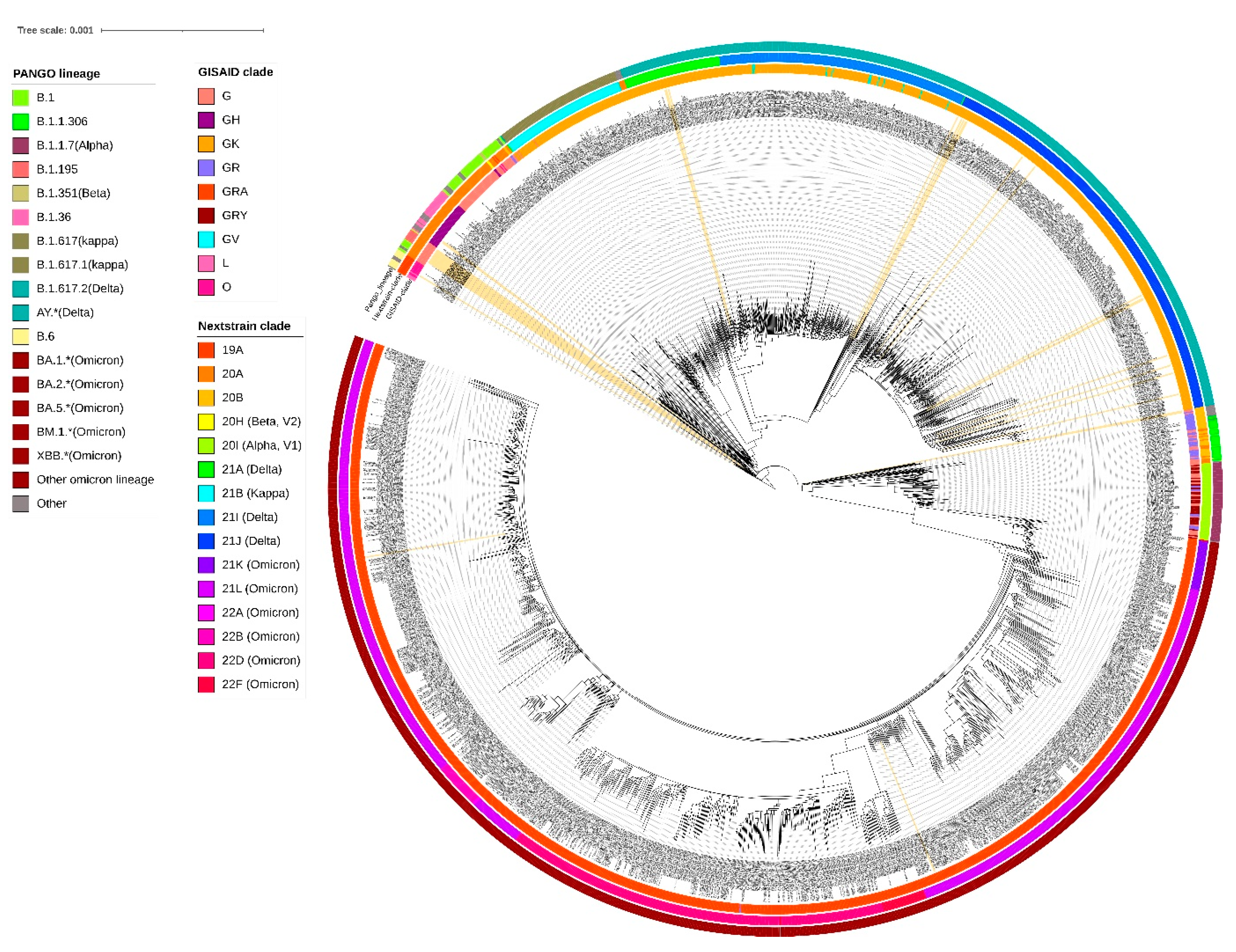
A neighbor-joining tree was constructed using n = 38 strains sequenced in this study (Figure 1), and their phylogenetic relationship, along with 2160 SARS-CoV-2, is shown in Figure 2, where the genomes sequenced in this study are shown in beige highlight. All the SARS-CoV-2 genomes reported so far from central India comprise 109 different SARS-CoV-2 Pangolin lineages. Importantly, these include B.1.1.7 (Alpha) (64/2160 = 2.96%), B.1.351 (Beta) (2/2160 = 0.09%), B.1.617.2 + AY.* (Delta) (625/2160 = 28.93%), and BA.* + BM.1.* + XBB.* (Omicron) (1212/2160 = 56.11%) VOCs as per the data submitted in GISAID until 31 March 2023 from central India.
A cluster of eight samples belonging to B.1.195 lineage (Figure 1) comprise three of the first four cases in Jabalpur (a family of three patients who had visited Dubai and tested positive on 20 March 2020) and their one contact (ICMR-NIRTH-S7, tested positive on 21 March 2020). The other three individuals in this cluster tested positive between 11 and 17 April 2020 and were also linked to each other. Another sample with a foreign travel history was ICMR-NIRTH-S3 (lineage B.1.1), who had returned from Germany and tested positive on 20 March 2020. We have analyzed this cluster, but this sample did not group with other strains reported from central India (Figure 2).
Another cluster represented B.1 lineage strains (n = 6 cases: three each from Chhindwara and Indore districts, which were epidemiologically linked) (Table 1). Two cases (ICMR-NIRTH-S1 and ICMR-NIRTH-S2) belonged to B.1.36.8 and had tested positive on the same day (27 March 2020). Sample ICMR-NIRTH-S14 (lineage B.6) belongs to a person who had traveled to Delhi and clusters together with other strains sequenced by the Defense Research and Development Establishment (DRDE), Gwalior (Figure 2).
The overall lineages distribution highlighted the dominant occurrence of B.1, B.1.36, and B.6 during the first wave, B.1.617.2 in the second, and omicron in the third wave in central India.

3.4. Distribution of Circulating Lineages

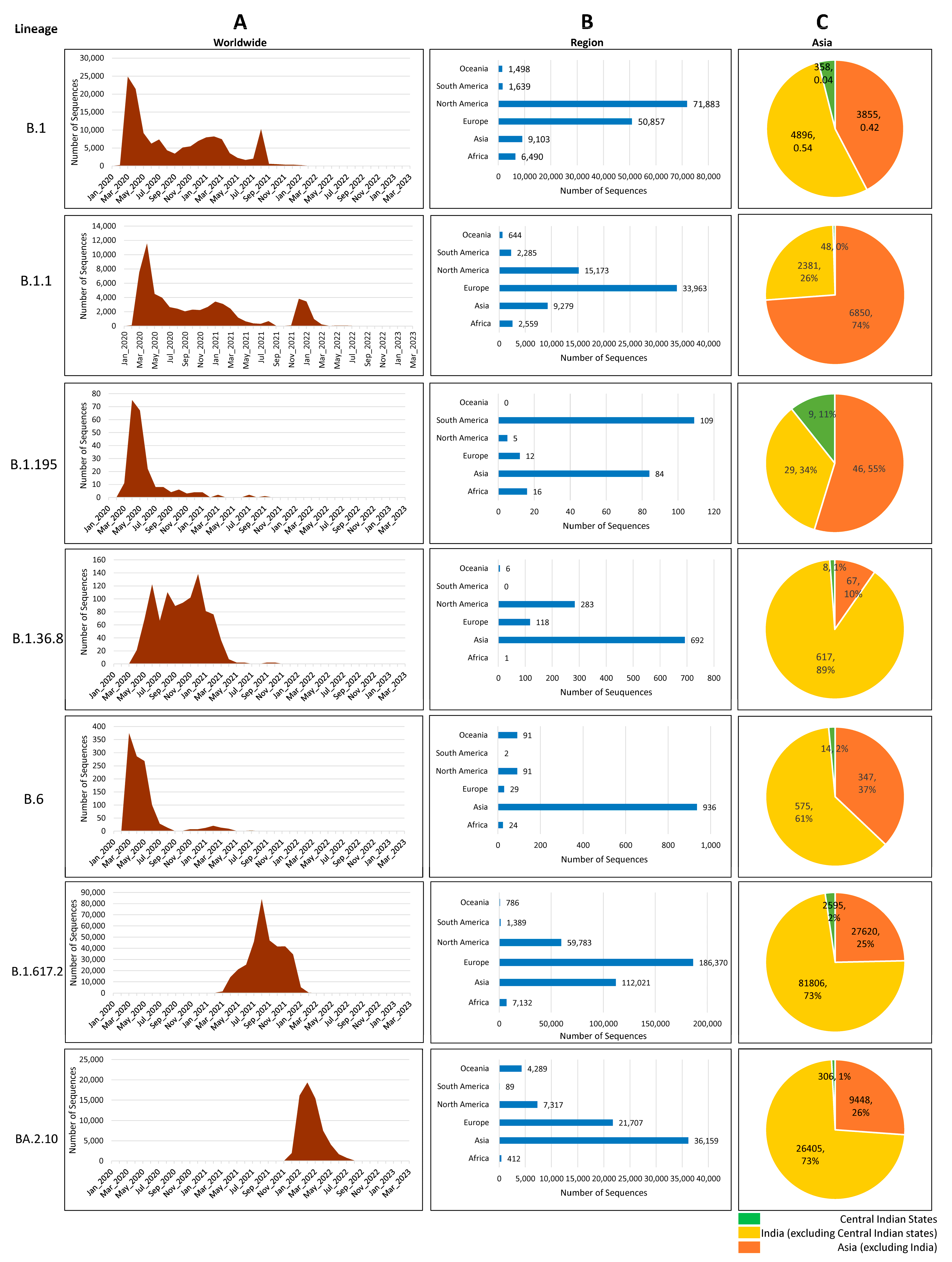
We have investigated the global prevalence of the seven lineages by analyzing the genomes from GISAID and found that, together, North America and Europe shared nearly 86% of the load of B.1 and 83% of that of B.1.1; Europe and Asia shared nearly 82% of the load of Delta and Omicron VOCs. Though only 6.43% of the genomes of B.1 were observed in Asia, among them, nearly half of (3.7%) the genomes were submitted from India, indicating the transmission of B.1 in this region. Interestingly, strains belonging to lineage B.1.195 have not been observed after 2021, whereas lineages B1 and B.1.1 have been reported until very recently in the year 2022. In fact, in central India, B.1.1 was present in the year 2022, indicating that these strains were in circulation after the first report. B.1.36 was present in a small percentage during the first wave but surged during the second wave. Lineages B.6 and B.1.195, however, were prevalent in the first wave, and an almost negligible number of genomes were reported after the first wave. Together, South America and Asia have submitted about 85.3% of lineage B.1.195 so far. Furthermore, Asia alone submitted 62.9% of lineage B.1.36.8 and 79.7% of B.6 SARS-CoV-2 sequences, with most of the sequences coming from India (Figure 3).

4. Discussion

Since the first case of SARS-CoV-2 was discovered in the state of Kerala, India has been on alert for the spread of airborne viruses. To contain and stop the spread of viral transmission, the Ministry of Home Affairs (MHAs) subsequently declared a nationwide lockdown that would last from 25 March 2020 to 14 April 2020. A single case regarding lineage B.1.195 (associated with migration) identified in this study may indicate that the initial period (March–April 2020) had effective containment measures, especially for those who had returned from abroad. This strain was associated with travel history to Dubai and was also closely related to other patients from Gujarat and Brazil. All these individuals had a travel history to Dubai around a similar time. However, the subsequent introduction of several lineages via interstate travel might have been the major driver of SARS-CoV-2 transmission in central India. This is supported by the fact that a significant genomic diversity was found during the initial WGS analyses of SARS-CoV-2 patients who traveled to India from other countries.
Another observation from the genomic analysis carried out in this study was that the earliest documented evidence of Delta variants in MP (GISAID ID: EPI_ISL_2461258, Lineage: AY.122) was in September 2020. However, the proportion of this variant among the newly detected strains after May 2021 has increased to over 90% in a short span of time. This indicates its selective advantage over the other circulating lineages [28]. Delta and Omicron variants have been reported to be more transmissible than the other variants, and strong evidence is available which states that they were behind the emergence of the second and third waves of COVID-19 in central India [29,30]. These observations suggested that the diverse genotypes of SARS-CoV-2 may have emerged as a process of convergent evolution. In addition, certain mutations may lead to changes in their surface antigenic structure, whereby the antibodies due to previous infection/vaccination might not bind to them as effectively, exerting an ‘immune evasion’ advantage to them which can serve as a selection pressure and might be associated with ‘breakthrough infections’.
The D614G (37/38 strains) mutation has been shown to enhance the replication rate and transmissibility of SARS-CoV-2 by the more efficient binding of SARS-CoV-2 spike protein to human angiotensin converting enzyme-2 (ACE2) receptors and subsequently increases viral entrance into host cells. Mutations in the spike can enhance viral transmission, disease severity, and the virus’s capacity to elude immune defensive responses [31].
Five immune escape mutations in the S gene at a codon position other than D614G were L452R, P681R/H, Q57H, E484A, and N501Y during the second and third waves. Substitution at positions P681 and E484 has become increasingly common among clinical isolates. Previous studies showed that the virulence and pathogenesis of the Delta variant could be impacted by D614G and P681R mutations [32]. Aside from D614G, the B.1.617.2 lineage, which has the spike protein mutations L452R and N501Y, may explain the transmission and surge in cases in central India in March 2021. Although the D614G mutation occurs in the Delta and Omicron variants, the additional presence of the P681H mutation may result in slow cleavage. Moreover, this may limit the Omicron virus replication to the upper respiratory tract, resulting in less fusion and infectivity compared to the Delta and D614G  +  P681R double mutants. A previous study reported that 90.5% of the samples had the D614G mutation, which has recently been linked to higher viral loads and accelerated replication on human lung epithelial cells [33]. Among the S protein mutations, S: L452R in the receptor-binding domain (RBD) has also been associated with vaccine breakthrough infections by decreasing the neutralization capability of vaccine-induced immune response [33,34]. Another mutation S:E484Q in the Spike RBD has also been associated with a similar clinical impact but was missing in the cases we sequenced [34,35]. Studies tracking the selection of such mutations using intra-host variation suggest that there was an early indication of Spike protein mutation playing a key role in increased virulence and transmissibility among the population [36].
Among 29 mutations, 20 mutations were only identified in the ORF1ab region, which was known to encompass mutational spectra. ORF1ab is a conserved area in the SARS-CoV-2 genome [37]. Out of these 29 mutations, 17 mutations were non-synonymous, 11 were synonymous, and 1 mutation was found in the non-coding region. By improving the virus’s adaptability to the utilization of human codons, synonymous SARS-CoV-2 mutations associated with the action of various mutational mechanisms may have a positive effect on viral evolution [37]. The non-synonymous mutation causes epitope loss, which might be connected to immune evasion, which would increase viral pathogenicity and propagation [38].
However, after the emergence of new Omicron sub-lineage BA.5, it is now unknown if SARS-CoV-2 is shifting to a more gradual adaptive mechanism or if it will continue to evolve in this beneficial fashion with the frequent emergence of highly divergent lineages [39,40].
In 2022, multiple lineages emerging within BA.2, BA.5, and Omicron were observed in a more step-wise fashion, with several amino acid changes and moderate transmission advantages, which could indicate a shift to a more gradual stepwise evolution. The formation of VOCs and possible future antigenically different lineages may be viewed as ‘shift-like events’, which are unanticipated, major alterations in the virus’ genetic make-up and, possibly, therapeutically relevant features [39,40]. It is impossible to foresee where future significant lineages will emerge from in viral genetic diversity and whether they will arise from ‘shift-like’ or more gradual ‘drift-like’ progression similar to that seen in the Omicron clade during 2022 [40,41].
However, these limitations do not affect our conclusions about the genetic diversity and phylogenetic relatedness of the strains from central India. Overall, our study provides useful insights into the extent of the genetic diversity of SARS-CoV-2 strains over the course of the COVID-19 pandemic right from the very beginning to know its origin, dissemination, and evolving pattern of genetic diversity.

Limitations of This Study

This study has a few limitations: (i) We have sequenced only a limited number of samples from central India. (ii) Although we identified new mutations in SARS-CoV-2 genomes, we were unable to link them with phenotypic effects. Despite these limitations, our study provides valuable insights into the genetic diversity of SARS-CoV-2 infections in central India and its trajectory through the two waves of the SARS-CoV-2 pandemic.

5. Conclusions

To our knowledge, this is among the very few studies in which the molecular surveillance-based phylogenetic trends based on whole-genome sequencing of SARS-CoV-2 has been undertaken from central India. In this study, we have performed WGS of 38 strains from Jabalpur and adjoining districts and compared them with over 2000 strains from various districts from central India representing different lineages. This study shows that the primary sources of COVID-19 introduction were those with a recent history of foreign travel in the affected countries. This was later on further amplified by interstate migration, which subsequently led to the emergence of newer strains.
The phylogenetic analysis of the SARS-CoV-2 strains in this study points to multiple introductions of the SARS-CoV-2 virus in central India. The genetic diversity, transmission, and evolution of SARS-CoV-2 exhibited a consistent pattern of increasing divergence within major lineages. Continued genomic surveillance strategies are needed for improved understanding of the SARS-CoV-2 pandemic. Furthermore, increased sequencing capacity is necessary for contact tracing and quicker identification of the transmission hotspots. This can be helpful for better preparedness, effective interventions, and surveillance activity.
The COVID-19 phenomena show that continuous and sustained monitoring of pathogens using next-generation sequencing is a useful tool for monitoring disease transmission and evolution patterns. The pandemic accelerated the capacity building at district level to enable molecular diagnostic with the capability of novel pathogen identification.

Supplementary Materials

The following supporting information can be downloaded at: https://www.mdpi.com/article/10.3390/v16101608/s1, Figure S1: Genomic map showing coverage of the 38 SARS-CoV-2 genomes sequenced in this study; Table S1: Metadata file with information about the SARS-CoV-2 genome sequences from Central India available in GISAID database till 31 March 2023; Table S2: Genome assembly features of the SARS-CoV-2 genomes sequenced in this study; Table S3: Nextclade results of SARS-CoV-2 classification of the 38 genomes sequenced in this study along with the mutational profile of each strain; Table S4: Detailed information with stability analysis of the 29 novel SNPs identified in this study.

Author Contributions

P.D. performed the NGS experiments: M.S., A.A. and A.G. wrote the manuscript, which was later updated and revised by S.K.R., S.K. and M.K. A.A. and A.G. analyzed the NGS data, whose interpretation was carried out by A.A., M.S. and A.G. using bioinformatics tools. S.C.B., A.K., L.S. and M.U. collected the samples and processed them. P.V.B., A.D. and P.S. conceptualized and supervised the study, P.S. led the overall conduction of the NGS and bioinformatics data analysis work along with manuscript writing and revisions. All authors have read and agreed to the published version of the manuscript.

Funding

The study has been carried out with the support of Indian Council of Medical Research (ICMR) and the Department of Health Research (DHR, grant number DHR/VDL/04/2018). The Laboratory and staff of P.S. is supported by LRI Netherlands (Grant number 708.20.09), R2STOP Canada, DBT (Ramalingaswami Fellowship Grant), DHR and ICMR. Infrastructure and funding support from ICMR are also gratefully acknowledged. P.D. and M.S. are recipients of Senior Research Fellowship and Research Associateship, respectively, from ICMR.

Institutional Review Board Statement

The Ethics approval of the State Level- Virus Research and Diagnostics Laboratory (SL-VRDL) Project under which this study was carried out was obtained from the Institutional Ethics Committee (IEC) of the ICMR-National Institute of Research in Tribal Health, Jabalpur (No. NIRTH/IEC/2229/2018, dated 1 November 2018). The manuscript has been approved by the Publication Screening Committee of ICMR-NIRTH Jabalpur and assigned with the number ICMR-NIRTH/PSC/40/2021.

Informed Consent Statement

Not applicable.

Data Availability Statement

All data generated or analyzed during this study are included in this article. The raw data used to support the findings of this study have been deposited in the NCBI with BioProject ID: PRJNA759056.

Acknowledgments

The contribution of the entire team of the Division of Virology and Zoonoses and all other staff of VRDL including Lab technicians, Data Entry Operators, and other support staff is sincerely acknowledged for handling and processing the patient samples and contributing to the diagnostic services amid challenging times. The resources and infrastructure at the Central Instrumentation Facility at ICMR-NIRTH was generously made available for this work, for which we are grateful. The authors are thankful to SK Raghav and Vidhan Jain and the members of Rapid Response team at ICMR-NIRTH for their help in this work; Sri Krishna and Sanchita Pacholi provided valuable technical assistance. We are also thankful to the patients and the state health authorities (clinicians and healthcare workers) for collecting patient samples and their associated epidemiological details. We gratefully acknowledge the GISAID database and all researchers who have submitted information and genomes to this valuable database.

Conflicts of Interest

The authors declare that they have no competing interests.

References

  1. Lu, R.; Zhao, X.; Li, J.; Niu, P.; Yang, B.; Wu, H.; Wang, W.; Song, H.; Huang, B.; Zhu, N.; et al. Genomic Characterisation and Epidemiology of 2019 Novel Coronavirus: Implications for Virus Origins and Receptor Binding. Lancet 2020, 395, 565–574. [Google Scholar] [CrossRef] [PubMed]
  2. Pedersen, S.F.; Ho, Y.-C. SARS-CoV-2: A Storm Is Raging. J. Clin. Investig. 2020, 130, 2202–2205. [Google Scholar] [CrossRef]
  3. Misra, G.; Manzoor, A.; Chopra, M.; Upadhyay, A.; Katiyar, A.; Bhushan, B.; Anvikar, A. Genomic Epidemiology of SARS-CoV-2 from Uttar Pradesh, India. Sci. Rep. 2023, 13, 14847. [Google Scholar] [CrossRef] [PubMed]
  4. COVID-19 Cases | WHO COVID-19 Dashboard. Available online: https://data.who.int/dashboards/covid19/cases?n=c (accessed on 4 June 2024).
  5. Zaki, A.M.; van Boheemen, S.; Bestebroer, T.M.; Osterhaus, A.D.; Fouchier, R.A. Isolation of a Novel Coronavirus from a Man with Pneumonia in Saudi Arabia. N. Engl. J. Med. 2012, 367, 1814–1820. [Google Scholar] [CrossRef] [PubMed]
  6. Andrews, M.A.; Areekal, B.; Rajesh, K.R.; Krishnan, J.; Suryakala, R.; Krishnan, B.; Muraly, C.P.; Santhosh, P.V. First Confirmed Case of COVID-19 Infection in India: A Case Report. Indian J. Med. Res. 2020, 151, 490–492. [Google Scholar] [CrossRef] [PubMed]
  7. Singh, S.; Sharma, A.; Gupta, A.; Joshi, M.; Aggarwal, A.; Soni, N.; Sana; Jain, D.; Verma, P.; Khandelwal, D.; et al. Demographic Comparison of the First, Second and Third Waves of COVID-19 in a Tertiary Care Hospital at Jaipur, India. Lung India 2022, 39, 525. [Google Scholar] [CrossRef] [PubMed]
  8. India COVID—Coronavirus Statistics—Worldometer. Available online: https://www.worldometers.info/coronavirus/country/india/ (accessed on 2 July 2024).
  9. Pattabiraman, C.; Habib, F.; Harsha, P.K.; Rasheed, R.; Prasad, P.; Reddy, V.; Dinesh, P.; Damodar, T.; Hosallimath, K.; George, A.K.; et al. Genomic Epidemiology Reveals Multiple Introductions and Spread of SARS-CoV-2 in the Indian State of Karnataka. PLoS ONE 2020, 15, e0243412. [Google Scholar] [CrossRef] [PubMed]
  10. Pattabiraman, C.; Desai, A.; Prasad, P.; George, A.K.; Sreenivas, D.; Rasheed, R.; Reddy, N.V.K.; Vasanthapuram, R. Importation, Circulation, and Emergence of Variants of SARS-CoV-2 in the South Indian State of Karnataka. Wellcome Open Res 2021, 6. [Google Scholar] [CrossRef]
  11. Singh, B.; Avula, K.; Chatterjee, S.; Datey, A.; Ghosh, A.; De, S.; Keshry, S.S.; Ghosh, S.; Suryawanshi, A.R.; Dash, R.; et al. Isolation and Characterization of Five Severe Acute Respiratory Syndrome Coronavirus 2 Strains of Different Clades and Lineages Circulating in Eastern India. Front. Microbiol. 2022, 13, 856913. [Google Scholar] [CrossRef]
  12. Radhakrishnan, C.; Divakar, M.K.; Jain, A.; Viswanathan, P.; Bhoyar, R.C.; Jolly, B.; Imran, M.; Sharma, D.; Rophina, M.; Ranjan, G.; et al. Initial Insights into the Genetic Epidemiology of SARS-CoV-2 Isolates from Kerala Suggest Local Spread from Limited Introductions. Front. Genet. 2021, 12, 630542. [Google Scholar] [CrossRef]
  13. Joshi, M.; Puvar, A.; Kumar, D.; Ansari, A.; Pandya, M.; Raval, J.; Patel, Z.; Trivedi, P.; Gandhi, M.; Pandya, L.; et al. Genomic Variations in SARS-CoV-2 Genomes From Gujarat: Underlying Role of Variants in Disease Epidemiology. Front. Genet. 2021, 12, 586569. [Google Scholar] [CrossRef] [PubMed]
  14. Limaye, S.; Kasibhatla, S.M.; Ramtirthkar, M.; Kinikar, M.; Kale, M.M.; Kulkarni-Kale, U. Circulation and Evolution of SARS-CoV-2 in India: Let the Data Speak. Viruses 2021, 13, 2238. [Google Scholar] [CrossRef] [PubMed]
  15. Gupta, A.; Sabarinathan, R.; Bala, P.; Donipadi, V.; Vashisht, D.; Katika, M.R.; Kandakatla, M.; Mitra, D.; Dalal, A.; Bashyam, M.D. A Comprehensive Profile of Genomic Variations in the SARS-CoV-2 Isolates from the State of Telangana, India. J. Gen. Virol. 2021, 102, 1562. [Google Scholar] [CrossRef] [PubMed]
  16. MoHFW | Home. Available online: https://www.mohfw.gov.in/#latest-update (accessed on 6 June 2024).
  17. Andrew, S. FastQC. A Quality Control Tool for High Throughput Sequence Data. 2010. Available online: https://www.bioinformatics.babraham.ac.uk/projects/fastqc/ (accessed on 15 March 2023).
  18. Bolger, A.M.; Lohse, M.; Usadel, B. Trimmomatic: A Flexible Trimmer for Illumina Sequence Data. Bioinformatics 2014, 30, 2114–2120. [Google Scholar] [CrossRef] [PubMed]
  19. Li, H. Aligning Sequence Reads, Clone Sequences and Assembly Contigs with BWA-MEM. arXiv 2013. [Google Scholar] [CrossRef]
  20. Danecek, P.; Bonfield, J.K.; Liddle, J.; Marshall, J.; Ohan, V.; Pollard, M.O.; Whitwham, A.; Keane, T.; McCarthy, S.A.; Davies, R.M.; et al. Twelve Years of SAMtools and BCFtools. Gigascience 2021, 10, giab008. [Google Scholar] [CrossRef] [PubMed]
  21. Aksamentov, I.; Roemer, C.; Hodcroft, E.B.; Neher, R.A. Nextclade: Clade Assignment, Mutation Calling and Quality Control for Viral Genomes. J. Open Source Softw. 2021, 6, 3773. [Google Scholar] [CrossRef]
  22. Chen, C.W.; Lin, M.H.; Liao, C.C.; Chang, H.P.; Chu, Y.W. IStable 2.0: Predicting Protein Thermal Stability Changes by Integrating Various Characteristic Modules. Comput. Struct. Biotechnol. J. 2020, 18, 622–630. [Google Scholar] [CrossRef]
  23. Hadfield, J.; Megill, C.; Bell, S.M. Genomic Epidemiology of Novel Coronavirus: Global Subsampling. Nextstrain: Real-Time Tracking of Pathogen Evolution, 2020. [Google Scholar]
  24. Hadfield, J.; Megill, C.; Bell, S.M.; Huddleston, J.; Potter, B.; Callender, C.; Sagulenko, P.; Bedford, T.; Neher, R.A. Nextstrain: Real-Time Tracking of Pathogen Evolution. Bioinformatics 2018, 34, 4121–4123. [Google Scholar] [CrossRef]
  25. Minh, B.Q.; Schmidt, H.A.; Chernomor, O.; Schrempf, D.; Woodhams, M.D.; Von Haeseler, A.; Lanfear, R. IQ-TREE 2: New Models and Efficient Methods for Phylogenetic Inference in the Genomic Era. Mol. Biol. Evol. 2020, 37, 1530–1534. [Google Scholar] [CrossRef]
  26. Kumar, S.; Stecher, G.; Suleski, M.; Blair Hedges, S. TimeTree: A Resource for Timelines, Timetrees, and Divergence Times. Mol. Biol. Evol. 2017, 34, 1812–1819. [Google Scholar] [CrossRef] [PubMed]
  27. Letunic, I.; Bork, P. Interactive Tree Of Life (ITOL) v5: An Online Tool for Phylogenetic Tree Display and Annotation. Nucleic Acids Res. 2021, 49, W293–W296. [Google Scholar] [CrossRef] [PubMed]
  28. Brown, K.A.; Gubbay, J.; Buchan, S.A.; Daneman, N.; Mishra, S.; Patel, S.; Day, T. Inflection in Prevalence of SARS-CoV-2 Infections Missing the N501Y Mutation as a Marker of Rapid Delta (B.1.617.2) Lineage Expansion in Ontario, Canada. medRxiv 2021. [Google Scholar] [CrossRef]
  29. Mohapatra, R.K.; Tiwari, R.; Sarangi, A.K.; Sharma, S.K.; Khandia, R.; Saikumar, G.; Dhama, K. Twin Combination of Omicron and Delta Variants Triggering a Tsunami Wave of Ever High Surges in COVID-19 Cases: A Challenging Global Threat with a Special Focus on the Indian Subcontinent. J. Med. Virol. 2022, 94, 1761–1765. [Google Scholar] [CrossRef]
  30. Ray, S.K.; Mukherjee, S. Divulging Incipient SARS-CoV-2 Delta (B.1.617.2) Variant: Possible with Global Scenario. Infect. Disord. Drug Targets 2022, 23, 35–38. [Google Scholar] [CrossRef]
  31. Jackson, C.B.; Zhang, L.; Farzan, M.; Choe, H. Functional Importance of the D614G Mutation in the SARS-CoV-2 Spike Protein. Biochem. Biophys. Res. Commun. 2021, 538, 108–115. [Google Scholar] [CrossRef]
  32. Khatri, R.; Siddqui, G.; Sadhu, S.; Maithil, V.; Vishwakarma, P.; Lohiya, B.; Goswami, A.; Ahmed, S.; Awasthi, A.; Samal, S. Intrinsic D614G and P681R/H Mutations in SARS-CoV-2 VoCs Alpha, Delta, Omicron and Viruses with D614G plus Key Signature Mutations in Spike Protein Alters Fusogenicity and Infectivity. Med. Microbiol. Immunol. 2023, 212, 103–122. [Google Scholar] [CrossRef]
  33. Franceschi, V.B.; Caldana, G.D.; de Menezes Mayer, A.; Cybis, G.B.; Neves, C.A.M.; Ferrareze, P.A.G.; Demoliner, M.; de Almeida, P.R.; Gularte, J.S.; Hansen, A.W.; et al. Genomic Epidemiology of SARS-CoV-2 in Esteio, Rio Grande Do Sul, Brazil. BMC Genom. 2021, 22, 371. [Google Scholar] [CrossRef]
  34. Ghosh, A.; Walia, S.; Rattan, R.; Kanampalliwar, A.; Jha, A.; Aggarwal, S.; Fatma, S.; Das, N.; Chayani, N.; Prasad, P.; et al. Genomic Profiles of Vaccine Breakthrough SARS-CoV-2 Strains from Odisha, India. Int. J. Infect. Dis. 2022, 119, 111–113. [Google Scholar] [CrossRef]
  35. Ferreira, I.A.T.M.; Kemp, S.A.; Datir, R.; Saito, A.; Meng, B.; Rakshit, P.; Takaori-Kondo, A.; Kosugi, Y.; Uriu, K.; Kimura, I.; et al. SARS-CoV-2 B.1.617 Mutations L452R and E484Q Are Not Synergistic for Antibody Evasion. J Infect Dis 2021, 224, 989–994. [Google Scholar] [CrossRef]
  36. Pathak, A.K.; Mishra, G.P.; Uppili, B.; Walia, S.; Fatihi, S.; Abbas, T.; Banu, S.; Ghosh, A.; Kanampalliwar, A.; Jha, A.; et al. Spatio-Temporal Dynamics of Intra-Host Variability in SARS-CoV-2 Genomes. Nucleic Acids Res 2022, 50, 1551–1561. [Google Scholar] [CrossRef]
  37. Rochman, N.D.; Wolf, Y.I.; Faure, G.; Mutz, P.; Zhang, F.; Koonin, E.V. Ongoing Global and Regional Adaptive Evolution of SARS-CoV-2. Proc. Natl. Acad. Sci. USA 2021, 118, 2104241118. [Google Scholar] [CrossRef] [PubMed]
  38. Gupta, A.M.; Chakrabarti, J.; Mandal, S. Non-Synonymous Mutations of SARS-CoV-2 Leads Epitope Loss and Segregates Its Variants. Microbes Infect. 2020, 22, 598–607. [Google Scholar] [CrossRef] [PubMed]
  39. Markov, P.V.; Ghafari, M.; Beer, M.; Lythgoe, K.; Simmonds, P.; Stilianakis, N.I.; Katzourakis, A. The Evolution of SARS-CoV-2. Nat. Rev. Microbiol. 2023, 21, 361–379. [Google Scholar] [CrossRef]
  40. Otto, S.P.; Day, T.; Arino, J.; Colijn, C.; Dushoff, J.; Li, M.; Mechai, S.; Van Domselaar, G.; Wu, J.; Earn, D.J.D.; et al. The Origins and Potential Future of SARS-CoV-2 Variants of Concern in the Evolving COVID-19 Pandemic. Curr. Biol. 2021, 31, R918–R929. [Google Scholar] [CrossRef] [PubMed]
  41. Bano, I.; Sharif, M.; Alam, S. Genetic Drift in the Genome of SARS-CoV-2 and Its Global Health Concern. J. Med. Virol. 2022, 94, 88–98. [Google Scholar] [CrossRef] [PubMed]
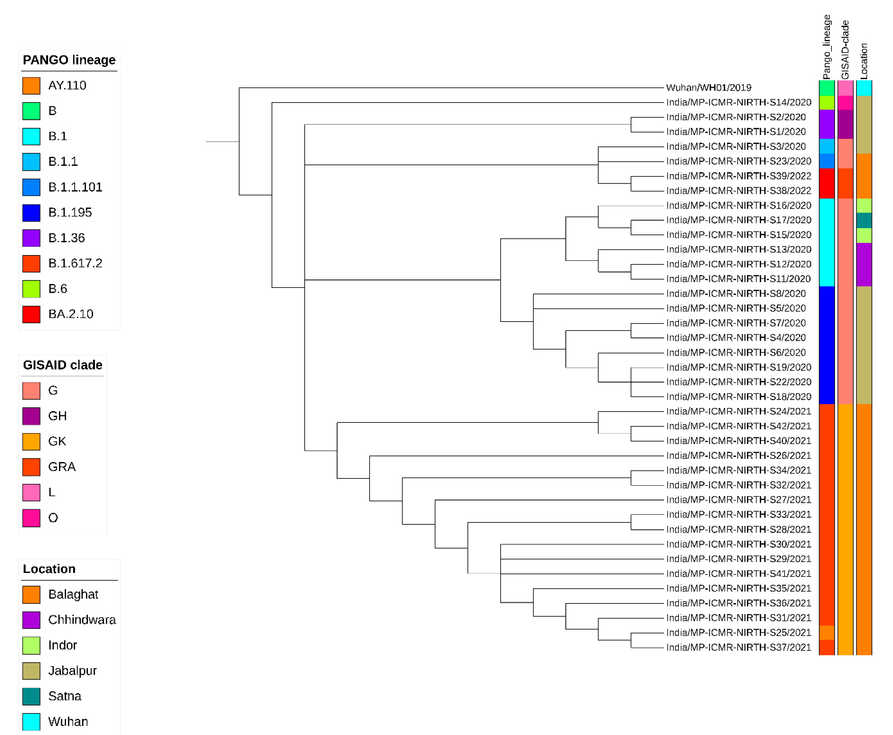
Figure 1. Phylogenetic analysis of the 38 SARS-CoV-2 genomes sequenced at ICMR-NIRTH with the reference genome (NC_045512) hCoV19/Wuhan/WH01/2019. Lineage distribution is depicted by different colors.
Viruses 16 01608 g001
Figure 2. Phylogenetic analysis of the 38 SARS-CoV-2 genomes sequenced at ICMR-NIRTH with the 2160 genomes obtained from GISAID. Classification of the genome sequences according to the Pangolin lineages, Nextstrain, and GISAID clade is shown in color. The labels corresponding to the ICMR-NIRTH genome sequences generated during this study are highlighted and have been marked with light yellow color.
Viruses 16 01608 g002
Figure 3. Global distribution of 5 lineages of SARS-CoV-2 from GISAID database until August 2022. (A) Month-wise distribution of lineages from worldwide submitted data. (B) Distribution of lineages across different continents of the world. (C) Comparison of lineages present in central Indian states with other Indian states and Asian countries.
Viruses 16 01608 g003
Table 2. Pangolin lineage, GISAID clade, and Nextstrain clade information of the 38 SARS-CoV-2 genomes sequenced in this study.
Sr NoSample IDDate of Sample CollectionGISAID CladeNextstrain CladePangolin LineageWHO Name
1ICMR-NIRTH-S127 March 2020 GH20AB.1.36.8unassigned
2ICMR-NIRTH-S227 March 2020 GH20AB.1.36.8unassigned
3ICMR-NIRTH-S320 March 2020 G20BB.1.1unassigned
4ICMR-NIRTH-S420 March 2020 G20AB.1.195unassigned
5ICMR-NIRTH-S520 March 2020 G20AB.1.195unassigned
6ICMR-NIRTH-S620 March 2020 G20AB.1.195unassigned
7ICMR-NIRTH-S721 March 2020 G20AB.1.195unassigned
8ICMR-NIRTH-S822 March 2020 G20AB.1.195unassigned
9ICMR-NIRTH-S111 April 2020 G20AB.1unassigned
10ICMR-NIRTH-S127 April 2020 G20AB.1unassigned
11ICMR-NIRTH-S137 April 2020 G20AB.1unassigned
12ICMR-NIRTH-S148 April 2020 O19AB.6unassigned
13ICMR-NIRTH-S1510 April 2020 G20AB.1unassigned
14ICMR-NIRTH-S1610 April 2020 G20AB.1unassigned
15ICMR-NIRTH-S1710 April 2020 G20AB.1unassigned
16ICMR-NIRTH-S1811 April 2020 G20AB.1.195unassigned
17ICMR-NIRTH-S1914 April 2020 G20AB.1.195unassigned
18ICMR-NIRTH-S2217 April 2020 G20AB.1.195unassigned
19ICMR-NIRTH-S2327 July 2020G20BB.1.1.101unassigned
20ICMR-NIRTH-S2416 April 2021GK21AB.1.617.2Delta
21ICMR-NIRTH-S2516 April 2021GK21JB.1.617.2Delta
22ICMR-NIRTH-S2617 April 2021GK21JB.1.617.2Delta
23ICMR-NIRTH-S2718 April 2021GK21JB.1.617.2Delta
24ICMR-NIRTH-S2819 April 2021GK21JB.1.617.2Delta
25ICMR-NIRTH-S2919 April 2021GK21JB.1.617.2Delta
26ICMR-NIRTH-S3020 April 2021GK21JB.1.617.2Delta
27ICMR-NIRTH-S319 April 2021GK21JB.1.617.2Delta
28ICMR-NIRTH-S329 April 2021GK21JB.1.617.2Delta
29ICMR-NIRTH-S3312 April 2021GK21JB.1.617.2Delta
30ICMR-NIRTH-S3413 April 2021GK21JB.1.617.2Delta
31ICMR-NIRTH-S3518 April 2021GK21JB.1.617.2Delta
32ICMR-NIRTH-S3621 April 2021GK21JB.1.617.2Delta
33ICMR-NIRTH-S3724 April 2021GK21JB.1.617.2Delta
34ICMR-NIRTH-S383 February 2022GRA21LBA.2.10Omicron
35ICMR-NIRTH-S394 February 2022GRA21LBA.2.10Omicron
36ICMR-NIRTH-S4020 April 2021GK21AB.1.617.2Delta
37ICMR-NIRTH-S4121 April 2021GK21JB.1.617.2Delta
38ICMR-NIRTH-S4227 April 2021GK21AB.1.617.2Delta
Disclaimer/Publisher’s Note: The statements, opinions and data contained in all publications are solely those of the individual author(s) and contributor(s) and not of MDPI and/or the editor(s). MDPI and/or the editor(s) disclaim responsibility for any injury to people or property resulting from any ideas, methods, instructions or products referred to in the content.