Article

CRISPR RNA-Guided Transposases Facilitate Dispensable Gene Study in Phage

1
School of Biology and Biological Engineering, South China University of Technology, Guangzhou 510006, China
2
Shenzhen Institute of Synthetic Biology, Shenzhen Institutes of Advanced Technology, Chinese Academy of Sciences, Shenzhen 518055, China
*
Authors to whom correspondence should be addressed.
Viruses 2024, 16(3), 422; https://doi.org/10.3390/v16030422
Submission received: 26 January 2024 / Revised: 5 March 2024 / Accepted: 7 March 2024 / Published: 9 March 2024
(This article belongs to the Section Bacterial Viruses)

Abstract

:
Phages provide a potential therapy for multi-drug-resistant (MDR) bacteria. However, a significant portion of viral genes often remains unknown, posing potential dangers. The identification of non-essential genes helps dissect and simplify phage genomes, but current methods have various limitations. In this study, we present an in vivo two-plasmid transposon insertion system to assess the importance of phage genes, which is based on the V. cholerae transposon Tn6677, encoding a nuclease-deficient type I-F CRISPR–Cas system. We first validated the system in Pseudomonas aeruginosa PAO1 and its phage S1. We then used the selection marker AcrVA1 to protect transposon-inserted phages from CRISPR-Cas12a and enriched the transposon-inserted phages. For a pool of selected 10 open-reading frames (2 known functional protein genes and 8 hypothetical protein genes) of phage S1, we identified 5 (2 known functional protein genes and 3 hypothetical protein genes) as indispensable genes and the remaining 5 (all hypothetical protein genes) as dispensable genes. This approach offers a convenient, site-specific method that does not depend on homologous arms and double-strand breaks (DSBs), holding promise for future applications across a broader range of phages and facilitating the identification of the importance of phage genes and the insertion of genetic cargos.

1. Introduction

Bacteriophages (phages)—viruses that specifically infect bacteria—are highly abundant in the biosphere, numbering up to 1031 [1,2]. With the rapid development of multi-drug-resistant (MDR) bacteria, phages have gained significance as a possible therapy due to their host specificity and self-amplifying features [3]. For safety, therapeutic phages are desirable to exclude toxin genes, virulence factors, and antibiotic resistance genes [4,5], and it is preferable for phages to carry a minimal number of genes with unknown functions to avoid potential dangers.
However, a substantial portion of the viral genetic information remains unknown, with 75% of the more than 2.79 million viral genes cataloged lacking identified functions and without any corresponding homologous genes found in public databases [6]. Even for the well-studied model phage T4, there is a significant gap in knowledge, as 45% of its genes are of unknown function [7]. Identifying non-essential genes for phage would aid in the analysis of phage genomes [5,8], and current functional investigation methods include homologous recombination, CRISPR-Cas, and complete genome reconstruction in non-host systems [9]. However, these approaches face various limitations. For homologous recombination, simultaneous serial insertions are challenging due to the specification of editing sites by homologous arms [10]. Additionally, short homologous arms result in low efficiency, but long arms may contain harmful phage genes [9]. Although efficiency can be improved by integrating CRISPR-Cas systems, this method risks causing cell death through double-strand breaks (DSBs) [11,12,13]. Complete genome reconstruction in non-host systems allows for genome design flexibility, but these reconstructed genomes often fail to reboot [14,15,16,17]. These emphasize the need for new phage genome engineering methods to identify the importance of phage genes.
Transposon insertion has, in the past, been used as a tool for phage gene characterization. It relies on randomly in vitro transposition using MuA transposase, followed by electroporation into host cells to identify mutant phages and determine transposon insertion sites. This method has only been applied to a few phages and its efficiency is mostly constrained by the transformation capacity of the host bacteria [18,19,20]. Recently, Klompe et al. reported a site-specific, programmable system for in vivo transposon insertion using the V. cholerae transposon Tn6677, which encodes a nuclease-deficient type I-F CRISPR–Cas system [21,22,23]. The system employs three plasmids: pQCascade, pTnsABC, and pDonor. pQCascade encodes the tniQ-cas8-cas7-cas6 operon (QCascade) alongside a synthetic CRISPR array; thereby, the Qcascade complex surveys and selectively binds to target DNA sites guided by crRNA. pTnsABC carries the tnsA-tnsB-tnsC operon, where the TnsA and TnsB transposase catalyze DNA insertion, and TnsC ATPase regulates target site selection. pDonor includes a mini-transposon donor containing the DNA cargo [10,21,24]. DNA integration occurs ~50 bp from the 3′ edge of the crRNA target site with a 5-bp target-site duplication (TSD). The transposon insertion system proved effective in E. coli, Klebsiella oxytoca, and Pseudomonas putida [10]. This success has prompted us to apply this system to explore phage genomes.
Given the prevalence of Pseudomonas aeruginosa (PA) as a critical antibiotic-resistant pathogen [25,26] and the need for an alternative treatment method, we chose a P. aeruginosa phage S1, which was isolated and purified for our study, for transposon insertion [25]. For simplicity, we reduced the three-plasmid transposon insertion system to two plasmids, validating the viability of the two-plasmid system on PAO1 and phage S1. We then used the selection marker AcrVA1 to protect the transposon-inserted phages from being eradicated, thereby enriching this group of phages. We selected 10 open-reading frames (orfs) from S1 and identified 5 as indispensable genes and the other 5 as dispensable genes. This study successfully demonstrates the feasibility of using the in vivo transposon insertion system to explore the importance of phage gene, providing new insights into a convenient, site-specific method that does not depend on homologous arms and DSBs.

2. Materials and Methods

2.1. Phage, Strains, and Media

The P. aeruginosa phage vB_PaeM_SCUT-S1 (S1, GenBank MK340760) served as the model organism for phage transposon insertion studies, and P. aeruginosa PAO1 was the host strain. P. aeruginosa PAO1 phzM::lacZ was used for transposon insertion test [27]. E. coli DH5α was employed for plasmid cloning. All strains were cultured in lysogeny broth (LB) medium at 37 °C. Tetracycline (10 μg/mL final concentration for E. coli, and 100 μg/mL for P. aeruginosa) or gentamicin (10 μg/mL for E. coli, and 50 μg/mL for P. aeruginosa) was added to the medium when required. Transposon insertion experiments were conducted using the overlay agar method with LB containing 2 mM CaCl2. For the top and the base agar, 0.6% and 1.5% agar were used, respectively. Plasmids pQCascade_entry (BsaI-stuffer), pTnsABC, and pDonor [21] were purchased from Addgene (numbered 130633 to 130635). Plasmids pCas12a-λRed and pCrRNA-V were stocked in our lab [27]. KOD FX DNA polymerase from TOYOBO Biotechnology (Dalian, China) was used for all cloning procedures. Restriction enzymes, T4 polynucleotide kinase, T4 DNA ligase, and Gibson assembly reagents [28] were purchased from New England Biolabs, Inc. (Beverly, MA, USA). DNA markers were acquired from Takara Bio Inc. (Dalian, China). All chemical reagents were of analytic grade and obtained from Sigma-Aldrich (St. Louis, MO, USA) or Sangon Biotechnology (Shanghai, China). The information about all the phage, bacterial strains, and plasmids used in this study is listed in Table S2, all spacer sequences are listed in Table S3, and all primers are listed in Table S4.

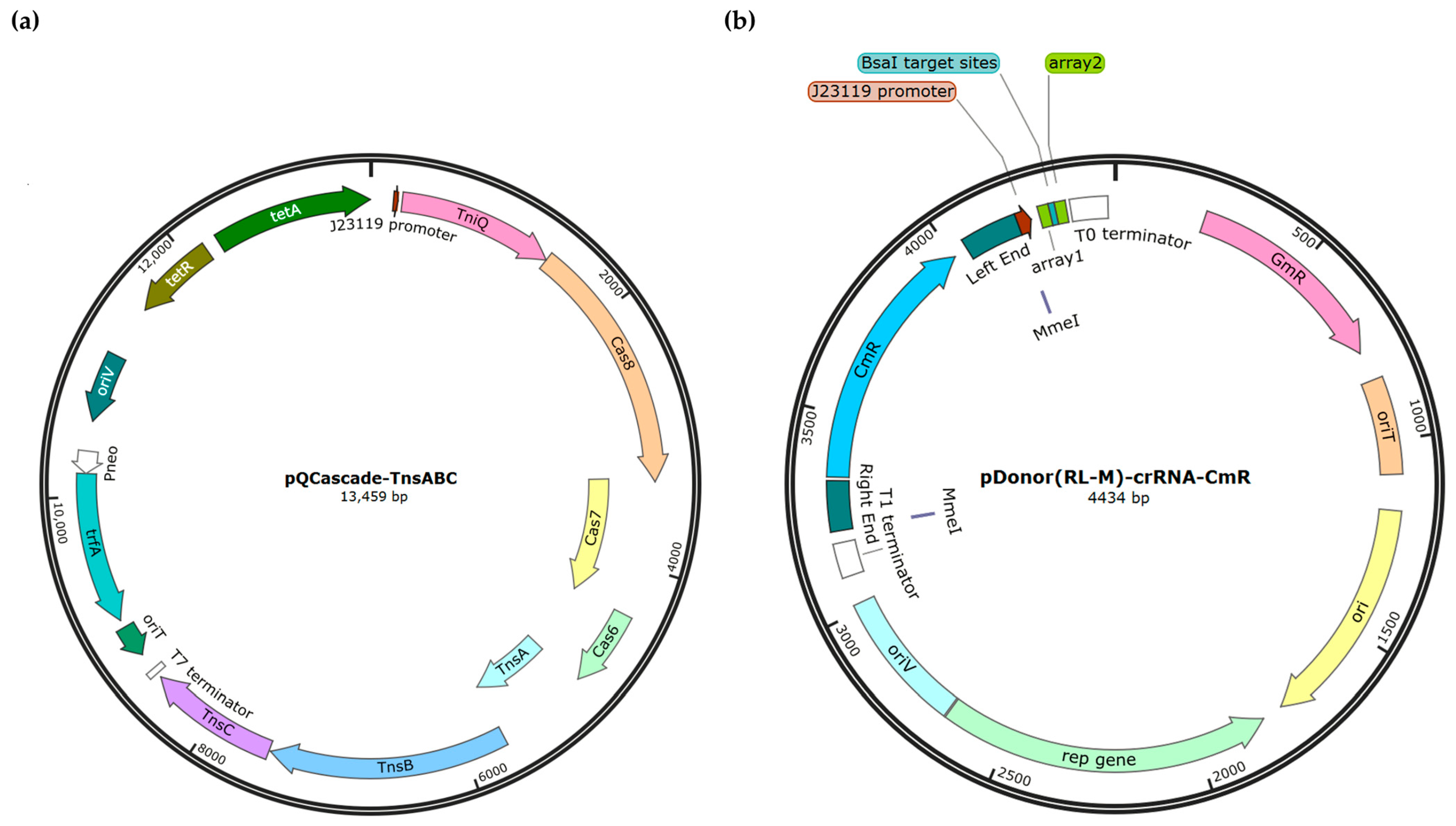
2.2. Plasmid Construction

Construction of pQCascade-TnsABC based on pCas12a-λRed [21,27]. The DNA fragment carrying PJ23119 and QCascade was amplified from plasmid pQCascade_entry (BsaI-stuffer) using primers ZZ21038/039; the DNA fragment harboring TnsABC and T7 terminator was amplified from plasmid pTnsABC using primers ZZ21040/177; the DNA fragment containing oriT, trfA, oriV and tetR was amplified from plasmid pCas12a-λRed using primers ZZ21075/173 and ZZ21075/174. pQCascade-TnsABC was generated by fusing these three DNA fragments by Gibson assembly. In this plasmid, QCascade-TnsABC was expressed under the constitutive promoter PJ23119.
Construction of the pDonor(RL-M)-crRNA-CmR plasmid series derived from pCrRNA-V [21,27]. The DNA fragment harboring two MmeI restriction enzyme cutting sites at both ends for Tn-seq, right end (RE), cargo sequence CmR, and left end (LE) was amplified from pDonor using primers ZZ21044/045 and digested by EcoRI/NdeI; the DNA fragment carrying the crRNA expression cassette with the spacer sequence replaced by two reversed BsaI sites was annealed from ZZ21046/047, ZZ21048/049, ZZ21050/051. The two BsaI sites enabled one-step insertion of the crRNA spacer using the Golden Gate cloning method (see below). Then, the two fragment, along with EcoRI/NdeI-digested pCrRNA-V, were ligated by T4 DNA ligase to yield plasmid pDonor(RL-M)-crRNA-CmR. In this plasmid, the crRNA was expressed as a processed repeat-spacer-repeat unit (a 32-nt spacer flanked by two 28-nt arrays) through the constitutive promoter PJ23119. Subsequently, a series of plasmids (pDonor(RL-M)-crRNA-CmR-X) for testing the transposon insertion system were constructed by inserting a 32-bp double-stranded overhang spacer between its two BsaI sites via Golden Gate cloning. The spacers were generated by annealing and phosphorylation with two primers with partial reverse complement sequences (Table S3). The spacer “5′-GTTGTCTGACACTTGTCACAAACCGCTAGGAG-3′” on pDonor(RL-M)-crRNA-CmR-NC did not target any sequence on the PAO1 or phage S1 genome.
The gene acrVA1 driven by Ptac [29] was synthesized by Sangon Biotechnology (Shanghai, China) and amplified by primers ZZ21159/160. The fragment, along with pDonor(RL-M)-crRNA-CmR, was digested by BamHI and XbaI, and ligated by T4 DNA ligase to yield plasmid pDonor(RL-M)-crRNA-acrVA1. Others operations were conducted as described above.

2.3. Design of crRNA Spacers

crRNAs were designed using the INTEGRATE guide RNA design tool via GitHub (https://github.com/sternberglab/INTEGRATE-guide-RNA-tool (accessed on 13 May 2021)) [10,21]. Spacers were selected based on the following criteria: (1) insertion sites located in the first half of the open-reading frame (orfs) to ensure loss of expression, (2) GC content between 30–70%, (3) ensuring that the designed crRNAs did not share similar sequences with the P. aeruginosa PAO1 genome (Table S3).

2.4. Preparation P. aeruginosa PAO1 Cells for Transposon Insertion

The plasmids pQCascade-TnsABC and pDonor-crRNA series were separately transferred into P. aeruginosa PAO1 by electroporation, as previously described [27]. After pulsing, cells were recovered in 1 mL antibiotic-free LB liquid medium and incubated at 37 °C for 1.5 h. Subsequently, they were plated and cultured at 37 °C for 24~48 h on LB agar plates supplemented with tetracycline 100 μg/mL and gentamycin 50 μg/mL.
For transposon insertion experiments, transformants were identified by colony PCR. The correct transformants were then selected and plated on LB agar plates with tetracycline (100 μg/mL) and gentamycin (50 μg/mL) for a second time round of enrichment instead of growing in liquid culture. Alternatively, after being plated at 37 °C for 24~48 h on LB agar plates supplemented with tetracycline and gentamycin as above. All the recovered cells were scraped from the plates using a spreader and suspended in 1 mL of LB (Ca2+) with tetracycline (100 μg/mL) and gentamycin (50 μg/mL).

2.5. Transposon Insertion Experiments

Inserted phages were obtained by transposition plaque assay. A mixture of 0.1 mL of phage S1 with suitable dilutions (106 pfu/mL, 105 pfu/mL, and 104 pfu/mL) and 0.3 mL of enriched cells was prepared. This mixture was then mixed with 5 mL of pre-warm top agar (0.6%, 50 °C) and poured onto LB agar. After drying at room temperature for about 10 min, the plates were incubated at 37 °C for approximately 20 h. Antibiotics were added to both the top and bottom agar during this step.

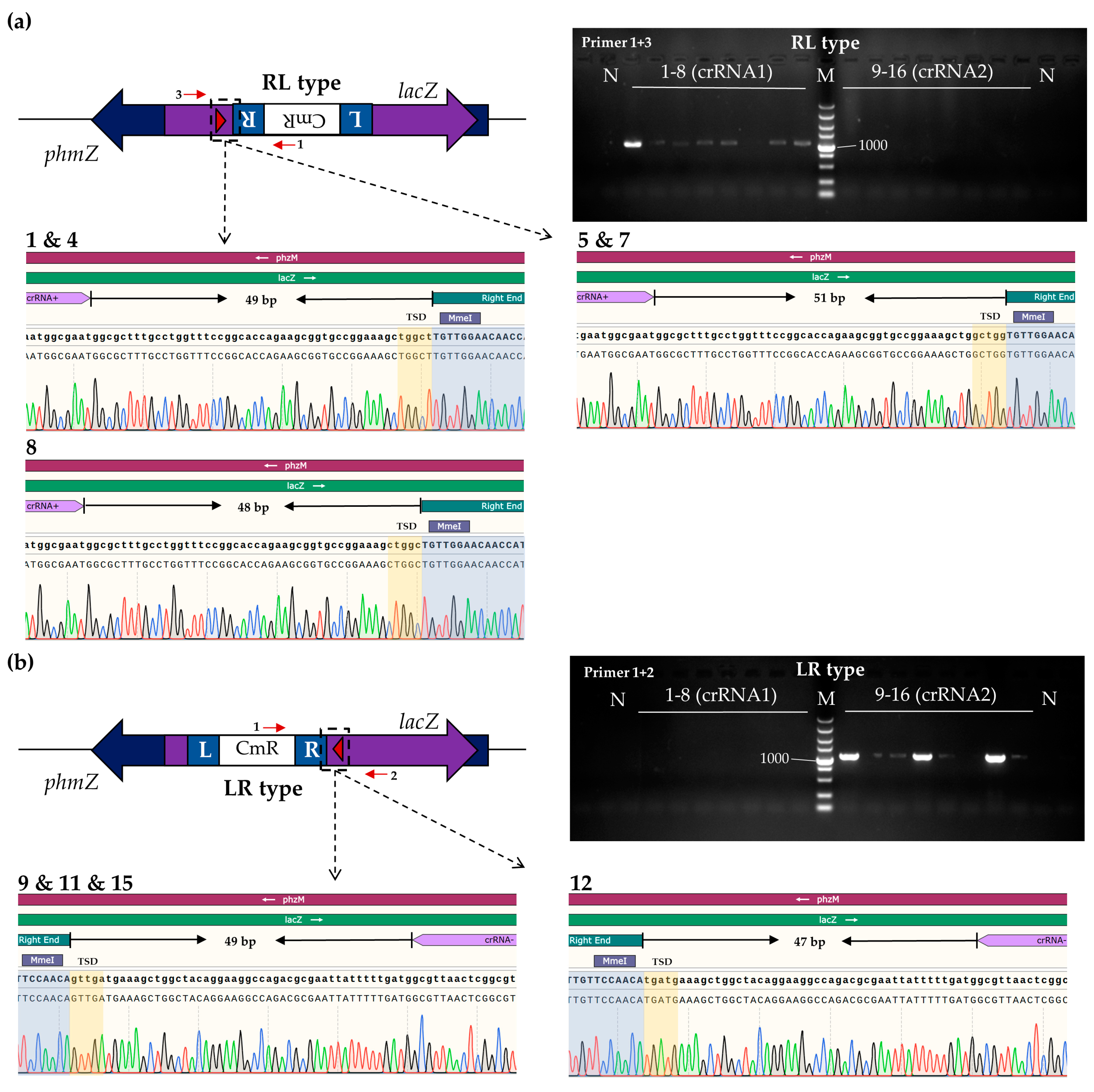
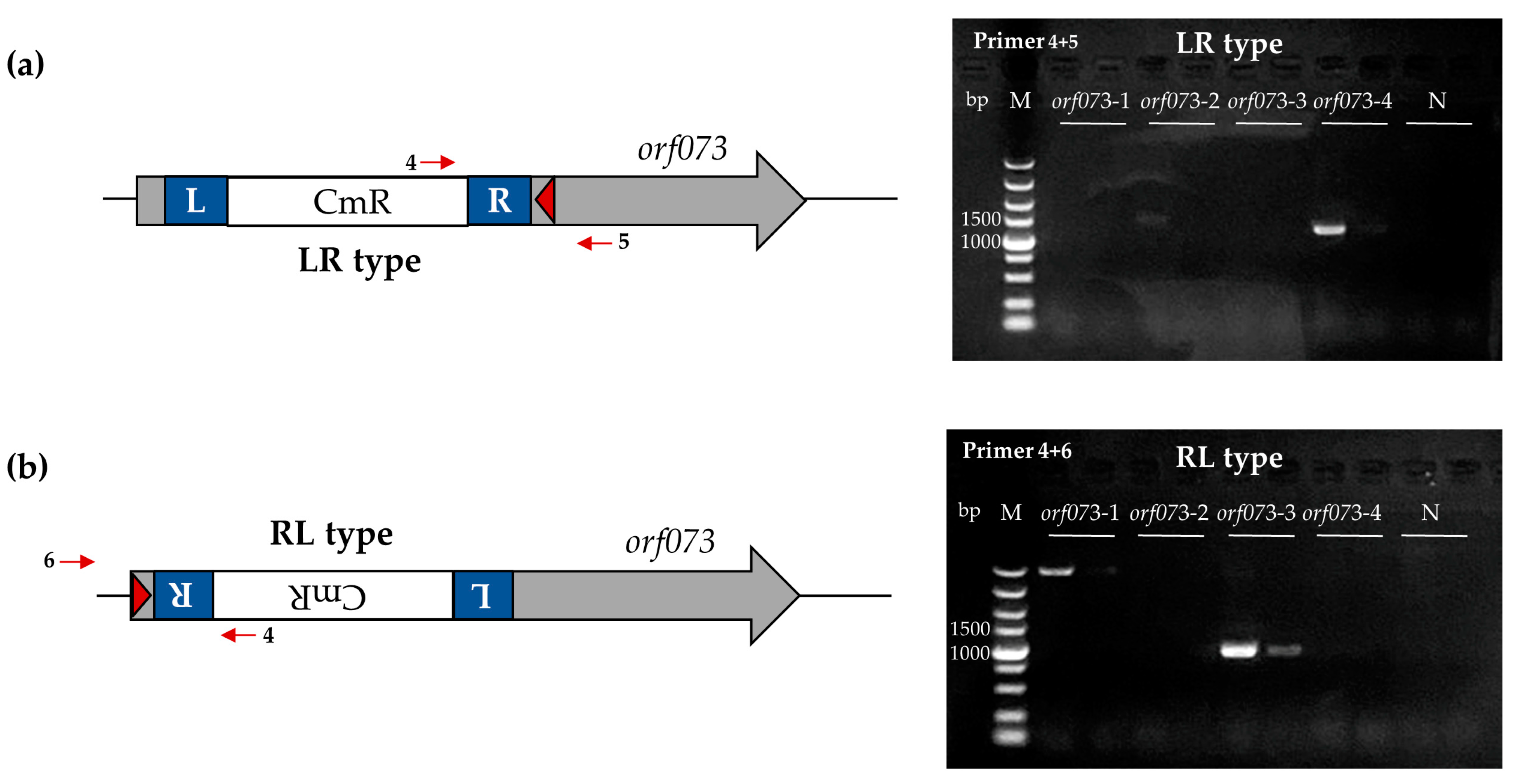
2.6. PCR and Sequencing Analysis of Transposon Insertion Products

After the transposon insertion experiments, the top and base agar of the double-layer agar plate was divided and transferred into 50 mL tubes containing 30 mL of SM buffer (100 nM NaCl, 8 mM MgSO4, and 50 mM Tris-HCl, pH 7.5) plus 300 μL of chloroform. After a 3 h incubation at room temperature, centrifugation and filtration (0.2 μm filter) removed agar and host cell debris to obtain a phage–lysate mix. For the enriched inserted phages, well-isolated plaques were transferred into 1.5 mL Eppedoff tubes containing 200 μL of SM buffer and 2 μL of chloroform. They were then incubated at room temperature for 1 h with mixing every few minutes.
A portion of the aliquots was denatured at 95 °C for 10 min, and 1.6 μL of the sample was used as template for PCR using KOD FX DNA polymerase [30]. The amplified DNA was verified by agarose gel electrophoresis and then sent for Sanger sequencing (Sangon Biotech (Shanghai, China) Co., Ltd.). Primer pairs contained one genome-specific primer and one transposon-specific primer, detecting both upstream and downstream of the target site for all possible insertion orientations.

2.7. Extraction of Phage Genomes

Genomic DNA was extracted as previous described with some modifications [25,31,32]. In brief, phage propagation was initially performed. A 100 mL of PAO1 culture (OD600 of 0.4–0.6) was infected with 100 μL of phage stocks (1010–1011 pfu/mL) and grown at 37 °C for lysis (4–5 h). Subsequently, 2 mL of chloroform was added, and the cells were incubated with shaking for 15 min at 37 °C to obtain the lysates. After centrifugation (8000× g, 10 min), the supernatant was transferred to new tubes. The genomic DNA and RNA of the host was removed by treating with 10 µg/mL DNase I and RNase for 1 h at 37 °C. The lysate was then supplemented with 10% (w/v) PEG 8000 and stored at 4 °C overnight to precipitate the phage particles.
After centrifugation (10,000× g, 30 min), the pellets were suspended in 600 μL of SM buffer, and 200 μL of chloroform was added to extract the PEG and the bacterial debris. Centrifuge at 8000× g for 5 min was performed, followed by treatment with 10 µg/mL DNase I and RNase for 1 h at 37 °C, and then incubation at 65 °C for 20 min to inactivate enzymes. The liquid was then added with 30 µL of 10% (w/v) SDS and 3 µL of 20 mg/mL proteinase K. The mixture was incubated for 20 min at 56 °C. Subsequently, 100 µL of 5 M NaCl and 80 µL of CTAB (cetyltrimethylammonium bromide)/NaCl solution were added and incubated for 10 min at 65 °C. Next, the mixture was sequentially treated with one volume of chloroform, one volume of phenol/chloroform/isoamyl alcohol (25:24:1), and one volume of chloroform, to gradually purify the genomic DNA. The aqueous phase was collected, and 0.7 volumes of isopropanol were added to precipitate the DNA. After centrifugation at 13,000× g for 15 min at 4 °C, the purified DNA pellet was washed thrice with 500 µL of ice-cold 70% ethanol and left to dry. The air-dried pellets were suspended in 100 µL ddH2O. The DNA concentration was measured using a Nanodrop2000 (Thermo Fisher Scientific, Waltham, MA, USA). The purified DNA was used for Tn-seq by Woosen Biotechnology (Hangzhou, China).

3. Results

3.1. Establishment of a Two-Plasmid Transposon Insertion System for P. aeruginosa Phage S1

In our initial efforts, we aimed to investigate the transposon insertion system for identifying non-essential genes within the phage S1 genome. To facilitate plasmid transformation and crRNA library construction, we streamlined the three-plasmid system (pQCascade, pTnsABC, and pDonor) into a two-plasmid system, pQCascade-TnsABC and pDonor(RL-M)-crRNA-CmR-X (referred to as pcrRNA-CmR-X), where X represents the specific target genes. Specifically, the plasmid pQCascade-TnsABC expressed the tniQ-cas8-cas7-cas6 operon (QCascade) and the tnsA-tnsB-tnsC operon (TnsABC) under the control of a constitutive promoter PJ23119 (Figure 1a). pcrRNA-CmR-X contained the right end (RE), a cargo sequence (chloramphenicol resistance gene CmR), the left end (LE), two MmeI cutting sites at both ends to facilitate Tn-seq, and the crRNA. The crRNA was expressed as a repeat-spacer-repeat unit, consisting of a 32-nt spacer flanked by two 28-nt arrays, also driven by PJ23119 (Figure 1b).
The functionality of the two-plasmid system was assessed in the P. aeruginosa strain PAO1 phzM::lacZ via blue-white selection [27]. Three crRNA plasmids, pcrRNA-CmR-lacZ-1/2, targeting lacZ genomic sites on the opposite strand, and the negative control plasmid pcrRNA-CmR-NC with no target site in the PAO1 or S1 genome, were constructed (Table S2). After separately introducing the plasmids pQCascade-TnsABC and pcrRNA-CmR into PAO1 cells through electroporation, cells were plated on X-gal plates at specific dilutions for 48 h, resulting in white and blue colonies (Figure S1a). All colonies on the negative control plate appeared blue (Figure S1b). White colonies on the pcrRNA-CmR-lacZ-1/2 plates suggested transposition occurring on lacZ guided by these two crRNAs, indicating that the two-plasmid transposon insertion system might function for PAO1. PCR was performed using a genome-specific primer and a transposon-specific primer, and only white colonies were found to product PCR fragments. Sanger sequencing confirmed transposition in white colonies, with integration events occurring 49, 51, and 48 bp from the 3′ edge of the target site for pcrRNA-CmR-lacZ-1 (Figure 2a), and 49 and 47 bp for pcrRNA-CmR-lacZ-2 (Figure 2b), consistent with the three-plasmid system [21]. The transposition efficiencies were approximately 8.5% for both pcrRNA-CmR-lacZ-1 (10/117) and pcrRNA-CmR-lacZ-2 (14/165) in insertion of PAO1 lacZ gene (Table S1). These results demonstrated the success of the two-plasmid transposon insertion system for gene insertion in P. aeruginosa strain PAO1.
We then investigated the applicability of the system for phage S1. We first targeted the non-essential gene orf073. We constructed four plasmids, pcrRNA-CmR-orf073-1~4, with half of the crRNAs located on the opposite strand. These crRNA plasmids were separately electroporated into PAO1 electrocompetent cells harboring pQCascade-TnsABC. Surprisingly, co-introduction of both plasmids resulted in cells being able to survive only on solid medium and not in liquid culture for unknown reasons. Consequently, solid medium was selected to support cell survival.
PCR of the phage lysate mix was used to determine the occurrence of transposon insertion in phage S1. PCR products (with a genome-specific primer and a transposon-specific primer, as described above) were observed in experiments with pcrRNA-CmR-orf073-3/4 but not with pcrRNA-CmR-NC or pcrRNA-CmR-orf073-1/2 (Figure 3), indicating that the two-plasmid transposon insertion system can be employed to insert target fragments into phage S1.
Although the two-plasmid system demonstrated the ability to catalyze insertion in phage S1, its efficiency was notably lower (<1%). It was reported that by eliminating the left end of MmeI cutting sites from the pDonor(RL-M)-crRNA-CmR-X could improve efficiency compared to RL m [21], but for our two-plasmid system, this modification failed to improve the efficiency (Figure S2).

3.2. Enrichment of Inserted Phages by AcrVA1

Unable to enhance the efficiency of the two-plasmid system by altering the number of MmeI sites, we sought a different method to enrich the inserted phages. Currently, there are limited selection makers available for phage engineering, and marker genes essential for phage propagation are scarce, found only in a few model phages [33,34,35]. Inspired by the editing approach of the Sulfolobus islandicus rod-shaped virus 2 (SIRV2), which uses the anti-CRISPR protein AcrID1 (SIRV3 gp02) to overcome type I-D CRISPR-Cas immunity [36], we chose AcrVA1, a broad-spectrum inhibitor of the commonly used Cas12a with modest inhibition [29], as the transposon cargo sequence for enriching inserted phages (Figure 4a). Given our establishment of CRISPR-Cas12a as an editing system for phage S1 (unpublished), this choice ensures that only phages carrying the inserted acrVA1 gene can evade CRISPR-Cas12a interference, enabling their survival, accumulation, and dominance in the enrichment field.
To this end, we constructed five crRNA plasmids, namely pcrRNA-acrVA1-NC and pcrRNA-acrVA1-orf073-1~4, replacing the cargo sequence CmR of pcrRNA-CmR-NC and pcrRNA-CmR-orf073-1~4 with Ptac-acrVA1. Subsequently, we cultured the phages on solid media and conducted plaque assays to assess the successful insertion of acrVA1. In experiments utilizing pQCascade-TnsABC and pcrRNA-acrVA1-orf073-2/3, PCR products (with a genome-specific primer and a transposon-specific primer, as described above) within the phage-lysate mixture were detected for acrVA1 insertion (Figure 4b,c).
To gauge the efficiency of AcrVA1, we needed to identify a high-activity crRNA (EOP < 0.1) capable of targeting the majority wild-type phage S1 in the presence of the Cas12a protein. pCrRNA(Cas12a) were one of the low EOP crRNAs we had already tested, so we selected this crRNA to enrich inserted phages (Table S2). Following interference by the CRISPR-Cas12a system for the inserted phages in experiment using pcrRNA-acrVA1-orf073-2, we randomly selected 24 plaques. Among them, 2 out of 24 displayed the expected PCR products (Figure 4d). Therefore, the efficiency of AcrVA1 enrichment was 8.33%, representing an improvement of more than 10 times compared to the non-selection-marker. In summary, these results indicated the success in using AcrVA1 as a selection maker to enrich inserted phages

3.3. Transposon Insertions of Phage S1

We next evaluated the capability of our two-plasmid transposon insertion system in identifying non-essential genes of the phage S1. Ten genes, including orf026 (putative major structural protein) and orf058 (putative DNA polymerase III alpha subunit), as well as orf010, orf014, orf043, orf055, orf072, orf075, orf081, and orf086 (all hypothetical proteins), were selected for testing. Four crRNAs were designed for each gene, targeting the opposite strand and all located in the first half of the genes to prevent expression of a disrupted gene. crRNA plasmids, pcrRNA-acrVA1-X-1~4, were constructed from pcrRNA-acrVA1-NC, where X represents the respective gene. Individual transpositions in phage S1 produced expected PCR products from orf010, orf014, orf072, orf081, and orf086 (Figure S3), indicating successful transposon insertions. However, orf026, orf058, orf043, orf055, and orf075 could not be inserted, suggesting their potential indispensability.
In contrast, simultaneous transposition of the selected ten open-reading frames using forty crRNA plasmids did not yield the expected PCR products (Figure S4a). This issue persists in smaller groups of crRNA plasmids, including orf010 + orf014, orf010 + orf014 + orf072, orf010 + orf014 + orf072 + orf081, and orf010 + orf014 + orf072 + orf081 + orf086 (Figure S4b). This challenge requires further investigation and resolution.

3.4. Discovering Site-Specific Integration by Tn-Seq

We utilized Tn-seq, a transposon insertion sequencing method, for accurately determining genetic interactions on a genome-wide scale [37]. More specifically, we constructed the pDonor(RL-M)-crRNA plasmid with two MmeI sites [10,21]. After transposition in phage S1, we amplified phage lysates, extracted phage genomes, and analysis of the Tn-seq data unveiled that the insertion sites of Tn-seq reads predominantly occurred in orf010, orf014, orf072, orf081, and orf086, accounting for approximately 99.4% of the total reads (Figure S5). This result demonstrates the exquisite specificity of our two-plasmid transposon insertion system, affirming confidence in identifying non-essential genes.

4. Discussion

In this work, we developed a two-plasmid CRISPR RNA-guided transposon insertion system for phage S1, utilizing the anti-CRISPR protein AcrVA1 as the transposon cargo to counteract CRISPR-Cas12a, enriching transposon inserted phages. Successful transposition of CmR into the PAO1 phzM::lacZ loci indicated compatibility in P. aeruginosa strain PAO1. Moreover, this system was applicable to phage S1. Additionally, acrVA1 was transposed into orf073, orf010, orf014, orf072, orf081, and orf086 of the phage S1 genome, affirming our confidence in identifying non-essential genes.
Our study presents the first site-specific, programmable in vivo phage transposon insertion system, holding promise for future applications across a broader range of phages and facilitating the insertion of genetic cargos. The ability to achieve site-specific DNA integration using the transposon insertion approach eliminates the need for homology arms in the donor DNA, double-strand breaks in the target DNA, and host or donor DNA repair factors [21]. This contributes a new tool for the identification of non-essential genes for phages.
Our initial design for the plasmid pDonor(RL-M)-crRNA aimed to generate large crRNA libraries, enabling high-throughput screening, like Tn-seq, of phage genes that are not easily accessible by random transposase strategies [10]. Unfortunately, due to the low efficiency of our two-plasmid system in phage S1, this purpose has not been realized. One potential contributing factor for this low efficiency may be the limited window phase of phage transposition. The two-plasmid system can only catalyze transposition in phages during the latent period, which is a short time, just a few minutes [38]. Additionally, plasmid loss is also a possible cause [39].
It is important to further improve the efficiency of our two-plasmid transposon insertion system. We attempted to replicate the reported single-plasmid system pSPIN, an advanced version of the three-plasmid system with greater efficiency [10]. However, constructing the pSPIN plasmid with a crRNA target for phage S1 failed, leading us to consider the possibility that the crRNA may have toxic effects on P. aeruginosa PAO1. Future studies might consider using an inducible promoter to prevent potential leaky expression of the QCascade-TnsABC complex, thereby preventing premature loss of the plasmid. The growing number of RNA-guided CRISPR-Cas transposase systems [40] suggests the potential to find a suitable and efficient transposase system for different phages. Efficient multiplexing insertions may also be achieved by employing multi-spacer CRISPR arrays in pDonor-crRNA following identical protocols as reported [10].

Supplementary Materials

The following supporting information can be downloaded at: https://www.mdpi.com/article/10.3390/v16030422/s1, Figure S1: Phenotypic analysis of the CmR-inserted PAO1 phzM::lacZ mutant; Figure S2: The efficiency of modified CRISPR RNA-guided transposon insertion system; Figure S3: Phage lysate PCR for the insertion of 10 open-reading frames; Figure S4: Simultaneous transposition of phage S1; Figure S5: Genome-wide analysis of the transposon insertion for phage S1; Table S1: Summary of the transposition efficiencies using transposon insertion system; Table S2: All the phage, bacterial strains, and plasmids used in this study; Table S3: Spacer sequences for CRISPR RNA-guided transposases; Table S4: The primers used for the construction of plasmids and colony or plaque PCR.

Author Contributions

Conceptualization, Y.L., Z.L. (Zizhen Liang) and Y.Y.; methodology, Z.L. (Zizhen Liang) validation, Y.L., Z.L. (Zizhen Liang) and S.Y.; investigation, Y.L. and Z.L. (Zizhen Liang); resources, Y.L., Z.L. (Zizhen Liang) and Z.L. (Zhanglin Lin); data curation, Y.L. and Z.L. (Zizhen Liang); writing—original draft preparation, Y.L. and Z.L. (Zizhen Liang); writing—review and editing, Z.L. (Zhanglin Lin); supervision, Y.Y.; project administration, Z.L. (Zhanglin Lin); funding acquisition, Z.L. (Zhanglin Lin). All authors have read and agreed to the published version of the manuscript.

Funding

This research was funded by National Key R&D Program of China (2018YFA0901000, 2022YFC2104800).

Institutional Review Board Statement

Not applicable.

Informed Consent Statement

Not applicable.

Data Availability Statement

The data presented in this study are available in the article.

Acknowledgments

We thank the members of Lin’s lab at the South China University of Technology for their helpful discussions.

Conflicts of Interest

The authors declare no conflicts of interest.

References

  1. Chevallereau, A.; Pons, B.J.; van Houte, S.; Westra, E.R. Interactions between bacterial and phage communities in natural environments. Nat. Rev. Microbiol. 2022, 20, 49–62. [Google Scholar] [CrossRef]
  2. Suttle, C.A. Viruses in the sea. Nature 2005, 437, 356–361. [Google Scholar] [CrossRef]
  3. Kortright, K.E.; Chan, B.K.; Koff, J.L.; Turner, P.E. Phage Therapy: A Renewed Approach to Combat Antibiotic-Resistant Bacteria. Cell Host Microbe 2019, 25, 219–232. [Google Scholar] [CrossRef]
  4. Pirnay, J.P.; Blasdel, B.G.; Bretaudeau, L.; Buckling, A.; Chanishvili, N.; Clark, J.R.; Corte-Real, S.; Debarbieux, L.; Dublanchet, A.; De Vos, D.; et al. Quality and safety requirements for sustainable phage therapy products. Pharm. Res. 2015, 32, 2173–2179. [Google Scholar] [CrossRef] [PubMed]
  5. Pires, D.P.; Monteiro, R.; Mil-Homens, D.; Fialho, A.; Lu, T.K.; Azeredo, J. Designing P. aeruginosa synthetic phages with reduced genomes. Sci. Rep. 2021, 11, 2164. [Google Scholar] [CrossRef] [PubMed]
  6. Lemay, M.L.; Tremblay, D.M.; Moineau, S. Genome Engineering of Virulent Lactococcal Phages Using CRISPR-Cas9. ACS Synth. Biol. 2017, 6, 1351–1358. [Google Scholar] [CrossRef]
  7. Miller, E.S.; Kutter, E.; Mosig, G.; Arisaka, F.; Kunisawa, T.; Rüger, W. Bacteriophage T4 genome. Microbiol. Mol. Biol. Rev. MMBR 2003, 67, 86–156. [Google Scholar] [CrossRef]
  8. Hatoum-Aslan, A. Phage Genetic Engineering Using CRISPR-Cas Systems. Viruses 2018, 10, 335. [Google Scholar] [CrossRef]
  9. Lemire, S.; Yehl, K.M.; Lu, T.K. Phage-Based Applications in Synthetic Biology. Annu. Rev. Virol. 2018, 5, 453–476. [Google Scholar] [CrossRef]
  10. Vo, P.L.H.; Ronda, C.; Klompe, S.E.; Chen, E.E.; Acree, C.; Wang, H.H.; Sternberg, S.H. CRISPR RNA-guided integrases for high-efficiency, multiplexed bacterial genome engineering. Nat. Biotechnol. 2021, 39, 480–489. [Google Scholar] [CrossRef] [PubMed]
  11. Jiang, Y.; Qian, F.; Yang, J.; Liu, Y.; Dong, F.; Xu, C.; Sun, B.; Chen, B.; Xu, X.; Li, Y.; et al. CRISPR-Cpf1 assisted genome editing of Corynebacterium glutamicum. Nat. Commun. 2017, 8, 15179. [Google Scholar] [CrossRef] [PubMed]
  12. Vento, J.M.; Crook, N.; Beisel, C.L. Barriers to genome editing with CRISPR in bacteria. J. Ind. Microbiol. Biotechnol. 2019, 46, 1327–1341. [Google Scholar] [CrossRef] [PubMed]
  13. Kosicki, M.; Tomberg, K.; Bradley, A. Repair of double-strand breaks induced by CRISPR-Cas9 leads to large deletions and complex rearrangements. Nat. Biotechnol. 2018, 36, 765–771. [Google Scholar] [CrossRef] [PubMed]
  14. Mahler, M.; Costa, A.R.; van Beljouw, S.P.B.; Fineran, P.C.; Brouns, S.J.J. Approaches for bacteriophage genome engineering. Trends Biotechnol. 2023, 41, 669–685. [Google Scholar] [CrossRef] [PubMed]
  15. Kilcher, S.; Loessner, M.J. Engineering Bacteriophages as Versatile Biologics. Trends Microbiol. 2019, 27, 355–367. [Google Scholar] [CrossRef] [PubMed]
  16. Kilcher, S.; Studer, P.; Muessner, C.; Klumpp, J.; Loessner, M.J. Cross-genus rebooting of custom-made, synthetic bacteriophage genomes in L-form bacteria. Proc. Natl. Acad. Sci. USA 2018, 115, 567–572. [Google Scholar] [CrossRef] [PubMed]
  17. Jia, H.J.; Jia, P.P.; Yin, S.; Bu, L.K.; Yang, G.; Pei, D.S. Engineering bacteriophages for enhanced host range and efficacy: Insights from bacteriophage-bacteria interactions. Front. Microbiol. 2023, 14, 1172635. [Google Scholar] [CrossRef] [PubMed]
  18. Vilen, H.; Aalto, J.M.; Kassinen, A.; Paulin, L.; Savilahti, H. A direct transposon insertion tool for modification and functional analysis of viral genomes. J. Virol. 2003, 77, 123–134. [Google Scholar] [CrossRef]
  19. Kiljunen, S.; Vilen, H.; Pajunen, M.; Savilahti, H.; Skurnik, M. Nonessential genes of phage phiYeO3-12 include genes involved in adaptation to growth on Yersinia enterocolitica serotype O:3. J. Bacteriol. 2005, 187, 1405–1414. [Google Scholar] [CrossRef]
  20. Krupovic, M.; Vilen, H.; Bamford, J.K.; Kivelä, H.M.; Aalto, J.M.; Savilahti, H.; Bamford, D.H. Genome characterization of lipid-containing marine bacteriophage PM2 by transposon insertion mutagenesis. J. Virol. 2006, 80, 9270–9278. [Google Scholar] [CrossRef]
  21. Klompe, S.E.; Vo, P.L.H.; Halpin-Healy, T.S.; Sternberg, S.H. Transposon-encoded CRISPR-Cas systems direct RNA-guided DNA integration. Nature 2019, 571, 219–225. [Google Scholar] [CrossRef]
  22. McDonald, N.D.; Regmi, A.; Morreale, D.P.; Borowski, J.D.; Boyd, E.F. CRISPR-Cas systems are present predominantly on mobile genetic elements in Vibrio species. BMC Genom. 2019, 20, 105. [Google Scholar] [CrossRef] [PubMed]
  23. Makarova, K.S.; Wolf, Y.I.; Koonin, E.V. Classification and Nomenclature of CRISPR-Cas Systems: Where from Here? CRISPR J. 2018, 1, 325–336. [Google Scholar] [CrossRef] [PubMed]
  24. Peters, J.E. Tn7. Microbiol. Spectr. 2014, 2. [Google Scholar] [CrossRef]
  25. Guo, Y.; Chen, P.; Lin, Z.; Wang, T. Characterization of Two Pseudomonas aeruginosa Viruses vB_PaeM_SCUT-S1 and vB_PaeM_SCUT-S2. Viruses 2019, 11, 318. [Google Scholar] [CrossRef]
  26. Breijyeh, Z.; Jubeh, B.; Karaman, R. Resistance of Gram-Negative Bacteria to Current Antibacterial Agents and Approaches to Resolve It. Molecules 2020, 25, 1340. [Google Scholar] [CrossRef]
  27. Lin, Z.; Li, H.; He, L.; Jing, Y.; Pistolozzi, M.; Wang, T.; Ye, Y. Efficient genome editing for Pseudomonas aeruginosa using CRISPR-Cas12a. Gene 2021, 790, 145693. [Google Scholar] [CrossRef] [PubMed]
  28. Gibson, D.G.; Young, L.; Chuang, R.Y.; Venter, J.C.; Hutchison, C.A., 3rd; Smith, H.O. Enzymatic assembly of DNA molecules up to several hundred kilobases. Nat. Methods 2009, 6, 343–345. [Google Scholar] [CrossRef]
  29. Marino, N.D.; Zhang, J.Y.; Borges, A.L.; Sousa, A.A.; Leon, L.M.; Rauch, B.J.; Walton, R.T.; Berry, J.D.; Joung, J.K.; Kleinstiver, B.P.; et al. Discovery of widespread type I and type V CRISPR-Cas inhibitors. Science 2018, 362, 240–242. [Google Scholar] [CrossRef]
  30. Tao, P.; Wu, X.; Tang, W.C.; Zhu, J.; Rao, V. Engineering of Bacteriophage T4 Genome Using CRISPR-Cas9. ACS Synth. Biol. 2017, 6, 1952–1961. [Google Scholar] [CrossRef]
  31. David, W.; Russell, J.S. Molecular Cloning: A Laboratory Manual, 3rd ed.; Cold Spring Harbor Laboratory Press: Cold Spring Harbor, NY, USA, 2001. [Google Scholar]
  32. Thurber, R.V.; Haynes, M.; Breitbart, M.; Wegley, L.; Rohwer, F. Laboratory procedures to generate viral metagenomes. Nat. Protoc. 2009, 4, 470–483. [Google Scholar] [CrossRef] [PubMed]
  33. Grigonyte, A.M.; Harrison, C.; MacDonald, P.R.; Montero-Blay, A.; Tridgett, M.; Duncan, J.; Sagona, A.P.; Constantinidou, C.; Jaramillo, A.; Millard, A. Comparison of CRISPR and Marker-Based Methods for the Engineering of Phage T7. Viruses 2020, 12, 193. [Google Scholar] [CrossRef] [PubMed]
  34. Ramirez-Chamorro, L.; Boulanger, P.; Rossier, O. Strategies for Bacteriophage T5 Mutagenesis: Expanding the Toolbox for Phage Genome Engineering. Front. Microbiol. 2021, 12, 667332. [Google Scholar] [CrossRef] [PubMed]
  35. Kiro, R.; Shitrit, D.; Qimron, U. Efficient engineering of a bacteriophage genome using the type I-E CRISPR-Cas system. RNA Biol. 2014, 11, 42–44. [Google Scholar] [CrossRef] [PubMed]
  36. Mayo-Muñoz, D.; He, F.; Jørgensen, J.B.; Madsen, P.K.; Bhoobalan-Chitty, Y.; Peng, X. Anti-CRISPR-Based and CRISPR-Based Genome Editing of Sulfolobus islandicus Rod-Shaped Virus 2. Viruses 2018, 10, 695. [Google Scholar] [CrossRef] [PubMed]
  37. van Opijnen, T.; Bodi, K.L.; Camilli, A. Tn-seq: High-throughput parallel sequencing for fitness and genetic interaction studies in microorganisms. Nat. Methods 2009, 6, 767–772. [Google Scholar] [CrossRef] [PubMed]
  38. Jariah, R.O.A.; Hakim, M.S. Interaction of phages, bacteria, and the human immune system: Evolutionary changes in phage therapy. Rev. Med. Virol. 2019, 29, e2055. [Google Scholar] [CrossRef]
  39. Cordes, C.; Meima, R.; Twiest, B.; Kazemier, B.; Venema, G.; van Dijl, J.M.; Bron, S. The expression of a plasmid-specified exported protein causes structural plasmid instability in Bacillus subtilis. J. Bacteriol. 1996, 178, 5235–5242. [Google Scholar] [CrossRef]
  40. Klompe, S.E.; Jaber, N.; Beh, L.Y.; Mohabir, J.T.; Bernheim, A.; Sternberg, S.H. Evolutionary and mechanistic diversity of Type I-F CRISPR-associated transposons. Mol. Cell 2022, 82, 616–628.e5. [Google Scholar] [CrossRef]
Figure 1. Maps of pQCascade-TnsABC and pDonor(RL-M)-crRNA-CmR plasmids created with SnapGene 4.1 (GSL Biotech LLC). (a) Plasmid pQCascade-TnsABC cintains the tniQ-cas8-cas7-cas6 operon (QCascade) and the tnsA-tnsB-tnsC operon (TnsABC), expressed under the control of the constitutive PJ23119 promoter. The tetracycline-resistance marker genes tetA and tetR are present for resistance in E. coli and P. aeruginosa; oriV, origin of replication of the broad-host-range plasmid RK2; trfA, plasmid replication initiator protein; oriT, origin of transfer for conjugation. (b) Plasmid pDonor(RL-M)-crRNA-CmR comprises the right end (RE), a cargo sequence (chloramphenicol resistance gene CmR), the left end (LE), two MmeI cutting sites at both ends, and the crRNA cassette transcribed through the J23119 promoter; BsaI, recognition sites of type IIS restriction endonuclease for the insertion of the 32-nt spacer in the crRNA; the crRNA was expressed as a processed repeat-spacer-repeat unit, spacer flanked by two 28-nt arrays; GmR gene, the gentamycin-resistance gene.
Viruses 16 00422 g001
Figure 2. Analysis of P. aeruginosa strain isolates containing lacZ-inserted transposons. (a) Upper left panel, schematic diagram of RL type insertion by the two-plasmid system; upper right panel, colony PCR for the insertion in lacZ; lower panel, Sanger sequencing results. (b) Upper left panel, schematic diagram of LR type insertion; upper right panel, colony PCR for the insertion in lacZ; lower panel, Sanger sequencing results. The red arrows indicate the primers used in colony PCR for verifying the transposon insertion. The red triangle represents the spacer. L, left transposon end; R, right transposon end. M, Takara 5000 bp DNA ladder; 1–8, randomly selected colonies grown on plate after transformation with plasmid pQCascade-TnsABC and pcrRNA-CmR-lacZ-1; 9–16, randomly selected colonies grown on plate after transformation with plasmid pQCascade-TnsABC and pcrRNA-CmR-lacZ-2; N, blue colony; TSD, target-site duplication.
Viruses 16 00422 g002
Figure 3. CRISPR RNA-guided transposon insertion system could function for phage S1. (a) Left panel, schematic diagram of LR type phage S1 orf073 CmR insertion by the two-plasmid transposon insertion system; right panel, PCR results of phage lysate for the insertion in orf073. (b) Left panel, schematic diagram of RL type insertion; right panel, PCR results of phage lysate for the insertion in orf073. The red arrows indicate the primers used in PCR for verifying the transposon insertion. The red triangle represents the spacer. L, left transposon end; R, right transposon end. M, Takara 5000 bp DNA ladder; orf073-1~4, phage lysate mix obtained after the transposon insertion experiment with plasmids pQCascade-TnsABC and pcrRNA-CmR-orf073-1~4; N, negative control.
Viruses 16 00422 g003
Figure 4. AcrVA1 can work as a selection marker for enriching integrated phages. (a) Schematic diagram illustrating the enrichment of inserted phages by AcrVA1. acrVA1-inserted phages can express AcrVA1 proteins, protecting them from interference. (b) Left panel, schematic diagram of LR type acrVA1 insertion; right panel, PCR results of the insertion in orf073. (c) Left panel, schematic diagram of RL type acrVA1 insertion; right panel, PCR results of the insertion. (d) PCR results of the enrichment of acrVA1-inserted phages. The red arrows indicate the primers used in PCR for verifying the transposon insertion. The red triangle represents the spacer. L, left transposon end; R, right transposon end. M, Takara 5000 bp DNA ladder; orf073-1~4, phage lysate mix obtained after the transposon insertion experiment with plasmids pQCascade-TnsABC and pcrRNA-acrVA1-orf073-1~4; 1–24, randomly selected plaques grown on plate after enrichment; N, negative control.
Viruses 16 00422 g004
Disclaimer/Publisher’s Note: The statements, opinions and data contained in all publications are solely those of the individual author(s) and contributor(s) and not of MDPI and/or the editor(s). MDPI and/or the editor(s) disclaim responsibility for any injury to people or property resulting from any ideas, methods, instructions or products referred to in the content.

