Brief Report

ddPCR for the Detection and Absolute Quantification of Oropouche Virus

Department of Infectious, Tropical Diseases and Microbiology, IRCCS Sacro Cuore Don Calabria Hospital, 37024 Negrar di Valpolicella, Verona, Italy
*
Author to whom correspondence should be addressed.
Viruses 2024, 16(9), 1426; https://doi.org/10.3390/v16091426
Submission received: 12 August 2024 / Revised: 3 September 2024 / Accepted: 6 September 2024 / Published: 7 September 2024
(This article belongs to the Special Issue Zoonotic and Vector-Borne Viral Diseases)

Abstract

:
Background: Oropouche virus (OROV) is a segmented RNA virus belonging to the genus Orthobunyavirus in the family Peribunyaviridae. Herein, an in-house droplet digital PCR (ddPCR) assay was used for the detection and quantification of OROV. Methods: The ddPCR reaction was assessed as duplex assay using the human housekeeping gene RPP30. Limit of detection (LoD) analysis was performed in whole blood, serum, and urine. The assay was executed on a total of 28 clinical samples (whole blood n = 9, serum n = 11, and urine n = 8), of which 16 specimens were tested positive at the routine molecular diagnostics (endpoint and real-time PCRs). Results: The LoD of the ddPCR performed using 10-fold serial dilution of OROV detected up to 1 cp/µL in all the biological matrices. Compared to the routine molecular diagnostics, the ddPCR assay showed 100% sensitivity for whole blood and serum and 75% for urine, highlighting higher positive rate of ddPCR. Conclusion: We have established a quantitative RNA detection method of OROV with high sensitivity and specificity based on ddPCR. This test is capable of quantitatively monitoring the viral load of OROV and can contribute, in addition to laboratory diagnosis, to shed light on the pathogenesis, filling in the knowledge gaps of this neglected disease and to the vector control programs.

1. Introduction

The Oropouche virus (OROV) is an important arthropod-borne virus in the Peribunyaviridae family that can cause febrile illnesses, and it is widely distributed in tropical regions such as Central and South America [1]. The Oropouche virus is spread to people primarily by the bite of an infected biting midge (Culicoides paraensis) in an urban cycle [1]. In May 2024, the Ministry of Public Health of Cuba reported the first ever outbreak of OROV disease [2]. Due to recent, very worrisome changes in the observed clinical and epidemiological characteristics of Oropouche fever, the Pan American Health Organization (PAHO) has raised the risk level for the region to high [3]. PAHO has highlighted the need to strengthen epidemiological and entomological surveillance and reinforce preventive measures in the population. It is critical to expand the scarce knowledge of this neglected disease, including new possible routes of transmission and new vectors that could affect both the general population and vulnerable groups, such as pregnant women. As of 30 July 2024, 8078 confirmed cases of OROV have been reported in the following five countries in the region of the Americas: the Plurinational State of Bolivia (n = 356), Brazil (n = 7284), Colombia (n = 74), Cuba (n = 74), and Peru (n = 290) [3]. The prevalence of OROV infections is likely underestimated in the Americas, where more than ten million cases of dengue were reported between epidemiological weeks 1 and 26 of 2024 [4,5]. In late May–early June 2024, OROV fever cases in two travelers from Cuba were notified as the first imported cases of OROV disease from our hospital in Italy [6], suggesting the importance of expanding the travel history for considering a diagnosis of OROV fever in returning travelers from Central or South America, including Cuba and other Caribbean countries too. As of 9 August 2024, between June and July 2024, 19 imported cases of OROV disease have been reported in Spain (n = 12), Italy (n = 5), and Germany (n = 2) [7]. It is noteworthy that PAHO has indicated, as diagnostic sampling, only serum and eventually cerebrospinal fluid (CSF) for cases presenting with aseptic meningitis (a rare complication of Oropouche fever) [8]. Based on our recent findings in the diagnosis of OROV infection in the EU, we also suggest the use of whole blood and urine to increase the possibility of a definite diagnosis [9]. OROV is a segmented RNA virus and, nowadays, the detection of viral RNA by a PCR-based molecular method (conventional RT-PCR and real-time RT-PCR) is the most frequently applied laboratory test due to its higher sensitivity and specificity. So far, no commercial PCR-based diagnostic tests have been developed in order to monitor and diagnose OROV, in-house OROV developed tests target a conserved 5′ region of the S segment [10,11], and one in particular has been recommended by PAHO [12]. In recent decades, a new generation of PCR (i.e., droplet digital PCR, ddPCR), has been introduced into the field of molecular virology for detection and, above all, the quantification of viruses [13,14]. Compared to the more commonly used real-time PCR (rt-PCR), ddPCR provides absolute quantification of nucleic acid copies in samples without the need for standard curves, and it also has the ability to detect lower genomic yield. The ddPCR is based on microfluidics technology, which allows for the generation of multiple reaction partitions that work as individual reactions. Based on the positive or negative fraction and following the Poisson distribution, it is possible to determine the absolute concentration of the target of interest in terms of the number of copies per microliter in the ddPCR reaction. In this study, we used the PAHO primers/probe set [12] to set up a ddPCR and to assess its analytical and clinical performance for detecting and quantifying genomic RNA of OROV on whole blood, serum, and urine matrices and clinical samples.

2. Materials and Methods

2.1. Cell Line and Virus

A viral isolate (named OROV-IRCCS-SCDC_1/2024) was obtained from the serum sample of a patient returning from Cuba on May 2024, with laboratory-confirmed OROV, in the IRCCS Sacro Cuore Don Calabria (SCDC) Hospital BLS3 (biosafety level 3) laboratories. Briefly, the serum sample was diluted 1:10 in minimum essential medium (MEM, Gibco, Thermo Fisher Scientific, Waltham, MA, USA) and then inoculated onto a semiconfluent monolayer of Vero E6 cells (ATCC CRL-1586) grown with MEM containing 1% penicillin-streptomycin (Gibco), 1% L-Glutamine (Gibco, Thermo Fisher Scientific, Waltham, MA, USA), and 2% fetal bovine serum (Gibco, Thermo Fisher Scientific, Waltham, MA, USA). Cytopathic effect (CPE) appearance was observed by light microscope. The culture medium was harvested, low-speed centrifuged, and the supernatant was aliquoted and stored at −80 °C.

2.2. Viral Titration

A day prior to the infection, Vero E6 cells were grown to semiconfluence with 10% FBS MEM in a 96-well tissue culture plate at 37 °C, 5% CO2.
On the day of infection, a series of 3-fold dilutions of the original virus sample were prepared in MEM without FBS. Each dilution was tested in four replicates. Once the dilution plate was prepared, the 10% FBS MEM medium was removed from the monolayers, and 100 µL of each dilution was added onto the cells. The plate was kept at 37 °C, 5% CO2.
In every plate, four wells were used as the no-virus control. The plate was incubated and observed daily to monitor the development of a cytopathic effect (CPE) by the light microscope. CPE was evaluated and viral titer was calculated using Reed and Muench’s [15] calculation and expressed as tissue culture infectious dose (TCID50)/mL.

2.3. Clinical Samples and in House PCRs

A total of 28 clinical samples (EDTA whole blood n = 9, serum n = 11, and urine n = 8) collected at diagnosis or during follow-up were previously screened in our laboratory with routine real-time PCR diagnostic tests specific for imported Flavivirus fevers (including Dengue, Zika, Chikungunya) and then for OROV, based on in-house endpoint PCR using Lambert primers [11] and the OROV real-time PCR (PAHO rtPCR) [12], with both targeting the S segment. Tests were performed using BioRad (Hercules, CA, USA) reverse transcriptase mixes (iScript Reverse Transcription Supermix for RT-qPCR or Reliance One-Step Multiplex RT-qPCR Supermix). Sanger sequencing was performed using the same amplification primers on positive endpoint samples to confirm OROV infection. Among the total of samples, n = 16 were tested positive and n = 12 negative to OROV. All samples were negative for Flaviviruses.

2.4. RNA Extraction

RNA was isolated from the whole blood and urine using EZ1 Advanced XL System with the EZ1 DSP Virus Kit (Qiagen, Hilden, Germany); both matrixes were pre-diluted 1:4 (V:V) with PBS and then mixed with ATL buffer following the manufacturer’s instructions and eluted in 60 μL. RNA from serum was instead isolated using the QIAamp Viral RNA Mini kit (Qiagen, Hilden, Germany) from 140 μL and eluted in 60 μL. The isolated RNA was stored at −80 °C until use by ddPCR.

2.5. ddPCR

The ddPCR procedure was performed with primers/FAM TAQman probe set targeting a tract of S segment of OROV genome used in PAHO-rtPCR [12]. In addition, the HEX probe assay for the human Ribonuclease P protein subunit p30 (RPP30) (ddPCR CNV Assay Validated, BioRad, Hercules, CA, USA) was used as the human housekeeping gene. The duplex PCR was performed using the One-step RT-ddPCR Advanced Kit for Probes (BioRad, Hercules, CA, USA) and the reaction mixture was assembled as follows: supermix 5.5 μL, reverse transcriptase 20 U/µL 2.2 μL, DTT 15 mM 1.1 μL, OROV primers at 900 nM, and probe at 250 nM as the final concentration, 20× RPP30 Assay 2 μL and RNA template 7 μL, in a final volume of 22 μL. The QX200 droplet generator (Bio-Rad, Hercules, CA, USA) was used to convert 20 μL of each reaction mix into droplets. The droplet-partitioned samples were transferred to a 96-well plate, sealed and processed in a C1000 touch Thermal Cycler (Bio-Rad) under the following cycling protocol: 25 °C hold for 3 min, 50 °C for 60 min reverse transcription, 95 °C for 10 min for enzyme activation, 95 °C for 30 s for denaturation, 60 °C for 60 s for annealing/extension for 50 cycles, 98 °C 10 min for enzyme deactivation, followed by 30 min at a 4 °C hold. The amplified samples were then transferred and read in the FAM (OROV) and HEX (RPP30) channels using the QX200 reader (BioRad, Hercules, CA, USA). The experiments were performed using a negative control (negative biological matrix), the no-template control (water) and a positive control (OROV strain isolate IRCCS-SCDC_1/2024). Discordant samples were repeated in duplicate (then merged for quantification). Data were analyzed using the QXManager 1.2 Standard Edition Software (Bio-Rad, Hercules, CA, USA) and expressed as copies/µL (cp/µL) in ddPCR reaction.

2.6. Analytical Sensitivity (Limit of Detection, LoD) Analysis

The LoD calculation of ddPCR was performed using ten-fold serial dilutions of OROV RNA extracted from the viral strain IRCCS-SCDC_1/2024 (stock titer 105 TCID50/mL) ranging from 104 to 10−2 TCID50/mL generated in nuclease-free water. Regarding whole blood and urine (both diluted 1:4 with PBS, V:V before extraction) [16], and serum, the serial dilutions were generated by spiking the OROV viral strain 105 TCID50/mL in each biological matrix (from healthy donors). An additional point was tested as negative control (no RNA or viral strain added) for both water and biological matrices. For the biological matrices, the RNA was then extracted as described above. The singleplex assay for OROV was performed in duplicate on water and the duplex OROV/RPP30 assay in triplicate on biological matrices. LoD was calculated as the last dilution with all replicates detected OROV positive. Standard deviation (SD) and coefficient of variation (CV%) were calculated. For each analysis, the results were expressed as copies/µL (cp/µL).

2.7. Statistics

Sensitivity and specificity for the clinical evaluation were analyzed by a two-way contingency table using a diagnostic test evaluation calculator (https://www.medcalc.org/calc/diagnostic_test.php, accessed on 12 July 2024). Correlation and regression analyses and graphs were performed using Jamovi software version 2.5.3.0. The Shapiro–Wilk test was used to test the normal distribution and Pearson’s correlation coefficient was used for the correlation analysis.

3. Results

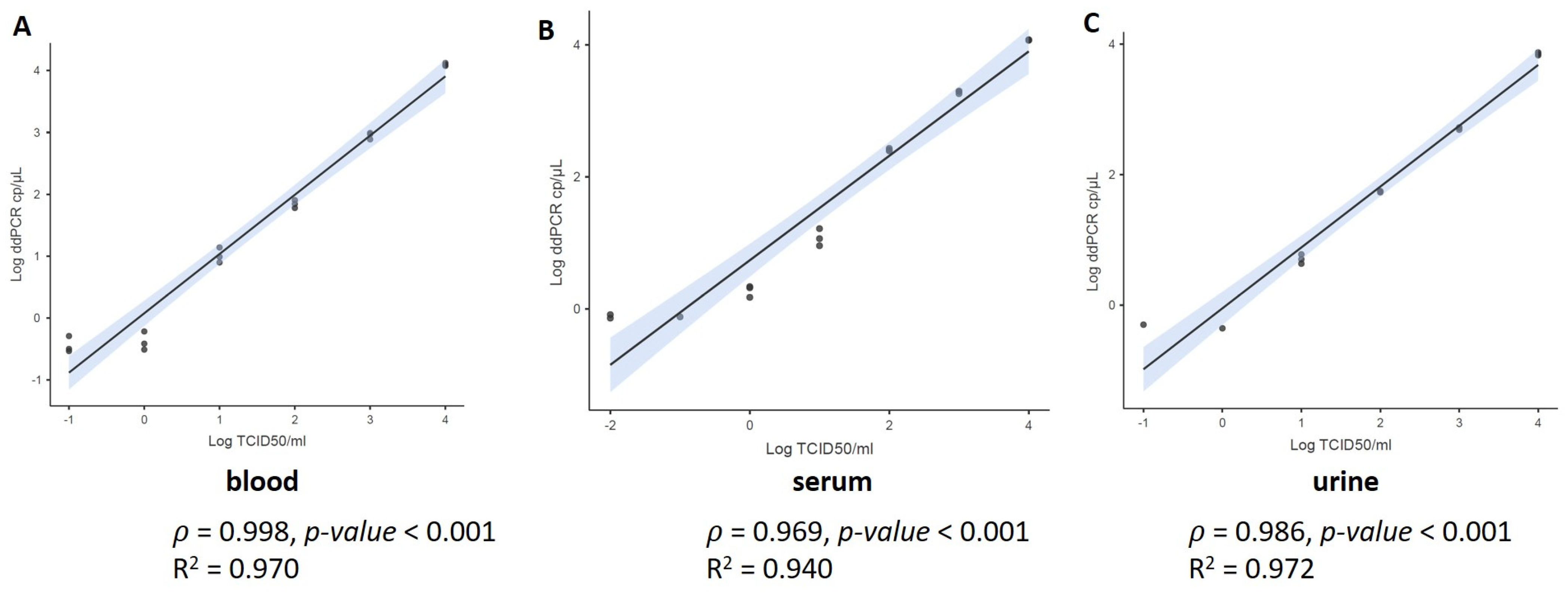
3.1. Limit of Detection Analysis (LoD)

The analytical sensitivity of the assay was assessed by determining the LoD as the lowest viral titer expressed in cp/µL that can be detected by ddPCR. The LoD of the ddPCR performed on 10-fold serial dilution of OROV RNA in water detected up to 1 cp/µL (in Supplementary Materials, Table S1). No OROV target was reported in the diluent control. Then, we performed the LoD analysis in different matrices by ddPCR spiking the viral strain in the whole blood, serum, and urine. We assessed the duplex assay targeting OROV and RPP30. The assay was performed in triplicate. The viral copies were detected up to 1 cp/µL in all replicates for all samples (data are reported in Supplementary Material Table S2). In particular, we observed a strong correlation between quantification (cp/μL) and viral titer (p > 0.98, p-value < 0.001) with a regression coefficient R2 >0.94 (Figure 1) for all the biological matrices. Concerning the variability of data, OROV and RPP30 results were equally repeatable, with CV generally ≤30% for all matrices.

3.2. Clinical Samples

We investigated a total of 28 clinical samples with positive (n = 16) or negative (n = 12) results at the routine diagnosis for OROV with rt-PCR. Table 1 summarizes the ddPCR results for each biological matrix (whole blood, serum, and urine). Positive detection was determined even if there were <3 droplets. Supplementary material reports the detailed data (Supplementary Material, Table S3). In order to evaluate the clinical value, we calculated the sensitivity (SE) and specificity (SP) using the results obtained by routine OROV rt-PCR as reference values. Based on these data, the SE and SP of OROV detection were, respectively, 100% [95% CI, 59.04 to 100] and 50% [95% CI, 1.26 to 98.74] for whole blood (with the routine rt-PCR Ct value range of 24.51–40.67). For serum, the SE and SP were 100% [95% CI, 47.82 to 100.00] and 100% [95% CI, 54.07 to 100.00], respectively (with the routine rtPCR Ct value range of 22.60–32.93). For urine, the SE and SP were both 75% [95% CI, 19.41 to 99.37]. Of note, we observed that the discordant results had Ct value ≥34 (by the routine rt-PCR). In particular, two whole blood samples (Ct 37.89 and 40.67) and one urine (Ct 39.13) sample were detected with <3 droplets. One urine specimen (Ct 38.50) tested negative even when repeated. Moreover, among the negative results, one whole blood sample and one urine sample were tested to be weakly positive (with <3 droplets) by ddPCR. Concerning RPP30, it was detected in all samples of the total cohort. For all the experiments, the negative and positive controls were tested and verified.

4. Discussion

Oropouche fever has clinical manifestations closely mimicking those of other arboviral infections as Dengue and Chikungunya viruses, this poses a significant challenge for diagnosis, necessitating laboratory confirmation. This scenario drives many One Health system challenges for vigilance and disease identification and control. As of today, no studies have reported the use of ddPCR for the detection of OROV. In order to support the monitoring programs, the purpose of this study was to provide preliminary data about the ddPCR using PAHO assay (designed and validated for Real-Time PCR) [12]. Overall, our results showed a strong correlation between TCID50/mL and number of RNA copies per µL, suggesting that the PAHO ddPCR assay can meet the need for a rapid and accurate determination of the viral titer, hence representing a good compromise for diagnostic or research laboratory. Overall, our LoD analyses detected 1 cp/µL, in line with Naveca et al.’s results [12] showing that PAHO Real-Time PCR assay had an LoD between 2 and 20 copies per reaction (Mean Ct values, 34.8–38.1) in serum. As a novel finding, here, we have shown the LoD measurements for urine and whole blood, suggesting the inclusion of these biological fluids, in addition to serum, for OROV investigation as we previously mentioned [9]. Of note, compared to the serum and whole blood data, the urine data were not completely equal in terms of quantification compared to the TCID50/mL, leading to further investigations to clarify whether the sample type may influence the detection rate as well as possibly find the optimal dilution to reduce potential reaction inhibitors [17].
Regarding the clinical samples, specificity and sensitivity suggest higher sensitivity of ddPCR compared to rt-PCR, but the estimated values may not be very precise because the CI intervals are wide. Of note, our analysis considered samples positive even if there were < 3 droplets and thus as detection data (positive/negative). On the other hand, for an accurate quantification, it is usually suggested to use a cut-off of at least three droplets as recommended by the manufacturer’s user guide [18]. Generally, for a weak positive sample, multiple well analysis and merge of quantification measurement could be indicated in order to achieve a more accurate analysis. Positive and negative controls are essential for clinical diagnosis [19], and they were tested and verified in our experiments, suggesting no false positivity in our cohort. Moreover, our work did not characterize viral load kinetics in OROV infection and, for a more accurate viral load quantification, it should be taken into account that viral loads can be expected to be relatively high in the diagnosis setting compared to the follow-up. The detection of OROV in the urine strongly suggests that OROV sheds into additional body fluids other than blood and CSF, as formerly reported [9]. To our knowledge, two previous studies have described the detection of OROV in the urine of infected patients [20,21] and, in light of our results, it is possible to recommend this specimen as a supplementary source for the detection of OROV.
Concerning the RPP30, it was generally measured to have a similar signal in all the biological matrices, highlighting a specific quantification measurement in the whole blood, serum, and urine. For the purpose of our study, we did not consider the use of additional reference genes for the different specimens analyzed. Indeed, we chose RPP30 as an indicator for cellular content [22,23,24] and, in order to assess it, a simple duplex assay was performed in a unique reaction. For arthropod studies, further investigation should include other vector genomic indicators.
To conclude, with this work, we have provided for the first time the usage of ddPCR for the RNA detection and quantification of OROV. Our results should be considered preliminary for the investigation on three different biological matrices, including whole blood and urine in addition to serum, recommended for the OROV clinical laboratory investigation. Based on the small number of our available OROV-infected cohort, a larger number of samples and data from other laboratories could increase the clinical value of ddPCR method presented herein. In particular, the wide CI intervals can reflect estimated values of sensitivity and specificity that are not very precise due to the small size of samples, and thus the analysis should be extended to a larger cohort. We also suggest, for example, the important application of monitoring the viral load dynamic of OROV during follow-up. In addition to the laboratory diagnosis, ddPCR can help to shed light on the pathogenesis, filling in the knowledge gaps of this neglected disease, and to formulate the vector control programs.

Supplementary Materials

The following supporting information can be downloaded at: https://www.mdpi.com/article/10.3390/v16091426/s1, Table S1: ddPCR LoD results in water; Table S2: ddPCR LoD results in biological matrix; Table S3: ddPCR results obtained in clinical positive samples.

Author Contributions

E.P. and C.C. conceived and designed the analyses. A.M. (Andrea Matucci) and A.M. (Antonio Mori) contributed to design the molecular experiments. S.A., R.P.M. and N.G. performed cell culture and viral titration. A.M. (Andrea Matucci), A.M. (Antonio Mori), S.A., R.P.M. and N.G. contributed to the collection of clinical samples. E.P. performed the ddPCR experiments and analyzed the data. E.P. draft the paper. All authors have read and agreed to the published version of the manuscript.

Funding

The study was partially funded by the Italian Ministry of Health “Fondi Ricerca Corrente” to IRCCS Sacro Cuore Don Calabria Hospital—L1P3 and L2P14 and “Fondi 5 per mille 2021” under the project “5M-2021-23684029” and by EU funding within the MUR PNRR Extended Partnership initiative on Emerging Infectious Diseases (Project no.PE00000007, INF-ACT).

Institutional Review Board Statement

The study was conducted according to the approval (n. 63961 24/10/2022) by the competent Ethics Committee for Clinical Research of Verona and Rovigo Provinces. Written informed consent was obtained from the patients and all research was performed in accordance with Helsinki declaration.

Informed Consent Statement

Not applicable.

Data Availability Statement

All data generated or analyzed during this study are included in this published article (and its supplementary information files).

Conflicts of Interest

The authors declare that they have no competing interests.

References

  1. Da Rosa, J.F.T.; De Souza, W.M.; de Paula Pinheiro, F.; Figueiredo, M.L.; Cardoso, J.F.; Acrani, G.O.; Nunes, M.R.T. Oropouche Virus: Clinical, Epidemiological, and Molecular Aspects of a Neglected Orthobunyavirus. Am. J. Trop. Med. Hyg. 2017, 96, 1019–1030. [Google Scholar] [CrossRef] [PubMed]
  2. WHO. Disease Outbreak News Oropouche Virus Disease—Cuba. Available online: https://www.who.int/emergencies/disease-outbreak-news/item/2024-DON521 (accessed on 12 August 2024).
  3. Pan American Health Organization; World Health Organization. Public Health Risk Assessment related to Oropouche Virus (OROV) in the Region of the Americas General Risk Statement, 3 August 2024. Washington, D.C. 2024. Available online: https://www.paho.org/en/documents/public-health-risk-assessment-related-oropouche-virus-orov-region-americas-3-august-2024 (accessed on 12 August 2024).
  4. Fujita, D.M.; Salvador, F.S.; da Silva Nali, L.H.; de Andrade Júnior, H.F. Oropouche in Brazil in 2024. J. Travel Med. 2024, 31, taae075. [Google Scholar] [CrossRef] [PubMed]
  5. Situation Report No 28—Dengue Epidemiological Situation in the Region of the Americas—Epidemiological Week 28. 2024. Available online: https://www.paho.org/en/documents/situation-report-no-32-dengue-epidemiological-situation-region-americas-epidemiological (accessed on 12 August 2024).
  6. Castilletti, C.; Mori, A.; Matucci, A.; Ronzoni, N.; Van Duffel, L.; Rossini, G.; Sponga, P.; D’Errico, M.L.; Rodari, P.; Cristini, F.; et al. Oropouche fever cases diagnosed in Italy in two epidemiologically non-related travellers from Cuba, late May to early June 2024. Eurosurveillance 2024, 29, 2400362. [Google Scholar] [CrossRef] [PubMed]
  7. ECDC, Threat Assessment Brief: Oropouche Virus Disease Cases Imported to the European Union. Available online: https://www.ecdc.europa.eu/en/publications-data/threat-assessment-brief-oropouche-virus-disease-cases-imported-european-union (accessed on 12 August 2024).
  8. Pan American Health Organisation Epidemiological Alert—Oropouche in the Region of the Americas—2 February 2024. Available online: https://www.paho.org/en/documents/epidemiological-alert-oropouche-region-americas-2-february-2024 (accessed on 16 July 2024).
  9. Castilletti, C.; Mori, A.; Pomari, E.; Matucci, A.; Martelli, G.; Curiale, S.; Gobbi, F.G. First diagnoses of Oropouche virus in Europe: How can we strengthen communication and preparedness globally? Lancet Infect. Dis. 2024. [Google Scholar] [CrossRef] [PubMed]
  10. Rojas, A.; Stittleburg, V.; Cardozo, F.; Bopp, N.; Cantero, C.; López, S.; Bernal, C.; Mendoza, L.; Aguilar, P.; Pinsky, B.A.; et al. Real-time RT-PCR for the detection and quantitation of Oropouche virus. Diagn. Microbiol. Infect. Dis. 2020, 96, 114894. [Google Scholar] [CrossRef] [PubMed]
  11. Lambert, A.J.; Lanciotti, R.S. Consensus amplification and novel multiplex sequencing method for S segment species identification of 47 viruses of the Orthobunyavirus, Phlebovirus, and Nairovirus genera of the family Bunyaviridae. J. Clin. Microbiol. 2009, 47, 2398–2404. [Google Scholar] [CrossRef] [PubMed]
  12. Naveca, F.G.; do Nascimento, V.A.; de Souza, V.C.; Nunes, B.T.D.; Rodrigues, D.S.G.; da Costa Vasconcelos, P.F. Multiplexed reverse transcription real-time polymerase chain reaction for simultaneous detection of Mayaro, Oropouche, and oropouche-like viruses. Mem. Inst. Oswaldo Cruz 2017, 112, 510–513. [Google Scholar] [CrossRef] [PubMed]
  13. Kojabad, A.A.; Farzanehpour, M.; Galeh, H.E.G.; Dorostkar, R.; Jafarpour, A.; Bolandian, M.; Nodooshan, M.M. Droplet digital PCR of viral DNA/RNA, current progress, challenges, and future perspectives. J. Med. Virol. 2021, 93, 4182–4197. [Google Scholar] [CrossRef] [PubMed]
  14. Rutsaert, S.; Bosman, K.; Trypsteen, W.; Nijhuis, M.; Vandekerckhove, L. Digital PCR as a tool to measure HIV persistence. Retrovirology 2018, 15, 16. [Google Scholar] [CrossRef] [PubMed]
  15. Reed, L.J.; Muench, H. A Simple Method Of Estimating Fifty Per Cent Endpoints12. Am. J. Epidemiol. 1938, 27, 493–497. [Google Scholar] [CrossRef]
  16. Mori, A.; Matucci, A.; Pomari, E.; Accordini, S.; Piubelli, C.; Donini, A.; Nicolini, L.; Castilletti, C. Urine: A Pitfall for Molecular Detection of Toscana Virus? An Analytical Proof-of-Concept Study. Viruses 2024, 16, 98. [Google Scholar] [CrossRef] [PubMed]
  17. Biel, S.S.; Held, T.K.; Landt, O.; Niedrig, M.; Gelderblom, H.R.; Siegert, W.; Nitsche, A. Rapid quantification and differentiation of human polyomavirus DNA in undiluted urine from patients after bone marrow transplantation. J. Clin. Microbiol. 2000, 38, 3689–3695. [Google Scholar] [CrossRef] [PubMed]
  18. BioRad, Droplet Digital PCR Applications Guide. Available online: https://www.bio-rad.com/webroot/web/pdf/lsr/literature/Bulletin_6407.pdf (accessed on 12 July 2024).
  19. Huggett, J.F. The Digital MIQE Guidelines Update: Minimum Information for Publication of Quantitative Digital PCR Experiments for 2020. Clin. Chem. 2020, 66, 1012–1029. [Google Scholar] [PubMed]
  20. do Nascimento, V.A.; Santos, J.H.A.; Monteiro, D.C.d.S.; Pessoa, K.P.; Cardoso, A.J.L.; de Souza, V.C.; Abdalla, L.F.; Naveca, F.G. Oropouche virus detection in saliva and urine. Mem. Inst. Oswaldo Cruz 2020, 115, e190338. [Google Scholar] [CrossRef] [PubMed]
  21. Fonseca, L.M.D.S.; Carvalho, R.H.; Bandeira, A.C.; Sardi, S.I.; Campos, G.S. Oropouche Virus Detection in Febrile Patients’ Saliva and Urine Samples in Salvador, Bahia, Brazil. Jpn. J. Infect. Dis. 2020, 73, 164–165. [Google Scholar] [CrossRef] [PubMed]
  22. de Kock, R.; Baselmans, M.; Scharnhorst, V.; Deiman, B. Sensitive detection and quantification of SARS-CoV-2 by multiplex droplet digital RT-PCR. Eur. J. Clin. Microbiol. Infect. Dis. 2021, 40, 807–813. [Google Scholar] [CrossRef] [PubMed]
  23. de Oliveira, M.F.; Gianella, S.; Letendre, S.; Scheffler, K.; Kosakovsky Pond, S.L.; Smith, D.M.; Strain, M.; Ellis, R.J. Comparative Analysis of Cell-Associated HIV DNA Levels in Cerebrospinal Fluid and Peripheral Blood by Droplet Digital PCR. PLoS ONE 2015, 10, e0139510. [Google Scholar] [CrossRef] [PubMed]
  24. Pomari, E.; Mori, A.; Accordini, S.; Donini, A.; Cordioli, M.; Tacconelli, E.; Castilletti, C. Evaluation of a ddPCR Commercial Assay for the Absolute Quantification of the Monkeypox Virus West Africa in Clinical Samples. Diagnostics 2023, 13, 1349. [Google Scholar] [CrossRef] [PubMed]
Figure 1. Evaluation of the ddPCR assay LoD performance. Standard curves were obtained from 10-fold serially diluted samples of the reference viral strain (105 TCID50/mL) in three biological matrices (whole blood (A), serum (B) and urine (C)). The curves include also the negative point (no viral strain). Pearson statistics was used.
Viruses 16 01426 g001
Table 1. Summarized results of ddPCR in the clinical samples detected using in house real time rtPCR. CI, confidence interval; SE, sensitivity; SP, specificity.
In House rt-PCR
(Reference)
SpecimenddPCR PositiveNegativeSE
[95% CI]
SP
[95% CI]
Whole bloodPositive71100%
[59.04 to 100]
50%
[1.26 to 98.74]
Negative01
SerumPositive50100%
[47.82 to 100.00]
100%
[54.07 to 100.00]
Negative06
UrinePositive3175%
[19.41 to 99.37]
75%
[19.41 to 99.37]
Negative13
Disclaimer/Publisher’s Note: The statements, opinions and data contained in all publications are solely those of the individual author(s) and contributor(s) and not of MDPI and/or the editor(s). MDPI and/or the editor(s) disclaim responsibility for any injury to people or property resulting from any ideas, methods, instructions or products referred to in the content.

