Review

Unleashing Nature’s Allies: Comparing the Vertical Transmission Dynamics of Insect-Specific and Vertebrate-Infecting Flaviviruses in Mosquitoes

by
Alyssa J. Peterson
1,2,
Roy A. Hall
2,3,
Jessica J. Harrison
2,
Jody Hobson-Peters
2,3 and
Leon E. Hugo
1,3,4,*
1
Mosquito Control Laboratory, QIMR Berghofer Medical Research Institute, Herston, QLD 4006, Australia
2
School of Chemistry and Molecular Biosciences, The University of Queensland, St. Lucia, QLD 4072, Australia
3
Australian Infectious Diseases Research Centre, Brisbane, QLD 4072, Australia
4
School of Biomedical Sciences, The University of Queensland, St. Lucia, QLD 4072, Australia
*
Author to whom correspondence should be addressed.
Viruses 2024, 16(9), 1499; https://doi.org/10.3390/v16091499
Submission received: 10 August 2024 / Revised: 13 September 2024 / Accepted: 18 September 2024 / Published: 23 September 2024
(This article belongs to the Special Issue Insect-Specific Viruses 2.0)

Abstract

:
Insect-specific viruses (ISVs) include viruses that are restricted to the infection of mosquitoes and are spread mostly through transovarial transmission. Despite using a distinct mode of transmission, ISVs are often phylogenetically related to arthropod-borne viruses (arboviruses) that are responsible for human diseases and able to infect both mosquitoes and vertebrates. ISVs can also induce a phenomenon called “superinfection exclusion”, whereby a primary ISV infection in an insect inhibits subsequent viral infections of the insect. This has sparked interest in the use of ISVs for the control of pathogenic arboviruses transmitted by mosquitoes. In particular, insect-specific flaviviruses (ISFs) have been shown to inhibit infection of vertebrate-infecting flaviviruses (VIFs) both in vitro and in vivo. This has shown potential as a new and ecologically friendly biological approach to the control of arboviral disease. For this intervention to have lasting impacts for biological control, it is imperative that ISFs are maintained in mosquito populations with high rates of vertical transmission. Therefore, these strategies will need to optimise vertical transmission of ISFs in order to establish persistently infected mosquito lines for sustainable arbovirus control. This review compares recent observations of vertical transmission of arboviral and insect-specific flaviviruses and potential determinants of transovarial transmission rates to understand how the vertical transmission of ISFs may be optimised for effective arboviral control.

1. Introduction

Arthropod-borne viruses, or arboviruses, are significant contributors to diseases of both medical and veterinary concern. Millions of cases of mosquito-borne viral infections occur every year, with over 390 million human infections by dengue virus (DENV) alone [1,2]. Arboviruses are primarily transmitted by mosquitoes, with transmission predominantly concentrated in tropical and subtropical regions [1,3,4]. However, in recent times, arboviruses that were historically localised to tropical areas have undergone ecological expansion due to urbanisation and global warming, leading to substantial outbreaks in more temperate regions, including West Nile virus (WNV), chikungunya virus (CHIKV), and Usutu virus outbreaks in Europe [5,6] and the outbreak of Japanese encephalitis virus (JEV) in temperate regions of Australia [7]. One of the biggest contributors to the global arbovirus burden is an increase in the global distribution of Aedes aegypti and Aedes albopictus mosquitoes, the main vectors of dengue (DENV), Zika (ZIKV), and yellow fever (YFV) viruses, due to climate change [8].
Approved vaccines are lacking for the majority of arboviral diseases, with mosquito management strategies being the primary means of disease control. While effective vaccines are available for YFV, JEV, and most recently CHIKV [9], vaccine development for DENV and ZIKV has been challenged by the complexities of the immune response [10]. The Dengvaxia vaccine (Sanofi Pasteur, Lyon, France) is only approved for dengue immune recipients, while the TAK-003/Qdenga (Takeda GmbH, Singen, Germany) vaccine has demonstrated greater efficacy and received limited regional approval [11]. The World Health Organisation launched the Global Arbovirus Initiative in March 2022 to design a multi-faceted and comprehensive plan to mitigate the spread of arboviruses with a focus on prevention, preparedness, and vector control [12]. A major challenge besetting vector control approaches includes insecticide resistance due to the widespread and indiscriminate use of insecticides [13,14,15].
An alternative approach to controlling the spread of arboviruses is through biological control, or biocontrol, the utilisation of biological resources to combat disease and pests. A precedent for the biocontrol of arboviral disease is the release of Wolbachia intracellular bacteria into mosquito populations to control dengue. Wolbachia alphaproteobacteria are naturally found in insects and nematodes and are maternally transmitted to offspring. Wolbachia can induce cytoplasmic incompatibility, leading to embryo lethality in crosses of infected males with uninfected females, which increases the proportion of infected individuals over time. Ae. aegypti mosquitoes, the primary vectors of YFV, DENV, and ZIKV, are naturally uninfected with Wolbachia. Microinjection of Ae. aegypti embryos with Wolbachia has produced stably infected lines that are resistant to subsequent infection of the mosquitoes with these arboviruses [16,17,18]. Wolbachia infections have been established in local Ae. aegypti populations in 11 countries by the World Mosquito Program (WMP) [19]. A randomised cluster trial in Indonesia showed that Wolbachia deployment was associated with a 77.1% protective efficacy against human DENV infection and an 86.2% protective efficacy against hospitalisation [20]. A different study conducted three years after Wolbachia infected Ae. aegypti were released in Niterói, Brazil, showed that Wolbachia had reduced dengue incidence by 69%, chikungunya incidence by 56%, and Zika incidence by 37% [21]. Wolbachia are model biological control agents for the arboviruses transmitted by Ae. aegypti, but Wolbachia infections have not been established in mosquito species that transmit several other important human pathogens [22].
In the past two decades, a novel group of viruses that exclusively replicate within insect cells has come to light, referred to as insect-specific viruses (ISVs). These viruses are sometimes referred to as mosquito-specific viruses when their replication and transmission are restricted to mosquitoes; however, for the sake of this review, we will utilise the more common term of ISVs. ISVs occur within virus families that also include arbovirus pathogens of humans, including the Flaviviridae (which includes DENV, ZIKV, and YFV) and Togaviridae (which includes CHIKV), and they have been isolated from various mosquito vectors [23,24]. While most ISVs can grow efficiently in mosquito cell lines, their restriction to insects has been demonstrated by their inability to replicate in a range of vertebrate cell lines, including those derived from human, monkey, rodent, porcine, avian, and reptilian tissue [25,26,27,28,29,30,31,32,33,34]. Unlike arboviruses, which are primarily transmitted in cycles that involve alternate transmission between vertebrates and mosquito vectors, ISVs can propagate entirely within mosquito populations via vertical transmission (VT), the transmission of virus from infected females to their progeny (Figure 1) [35,36]. ISVs can also induce a phenomenon termed “superinfection exclusion”, in which a primary viral infection inhibits a secondary viral infection of a cell. Of particular interest is the capacity for certain ISVs to suppress the infection of mosquito cells by human arboviral pathogens, both in vivo and in vitro [26,37,38,39,40], raising the potential for ISVs to be employed as alternative biocontrol agents against arboviral diseases.
Arboviruses and ISVs belonging to the Flaviviridae comprise the largest proportion of the mosquito virome [41]. Given that superinfection exclusion is more likely to occur between homologous viruses [42], it is important to understand the interactions and transmission dynamics of insect-specific flaviviruses (ISFs) and flaviviral arboviruses to develop ISV-based control strategies against flaviviral pathogens.

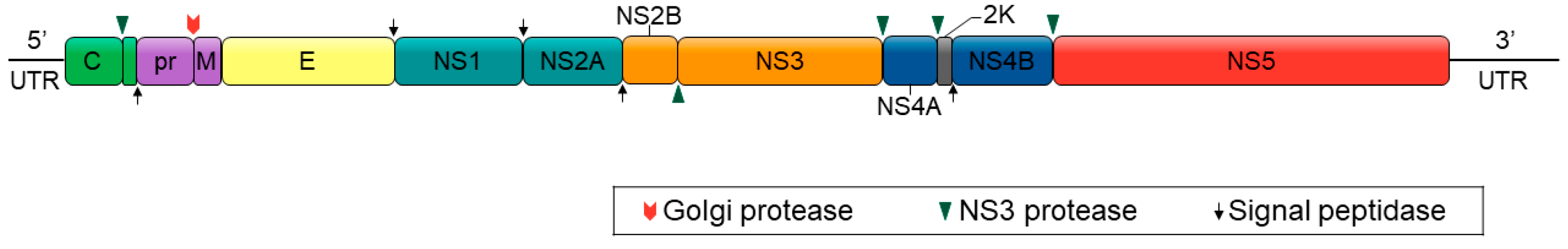
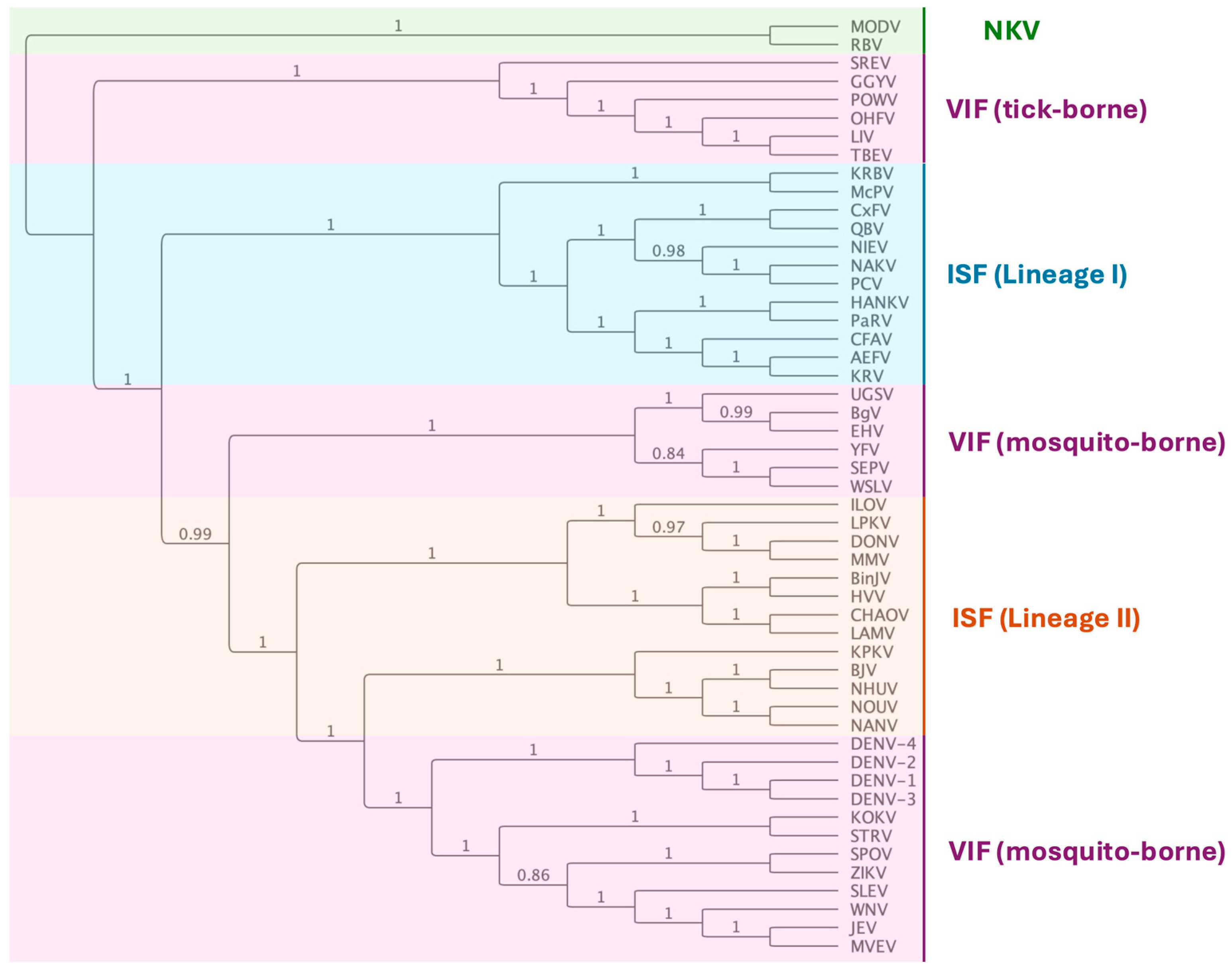
2. Insect-Specific Flaviviruses (ISFs)

Flaviviruses are single-stranded viruses with positive sense RNA genomes of approximately 11 kb [43]. The genomes of flaviviruses traditionally have a 5′ untranslated region of ~100 nt, a single open reading frame (ORF), and a 3′ untranslated region (3′ UTR) of 400–700 nt. The ORF encodes for a large polyprotein that once cleaved generates three structural proteins (capsid (C), pre-membrane/membrane (prM), and envelope (E)) and seven non-structural proteins in the order: 5′–C–prM(M)–E–NS1–NS2A–NS2B–NS3–NS4A–NS4B–NS5-3′ [44] (Figure 2). The flaviviruses can be divided into three phylogenetic groups: vertebrate-infecting viruses (VIFs) (flaviviruses that are also arboviruses), no-known vector (NKV) flaviviruses, and the ISFs [23] (Figure 3). The ISFs can be further divided into two distinct phylogenetic groups—lineage I (classical or cISFs) and lineage II (dual-host associated or dISFs), which are more closely related to VIFs [23,25,45].
The first lineage I ISF discovered, cell fusing agent virus (CFAV), was identified in a persistently infected Aag2 mosquito cell culture [46]. The subsequent detection of Kamiti River virus (KRV) in wild-caught Aedes macintoshi mosquitoes in Kenya [47,48] initiated the discovery of ISFs through interrogation of natural mosquito microbiomes, which has been further propelled through the advancements of deep sequencing technologies. Indeed, such discoveries have driven research into the consequences of persistent infection of ISFs (and other ISVs) in mosquito populations on the transmission dynamics of arboviral pathogens [26,38,49,50]. Such studies have revealed the ability of ISFs to exhibit superinfection exclusion of VIFs, including DENV, ZIKV, and WNV [25,51,52,53,54]. Initial studies reported by Bolling et al. [50] showed that Culex pipiens mosquitoes naturally infected with Culex flavivirus (CxFV) exhibited a significant delay in the transmission of WNV compared to controls. Subsequently, intrathoracic injection of Culex annulirostris mosquitoes with Palm Creek virus (PCV) reduced the infection of mosquitoes by the Kunjin strain of West Nile virus (WNVKUN) by 25% when compared to control (ISF-free) mosquitoes [40]. Further, titres of DENV-1 in peripheral tissues of Ae. albopictus were significantly lower when CFAV was injected into the mosquitoes six days before an infectious blood meal compared to mosquitoes injected two days before infection [54].
Figure 2. Flavivirus genome structure. Flaviviruses have a genome of approximately 11 kbp, with a single open reading frame (ORF) coding for seven non-structural proteins and three structural proteins. The polyprotein ORF is flanked on either side by 5′ and 3′ untranslated regions (UTRs) and is cleaved post-translationally by host and viral proteases. Image adapted from [55].
Viruses 16 01499 g002
Figure 3. Flavivirus phylogenic tree created using maximum likelihood analysis of the complete polyprotein amino acid sequence. The tree was constructed using the methods described in [45], applying a Whelan and Goldman [56] evolutionary model optimised for Gamma likelihood with 1000 bootstrap iterations and using NKV as the outgroup. Sequences were derived from Genbank accession numbers: AB488408, Aedes flavivirus (AEFV); AY898809, Alfuy virus (ALFV); KU308380, Bamaga virus (BgV); KC496020, Barkedji virus (BJV); MG587038, Binjari virus (BinJV); KJ741267, cell fusing agent virus (CFAV); JQ308185, Chaoyang virus (CHAOV), AB262759, Culex flavivirus (CxFV); HE574574, Culex theileri flavivirus (CTFV); U88536, dengue virus serotype 1 (DENV-1); U87411, dengue virus serotype 2 (DENV-2); AY099336, dengue virus serotype 3 (DENV-3); AF326825, dengue virus serotype 4 (DENV-4); NC_016997, Donggang virus (DONV); DQ859060, Edge Hill virus (EHV); DQ235145, Gadgets Gully virus (GGYV); NC_030401, Hanko virus (HANKV); MN954647, Hidden Valley virus (HVV), KC692067, Ilomantsi virus (ILOV), AF217620, Japanese encephalitis virus (JEV), AY149905, Kamiti River virus (KRV); KY320648, Kampung Karu virus (KPKV); NC_035118, Karumba virus (KRBV); AY632541, Kokoberra virus (KOKV), KC692068, Lammi virus (LAMV); Y07863, Louping ill virus (LIV), KY290256, Long Pine Key virus (LPKV); NC_035187, Mac Peak virus (McPV); MF139576, Marisma mosquito virus (MMV); AJ242984, Modoc virus (MODV); AF161266, Murray Valley encephalitis virus (MVEV); NC_030400, Nakiwogo virus (NAKV), MF139575, Nanay virus (NANV); KJ210048, Nhumirim virus (NHUV); JQ957875, Nienokoue virus (NIEV); EU159426, Nounane virus (NOUV); AY193805, Omsk haemorrhagic fever virus (OHFV); KC505248, Palm Creek virus (PCV); KT192549, Parramatta River virus (PaRV); L06436, Powassan virus (POWV); FJ644291, Quang Binh virus (QBV); NC_003675, Rio Bravo virus (RBV); DQ235150, Saumarez Reef virus (SREV); DQ837642, Sepik virus (SEPV); KM225263, Stratford virus (STRV); DQ859064, Spondeweni virus (SPOV); DQ525916, St Louis encephalitis virus (SLEV); U27495, tick-borne encephalitis virus (TBEV); DQ859065, Uganda S virus (UGSV); JN226796, Wesselsbron virus (WSLV); KY229074, West Nile virus (WNV); MN1062241, yellow fever virus (YFV); AY632535, Zika virus (ZIKV).
Viruses 16 01499 g003
Lineage II ISFs, or dual-host-affiliated ISFs, are also unable to replicate in vertebrate cells, but their phylogenetic position places them more closely to the VIFs [25]. This may indicate that they have evolved from vertebrate-infecting viruses and have adopted an insect-specific phenotype [23]. Despite the genetic and antigenic similarity of Lineage II ISFs to VIFs, they share a similar host restriction phenotype to Lineage I ISFs, with restriction occurring at multiple levels pre- and post-entry [45]. Unsurprisingly, the comparably fewer studies that have been performed for the Lineage II ISFs show similar trends of superinfection exclusion of VIFs in vivo. Co-inoculation of Culex quinquefasciatus mosquitoes with WNV and Nhumirim virus (NHUV) demonstrated replication inhibition that resulted in a 40% reduction in the ability of NHUV-infected mosquitoes to transmit WNV [57].

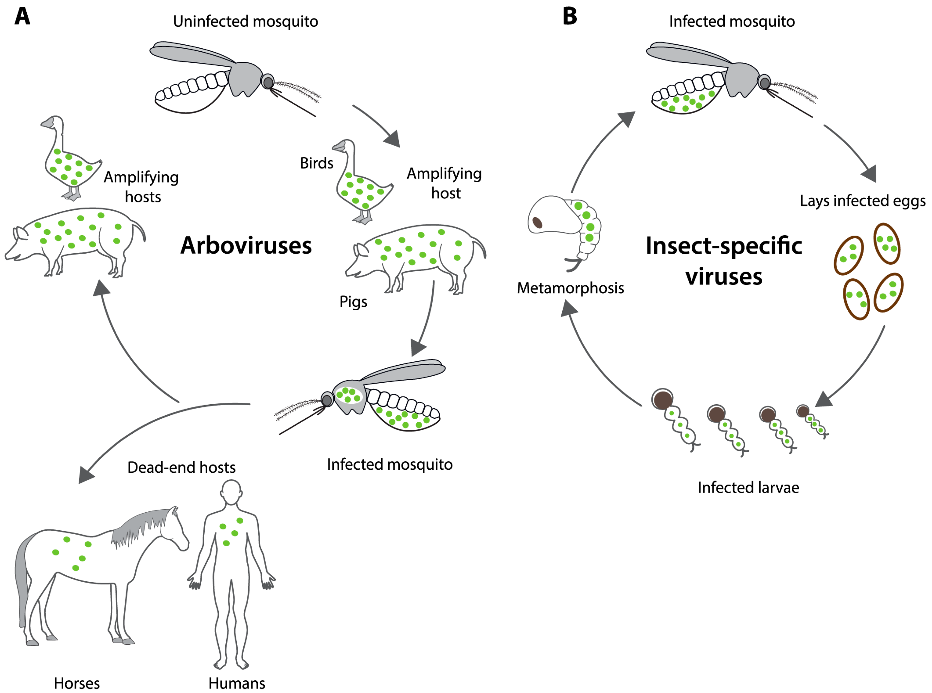
3. Vertical Transmission of Flaviviruses in Mosquitoes

Vertical transmission is a secondary route of transmission of VIFs but is considered to be the main method of transmission for the ISFs. VT can occur by one of two routes: transovarial transmission or transovum transmission [58] (Figure 4). Transovarial transmission involves the transmission of virus from infected female mosquitoes to their developing eggs while within the ovaries [59]. Transovum transmission involves the infection of the egg during oviposition [60,61]. Infection occurs as the eggs exit the infected common oviduct during oviposition, and the virus infects the outer layers of the egg, thereby transmitting the virus from the outside of the eggshell onto the larvae after hatching [60].
Vertical transmission is usually characterised by two properties: the vertical transmission rate, defined as the proportion of infected females that transmit virus to any of their offspring, and the filial infection rate (FIR), defined as the proportion of infected larvae among all progeny from an infected mother. In field studies, however, testing for VT requires testing immature or male adult mosquitoes, for which it is impossible to know the infection status of the mother. The prevalence of immature mosquitoes that are positive for virus is often low, particularly for the VIFs (<1%), and therefore, pools of adult mosquitoes are tested to save resources. When pools of mosquitoes are tested, VT is quoted as the minimum filial infection rate (MFIR), which is calculated as the number of positive pools per total number of mosquitoes and expressed as a rate per 1000 offspring.
Understanding the differences in VT efficiency between the VIFs and ISVs and the underlying mechanisms may illuminate factors defining flavivirus host restriction and the suitability of ISVs for biocontrol. The VT of arboviruses, including VIFs, has been extensively reviewed [62,63,64,65,66]. Here, we contrast observations of natural VT between VIFs and ISFs. For the VIFs, we focus on studies occurring since the last review of arbovirus VT [64]. We also compare observations of natural VT occurring in the field and experimentally induced VT, in which the female mosquito was artificially infected with the virus.

3.1. Natural Occurrence of VIF and ISF Vertical Transmission in Mosquitoes

The natural occurrence of VT of flaviviruses has been demonstrated by virus detection in field-collected mosquitoes across several continents (Table 1). In these studies, VT is determined by the detection of virus in mosquitoes that have been collected in their immature stages, either as eggs, larvae, or pupae, or in male mosquitoes that do not feed on blood. Virus detections were made directly from immature stages (larvae or pupae) that were brought to the laboratory and reared to the adult stage before being tested for the presence of virus. The presence of virus was most frequently tested using variations of the reverse transcription polymerase chain reaction (RT-PCR) that detects viral genomic RNA, though some detections were made using tissue culture-based assays for virus isolation.
Sixteen investigations of the natural VT rates of VIFs since 2018 were performed on different serotypes of DENV and ZIKV on collections of up to 21,390 immature mosquitoes (Table 1). The MFIR reported for DENV ranged from 0 for DENV-1 in Ae. aegypti from Brazil [67] to 22.3 per 1000 for DENV-3 in Ae. aegypti from Mexico [63]. MFIRs for Zika virus (ZIKV) ranged from 0.5 per 1000 Ae. aegypti progeny from Amazonas, Brazil [58], to 7.2 per 1000 Ae. albopictus progeny from Mato Grosso, Brazil [68]. While many VIFs have been found to be vertically transmitted, mathematical models have shown that VT rates need to exceed 20% in order to have any impact on DENV endemicity [69]. Therefore, field observations of MIRs produced by VIFs indicate that VT is not likely to be sufficient to sustain viral presence alone.
Table 1. Naturally occurring flavivirus vertical transmission rates in mosquito populations.
Virus aMosquitoStrainDesign bDetected cFIR d
/100
MFIR
/1000
NReference
Vertebrate-infecting flaviviruses
DENV(1–4)Ae. aegyptiHavana, CubaLqRT 9.04102[70]
DENV-1Ae. aegyptiSao Paulo, BrazilLqRT 0.0910[67]
DENV-1Ae. aegyptiMexicoAnRT 0.12 390[71]
DENV-2Ae. aegyptiSinaloa, MexicoLRT 6.5308[72]
DENV-2Ae. aegyptiMexicoAnRT 1.92 390[71]
DENV-3Ae. aegyptiMexicoAnRT 2.52 390[71]
DENV-3Ae. albopictusSao Paulo, BrazilAqRT 0.63270[64]
DENV-4Ae. aegyptiMato Grosso, BrazilLqRT 2.14490[68]
DENV-4Ae. albopictusMato Grosso, BrazilLqRT 7.0296[68]
DENV-4Ae. aegyptiSinaloa, MexicoART 22.3672[73]
DENVAe. aegyptiYucatan, MexicoAqRT0.9 1278[74]
ZIKVAe. aegyptiAmazonas, BrazilLqRT 0.52057[58]
ZIKVAe. aegyptiMato Grosso, BrazilLqRT 1.84490[68]
ZIKVAe. albopictusMato Grosso, BrazilLqRT 7.2296[68]
ZIKVAe. aegyptiMorelos, MexicoLqRT 3.94300[75]
ZIKVAe. aegyptiYucatan, MexicoAqRT0.1 1278[74]
Lineage I Insect-specific flaviviruses
AeFVAe. luteocephalusWest KenyaAqRT 44.168[76]
AeFVAe. albopictusBangkokCRT100 130[77]
An(g)FVAn. gambiaeWest KenyaAqRT 85.735[76]
CFAVAe. aegyptiWest KenyaAqRT 4.1729[76]
CFAVAe. aegyptiGalveston CRT100 56[78]
CFAVAe. aegyptiGalveston ♀ × Iquitos ♂CRT92.6 68[78]
CFAVAe. aegyptiGalveston ♂ × Iquitos ♀CRT75.9 104[78]
CxFVCx. pipiensColoradoCqRT90.3 52[79]
CxFVCx. pipiensIowaAqRT97.4 540[80]
PARVAe. vigilaxSydneyATC 141.9 [39]
PARVAe. vigilaxBrisbaneATC 29.64 [39]
a AeFV, Aedes flavivirus. An(g)FV, Anopheles gambiae flavivirus. DENV, dengue virus. CFV, cell-fusing agent virus. PARV, Parramatta River Virus. ZIKV, Zika virus. b Experiment design. L: progeny were collected as immature mosquitoes and tested as larvae. A: progeny were collected from the wild as eggs or immature mosquitoes and tested as adult mosquitoes. C: mosquitoes were obtained from a laboratory colony. c qRT, quantitative reverse transcription PCR. RT, reverse transcription PCR. nRT, nested reverse transcription PCR. TC, tissue culture. d FIR, filial infection rate. MFIR, minimum filial infection rate.
In contrast to the low VT rates observed for VIFs, lineage I ISFs were frequently observed to lead to substantially higher rates of VT. There have been few studies into the natural VT of CFAV, although a MFIR of 4.1 per 1000 was observed among Ae. aegypti. progeny in West Kenya [76]. However, the capacity of CFAV for substantial VT in nature is likely to be substantially higher, as CFAV was detected in more than 50% of pools of Ae. aegypti collected in Texas [81], and VT rates of 90% are observed in laboratory colonies (below). Very high rates of VT have been recorded for CxFv, for which near complete penetrance was observed for a population of Cx. pipiens in Iowa (97.4% of offspring infected) [80]. The Australian ISF Parramatta River virus (PaRV) was detected in Aedes vigilax progeny to a MFIR of 141.9/1000 larvae after field-collected mosquitoes were offered a single non-infectious blood meal [39]. In west Kenya, Aedes flavivirus (AeFV) was detected from Aedes luteocephalus at a MFIR of 44.1 per 1000 offspring and Anopheles flavivirus in Anopheles gambiae at an MFIR of 86.7 [76]. There is a high likelihood that vertical transmission plays an important role in the maintenance of another Anopheles-infecting lineage I flavivirus, Karumba virus (KRBV), from northern Australia, given high rates of detection in mosquito pools [82]. KRBV was detected at prevalences of 53–100% in mosquito pools from three locations across the region.
Laboratory strains of mosquitoes that were persistently infected with ISFs, in which progeny were infected via naturally occurring transovarial or transovum transmission, produced FIRs of between 66 and 100% (Table 1, experimental design C). Filial infection rates of 100% were observed from a strain of Ae. albopictus persistently infected with Aedes flavivirus (AeFV) [77] and the Galveston strain of Ae. aegypti that is persistently infected with CFAV [78]. The Galveston strain provided an opportunity to investigate the CFAV VT efficiency in mating crosses with uninfected Ae. aegypti. Crosses of Galveston strain females with the uninfected Iquitos strain of Ae. aegypti produced a 92.6% FIR among progeny, whereas crosses between Galveston males and Iquitos females produced a FIR of 75.9%. The latter result demonstrated CFAV can also be transmitted horizontally by venereal transmission, and that infection of females by this route also leads to efficient VT.
The natural VT of lineage II ISFs has not been specifically tested. However, Binjari virus (BinJV) was detected in one out of two hundred pools of Aedes normanensi mosquitoes, indicating low rates of VT [45]. In contrast, Hidden Valley virus (HVV) was detected in 60% of pools of Aedeomyia catasticta mosquitoes tested, indicating that VT may be the prominent means of transmission for this virus [45].

3.2. Experimentally Induced VIF and ISF Vertical Transmission Rates in Mosquitoes

The potential for VT can be inferred from experimental studies in which female mosquitoes are inoculated with the virus and the resulting proportion of infected progeny is determined. For the VIFs, female mosquitoes were most frequently challenged with virus orally, via artificial membrane feeding, but occasionally by intrathoracic (IT) microinjection (Table 2). The VT of ISFs was most often tested following virus challenge of the female by IT inoculation. In several instances, the recipient mosquitoes are not from the same species or genus as the species the virus was isolated from [40,57,83]. Viral detections were made predominantly from adult mosquitoes using quantitative RT-PCR or occasionally tissue culture ELISA methods. An emerging theme is that artificial inoculation of ISVs known to be vertically transmitted in nature has frequently not induced vertical transmission in offspring [84,85], although there are prominent examples where IT inoculation has successfully led to VT [86,87].
The VT prevalence of VIFs resulting from experimental inoculation of female mosquitoes (Table 2) was generally higher than the prevalence observed naturally for the same viruses (Table 1). Oral exposure of Ae. albopictus from Guangdong, China, to DENV-1 resulted in the highest recorded VT rate for VIFs, with 17.6% of offspring infected with the virus [88]. Oral exposure of a strain of Ae. aegypti from Mexico with DENV-2 resulted in an MFIR of between 0 and 34.4/1000 infected progeny [89]. Interestingly, there was no VT in progeny resulting from the first gonotrophic cycle (the cycle obtained from the first blood meal), but VT occurred from the second and third gonotrophic cycles, and successive cycles produced an increasing MFIR. This indicated that the likelihood of VT increased with the time available for the virus to disseminate through the mosquito and infect ovaries and developing eggs. Experimental oral exposure of Ae. aegypti and Ae. albopictus to ZIKV also resulted in substantially higher infection rates in progeny than has been observed from field collections (Table 2). Exposure of the Ae. aegypti UGAL strain to ZIKV resulted in an FIR of 14.3% of progeny [90]. Oral exposure of GUA and GT strains of Ae. albopictus from China to ZIKV resulted in increasing MIR over successive egg-laying cycles, reaching a mean of approximately 5–6% [91]. The GUA strain harbours the Wolbachia wAlbA and wAlbB strains that naturally infect this species, and a third mosquito strain, HC, was also transinfected with the wPip Wolbachia strain isolated from Cx. pipiens. There was a decrease in ZIKV infection in mosquito ovaries as the number of Wolbachia strains increased, and complete blockage of vertical transmission occurred in the HC strain containing the triple infection. Adding Wolbachia strains therefore caused a cumulative reduction in ISV vertical transmission.
For mosquito strains not harbouring ISF infections, attempts have been made to establish new infection through experimental inoculation (Table 2). One of the most commonly applied inoculation methods was IT microinjection. Experiments with CFAV demonstrated the ability to induce VT of an uninfected strain of Ae. aegypti via IT microinjection [86]. The filial infection rate among the progeny of the injected Bangkok strain females was low (30.0%, F1 generation); however, the authors were able to increase the filial infection rate to 100% among the F2 generation by selective breeding. More recent attempts to induce VT in uninfected strains of Ae. aegypti by IT microinjection have not been successful [84,85]. In both instances, CFAV was obtained from long-term cell cultures (persistently infected Aag2 cells [84] and C6/36 cells [85]) in contrast to the study by Contreras-Gutierrez et al. [86], who utilised CFAV that had been more recently isolated from mosquitoes. Similarly, attempts to induce VT in uninfected Cx. pipiens by IT microinjection resulted in detectable virus in ovaries but not in the F1 offspring [80]. This was in contrast to the high rates of VT observed from naturally infected Cx. pipiens obtained from the field in the same study. A possible hypothesis to explain these discrepancies is that the capacity of ISFs for VT is determined by genomic factors, and these are selected against during long-term cell culture. The lineage I ISF Palm Creek virus (PCV) was obtained after four passages in C6/36 cells and injected into Cx. annulirostris females [39]. Infection rates of 95% or higher were observed in the injected mosquitoes; however, no PCV RNA was detected from the 1038 progeny tested.
Experimental inoculation of lineage II ISFs has produced varied results, although these viruses have notably been less studied. Inoculation of the UGAL strain of Ae. aegypti with the lineage II ISF Chaoyang virus (CHAOV) by IT microinjection resulted in a filial infection rate of 100% [87]. Another study injected NHUV from Culex maxi mosquitoes into Cx. pipiens and Cx. quinquefasciatus females [57]. From the progeny (n = 3) of one of the positive Cx. pipiens mosquitoes that were reared to adulthood, only one tested positive for VT of the virus.
Table 2. Experimentally achieved flavivirus vertical transmission rates in mosquito strains.
Virus aMosquitoStrainDesign bTested cDetection dFIR e
/100
MFIR e
/1000
nReference
Vertebrate-infecting flaviviruses
DENV-1Ae. albopictusGuangdongOrLqRT17.6 154[88]
DENV-2Ae. aegyptiMexicoOr (6)L (GC1)TC 0500[89]
DENV-2Ae. aegyptiMexicoOr (6)L (GC2, 10 d)TC 27.7980[89]
DENV-2Ae. aegyptiMexicoOr (6)L (GC2, 21 d)TC 34.41180[89]
DENV-2Ae. aegyptiHanoOrAqRT0.2 222[92]
WNVAe. vexansConnecticutOr (5)AqRT 21.71389[93]
ZIKVAe. aegyptiManausOr (5)AqRT 43.3600[94]
ZIKVAe. aegyptiUGALOrAqRT14.3 15[90]
ZIKVAe. aegyptiFloridaOr (6)AqRT10 56[95]
ZIKVAe. aegyptiFloridaOr (6)AqRT6.4 20[95]
ZIKVAe. albopictusGuangdongOr (5)LRT 53.375[96]
ZIKVAe. albopictusBeijingOr (8)AqRT 32.6120[97]
ZIKVAe. albopictusGUA, GTOr (6)AqRT≈5–6 ≈15[91]
ZIKVAe. albopictusHCOr (6)AqRT0 ≈15[91]
ZIKVAe. albopictusFloridaOr (6)AqRT6.4 20[95]
ZIKVAe. albopictusCataloniaIT (8)ATC 50.25559[98]
Lineage I Insect-specific flaviviruses
CFAVAe. aegyptiBangkokITA (F1)RT78.360 [86]
CFAVAe. aegyptiBangkokITA (F2)RT100.034 [86]
CFAVAe. aegyptiMengeiIT (5)AqRT0 60[84]
CFAVAe. aegyptiHaikouIT (7)AqRT0 120[84]
CFAVAe. aegyptiPoza Rico, Florida, BangkokIT (3)LqRT0.05 1962[85]
CxFVCx. pipiensIowaITART0 950[80]
KRVAe. aegyptiNairobiOr (7)ATC3.9 410[83]
PCVCx. annulirostrisBrisbaneIT (5)ART0 1038[40]
Lineage II Insect-specific flaviviruses
CHAOVAe. aegyptiUGALIT (2)AqRT100 27[87]
CHAOV-ZIKVAe. aegyptiUGALIT (2)AqRT≈15 27[87]
NHUVCx. pipiensFloridaIT (9)ART66 3[57]
Filial or minimum filial infection rates among offspring resulting from the artificial inoculation of the parental generation or within persistently infected laboratory colonies. a AeFV, Aedes flavivirus. An(g)FV, Anopheles gambiae flavivirus. DENV, dengue virus. CFAV, cell fusing agent virus. CHAOV. Chaoyang virus. CxFV. Culex flavivirus. NHUV, Nhumirim virus. PARV, Parramatta River Virus. PCV, Palm Creek virus. WNV, West Nile virus. ZIKV, Zika virus. b Experiment design. Or: mosquitoes challenged with ISF orally, by feeding. IT: mosquitoes inoculated with the ISF by intrathoracic microinjection. Numbers in parentheses denote the log value of the inoculum (pfu/mL or TCID50/mL). c Life-stage tested. A, adult. L, larvae. GC1, first gonotrophic cycle. GC2, second gonotrophic cycle. d qRT, quantitative reverse transcription PCR. RT, reverse transcription PCR. nRT, nested reverse transcription PCR. TC, tissue culture. e FIR, filial infection rate. MFIR, minimum filial infection rate.

4. Potential Determinants of ISF Vertical Transmission

4.1. Host Factors

The rates of VT have varied for individual flaviviruses between mosquito species, both in nature (Table 1) and following experimental inoculation (Table 2). The variation may be attributed to mosquito factors determining host restriction of arboviruses and ISVs and whether these vary between mosquito tissues. The susceptibility of a mosquito species to infection by viruses is determined by factors, including the presence of appropriate cell surface receptors and/or co-receptors required for viral attachment and cell entry and the permissiveness of the intracellular environment to viral replication for the production of new virions [34]. Host cell receptors have been identified for prominent flaviviral arboviruses [42] but are unknown for ISFs in mosquitoes. Being acquired through blood feeding, VIFs particularly require permissiveness of the mosquito midgut. Little is known of the relative importance of different mosquito tissues to ISF infection, though it is likely that the susceptibility of ovarian and follicular tissue is a requirement for VT. Interestingly, we have detected lineage I ISFs in mosquito midgut epithelia [39,40], an important site for the establishment of arbovirus infection. Further, studies with lineage II Binjari reporter virus revealed a likely salivary gland infection [99]. This raises the possibility of inhibitory interactions with arboviruses in these important niches for arbovirus infection and transmission.
Host innate immune mechanisms determine the trajectory of VIF and ISF infection, and recent studies indicate a role of innate immunity in VT. Once inside the insect cell, viruses are subject to host innate immune responses against viral infection [100,101]. The main antiviral immune responses are the innate JAK–STAT, Toll, IMD, and RNA interference (RNAi) pathways. RNAi pathways include the small interfering RNA (siRNA) and PIWI interfering (piRNA) pathways that can induce sequence-specific virus cleavage. In the siRNA pathway, dsRNA produced during viral replication is processed into 21 nt long siRNA by the RNAse III enzyme Dicer 2, which is then loaded into the RNA-induced silencing complex (RISC) that cleaves complementary viral RNA sequences. The piRNA pathway was first known for its role in the protection of the Drosophila genomes against transposable elements (TE) in germline tissues, involving the interaction of P-element-induced wimpy testis (PIWI) family proteins, Aubergine (Aub), and Agonaut 3 with genome-encoded 21–24 nt piRNAs to degrade TE. In mosquitoes, the pathway has expanded to target viral RNA genomes, guided by virus-derived piRNAs (vpiRNAs) [102].
Recent findings indicate the existence of an immune pathway involving piRNA and viral DNA (vDNA) in mosquito ovaries that may play a role in limiting VT [103,104]. In mosquitoes, viral RNA is reverse transcribed into viral DNA through endogenous cellular reverse transcriptase activity, likely arising from endogenous retrotransposons [105]. The viral DNA can occur as episomes or be integrated into mosquito genomes as non-retroviral endogenous viral elements (EVEs). EVEs are accumulated within the genome over many generations and are inherited as host alleles [106]. EVEs from the ISFs CFAV and Kamiti River virus (KRV) are present within the Ae. aegypti genome [107]. Viral DNA can prime early phases of small RNA responses against the cognate virus, including the production of virus-specific siRNA and PIWI-interacting RNA (piRNA) [105]. Recent studies have identified a role of a CFAV EVE in the suppression of CFAV viral replication in mosquito ovaries via the piRNA pathway [103,104]. Ablation of a CFAV EVE by genomic editing substantially reduced EVE-derived piRNAs and increased CFAV virus replication in ovaries.
A similar phenomenon of vertically transmitted viral immunity was recently demonstrated for alphaviruses (positive sense RNA viruses within the family Togaviridae) [103]. The progeny of Ae. aegypti infected with Semliki Forest virus (SFV) and Ross River virus contained no detectable “infectious” virus but did possess episomal and integrated viral DNA. The presence of episomal DNA was associated with resistance to subsequent challenge of the progeny with SFV, and the episomal DNA-associated resistance was inherited over four generations in the absence of further virus challenge. Treatment of the parent mosquitoes with the reverse transcriptase inhibitor Zidovudine (AZT) eliminated viral DNA from ovaries and installed VT of SFV in a small proportion of offspring.

4.2. Viral Factors

The differing rates of VT between VIFs and ISFs have raised questions about the contribution of specific regions of the flavivirus genome to VT. One way to assess the contribution of different viral genes is by swapping genes with other viruses using reverse genetics approaches to create viral chimeras and testing these in vitro. Such approaches have been applied to ISFs to determine the viral genomic elements responsible for host restriction, although most host-restriction work has been limited to various vertebrate cell lines [45,108,109,110,111]. These studies have enabled comparisons of host restriction of lineage I and II ISFs. Restriction of the lineage I ISFs PCV and Niénokoué virus (NIEV) in vertebrate cells occurs at multiple stages of cellular infection, including attachment/entry and assembly/release [108,111]. Chimeric viruses based on the lineage II flavivirus BinJV are capable of limited entry into vertebrate cells but face strong restriction of virus replication that is mediated by non-interferon immune and temperature-dependent mechanisms [45]. Recent studies using reporter virus particles have confirmed the capacity of ISFs for vertebrate cell entry and that strong restriction of replication occurs due to interactions between host proteins and the viral UTRs [109,110]. Similar molecular genetics approaches could be applied to establish the contribution of specific genomic regions to VT in mosquitoes.
ISF chimeras are now being applied to test the contribution of viral proteins to VT in vivo. One recent study tested the capacity of a lineage II ISF-VIF chimeric vaccine candidate to be vertically transmitted by mosquitoes [87]. Aedes aegypti mosquitoes microinjected with the wild-type CHAOV transmitted the virus to 100% of their offspring. In contrast, VT was almost completely suppressed in mosquitoes injected with the vaccine chimera, in which the antigenic structural proteins (prM-E) of CHAOV were replaced with the prM-E of ZIKV. Therefore, while the non-structural proteins are important for host restriction, it appears that structural proteins may have a greater role in VT for some ISFs.

4.3. Microbiome

Mosquito microbiota also contribute to the likelihood of ISF VT. The suppression of ZIKV VT by Wolbachia in the Ae. albopictus HC strain demonstrates the influence of microbiome in determining VT efficiency (Table 2) [91]. Wolbachia induced strong inhibition of CFAV in vitro [112], and interactions with other ISVs in wild mosquitoes have been reported. Virus inhibition may also be induced by other bacterial microbiota in the mosquito reproductive tract, including Enterobacter, Serratia, Asaia [113], and Rosenbergiella [114]. Interactions between these taxa and flaviviruses in mosquitoes have been reported [114,115,116]; however, it is unclear how these may affect VT. Infection of mosquitoes with pre-existing ISFs may influence the establishment of persistent infection with a desired ISF through superinfection exclusion, particularly if the endogenous virus has established a prior infection in ovaries. An appraisal of the existing bacterial and viral microbiota of a target mosquito vector by deep sequencing should be conducted before engaging in efforts to establish ISF infections.

5. Conclusions

Research into the VT of flaviviruses has illuminated differences between the VT of VIFs and ISFs that may guide the optimisation of sustainable ISF biocontrol strategies against arboviruses. The unique ability of certain ISFs to hinder VIF replication in mosquitoes, coupled with their exclusive infectivity to insect hosts, positions them as compelling biocontrol candidates for the control of arboviral diseases. However, attaining high VT rates will be key to achieving the sustainability of such a strategy. Observations of near-complete penetrance of mosquito populations by ISFs highlight their potential as naturally propagating biological control agents.
VT rates were consistently higher for ISFs than for VIFs, reflecting the importance of this pathway to ISF propagation. The highest rates of ISF VT were observed from select laboratory mosquito colonies that are persistently infected with ISFs. For these strains, the ISF infection was transmitted to up to 100% of the progeny. Other associations lead to incomplete penetrance of virus infection among progeny. Research on the determinants of this variation indicates that host, virus, and microbiome factors may play a role in determining the propensity of ISFs for VT. Understanding these factors is essential to optimising the VT of ISFs for biological control applications.

Author Contributions

Conceptualisation, A.J.P., J.H.-P. and L.E.H.; writing—original draft preparation, A.J.P.; writing—review and editing, A.J.P., R.A.H., J.J.H., J.H.-P. and L.E.H.; visualisation, A.J.P.; funding acquisition, A.J.P., R.A.H., J.H.-P. and L.E.H.; supervision, R.A.H., J.J.H., J.H.-P. and L.E.H. All authors have read and agreed to the published version of the manuscript.

Funding

This research was funded by the National Health and Medical Research Council, grant number 2012500. A.J.P. was funded by a Research Training Program stipend scholarship from the University of Queensland and an American Australian Association (AAA) Graduate Education Fund Scholarship.

Acknowledgments

Figure 1 and Figure 4 illustrations were prepared by Katharine Trenholme (FivePrime Design), and Figure 2 was prepared by Christopher Ang (The University of Queensland).

Conflicts of Interest

The authors declare no conflict of interest.

References

  1. Messina, J.P.; Brady, O.J.; Golding, N.; Kraemer, M.U.G.; Wint, G.R.W.; Ray, S.E.; Pigott, D.M.; Shearer, F.M.; Johnson, K.; Earl, L.; et al. The current and future global distribution and population at risk of dengue. Nat. Microbiol. 2019, 4, 1508–1515. [Google Scholar] [CrossRef] [PubMed]
  2. Bhatt, S.; Gething, P.W.; Brady, O.J.; Messina, J.P.; Farlow, A.W.; Moyes, C.L.; Drake, J.M.; Brownstein, J.S.; Hoen, A.G.; Sankoh, O.; et al. The global distribution and burden of dengue. Nature 2013, 496, 504–507. [Google Scholar] [CrossRef] [PubMed]
  3. Gubler, D.J. Dengue, urbanization and globalization: The unholy trinity of the 21(st) century. Trop. Med. Health 2011, 39, 3–11. [Google Scholar] [CrossRef]
  4. Kramer, L.D.; Styer, L.M.; Ebel, G.D. A global perspective on the epidemiology of West Nile virus. Annu. Rev. Entomol. 2008, 53, 61–81. [Google Scholar] [CrossRef] [PubMed]
  5. Mourad, O.; Makhani, L.; Chen, L.H. Chikungunya: An emerging public health concern. Curr. Infect. Dis. Rep. 2022, 24, 217–228. [Google Scholar] [CrossRef]
  6. Simonin, Y. Circulation of West Nile virus and Usutu virus in Europe: Overview and challenges. Viruses 2024, 16, 599. [Google Scholar] [CrossRef]
  7. Yakob, L.; Hu, W.; Frentiu, F.D.; Gyawali, N.; Hugo, L.E.; Johnson, B.; Lau, C.; Furuya-Kanamori, L.; Magalhaes, R.S.; Devine, G. Japanese encephalitis emergence in Australia: The potential population at risk. Clin. Infect. Dis. 2023, 76, 335–337. [Google Scholar] [CrossRef]
  8. de Souza, W.M.; Weaver, S.C. Effects of climate change and human activities on vector-borne diseases. Nat. Rev. Microbiol. 2024, 22, 476–491. [Google Scholar] [CrossRef]
  9. Mullard, A. FDA approves first chikungunya vaccine. Nat. Rev. Drug Discov. 2024, 23, 8. [Google Scholar] [CrossRef]
  10. Huang, Z.; Zhang, Y.; Li, H.; Zhu, J.; Song, W.; Chen, K.; Zhang, Y.; Lou, Y. Vaccine development for mosquito-borne viral diseases. Front. Immunol. 2023, 14, 1161149. [Google Scholar] [CrossRef]
  11. Thomas, S.J. Is new dengue vaccine efficacy data a relief or cause for concern? NPJ Vaccines 2023, 8, 55. [Google Scholar] [CrossRef] [PubMed]
  12. WHO. Launch of the WHO Global Arbovirus Initiative. Meeting Report; WHO: Geneva, Switzerland, 2022. [Google Scholar]
  13. Corbel, V.; Achee, N.L.; Chandre, F.; Coulibaly, M.B.; Dusfour, I.; Fonseca, D.M.; Grieco, J.; Juntarajumnong, W.; Lenhart, A.; Martins, A.J.; et al. Tracking insecticide resistance in mosquito vectors of arboviruses: The worldwide insecticide resistance network (WIN). PLoS Negl. Trop. Dis. 2016, 10, e0005054. [Google Scholar] [CrossRef]
  14. Liu, H.; Xu, Q.; Zhang, L.; Liu, N. Chlorpyrifos resistance in mosquito Culex quinquefasciatus. J. Med. Entomol. 2005, 42, 815–820. [Google Scholar] [CrossRef]
  15. Silva, J.V.J., Jr.; Lopes, T.R.R.; Oliveira-Filho, E.F.; Oliveira, R.A.S.; Duraes-Carvalho, R.; Gil, L. Current status, challenges and perspectives in the development of vaccines against yellow fever, dengue, Zika and chikungunya viruses. Acta Trop. 2018, 182, 257–263. [Google Scholar] [CrossRef]
  16. Walker, T.; Johnson, P.H.; Moreira, L.A.; Iturbe-Ormaetxe, I.; Frentiu, F.D.; McMeniman, C.J.; Leong, Y.S.; Dong, Y.; Axford, J.; Kriesner, P.; et al. The wMel Wolbachia strain blocks dengue and invades caged Aedes aegypti populations. Nature 2011, 476, 450–453. [Google Scholar] [CrossRef] [PubMed]
  17. Moreira, L.A.; Iturbe-Ormaetxe, I.; Jeffery, J.A.; Lu, G.; Pyke, A.T.; Hedges, L.M.; Rocha, B.C.; Hall-Mendelin, S.; Day, A.; Riegler, M.; et al. A Wolbachia symbiont in Aedes aegypti limits infection with dengue, Chikungunya, and Plasmodium. Cell 2009, 139, 1268–1278. [Google Scholar] [CrossRef]
  18. van den Hurk, A.F.; Hall-Mendelin, S.; Pyke, A.T.; Frentiu, F.D.; McElroy, K.; Day, A.; Higgs, S.; O’Neill, S.L. Impact of Wolbachia on infection with chikungunya and yellow fever viruses in the mosquito vector Aedes aegypti. PLoS Negl. Trop. Dis. 2012, 6, e1892. [Google Scholar] [CrossRef]
  19. O’Neill, S.L. World Mosquito Program Annual Review 2022; The World Mosquito Program: Clayton, Australia, 2022. [Google Scholar]
  20. Utarini, A.; Indriani, C.; Ahmad, R.A.; Tantowijoyo, W.; Arguni, E.; Ansari, M.R.; Supriyati, E.; Wardana, D.S.; Meitika, Y.; Ernesia, I.; et al. Efficacy of Wolbachia-infected mosquito deployments for the control of dengue. N. Engl. J. Med. 2021, 384, 2177–2186. [Google Scholar] [CrossRef] [PubMed]
  21. Pinto, S.B.; Riback, T.I.S.; Sylvestre, G.; Costa, G.; Peixoto, J.; Dias, F.B.S.; Tanamas, S.K.; Simmons, C.P.; Dufault, S.M.; Ryan, P.A.; et al. Effectiveness of Wolbachia-infected mosquito deployments in reducing the incidence of dengue and other Aedes-borne diseases in Niteroi, Brazil: A quasi-experimental study. PLoS Negl. Trop. Dis. 2021, 15, e0009556. [Google Scholar] [CrossRef]
  22. Ross, P.A.; Wiwatanaratanabutr, I.; Axford, J.K.; White, V.L.; Endersby-Harshman, N.M.; Hoffmann, A.A. Wolbachia Infections in Aedes aegypti differ markedly in their response to cyclical heat stress. PLoS Pathog. 2017, 13, e1006006. [Google Scholar] [CrossRef]
  23. Blitvich, B.J.; Firth, A.E. Insect-specific flaviviruses: A systematic review of their discovery, host range, mode of transmission, superinfection exclusion potential and genomic organization. Viruses 2015, 7, 1927–1959. [Google Scholar] [CrossRef]
  24. Patterson, E.I.; Villinger, J.; Muthoni, J.N.; Dobel-Ober, L.; Hughes, G.L. Exploiting insect-specific viruses as a novel strategy to control vector-borne disease. Curr. Opin. Insect Sci. 2020, 39, 50–56. [Google Scholar] [CrossRef]
  25. Hall, R.A.; Bielefeldt-Ohmann, H.; McLean, B.J.; O’Brien, C.A.; Colmant, A.M.; Piyasena, T.B.; Harrison, J.J.; Newton, N.D.; Barnard, R.T.; Prow, N.A.; et al. Commensal viruses of mosquitoes: Host restriction, transmission, and interaction with arboviral pathogens. Evol. Bioinform. Online 2016, 12, 35–44. [Google Scholar] [CrossRef]
  26. Hobson-Peters, J.; Yam, A.W.; Lu, J.W.; Setoh, Y.X.; May, F.J.; Kurucz, N.; Walsh, S.; Prow, N.A.; Davis, S.S.; Weir, R.; et al. A new insect-specific flavivirus from northern Australia suppresses replication of West Nile virus and Murray Valley encephalitis virus in co-infected mosquito cells. PLoS ONE 2013, 8, e56534. [Google Scholar] [CrossRef]
  27. Carvalho, V.L.; Long, M.T. Insect-Specific Viruses: An overview and their relationship to arboviruses of concern to humans and animals. Virology 2021, 557, 34–43. [Google Scholar] [CrossRef]
  28. Nouri, S.; Matsumura, E.E.; Kuo, Y.W.; Falk, B.W. Insect-specific viruses: From discovery to potential translational applications. Curr. Opin. Virol. 2018, 33, 33–41. [Google Scholar] [CrossRef]
  29. Vasilakis, N.; Guzman, H.; Firth, C.; Forrester, N.L.; Widen, S.G.; Wood, T.G.; Rossi, S.L.; Ghedin, E.; Popov, V.; Blasdell, K.R.; et al. Mesoniviruses are mosquito-specific viruses with extensive geographic distribution and host range. Virol. J. 2014, 11, 97. [Google Scholar] [CrossRef]
  30. Vasilakis, N.; Forrester, N.L.; Palacios, G.; Nasar, F.; Savji, N.; Rossi, S.L.; Guzman, H.; Wood, T.G.; Popov, V.; Gorchakov, R.; et al. Negevirus: A proposed new taxon of insect-specific viruses with wide geographic distribution. J. Virol. 2013, 87, 2475–2488. [Google Scholar] [CrossRef]
  31. Boshra, H. An Overview of the Infectious Cycle of Bunyaviruses. Viruses 2022, 14, 2139. [Google Scholar] [CrossRef] [PubMed]
  32. Newton, N.D.; Colmant, A.M.G.; O’Brien, C.A.; Ledger, E.; Paramitha, D.; Bielefeldt-Ohmann, H.; Watterson, D.; McLean, B.J.; Hall-Mendelin, S.; Warrilow, D.; et al. Genetic, Morphological and Antigenic Relationships between Mesonivirus Isolates from Australian Mosquitoes and Evidence for Their Horizontal Transmission. Viruses 2020, 12, 1159. [Google Scholar] [CrossRef] [PubMed]
  33. Nasar, F.; Haddow, A.D.; Tesh, R.B.; Weaver, S.C. Eilat virus displays a narrow mosquito vector range. Parasit. Vectors 2014, 7, 595. [Google Scholar] [CrossRef]
  34. Elrefaey, A.M.; Abdelnabi, R.; Rosales Rosas, A.L.; Wang, L.; Basu, S.; Delang, L. Understanding the mechanisms underlying host restriction of insect-specific viruses. Viruses 2020, 12, 964. [Google Scholar] [CrossRef]
  35. Cook, S.; Moureau, G.; Kitchen, A.; Gould, E.A.; de Lamballerie, X.; Holmes, E.C.; Harbach, R.E. Molecular evolution of the insect-specific flaviviruses. J. Gen. Virol. 2012, 93, 223–234. [Google Scholar] [CrossRef]
  36. Kurnia, N.; Kaitana, Y.; Salaki, C.L.; Mandey, L.C.; Tuda, J.S.B.; Tallei, T.E. Study of dengue virus transovarial transmission in Aedes spp. in Ternate City using streptavidin-biotin-peroxidase complex immunohistochemistry. Infect. Dis. Rep. 2022, 14, 765–771. [Google Scholar] [CrossRef]
  37. Goenaga, S.; Goenaga, J.; Boaglio, E.R.; Enria, D.A.; Levis, S.D.C. Superinfection exclusion studies using West Nile virus and Culex flavivirus strains from Argentina. Mem. Inst. Oswaldo Cruz. 2020, 115, e200012. [Google Scholar] [CrossRef]
  38. Kenney, J.L.; Solberg, O.D.; Langevin, S.A.; Brault, A.C. Characterization of a novel insect-specific flavivirus from Brazil: Potential for inhibition of infection of arthropod cells with medically important flaviviruses. J. Gen. Virol. 2014, 95, 2796–2808. [Google Scholar] [CrossRef]
  39. McLean, B.J.; Hall-Mendelin, S.; Webb, C.E.; Bielefeldt-Ohmann, H.; Ritchie, S.A.; Hobson-Peters, J.; Hall, R.A.; van den Hurk, A.F. The insect-specific Parramatta River virus is vertically transmitted by Aedes vigilax mosquitoes and suppresses replication of pathogenic flaviviruses in vitro. Vector Borne Zoonotic Dis. 2021, 21, 208–215. [Google Scholar] [CrossRef]
  40. Hall-Mendelin, S.; McLean, B.J.; Bielefeldt-Ohmann, H.; Hobson-Peters, J.; Hall, R.A.; van den Hurk, A.F. The insect-specific Palm Creek virus modulates West Nile virus infection in and transmission by Australian mosquitoes. Parasit. Vectors 2016, 9, 414. [Google Scholar] [CrossRef]
  41. de Almeida, J.P.; Aguiar, E.R.; Armache, J.N.; Olmo, R.P.; Marques, J.T. The virome of vector mosquitoes. Curr. Opin. Virol. 2021, 49, 7–12. [Google Scholar] [CrossRef]
  42. Laureti, M.; Narayanan, D.; Rodriguez-Andres, J.; Fazakerley, J.K.; Kedzierski, L. Flavivirus receptors: Diversity, identity, and cell entry. Front. Immunol. 2018, 9, 2180. [Google Scholar] [CrossRef]
  43. Lindenbach, B.D.; Randal, G.; Bartenschlager, R.; Rice, C.M. Flaviviridae: The viruses and their replication. In Fields Virology: Emerging Viruses; Howley, P.M., Knipe, D.M., Whelan, S., Eds.; Lippincott Williams & Wilkins: Philadelphia, PA, USA, 2020. [Google Scholar]
  44. Howley, P.M. Fields Virology, 7th ed.; Whelan, S., Ed.; Wolters Kluwer: Alphen aan den Rijn, The Netherlands, 2020; Volume 1, p. 840. [Google Scholar]
  45. Harrison, J.J.; Hobson-Peters, J.; Colmant, A.M.G.; Koh, J.; Newton, N.D.; Warrilow, D.; Bielefeldt-Ohmann, H.; Piyasena, T.B.H.; O’Brien, C.A.; Vet, L.J.; et al. Antigenic characterization of new lineage II insect-specific flaviviruses in Australian mosquitoes and identification of host restriction factors. mSphere 2020, 5. [Google Scholar] [CrossRef]
  46. Stollar, V.; Thomas, V.L. An agent in the Aedes aegypti cell line (Peleg) which causes fusion of Aedes albopictus cells. Virology 1975, 64, 367–377. [Google Scholar] [CrossRef]
  47. Sang, R.C.; Gichogo, A.; Gachoya, J.; Dunster, M.D.; Ofula, V.; Hunt, A.R.; Crabtree, M.B.; Miller, B.R.; Dunster, L.M. Isolation of a new flavivirus related to cell fusing agent virus (CFAV) from field-collected flood-water Aedes mosquitoes sampled from a dambo in central Kenya. Arch. Virol. 2003, 148, 1085–1093. [Google Scholar] [CrossRef]
  48. Crabtree, M.B.; Sang, R.C.; Stollar, V.; Dunster, L.M.; Miller, B.R. Genetic and phenotypic characterization of the newly described insect flavivirus, Kamiti River virus. Arch. Virol. 2003, 148, 1095–1118. [Google Scholar] [CrossRef]
  49. Olmo, R.P.; Todjro, Y.M.H.; Aguiar, E.; de Almeida, J.P.P.; Ferreira, F.V.; Armache, J.N.; de Faria, I.J.S.; Ferreira, A.G.A.; Amadou, S.C.G.; Silva, A.T.S.; et al. Mosquito vector competence for dengue is modulated by insect-specific viruses. Nat. Microbiol. 2023, 8, 135–149. [Google Scholar] [CrossRef]
  50. Bolling, B.G.; Olea-Popelka, F.J.; Eisen, L.; Moore, C.G.; Blair, C.D. Transmission dynamics of an insect-specific flavivirus in a naturally infected Culex pipiens laboratory colony and effects of co-infection on vector competence for West Nile virus. Virology 2012, 427, 90–97. [Google Scholar] [CrossRef]
  51. Barbanti-Brodano, G.; Swetly, P.; Koprowski, H. Superinfection of simian virus 40-transformed permissive cells with simian virus 40. J. Virol. 1970, 6, 644–651. [Google Scholar] [CrossRef]
  52. Burivong, P.; Pattanakitsakul, S.N.; Thongrungkiat, S.; Malasit, P.; Flegel, T.W. Markedly reduced severity of Dengue virus infection in mosquito cell cultures persistently infected with Aedes albopictus densovirus (AalDNV). Virology 2004, 329, 261–269. [Google Scholar] [CrossRef]
  53. Pepin, K.M.; Lambeth, K.; Hanley, K.A. Asymmetric competitive suppression between strains of dengue virus. BMC Microbiol. 2008, 8, 28. [Google Scholar] [CrossRef]
  54. Baidaliuk, A.; Miot, E.F.; Lequime, S.; Moltini-Conclois, I.; Delaigue, F.; Dabo, S.; Dickson, L.B.; Aubry, F.; Merkling, S.H.; Cao-Lormeau, V.M.; et al. Cell-fusing agent virus reduces arbovirus dissemination in Aedes aegypti mosquitoes In Vivo. J. Virol. 2019, 93. [Google Scholar] [CrossRef] [PubMed]
  55. Pierson, T.C.; Diamond, M.S. The continued threat of emerging flaviviruses. Nat. Microbiol. 2020, 5, 796–812. [Google Scholar] [CrossRef] [PubMed]
  56. Whelan, S.; Goldman, N. A general empirical model of protein evolution derived from multiple protein families using a maximum-likelihood approach. Mol. Biol. Evol. 2001, 18, 691–699. [Google Scholar] [CrossRef]
  57. Goenaga, S.; Kenney, J.L.; Duggal, N.K.; Delorey, M.; Ebel, G.D.; Zhang, B.; Levis, S.C.; Enria, D.A.; Brault, A.C. Potential for co-infection of a mosquito-specific flavivirus, Nhumirim virus, to block West Nile virus transmission in mosquitoes. Viruses 2015, 7, 5801–5812. [Google Scholar] [CrossRef]
  58. da Costa, C.F.; da Silva, A.V.; do Nascimento, V.A.; de Souza, V.C.; Monteiro, D.; Terrazas, W.C.M.; Dos Passos, R.A.; Nascimento, S.; Lima, J.B.P.; Naveca, F.G. Evidence of vertical transmission of Zika virus in field-collected eggs of Aedes aegypti in the Brazilian Amazon. PLoS Negl. Trop. Dis. 2018, 12, e0006594. [Google Scholar] [CrossRef]
  59. Rosen, L.; Shroyer, D.A.; Tesh, R.B.; Freier, J.E.; Lien, J.C. Transovarial transmission of dengue viruses by mosquitoes: Aedes albopictus and Aedes aegypti. Am. J. Trop. Med. Hyg. 1983, 32, 1108–1119. [Google Scholar] [CrossRef]
  60. Rosen, L. Further observations on the mechanism of vertical transmission of flaviviruses by Aedes mosquitoes. Am. J. Trop. Med. Hyg. 1988, 39, 123–126. [Google Scholar] [CrossRef]
  61. Agboli, E.; Leggewie, M.; Altinli, M.; Schnettler, E. Mosquito-specific viruses-transmission and interaction. Viruses 2019, 11, 873. [Google Scholar] [CrossRef]
  62. Lequime, S.; Paul, R.E.; Lambrechts, L. Determinants of arbovirus vertical transmission in mosquitoes. PLoS Pathog. 2016, 12, e1005548. [Google Scholar] [CrossRef]
  63. Grunnill, M.; Boots, M. How important is vertical tansmission of dengue viruses by mosquitoes (Diptera: Culicidae)? J. Med. Entomol. 2016, 53, 1–19. [Google Scholar] [CrossRef]
  64. Ferreira-de-Lima, V.H.; Lima-Camara, T.N. Natural vertical transmission of dengue virus in Aedes aegypti and Aedes albopictus: A systematic review. Parasit. Vectors 2018, 11, 77. [Google Scholar] [CrossRef]
  65. Bergren, N.A.; Kading, R.C. The ecological significance and implications of transovarial transmission among the vector-borne bunyaviruses: A review. Insects 2018, 9, 173. [Google Scholar] [CrossRef]
  66. Dahiya, N.; Yadav, M.; Yadav, A.; Sehrawat, N. Zika virus vertical transmission in mosquitoes: A less understood mechanism. J. Vector Borne Dis. 2022, 59, 37–44. [Google Scholar] [CrossRef]
  67. Moraes, A.; Cortelli, F.C.; Miranda, T.B.; Aquino, D.R.; Cortelli, J.R.; Guimaraes, M.I.A.; Costa, F.O.; Cortelli, S.C. Transovarial transmission of dengue 1 virus in Aedes aegypti larvae: Real-time PCR analysis in a Brazilian city with high mosquito population density. Can. J. Microbiol. 2018, 64, 393–400. [Google Scholar] [CrossRef]
  68. Maia, L.M.S.; Bezerra, M.C.F.; Costa, M.C.S.; Souza, E.M.; Oliveira, M.E.B.; Ribeiro, A.L.M.; Miyazaki, R.D.; Slhessarenko, R.D. Natural vertical infection by dengue virus serotype 4, Zika virus and Mayaro virus in Aedes (Stegomyia) aegypti and Aedes (Stegomyia) albopictus. Med. Vet. Entomol. 2019, 33, 437–442. [Google Scholar] [CrossRef]
  69. Adams, B.; Boots, M. How important is vertical transmission in mosquitoes for the persistence of dengue? Insights from a mathematical model. Epidemics 2010, 2, 1–10. [Google Scholar] [CrossRef]
  70. Gutierrez-Bugallo, G.; Rodriguez-Roche, R.; Diaz, G.; Perez, M.; Mendizabal, M.E.; Peraza, I.; Vazquez, A.A.; Alvarez, M.; Rodriguez, M.; Bisset, J.A.; et al. Spatio-temporal distribution of vertically transmitted dengue viruses by Aedes aegypti (Diptera: Culicidae) from Arroyo Naranjo, Havana, Cuba. Trop. Med. Int. Health 2018, 23, 1342–1349. [Google Scholar] [CrossRef]
  71. Danis-Lozano, R.; Diaz-Gonzalez, E.E.; Malo-Garcia, I.R.; Rodriguez, M.H.; Ramos-Castaneda, J.; Juarez-Palma, L.; Ramos, C.; Lopez-Ordonez, T.; Mosso-Gonzalez, C.; Fernandez-Salas, I. Vertical transmission of dengue virus in Aedes aegypti and its role in the epidemiological persistence of dengue in Central and Southern Mexico. Trop. Med. Int. Health 2019, 24, 1311–1319. [Google Scholar] [CrossRef]
  72. Apodaca-Medina, A.I.; Torres-Avendano, J.I.; Rendon-Maldonado, J.G.; Torres-Montoya, E.H.; Flores-Lopez, B.A.; Del Angel, R.M.; Velarde-Felix, J.S.; Salomon-Soto, V.M.; Castillo-Ureta, H. First evidence of vertical infection of dengue Virus 2 in Aedes aegypti mosquitoes from Sinaloa, Mexico. Vector Borne Zoonotic Dis. 2018, 18, 231–233. [Google Scholar] [CrossRef]
  73. Torres-Avendano, J.I.; Apodaca-Medina, A.I.; Castillo-Ureta, H.; Rendon-Maldonado, J.G.; Torres-Montoya, E.H.; Cota-Medina, A.; Rios-Tostado, J.J.; Zazueta-Moreno, J.M. Natural vertical transmission of dengue virus serotype 4 in Aedes aegypti larvae from urban areas in Sinaloa, Mexico. Vector Borne Zoonotic Dis. 2021, 21, 478–481. [Google Scholar] [CrossRef] [PubMed]
  74. Kirstein, O.D.; Talavera, G.A.; Wei, Z.; Ciau-Carrilo, K.J.; Koyoc-Cardena, E.; Puerta-Guardo, H.; Rodriguez-Martin, E.; Medina-Barreiro, A.; Mendoza, A.C.; Piantadosi, A.L.; et al. Natural Aedes-borne virus infection detected in male adult Aedes aegypti (Diptera: Culicidae) collected from urban settings in Merida, Yucatan, Mexico. J. Med. Entomol. 2022, 59, 1336–1346. [Google Scholar] [CrossRef] [PubMed]
  75. Izquierdo-Suzan, M.; Zarate, S.; Torres-Flores, J.; Correa-Morales, F.; Gonzalez-Acosta, C.; Sevilla-Reyes, E.E.; Lira, R.; Alcaraz-Estrada, S.L.; Yocupicio-Monroy, M. Natural vertical transmission of Zika virus in larval Aedes aegypti populations, Morelos, Mexico. Emerg. Infect. Dis. 2019, 25, 1477–1484. [Google Scholar] [CrossRef]
  76. Ajamma, Y.U.; Onchuru, T.O.; Ouso, D.O.; Omondi, D.; Masiga, D.K.; Villinger, J. Vertical transmission of naturally occurring Bunyamwera and insect-specific flavivirus infections in mosquitoes from islands and mainland shores of Lakes Victoria and Baringo in Kenya. PLoS Negl. Trop. Dis. 2018, 12, e0006949. [Google Scholar] [CrossRef]
  77. Peinado, S.A.; Aliota, M.T.; Blitvich, B.J.; Bartholomay, L.C. Biology and transmission dynamics of Aedes flavivirus. J. Med. Entomol. 2022, 59, 659–666. [Google Scholar] [CrossRef]
  78. Logan, R.A.E.; Quek, S.; Muthoni, J.N.; von Eicken, A.; Brettell, L.E.; Anderson, E.R.; Villena, M.E.N.; Hegde, S.; Patterson, G.T.; Heinz, E.; et al. Vertical and horizontal transmission of cell fusing agent virus in Aedes aegypti. Appl. Environ. Microbiol. 2022, 88, e0106222. [Google Scholar] [CrossRef]
  79. Bolling, B.G.; Eisen, L.; Moore, C.G.; Blair, C.D. Insect-specific flaviviruses from Culex mosquitoes in Colorado, with evidence of vertical transmission. Am. J. Trop. Med. Hyg. 2011, 85, 169–177. [Google Scholar] [CrossRef] [PubMed]
  80. Saiyasombat, R.; Bolling, B.G.; Brault, A.C.; Bartholomay, L.C.; Blitvich, B.J. Evidence of efficient transovarial transmission of Culex flavivirus by Culex pipiens (Diptera: Culicidae). J. Med. Entomol. 2011, 48, 1031–1038. [Google Scholar] [CrossRef] [PubMed]
  81. Martin, E.; Tang, W.; Briggs, C.; Hopson, H.; Juarez, J.G.; Garcia-Luna, S.M.; de Valdez, M.W.; Badillo-Vargas, I.E.; Borucki, M.K.; Frank, M.; et al. Cell fusing agent virus (Flavivirus) infection in Aedes aegypti in Texas: Seasonality, comparison by trap type, and individual viral loads. Arch. Virol. 2020, 165, 1769–1776. [Google Scholar] [CrossRef]
  82. Colmant, A.M.G.; Hobson-Peters, J.; Bielefeldt-Ohmann, H.; van den Hurk, A.F.; Hall-Mendelin, S.; Chow, W.K.; Johansen, C.A.; Fros, J.; Simmonds, P.; Watterson, D.; et al. A new clade of insect-specific faviviruses from Australian Anopheles mosquitoes displays species-specific host restriction. mSphere 2017, 2. [Google Scholar] [CrossRef]
  83. Lutomiah, J.J.; Mwandawiro, C.; Magambo, J.; Sang, R.C. Infection and vertical transmission of Kamiti river virus in laboratory bred Aedes aegypti mosquitoes. J. Insect Sci. 2007, 7, 1–7. [Google Scholar] [CrossRef] [PubMed]
  84. Zhou, N.; Huang, E.; Guo, X.; Xiong, Y.; Xie, J.; Cai, T.; Du, Y.; Wu, Q.; Guo, S.; Han, W.; et al. Cell fusing agent virus isolated from Aag2 cells does not vertically transmit in Aedes aegypti via artificial infection. Parasit. Vectors 2023, 16, 402. [Google Scholar] [CrossRef] [PubMed]
  85. Nag, D.K.; Efner, K. Cell fusing agent virus rarely transmits vertically in artificially infected laboratory-colonized Aedes aegypti mosquitoes. Parasit. Vectors 2024, 17, 177. [Google Scholar] [CrossRef] [PubMed]
  86. Contreras-Gutierrez, M.A.; Guzman, H.; Thangamani, S.; Vasilakis, N.; Tesh, R.B. Experimental Infection with and maintenance of cell fusing agent virus (Flavivirus) in Aedes aegypti. Am. J. Trop. Med. Hyg. 2017, 97, 299–304. [Google Scholar] [CrossRef]
  87. Wen, D.; Ding, L.S.; Zhang, Y.; Li, X.; Zhang, X.; Yuan, F.; Zhao, T.; Zheng, A. Suppression of flavivirus transmission from animal hosts to mosquitoes with a mosquito-delivered vaccine. Nat. Commun. 2022, 13, 7780. [Google Scholar] [CrossRef]
  88. Chen, Y.; Gao, J.; Yang, L.; Li, C.; Chen, R.; Xie, Z.; Ren, R. A predominant dengue virus-1 endemic strain and the vector competence of Aedes albopictus from Guangzhou City, China. Acta Trop. 2019, 199, 104975. [Google Scholar] [CrossRef] [PubMed]
  89. Sanchez-Vargas, I.; Harrington, L.C.; Doty, J.B.; Black, W.C.t.; Olson, K.E. Demonstration of efficient vertical and venereal transmission of dengue virus type-2 in a genetically diverse laboratory strain of Aedes aegypti. PLoS Negl. Trop. Dis. 2018, 12, e0006754. [Google Scholar] [CrossRef]
  90. Comeau, G.; Zinna, R.A.; Scott, T.; Ernst, K.; Walker, K.; Carriere, Y.; Riehle, M.A. Vertical transmission of Zika virus in Aedes aegypti produces potentially infectious progeny. Am. J. Trop. Med. Hyg. 2020, 103, 876–883. [Google Scholar] [CrossRef] [PubMed]
  91. Guo, Y.; Guo, J.; Li, Y. Wolbachia wPip blocks Zika virus transovarial transmission in Aedes albopictus. Microbiol. Spectr. 2022, 10, e0263321. [Google Scholar] [CrossRef]
  92. Goncalves, D.D.S.; Hue, K.D.T.; Thuy, V.T.; Tuyet, N.V.; Thi, G.N.; Thi Thuy, V.H.; Xuan, T.H.T.; Thi, D.L.; Vo, L.T.; Le Anh Huy, H.; et al. Assessing the vertical transmission potential of dengue virus in field-reared Aedes aegypti using patient-derived blood meals in Ho Chi Minh City, Vietnam. Parasit. Vectors 2020, 13, 468. [Google Scholar] [CrossRef]
  93. Anderson, J.F.; Main, A.J.; Ferrandino, F.J. Horizontal and vertical transmission of West Nile virus by Aedes vexans (Diptera: Culicidae). J. Med. Entomol. 2020, 57, 1614–1618. [Google Scholar] [CrossRef]
  94. Chaves, B.A.; Junior, A.B.V.; Silveira, K.R.D.; Paz, A.D.C.; Vaz, E.; Araujo, R.G.P.; Rodrigues, N.B.; Campolina, T.B.; Orfano, A.D.S.; Nacif-Pimenta, R.; et al. Vertical transmission of Zika virus (Flaviviridae, Flavivirus) in Amazonian Aedes aegypti (Diptera: Culicidae) delays egg hatching and larval development of progeny. J. Med. Entomol. 2019, 56, 1739–1744. [Google Scholar] [CrossRef]
  95. Zimler, R.A.; Alto, B.W. Vertical transmission of Zika virus by Florida Aedes aegypti and Ae. albopictus. Insects 2023, 14, 289. [Google Scholar] [CrossRef] [PubMed]
  96. Guo, X.; Li, C.; Deng, Y.; Jiang, Y.; Sun, A.; Liu, Q.; Dong, Y.; Xing, D.; Cao, W.; Qin, C.; et al. Vector competence and vertical transmission of Zika virus in Aedes albopictus (Diptera: Culicidae). Vector Borne Zoonotic Dis. 2020, 20, 374–379. [Google Scholar] [CrossRef] [PubMed]
  97. Zhang, Q.; Jiang, Y.; Li, C.; Gao, J.; Zhao, T.; Zhang, H.; Li, C.; Xing, D.; Dong, Y.; Zhao, T.; et al. Survival and replication of Zika virus in diapause eggs of Aedes albopictus from Beijing, China. Front. Microbiol. 2022, 13, 924334. [Google Scholar] [CrossRef] [PubMed]
  98. Nunez, A.I.; Talavera, S.; Birnberg, L.; Rivas, R.; Pujol, N.; Verdun, M.; Aranda, C.; Berdugo, M.; Busquets, N. Evidence of Zika virus horizontal and vertical transmission in Aedes albopictus from Spain but not infectious virus in saliva of the progeny. Emerg. Microbes. Infect. 2020, 9, 2236–2244. [Google Scholar] [CrossRef]
  99. Torres, F.J.; Parry, R.; Hugo, L.E.; Slonchak, A.; Newton, N.D.; Vet, L.J.; Modhiran, N.; Pullinger, B.; Wang, X.; Potter, J.; et al. Reporter flaviviruses as tools to demonstrate homologous and heterologous superinfection Exclusion. Viruses 2022, 14, 1501. [Google Scholar] [CrossRef]
  100. Lee, W.S.; Webster, J.A.; Madzokere, E.T.; Stephenson, E.B.; Herrero, L.J. Mosquito antiviral defense mechanisms: A delicate balance between innate immunity and persistent viral infection. Parasit. Vectors 2019, 12, 165. [Google Scholar] [CrossRef]
  101. Samuel, G.H.; Adelman, Z.N.; Myles, K.M. Antiviral immunity and virus-mediated antagonism in disease vector mosquitoes. Trends Microbiol. 2018, 26, 447–461. [Google Scholar] [CrossRef] [PubMed]
  102. Blair, C.D. Deducing the role of virus genome-derived PIWI-associated RNAs in the mosquito-arbovirus arms race. Front. Genet. 2019, 10, 1114. [Google Scholar] [CrossRef]
  103. Rodriguez-Andres, J.; Axford, J.; Hoffmann, A.; Fazakerley, J. Mosquito transgenerational antiviral immunity is mediated by vertical transfer of virus DNA sequences and RNAi. iScience 2024, 27, 108598. [Google Scholar] [CrossRef]
  104. Suzuki, Y.; Baidaliuk, A.; Miesen, P.; Frangeul, L.; Crist, A.B.; Merkling, S.H.; Fontaine, A.; Lequime, S.; Moltini-Conclois, I.; Blanc, H.; et al. Non-retroviral endogenous viral element limits cognate virus replication in Aedes aegypti ovaries. Curr. Biol. 2020, 30, 3495–3506.e6. [Google Scholar] [CrossRef]
  105. Goic, B.; Stapleford, K.A.; Frangeul, L.; Doucet, A.J.; Gausson, V.; Blanc, H.; Schemmel-Jofre, N.; Cristofari, G.; Lambrechts, L.; Vignuzzi, M.; et al. Virus-derived DNA drives mosquito vector tolerance to arboviral infection. Nat. Commun. 2016, 7, 12410. [Google Scholar] [CrossRef] [PubMed]
  106. Palatini, U.; Contreras, C.A.; Gasmi, L.; Bonizzoni, M. Endogenous viral elements in mosquito genomes: Current knowledge and outstanding questions. Curr. Opin. Insect Sci. 2022, 49, 22–30. [Google Scholar] [CrossRef] [PubMed]
  107. Crochu, S.; Cook, S.; Attoui, H.; Charrel, R.N.; De Chesse, R.; Belhouchet, M.; Lemasson, J.J.; de Micco, P.; de Lamballerie, X. Sequences of flavivirus-related RNA viruses persist in DNA form integrated in the genome of Aedes spp. mosquitoes. J. Gen. Virol. 2004, 85, 1971–1980. [Google Scholar] [CrossRef]
  108. Piyasena, T.B.H.; Newton, N.D.; Hobson-Peters, J.; Vet, L.J.; Setoh, Y.X.; Bielefeldt-Ohmann, H.; Khromykh, A.A.; Hall, R.A. Chimeric viruses of the insect-specific flavivirus Palm Creek with structural proteins of vertebrate-infecting flaviviruses identify barriers to replication of insect-specific flaviviruses in vertebrate cells. J. Gen. Virol. 2019, 100, 1580–1586. [Google Scholar] [CrossRef]
  109. Zhang, Y.; Liang, D.; Yuan, F.; Yan, Y.; Wang, Z.; Liu, P.; Yu, Q.; Zhang, X.; Wang, X.; Zheng, A. Replication is the key barrier during the dual-host adaptation of mosquito-borne flaviviruses. Proc. Natl. Acad. Sci. USA 2022, 119, e2110491119. [Google Scholar] [CrossRef]
  110. Zhang, Y.; Yan, Y.; Li, S.; Yuan, F.; Wen, D.; Jia, N.; Xiong, T.; Zhang, X.; Zheng, A. Broad host tropism of flaviviruses during the entry stage. Microbiol. Spectr. 2023, 11, e0528122. [Google Scholar] [CrossRef] [PubMed]
  111. Junglen, S.; Korries, M.; Grasse, W.; Wieseler, J.; Kopp, A.; Hermanns, K.; Leon-Juarez, M.; Drosten, C.; Kummerer, B.M. Host range restriction of insect-specific flaviviruses occurs at several levels of the viral life cycle. mSphere 2017, 2. [Google Scholar] [CrossRef]
  112. Schnettler, E.; Sreenu, V.B.; Mottram, T.; McFarlane, M. Wolbachia restricts insect-specific flavivirus infection in Aedes aegypti cells. J. Gen. Virol. 2016, 97, 3024–3029. [Google Scholar] [CrossRef]
  113. Diaz, S.; Camargo, C.; Avila, F.W. Characterization of the reproductive tract bacterial microbiota of virgin, mated, and blood-fed Aedes aegypti and Aedes albopictus females. Parasit. Vectors 2021, 14, 592. [Google Scholar] [CrossRef]
  114. Zhang, L.; Wang, D.; Shi, P.; Li, J.; Niu, J.; Chen, J.; Wang, G.; Wu, L.; Chen, L.; Yang, Z.; et al. A naturally isolated symbiotic bacterium suppresses flavivirus transmission by Aedes mosquitoes. Science 2024, 384, eadn9524. [Google Scholar] [CrossRef]
  115. do Nascimento, R.M.; Campolina, T.B.; Chaves, B.A.; Delgado, J.L.F.; Godoy, R.S.M.; Pimenta, P.F.P.; Secundino, N.F.C. The influence of culture-dependent native microbiota in Zika virus infection in Aedes aegypti. Parasit. Vectors 2022, 15, 57. [Google Scholar] [CrossRef]
  116. Garrigos, M.; Garrido, M.; Panisse, G.; Veiga, J.; Martinez-de la Puente, J. Interactions between West Nile virus and the microbiota of Culex pipiens vectors: A literature review. Pathogens 2023, 12, 1287. [Google Scholar] [CrossRef]
Figure 1. Differences in transmission of arboviruses and insect-specific viruses. (A). Cycle of arbovirus transmission, with an uninfected mosquito feeding on an amplifying host and spreading the virus to other vertebrate hosts. Some host species may develop sufficient viraemia to infect feeding mosquitoes, referred to as amplifying hosts, while other host species do not develop viremias that are sufficient to infect mosquitoes and are referred to as “dead end hosts”. (B). Vertical transmission of insect-specific viruses within mosquito populations showing the transmission of the virus directly from infected mother to offspring, without the need for virus amplification in vertebrate hosts. Green dots represent viral infection.
Viruses 16 01499 g001
Figure 4. Different mechanisms of vertical transmission in mosquitoes. (A) Transovarial transmission occurs in the ovaries during development, where the eggs become infected with the virus internally. (B) Transovum transmission occurs when the eggs are infected through an infected common oviduct, in which larvae become infected after hatching. Green dots represent viral infection.
Viruses 16 01499 g004
Disclaimer/Publisher’s Note: The statements, opinions and data contained in all publications are solely those of the individual author(s) and contributor(s) and not of MDPI and/or the editor(s). MDPI and/or the editor(s) disclaim responsibility for any injury to people or property resulting from any ideas, methods, instructions or products referred to in the content.

