Review

Electrocochleography in Auditory Neuropathy Related to Mutations in the OTOF or OPA1 Gene

by
Rosamaria Santarelli
1,2,*,
Pietro Scimemi
1,2,
Chiara La Morgia
3,4,
Elona Cama
1,2,
Ignacio del Castillo
5,6 and
Valerio Carelli
3,4
1
Department of Neurosciences, University of Padova, Via Belzoni 160, 35121 Padova, Italy
2
Audiology Service, Santi Giovanni e Paolo Hospital, Campo Santi Giovanni e Paolo, Castello 6777, 30122 Venezia, Italy
3
Department of Biomedical and Neuromotor Sciences (DIBINEM), University of Bologna, Via Ugo Foscolo 7, 40123 Bologna, Italy
4
IRCCS Institute of Neurological Sciences of Bologna, Bellaria Hospital, Via Altura 3, 40139 Bologna, Italy
5
Servicio de Genética, Hospital Universitario Ramón y Cajal, IRYCIS, 28034 Madrid, Spain
6
Centro de Investigación Biomédica en Red de Enfermedades Raras (CIBERER), 28034 Madrid, Spain
*
Author to whom correspondence should be addressed.
Audiol. Res. 2021, 11(4), 639-652; https://doi.org/10.3390/audiolres11040059
Submission received: 8 September 2021 / Revised: 18 November 2021 / Accepted: 19 November 2021 / Published: 26 November 2021
(This article belongs to the Special Issue Genetics of Hearing Loss)

Abstract

:
Auditory Neuropathy (AN) is characterized by disruption of temporal coding of acoustic signals in auditory nerve fibers resulting in alterations of auditory perceptions. Mutations in several genes have been associated to the most forms of AN. Underlying mechanisms include both pre-synaptic and post-synaptic damage involving inner hair cell (IHC) depolarization, neurotransmitter release, spike initiation in auditory nerve terminals, loss of auditory fibers and impaired conduction. In contrast, outer hair cell (OHC) activities (otoacoustic emissions [OAEs] and cochlear microphonic [CM]) are normal. Disordered synchrony of auditory nerve activity has been suggested as the basis of both the alterations of auditory brainstem responses (ABRs) and reduction of speech perception. We will review how electrocochleography (ECochG) recordings provide detailed information to help objectively define the sites of auditory neural dysfunction and their effect on receptor summating potential (SP) and neural compound action potential (CAP), the latter reflecting disorders of ribbon synapses and auditory nerve fibers.

1. Introduction

Auditory neuropathy (AN) is a disorder characterized by alteration of the temporal coding of acoustic signals in auditory fibers with consequent reducction of auditory perceptions [1,2,3]. Disordered synchrony of auditory nerve activity has been suggested as the basis of both the profound alterations of auditory brainstem responses (ABRs) and reduction of speech perception [4].
Auditory neuropathy may be congenital or postlingual in onset. Congenital forms affect the development of language (prelingual AN) [5]. When the onset of AN is delayed to childhood or adult life (postlingual AN), alteration of temporal coding results in severe impairment of speech perception and possible deterioration of acquired language skills [2] Both congenital and postlingual forms of the disorder may be underlain by genetic disorders or result from a huge range of other etiologies (infectious, toxic, metabolic, immune) [2].
Diagnosis relies on decrease of speech perception beyond that expected for the hearing loss, absence or profound abnormality of auditory brainstem responses, and normal outer hair cell activities (otoacoustic emissions and/or cochlear microphonic). However, in some AN patients hearing thresholds are normal and the impairment of speech perception is apparent only in the presence of noise [2,6,7,8]. In these cases, the evaluation of speech perception in noise and psychoacoustical testing (gap detection, frequency discrimination) [3] is mandatory.
Cochlear implantation is the only rehabilitative tool potentially able to restore speech perception in patients affected by pre-synaptic AN [9,10] or post-synaptic AN involving the distal portion of auditory fibers [11,12].
The most well-known forms of AN are due to gene mutations and the mechanisms believed to be involved are functional alterations at pre- and post-synaptic sites, including neurotransmitter release from synapses, spike initiation in auditory nerve fibers and the neural dys-synchrony accompanying demyelination and axonal loss, all resulting in impairment of auditory nerve discharge in response to acoustic stimuli [2,7].
In the last two decades, the identification of many genes involved in the pathogenesis of AN has greatly contributed to the diagnosis and elucidation of the mechanisms underlying this disorder (Table 1). In these disorders AN may present as an isolated hearing disorder (non-syndromic AN) or be associated with multisystem involvement (syndromic AN) [2,7]. In general, most isolated forms of AN are underlain by presynaptic lesions, whereas AN disorders with multisystem involvement are typically due to post-synaptic damage affecting the auditory nerve [7].
We will review how electrocochleography (ECochG) can provide detailed information to help objectively define the sites of auditory neural dysfunction as affecting inner hair cell receptor summating potential (SP) and compound action potential (CAP), the latter reflecting disorders of ribbon synapsis and auditory nerve fibers. This study mostly reflects the experiences from the University of Padua Service of Audiology and Phoniatrics together with the Institute of Neurological Sciences of the University of Bologna and the Servicio de Genética of the Hospital Universitario Ramón y Cajal in Spain.

2. Electrocochleography (ECochG)

Electrocochleography (ECochG) recording is acquiring importance in the diagnosis of auditory neuropathy since it allows us to define the details of both neural and receptor responses in the various forms of the disorder [7,35,36]. The identification of specific gene mutations combined with typical electrophysiological patterns may be the key factor in localizing the site of lesions and in revealing how the failure of different molecular processes underlies the varieties of AN.
The ECochG potentials evoked in response to acoustic stimuli result from the superimposition of three components, two originating from receptor elements, the cochlear microphonic (CM) and summating potential (SP); and the other, the compound action potential (CAP), arising from auditory nerve fibers ([36] for a review). These components are intermingled in the recordings obtained in response to stimuli of a given polarity and, depending on the type and intensity of acoustic stimulation, cannot easily be distinguished from one another. Figure 1 reports an example of ECochG potentials recorded from the promontory wall in one subject with normal hearing in response to high-level 0.1 ms click stimuli (110 dB peSPL). The recorded signals were amplified (50,000 times), filtered (5–8000 Hz) and digitized (25 µs) and then averaged (500 trials). The procedure of averaging the responses evoked separately by condensation and rarefaction clicks is applied to extract the CAP with the superimposed SP [36]. The top panel displays the ECochG potentials evoked by stimuli of opposite polarity. Superimposed responses to condensation and rarefaction clicks show the phase-reversed CMs intermixed with negative in-phase SP and CAP. Since CM activity is related to the basilar membrane motion, the procedure of averaging the responses evoked separately by condensation, and rarefaction stimuli is applied to extract the CAP together with the superimposed SP. This is shown in the middle panel, where the condensation and rarefaction waveforms have been averaged to cancel the CM and reveal the SP and CAP components. The averaged curve is then subtracted from the response evoked by condensation (or rarefaction) stimuli to obtain the CM (lower panel).
CM is believed to originate mainly from the sum of the extra-cellular components of receptor potentials arising in inner (IHCs) and outer hair cells (OHCs), with the latter contributing more to CM generation because of their greater number [37].
SP is considered to be a gross reflection of the DC component of receptor potentials, which results from asymmetries in the hair-cell transducer function. Because the SP recorded at the round window in chinchillas has been found to decrease in amplitude by over 50% after selective destruction of IHCs, the main contribution to SP generation is believed to arise from activation of IHCs located in the basal cochlear turn Durrant et al. [38]. More recently, Pappa et al. [39] have reported that SP recorded at the round window in both gerbils and human beings results from the contribution of OHC and IHC receptor potentials together with auditory nerve activation. Nevertheless, the SP recorded in normally-hearing individuals in response to click stimuli at high intensity shows peak latencies and mean peak latencies (<1 ms) [36], which rules out synaptic transmission and then neural contribution to SP generation.
CAP results from the weighted sum of the extracellular components of the action potentials generated by individual auditory nerve fibers in response to acoustic stimulation, with the main contribution coming from fibers showing high characteristic frequency and short latency of activation localized to the basal portion of the cochlea [40]. Recently, Bourien et al. [41] have shown that the CAP mostly reflects the contribution of high- and medium-SR (spontaneous rate) fibers, while there is little to no contribution of low-SR fibers.

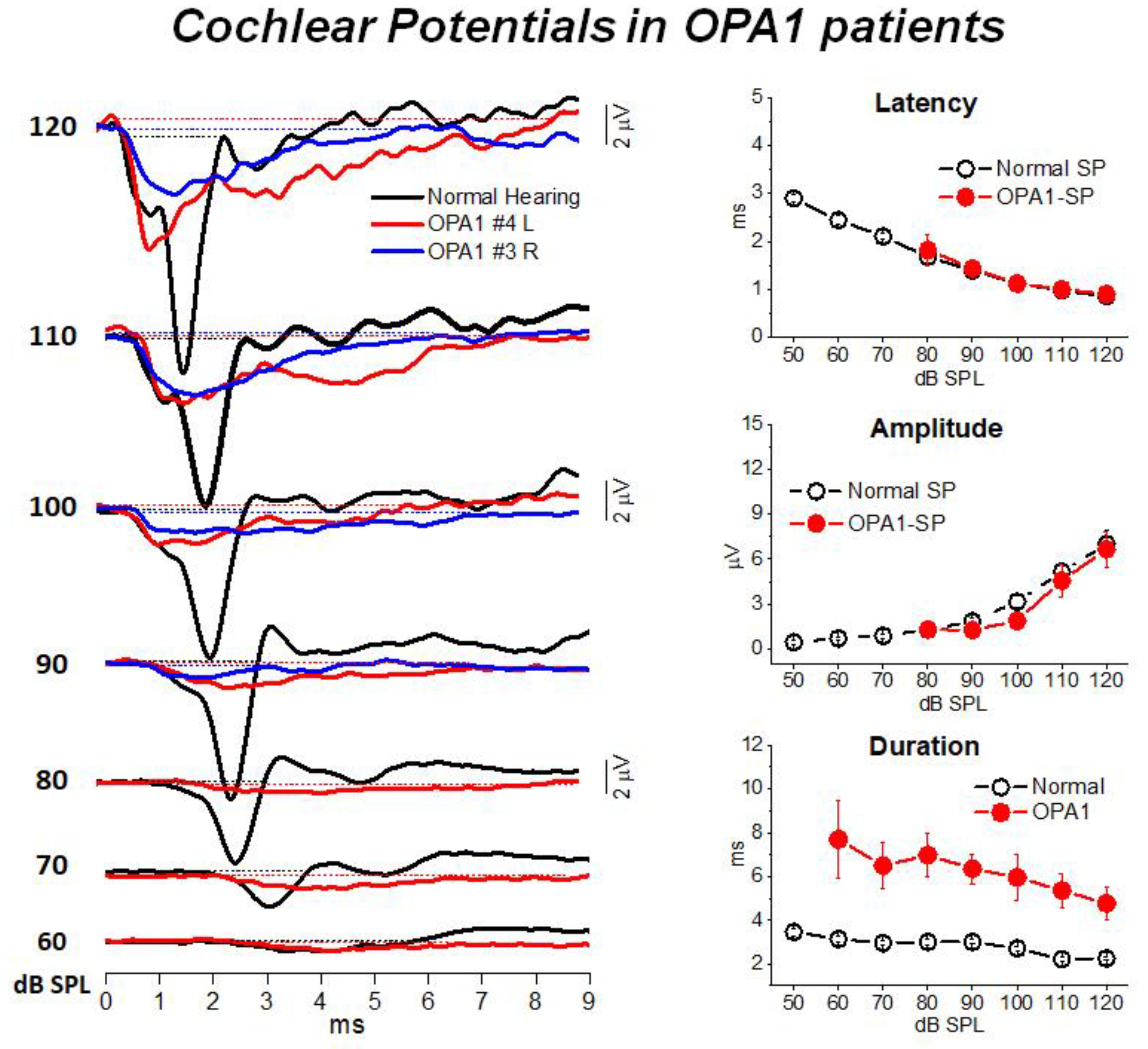
3. Mutations in the OPA1 Gene: A Model of Post-Synaptic AN

Dominant optic atrophy (DOA) is the most common genetic optic neuropathy. The hallmark of the disease is progressive visual loss beginning in childhood [42]. About 60–70% of DOA cases are caused by mutations in the nuclear gene encoding the OPA1 protein, a mitochondria GTPase embedded in the mitochondrial membrane. This protein is involved in fusion of the inner mitochondrial membrane [43], preservation of the structure of mitochondrial cristae [44], maintenance of membrane potential and oxidative phosphorylation [45].
Patients carrying missense mutations localized to the GTPase domain show a syndromic form of DOA associated with several extra-ocular manifestations, such as sensorineural hearing loss, peripheral neuropathy, ataxia, external ophthalmoplegia and myopathy [46,47]. Sensorineural hearing loss affects about 60% of OPA1 patients and AN has been proposed as the mechanism underlying the hearing disorder [11,25,47].
In the last decade, nine hearing-impaired subjects (age range of five–58 years) harboring missense mutations in the OPA1 gene have been diagnosed and followed up at the University of Padua Service of Audiology and Phoniatrics, while their neurological and ophthalmologic evaluation together with genetic analyses were performed at the Institute of Neurological Sciences of University of Bologna [12]. In all patients vision problems and difficulties in understanding speech began in childhood or adolescence. Audiological assessment showed severe impairment in speech perception, absence or profound abnormalities of auditory brainstem responses and the presence of otoacoustic emissions.
Transtympanic electrocochleography was performed in patients carrying missense mutations in the OPA1 gene at decreasing stimulus intensities from 120 to 60 dB SPL. ECochG waveforms were compared to the corresponding recordings obtained in 20 normally-hearing controls. CM potentials were significantly larger in OPA1 patients compared to controls, thus confirming preservation of outer hair cell activities. We hypothesized that The increase of CM amplitude might result from reduced activity of the efferent system resulting in turn from abnormal activation of auditory fibers.
Figure 2 illustrates the cochlear potentials obtained after CM cancellation from two representative OPA1 patients at decreasing stimulus intensities. The recordings are superimposed to the ECochG waveforms recorded in one normally-hearing control. In the latter, the responses consist of the rapid receptor summating potential (SP) followed by the synchronous neural compound action potential (CAP). Decreasing the stimulus level results in progressive latency increase and amplitude reduction of both SP and CAP peaks. In OPA1 patients the responses recorded at high intensity (120–100 dB SPL) begin with the SP potential, which shows comparable amplitudes and latencies with respect to controls. This finding points to preservation of IHC function. However, differently from controls, no CAP was recorded as SP was followed by a low-amplitude prolonged negative potential, which returned to baseline at 8–9 ms from response onset. This prolonged potential has been interpreted as resulting from abnormal activation of the terminal dendrites of auditory nerve fibers. This hypothesis was confirmed by the reduction in both amplitude and duration of ECochG potentials during high-rate stimulation, which is consistent with their neural generation.
All patients had tried hearing aids without benefit. Eight patients received unilateral cochlear implants in order to improve speech perception by electrical stimulation of auditory fibers through the cochlear implant. Overall, speech perception remarkably improved in all cochlear implant recipients also in the presence of competing noise, although considerable variation of speech perception scores between was observed. Interestingly, no compound action potential was recorded in response to electrical stimulation through the cochlear implant, which confirmed the presence of neural damage. In contrast, brainstem potentials were restored in response to electrical stimulation. This finding supports the hypothesis that the hearing disorder affecting OPA1 patients is underlain by degeneration of the distal portion of auditory nerve fibers. Cochlear implantation restores synchronous activation of auditory pathways by by-passing the site of the lesion.
Overall, these findings indicate that in patients carrying OPA1 missense mutations neural degeneration affecting the terminal dendrites results in a disruption of synchrony of auditory nerve fiber activity. Cochlear implantation improves speech perception in post-synaptic AN associated with OPA1 disease. However, a positive outcome of cochlear implant use was not invariably reported for all forms of post-synaptic AN, variability being possibly dependent on both the sites and extension of the lesion along auditory nerve fibers. Indeed, a variable improvement of speech perception scores has been reported with cochlear implant use in patients with AN associated with Friedreich ataxia [48,49].
The findings collected in families carrying mutations in the OPA1 gene indicated that combination of findings from genetic research and high-sensitive neurophysiologic recordings plays a crucial role in clarifying both mechanisms and sites underlying the abnormal synchrony of auditory fiber firing.

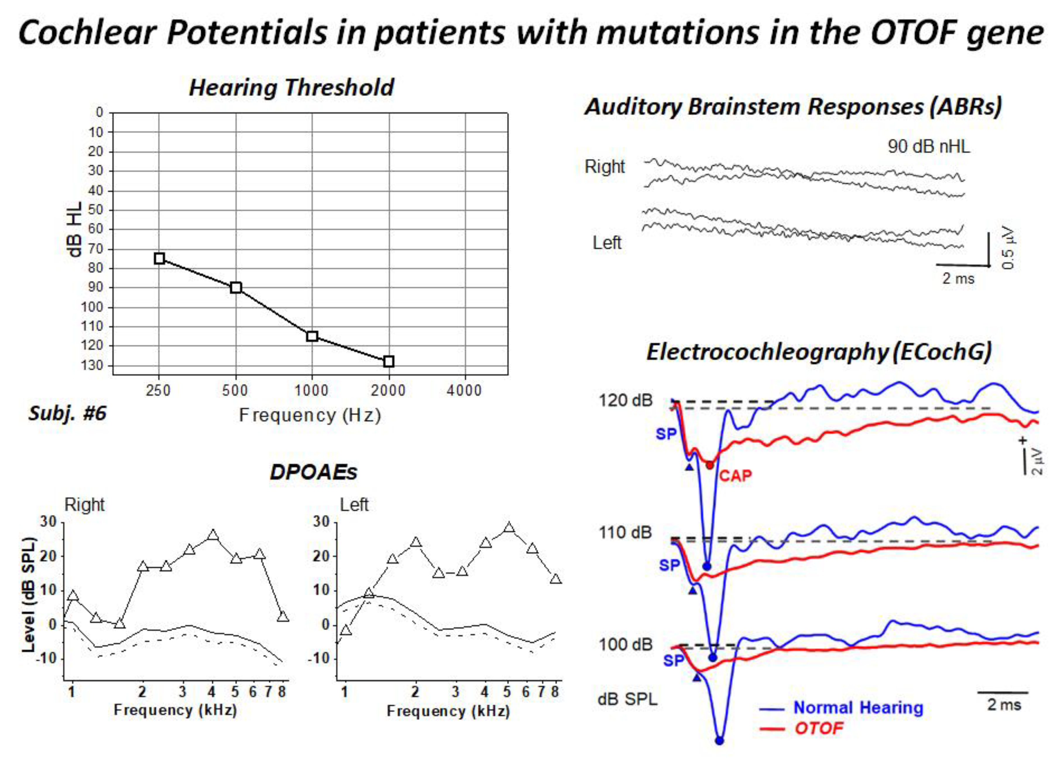
4. Mutations in the OTOF Gene: A Model of Synaptopathy

One well-studied form of presynaptic AN is caused by mutations in the OTOF gene encoding otoferlin [13,50]. Otoferlin is a transmembrane protein, localized to the synaptic pole in mature IHCs, which has been implicated in multivesicular release at the synapse between the IHCs and auditory nerve fibers [51] as well as in fast vesicle replenishment [52].
Mutations in the OTOF gene result in a disrupted function of the ribbon synapses with an impaired multivesicular glutamate release and vesicle replenishment. The majority of subjects carrying mutations in the OTOF gene present with the phenotype of congenital profound hearing loss, the absence of auditory brainstem potentials associated with preserved cochlear hair cell activities (OAEs, CM) [13,50].
In the last ten years, ten hearing-impaired children (age range of four month to three years) harboring biallelic mutations of the OTOF gene have been diagnosed and followed up at the University of Padua Service of Audiology and Phoniatrics, while their genetic analyses were performed at the Hospital Ramón y Cajal in Madrid [9,53]. All children showed profound hearing loss and absent ABRs and presence of distortion product otoacoustic emissions (DPOAEs). Moreover, ECochG recordings showed no differences in CM amplitude between OTOF patients and a group of 20 normally-hearing controls, thus confirming the preservation of OHC function. The audiological and electrophysiological findings collected in a representative patient are displayed in Figure 3. ECochG waveforms obtained after CM cancellation at high intensity (120–100 dB peSPL) are superimposed on the corresponding responses collected from one normally-hearing control. ECochG potentials begin with a rapid negative deflection that peaks at the same SP peak latency as in the normal control, and is of a comparable amplitude, which points to preservation of the IHC function. No CAP was recorded in the hearing-impaired child carrying mutations in the OTOF gene, as the SP is followed by a low-amplitude negative potential showing a markedly prolonged duration. Nevertheless, a small CAP is superimposed on the prolonged activity at high intensity in some children.
The prolonged potentials are likely to result from the sum of EPSPs arising in the terminal dendrites, which are dispersed in time as a consequence of the impaired multivesicle release. EPSPs only occasionally reach the threshold for triggering action potentials in some OTOF patients, and are recorded as high-threshold CAPs superimposed on the prolonged activity at high intensity. Therefore, the impaired multivesicular release is likely to result in a lesser probability of synchronized neural spiking and in a reduced signaling to the auditory brainstem pathways in comparison with normal hearing.
All children harboring OTOF mutations diagnosed at our department underwent unilateral cochlear implantation. They showed a remarkable improvement in hearing sensitivity and all reached speech perception scores of 90–100% by one year of cochlear implant use. Moreover, language development was in line with the language skills of their peers using cochlear implants whose deafness was related to causes other than mutations in the OTOF gene.
Unlike patients with post-synaptic AN, the implanted children with otoferlin-related deafness showed electrically-evoked auditory nerve potentials (e-CAPs) in response to electrical stimulation through the cochlear implant, consistent with preservation of auditory nerve function. Based on these findings, the outcome of cochlear implantation in patients with presynaptic AN due to OTOF mutations is expected to be successful.
In addition, it is worthy of note that recent studies on animal models of OTOF showed successful preliminary results by using gene therapy [54,55].

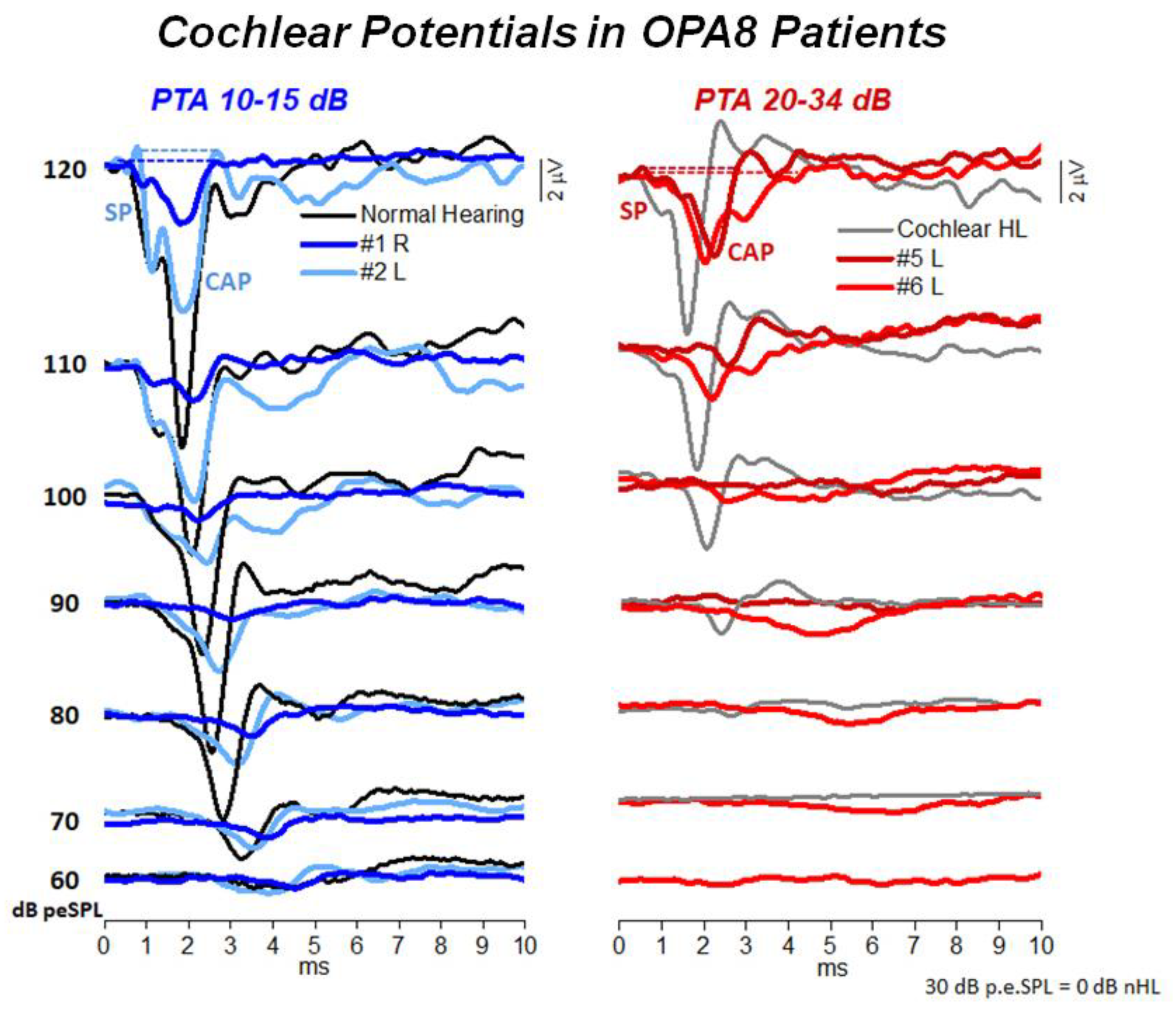
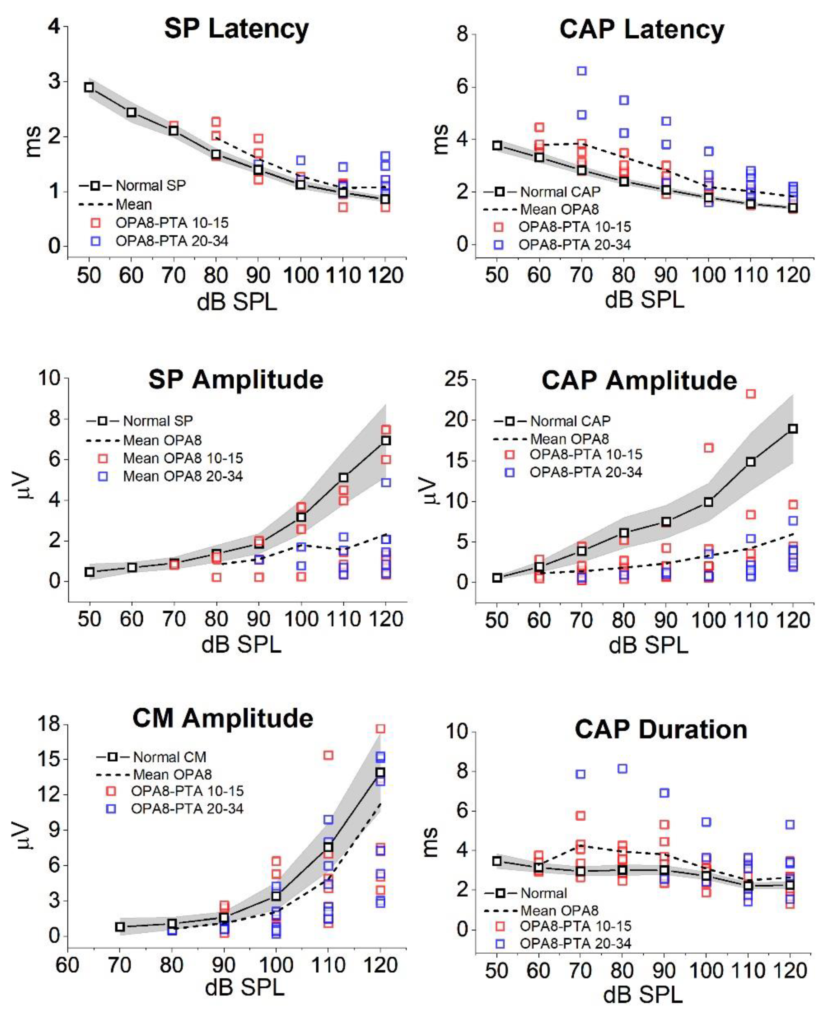
5. Hearing Dysfunction Related to the OPA8 Locus: A Model of Hidden AN

Hidden auditory neuropathy is characterized by reduced performances in challenging auditory tasks with preservation of hearing thresholds. This condition results from loss of synapses which ensues from degeneration of the unmyelinated portion of auditory nerve fibers [56].
Hidden AN has been observed in ten members of a large Italian family affected by DOA associated with the OPA8 locus (age range 19–72 years) [8,26]. Their neurological and ophthalmologic assessment was performed at the Institute of Neurological Sciences of University of Bologna.
Affected subjects complained of difficulties in understanding speech in the presence of noise. Hearing thresholds and speech perception were normal in the youngest members of this family, whereas the oldest patients showed mild hearing loss at high frequencies and decreased speech perception scores in the presence of noise. OAEs and CM recordings were within normal limits, thus indicating preservation of OHC function in all patients. In contrast, ABRs showed attenuated amplitudes in the youngest patients and severe abnormalities in the oldest. In addition, Wave I amplitude was significantly reduced compared to normally-hearing individuals, consistent with degeneration of IHCs synapses [57].
All subjects were submitted to ECochG recording. Figure 4 reports the ECochG potentials obtained in four OPA8 subjects, two with normal hearing thresholds and two with mild hearing loss, superimposed on the corresponding ECochG waveforms recorded in two controls. Of these, one had normal hearing while the other had a mild hearing loss of cochlear origin. In OPA8 patients the ECochG waveforms begin with the SP potential, which showed reduced amplitudes and increased latencies in comparison to recordings obtained from controls. SP potential was followed by the CAP, which showed a smaller amplitude and an increased peak latency in comparison with controls. The reduction in amplitude and the delay in peak latency of ECochG components were more pronounced in the group of older OPA8 patients showing mild hearing loss (Figure 5). These findings support the hypothesis that in the youngest members of the OPA8 family synaptic damage was far less pronounced [8].
The decrease in amplitude of both SP and CAP points to a decrease of the cochlear output, whereas the increase of both CAP peak latency and duration points to a selective reduction of auditory fibers from the basal portion of the cochlea. This is consistent with the audiometric profile of the oldest OPA8 patients showing mild hearing loss at high frequencies.
These findings indicate that in OPA8 patients hidden AN results from loss of IHCs synapses. At an early stage of the disease, the functional alterations only consist of abnormalities of ABR Wave I while the cochlear potentials show a decrease in amplitude and prolongation in latency of both SP and CAP. At this stage speech perception is relatively preserved. A reduction in speech perception scores and the worsening of cochlear potentials alterations become apparent only with progression of the disease.

6. Conclusions and Future Directions

AN disorders associated with gene mutations constitute a group of hearing dysfunctions resulting from different pathophysiological mechanisms. The identification of specific gene mutations combined with typical electrophysiological patterns may be the key factor in revealing how the failure of different molecular processes underlies the varieties of AN. This information is crucial for selecting effective rehabilitative options. In addition, the development of new electrophysiological tools may help in identifying neural and receptor components in the ECochG recordings collected from AN patients [58].

Author Contributions

All authors contributed to this work. R.S., I.d.C. and V.C. designed the review plan; R.S. wrote the text; P.S. and E.C. contributed to figure and table preparation; C.L.M. provided critical revision and commented on the manuscript. All authors have read and agreed to the published version of the manuscript.

Funding

This work was supported in part by the "Department of excellence 2018–2022" initiative of the Italian Ministry of education (MIUR) awarded to the Department of Neuroscience—University of Padua.

Institutional Review Board Statement

The study was conducted according to the guidelines of the Declaration of Helsinki.

Informed Consent Statement

Informed consent was obtained from all subjects involved in the study.

Acknowledgments

We are greatly indebted to Roberto Bovo and Franco Trabalzini who performed surgery. We are also grateful to engineer Antonio Selmo who supported us in setting up the recording apparatus.

Conflicts of Interest

The authors declare no conflict of interest.

References

  1. Starr, A.; Picton, T.W.; Sininger, Y.; Hood, L.J.; Berlin, C.I. Auditory neuropathy. Brain 1996, 119, 741–753. [Google Scholar] [CrossRef]
  2. Starr, A.; Zeng, F.; Michalewski, H.; Moser, T. Perspectives on auditory neuropathy: Disorders of inner hair cell, auditory nerve, and their synapse. In The Senses: A Comprehensive Reference; Basbaum, A.I., Kaneko, A., Shepherd, G.M., Westheimer, G., Albright, T.D., Masland, A., Eds.; Elsevier: Amsterdam, The Netherlands, 2008; Volume 3: Audition, pp. 397–412. [Google Scholar]
  3. Zeng, F.-G.; Kong, Y.-Y.; Michalewski, H.J.; Starr, A. Perceptual Consequences of Disrupted Auditory Nerve Activity. J. Neurophysiol. 2005, 93, 3050–3063. [Google Scholar] [CrossRef] [PubMed] [Green Version]
  4. Moser, T.; Starr, A. Auditory neuropathy—Neural and synaptic mechanisms. Nat. Rev. Neurol. 2016, 12, 135–149. [Google Scholar] [CrossRef]
  5. Johnson, J.S.; Newport, E.L. Critical period effects on universal properties of language: The status of subjacency in the acquisition of a second language. Cognition 1991, 39, 215–258. [Google Scholar] [CrossRef]
  6. Butinar, D.; Zidar, J.; Leonardis, L.; Popovic, M.; Kalaydjieva, L.; Angelicheva, D.; Sininger, Y.; Keats, B.; Starr, A. Hereditary auditory, vestibular, motor, and sensory neuropathy in a Slovenian Roma (Gypsy) kindred. Ann. Neurol. 1999, 46, 36–44. [Google Scholar] [CrossRef]
  7. Santarelli, R. Information from cochlear potentials and genetic mutations helps localize the lesion site in auditory neuropathy. Genome Med. 2010, 2, 91. [Google Scholar] [CrossRef] [PubMed] [Green Version]
  8. Santarelli, R.; La Morgia, C.; Valentino, M.L.; Barboni, P.; Monteleone, A.; Scimemi, P.; Carelli, V. Hearing Dysfunction in a Large Family Affected by Dominant Optic Atrophy (OPA8-Related DOA): A Human Model of Hidden Auditory Neuropathy. Front. Neurosci. 2019, 13, 501. [Google Scholar] [CrossRef] [Green Version]
  9. Santarelli, R.; del Castillo, I.; Cama, E.; Scimemi, P.; Starr, A. Audibility, speech perception and processing of temporal cues in ribbon synaptic disorders due to OTOF mutations. Hear. Res. 2015, 330, 200–212. [Google Scholar] [CrossRef] [PubMed] [Green Version]
  10. Santarelli, R.; Scimemi, P.; Costantini, M.; Domínguez-Ruiz, M.; Rodríguez-Ballesteros, M.; del Castillo, I. Cochlear Synaptopathy due to Mutations in OTOF Gene May Result in Stable Mild Hearing Loss and Severe Impairment of Speech Perception. Ear Hear. 2021, 42, 1627–1639. [Google Scholar] [CrossRef]
  11. Huang, T.; Santarelli, R.; Starr, A. Mutation of OPA1 gene causes deafness by affecting function of auditory nerve terminals. Brain Res. 2009, 1300, 97–104. [Google Scholar] [CrossRef] [PubMed] [Green Version]
  12. Santarelli, R.; Rossi, R.; Scimemi, P.; Cama, E.; Valentino, M.L.; La Morgia, C.; Caporali, L.; Liguori, R.; Magnavita, V.; Monteleone, A.; et al. OPA1-related auditory neuropathy: Site of lesion and outcome of cochlear implantation. Brain 2015, 138, 563–576. [Google Scholar] [CrossRef] [Green Version]
  13. Rodríguez-Ballesteros, M.; del Castillo, F.J.; Martín, Y.; Moreno-Pelayo, M.A.; Morera, C.; Prieto, F.; Marco, J.; Morant, A.; Gallo-Terán, J.; Morales-Angulo, C.; et al. Auditory neuropathy in patients carrying mutations in the otoferlin gene (OTOF). Hum. Mutat. 2003, 22, 451–456. [Google Scholar] [CrossRef]
  14. Delmaghani, S.; del Castillo, F.; Michel, V.; Leibovici, M.; Aghaie, A.; Ron, U.; Van Laer, L.; Ben-Tal, N.; Van Camp, G.; Weil, D.; et al. Mutations in the gene encoding pejvakin, a newly identified protein of the afferent auditory pathway, cause DFNB59 auditory neuropathy. Nat. Genet. 2006, 38, 770–778. [Google Scholar] [CrossRef]
  15. Schoen, C.J.; Emery, S.B.; Thorne, M.C.; Ammana, H.R.; Sliwerska, E.; Arnett, J.; Hortsch, M.; Hannan, F.; Burmeister, M.; Lesperance, M. Increased activity of Diaphanous homolog 3 (DIAPH3)/diaphanous causes hearing defects in humans with auditory neuropathy and in Drosophila. Proc. Natl. Acad. Sci. USA 2010, 107, 13396–13401. [Google Scholar] [CrossRef] [Green Version]
  16. Wang, Q.J.; Li, Q.Z.; Rao, S.Q.; Lee, K.; Huang, X.S.; Yang, W.Y.; Zhai, S.Q.; Guo, W.W.; Guo, Y.F.; Yu, N.; et al. AUNX1, a novel locus responsible for X linked recessive auditory and peripheral neuropathy, maps to Xq23-27.3. J. Med. Genet. 2005, 43, e33. [Google Scholar] [CrossRef] [Green Version]
  17. Thirlwall, A.S.; Brown, D.J.; McMillan, P.M.; Barker, S.E.; Lesperance, M.M. Phenotypic Characterization of Hereditary Hearing Impairment Linked to DFNA25. Arch. Otolaryngol.-Head Neck Surg. 2003, 129, 830–835. [Google Scholar] [CrossRef] [Green Version]
  18. Jang, M.W.; Oh, D.-Y.; Yi, E.; Liu, X.; Ling, J.; Kim, N.; Sharma, K.; Kim, T.Y.; Lee, S.; Kim, A.-R.; et al. A nonsense TMEM43 variant leads to disruption of connexin-linked function and autosomal dominant auditory neuropathy spectrum disorder. Proc. Natl. Acad. Sci. USA 2021, 118, e2019681118. [Google Scholar] [CrossRef]
  19. Kovach, M.; Campbell, K.; Herman, K.; Waggoner, B.; Gelber, D.; Hughes, L.; Kimonis, V. Anticipation in a unique family with Charcot-Marie-Tooth syndrome and deafness: Delineation of the clinical features and review of the literature. Am. J. Med. Genet. 2002, 108, 295–303. [Google Scholar] [CrossRef]
  20. Starr, A.; Michalewski, H.J.; Zeng, F.; Fujikawa-Brooks, S.; Linthicum, F.; Kim, C.S.; Winnier, D.; Keats, B. Pathology and physiology of auditory neuropathy with a novel mutation in the MPZ gene (Tyr145->Ser). Brain 2003, 126, 1604–1619. [Google Scholar] [CrossRef]
  21. Butinar, D.; Starr, A.; Zidar, J.; Koutsou, P.; Christodoulou, K. Auditory nerve is affected in one of two different point mutations of the neurofilament light gene. Clin. Neurophysiol. 2008, 119, 367–375. [Google Scholar] [CrossRef] [Green Version]
  22. Kalaydjieva, L.; Gresham, D.; Gooding, R.; Heather, L.; Baas, F.; de Jonge, R.; Blechschmidt, K.; Angelicheva, D.; Chandler, D.; Worsley, P.; et al. N-myc Downstream-Regulated Gene 1 Is Mutated in Hereditary Motor and Sensory Neuropathy–Lom. Am. J. Hum. Genet. 2000, 67, 47–58. [Google Scholar] [CrossRef] [Green Version]
  23. Lopez-Bigas, N.; Olive, M.; Rabionet, R.; Ben-David, O.; Martínez-MatosJ, A.; Bravo, O.; Banchs, I.; Volpini, V.; Gasparini, P.; Avraham, K.B.; et al. Connexin 31 (GJB3) is expressed in the peripheral and auditory nerves and causes neuropathy and hearing impairment. Hum. Mol. Genet. 2001, 10, 947–952. [Google Scholar] [CrossRef] [Green Version]
  24. Bähr, M.; Andres, F.; Timmerman, V.; E Nelis, M.; Van Broeckhoven, C.; Dichgans, J. Central visual, acoustic, and motor pathway involvement in a Charcot-Marie-Tooth family with an Asn205Ser mutation in the connexin 32 gene. J. Neurol. Neurosurg. Psychiatry 1999, 66, 202–206. [Google Scholar] [CrossRef] [Green Version]
  25. Amati-Bonneau, P.; Guichet, A.; Olichon, A.; Chevrollier, A.; Viala, F.; Miot, S.; Ayuso, C.; Odent, S.; Arrouet, C.; Verny, C.; et al. OPA1 R445H mutation in optic atrophy associated with sensorineural deafness. Ann. Neurol. 2005, 58, 958–963. [Google Scholar] [CrossRef]
  26. Carelli, V.; Schimpf, S.; Fuhrmann, N.; Valentino, M.L.; Zanna, C.; Iommarini, L.; Papke, M.; Schaich, S.; Tippmann, S.; Baumann, B.; et al. A clinically complex form of dominant optic atrophy (OPA8) maps on chromosome 16. Hum. Mol. Genet. 2011, 20, 1893–1905. [Google Scholar] [CrossRef] [PubMed] [Green Version]
  27. Meyer, E.; Michaelides, M.; Tee, L.J.; Robson, A.; Rahman, F.; Pasha, S.; Luxon, L.M.; Moore, A.T.; Maher, E.R. Nonsense mutation in TMEM126A causing autosomal recessive optic atrophy and auditory neuropathy. Mol. Vis. 2010, 16, 650–664. [Google Scholar] [PubMed]
  28. Rance, G.; Fava, R.; Baldock, H.; Chong, A.; Barker, E.; Corben, L.; Delatycki, M.B. Speech perception ability in individuals with Friedreich ataxia. Brain 2008, 131, 2002–2012. [Google Scholar] [CrossRef] [Green Version]
  29. Wang, Q.; Li, R.; Zhao, H.; Peters, J.L.; Liu, Q.; Yang, L.; Han, D.; Greinwald, J.H., Jr.; Young, W.-Y.; Guan, M.-X. Clinical and molecular characterization of a Chinese patient with auditory neuropathy associated with mitochondrial 12S rRNA T1095C mutation. Am. J. Med. Genet. Part A 2005, 133A, 27–30. [Google Scholar] [CrossRef] [Green Version]
  30. Bahmad, F.; Merchant, S.N.; Nadol, J.B.; Tranebjaerg, L. Otopathology in Mohr-Tranebjaerg syndrome. Laryngoscope 2007, 117, 1202–1208. [Google Scholar] [CrossRef] [Green Version]
  31. Ceranić, B.; Luxon, L.M. Progressive auditory neuropathy in patients with Leber’s hereditary optic neuropathy. J. Neurol. Neurosurg. Psychiatr. 2004, 75, 626–630. [Google Scholar] [CrossRef] [PubMed] [Green Version]
  32. Ołdak, M.; Oziębło, D.; Pollak, A.; Stępniak, I.; Lazniewski, M.; Lechowicz, U.; Kochanek, K.; Furmanek, M.; Tacikowska, G.; Plewczynski, D.; et al. Novel neuro-audiological findings and further evidence for TWNK involvement in Perrault syndrome. J. Transl. Med. 2017, 15, 25. [Google Scholar] [CrossRef]
  33. Dulon, D.; Papal, S.; Patni, P.; Cortese, M.; Vincent, M.; Tertrais, M.; Emptoz, A.; Tlili, A.; Bouleau, Y.; Michel, V.; et al. Clarin-1 gene transfer rescues auditory synaptopathy in model of Usher syndrome. J. Clin. Investig. 2018, 128, 3382–3401. [Google Scholar] [CrossRef]
  34. Tranebjærg, L.; Strenzke, N.; Lindholm, S.; Rendtorff, N.D.; Poulsen, H.; Khandelia, H.; Kopec, W.; Lyngbye, T.J.B.; Hamel, C.; Delettre, C.; et al. The CAPOS mutation in ATP1A3 alters Na/K-ATPase function and results in auditory neuropathy which has implications for management. Qual. Life Res. 2018, 137, 111–127. [Google Scholar] [CrossRef] [Green Version]
  35. Santarelli, R.; Starr, A.; Michalewski, H.J.; Arslan, E. Neural and receptor cochlear potentials obtained by transtympanic electrocochleography in auditory neuropathy. Clin. Neurophysiol. 2008, 119, 1028–1041. [Google Scholar] [CrossRef] [Green Version]
  36. Santarelli, R.; Arslan, E. Electrocochleography. In Handbook of Clinical Neurophysiology; Celesia, G.G., Ed.; Disorders of Peripheral and Central Auditory Processing; Elsevier: Amsterdam, The Netherlands, 2013; pp. 83–113. [Google Scholar]
  37. Dallos, P.; Cheatham, M.A. Production of cochlear potentials by inner and outer hair cells. J. Acoust. Soc. Am. 1976, 60, 510–512. [Google Scholar] [CrossRef]
  38. Durrant, J.D.; Wang, J.; Ding, D.L.; Salvi, R.J. Are inner or outer hair cells the source of summating potentials recorded from the round wi‘ndow? J. Acoust. Soc. Am. 1998, 104, 370–377. [Google Scholar] [CrossRef]
  39. Pappa, A.K.; Hutson, K.A.; Scott, W.C.; Wilson, J.D.; Fox, K.E.; Masood, M.M.; Giardina, C.K.; Pulver, S.H.; Grana, G.D.; Askew, C.; et al. Hair cell and neural contributions to the cochlear summating potential. J. Neurophysiol. 2019, 121, 2163–2180. [Google Scholar] [CrossRef]
  40. Goldstein, M.H.; Kiang, N.Y. Synchrony of Neural Activity in Electric Responses Evoked by Transient Acoustic Stimuli. J. Acoust. Soc. Am. 1958, 30, 107–114. [Google Scholar] [CrossRef]
  41. Bourien, J.; Tang, Y.; Batrel, C.; Huet, A.; Lenoir, M.; Ladrech, S.; Desmadryl, G.; Nouvian, R.; Puel, J.-L.; Wang, J. Contribution of auditory nerve fibers to compound action potential of the auditory nerve. J. Neurophysiol. 2014, 112, 1025–1039. [Google Scholar] [CrossRef] [Green Version]
  42. Carelli, V.; Ross-Cisneros, F.N.; Sadun, A.A. Mitochondrial dysfunction as a cause of optic neuropathies. Prog. Retin. Eye Res. 2004, 23, 53–89. [Google Scholar] [CrossRef]
  43. Olichon, A.; Guillou, E.; Delettre, C.; Landes, T.; Arnauné-Pelloquin, L.; Emorine, L.J.; Mils, V.; Daloyau, M.; Hamel, C.; Amati-Bonneau, P.; et al. Mitochondrial dynamics and disease, OPA1. Biochim. Biophys. Acta 2006, 1763, 500–509. [Google Scholar] [CrossRef] [PubMed] [Green Version]
  44. Frezza, C.; Cipolat, S.; De Brito, O.M.; Micaroni, M.; Beznoussenko, G.V.; Rudka, T.; Bartoli, D.; Polishuck, R.S.; Danial, N.N.; De Strooper, B.; et al. OPA1 Controls Apoptotic Cristae Remodeling Independently from Mitochondrial Fusion. Cell 2006, 126, 177–189. [Google Scholar] [CrossRef] [Green Version]
  45. Lodi, R.; Tonon, C.; Valentino, M.L.; Iotti, S.; Clementi, V.; Malucelli, E.; Barboni, P.; Longanesi, L.; Schimpf, S.; Wissinger, B.; et al. Deficit of in vivo mitochondrial ATP production in OPA1-related dominant optic atrophy. Ann. Neurol. 2004, 56, 719–723. [Google Scholar] [CrossRef]
  46. Amati-Bonneau, P.; Valentino, M.L.; Reynier, P.; Gallardo, M.E.; Bornstein, B.; Boissière, A.; Campos, Y.; Rivera, H.; de la Aleja, J.G.; Carroccia, R.; et al. OPA1 mutations induce mitochondrial DNA instability and optic atrophy ’plus’ phenotypes. Brain 2008, 131, 338–351. [Google Scholar] [CrossRef] [Green Version]
  47. Yu-Wai-Man, P.; Griffiths, P.G.; Gorman, G.S.; Lourenco, C.M.; Wright, A.F.; Auer-Grumbach, M.; Toscano, A.; Musumeci, O.; Valentino, M.L.; Caporali, L.; et al. Multi-system neurological disease is common in patients with OPA1 mutations. Brain A J. Neurol. 2010, 133, 771–786. [Google Scholar] [CrossRef]
  48. Miyamoto, R.T.; Kirk, K.H.; Renshaw, J.; Hussain, D. Cochlear Implantation in Auditory Neuropathy. Laryngoscope 1999, 109, 181–185. [Google Scholar] [CrossRef]
  49. Frewin, B.; Chung, M.; Donnelly, N. Bilateral cochlear implantation in Friedreich’s ataxia: A case study. Cochlear Implants Int. 2013, 14, 287–290. [Google Scholar] [CrossRef]
  50. Rodríguez-Ballesteros, M.; Reynoso, R.; Olarte, M.; Villamar, M.; Morera, C.; Santarelli, R.; Arslan, E.; Medá, C.; Curet, C.; Völter, C.; et al. A multicenter study on the prevalence and spectrum of mutations in the otoferlin gene (OTOF) in subjects with nonsyndromic hearing impairment and auditory neuropathy. Hum. Mutat. 2008, 29, 823–831. [Google Scholar] [CrossRef]
  51. Roux, I.; Safieddine, S.; Nouvian, R.; Grati, M.; Simmler, M.-C.; Bahloul, A.; Perfettini, I.; Le Gall, M.; Rostaing, P.; Hamard, G.; et al. Otoferlin, Defective in a Human Deafness Form, Is Essential for Exocytosis at the Auditory Ribbon Synapse. Cell 2006, 127, 277–289. [Google Scholar] [CrossRef]
  52. Pangršič, T.; Lasarow, L.; Reuter, K.; Takago, H.; Schwander, M.; Riedel, D.; Frank, T.; Tarantino, L.M.; Bailey, J.S.; Strenzke, N.; et al. Hearing requires otoferlin-dependent efficient replenishment of synaptic vesicles in hair cells. Nat. Neurosci. 2010, 13, 869–876. [Google Scholar] [CrossRef]
  53. Santarelli, R.; del Castillo, I.; Rodríguez-Ballesteros, M.; Scimemi, P.; Cama, E.; Arslan, E.; Starr, A. Abnormal Cochlear Potentials from Deaf Patients with Mutations in the Otoferlin Gene. J. Assoc. Res. Otolaryngol. 2009, 10, 545–556. [Google Scholar] [CrossRef] [Green Version]
  54. Akil, O.; Dyka, F.; Calvet, C.; Emptoz, A.; Lahlou, G.; Nouaille, S.; de Monvel, J.B.; Hardelin, J.-P.; Hauswirth, W.W.; Avan, P.; et al. Dual AAV-mediated gene therapy restores hearing in a DFNB9 mouse model. Proc. Natl. Acad. Sci. USA 2019, 116, 4496–4501. [Google Scholar] [CrossRef] [Green Version]
  55. Rankovic, V.; Vogl, C.; Dörje, N.M.; Bahader, I.; Duque-Afonso, C.J.; Thirumalai, A.; Weber, T.; Kusch, K.; Strenzke, N.; Moser, T. Overloaded Adeno-Associated Virus as a Novel Gene Therapeutic Tool for Otoferlin-Related Deafness. Front. Mol. Neurosci. 2021, 13, 253. [Google Scholar] [CrossRef]
  56. Schaette, R.; McAlpine, D. Tinnitus with a Normal Audiogram: Physiological Evidence for Hidden Hearing Loss and Computational Model. J. Neurosci. 2011, 31, 13452–13457. [Google Scholar] [CrossRef] [PubMed] [Green Version]
  57. Liberman, M.C.; Kujawa, S.G. Cochlear synaptopathy in acquired sensorineural hearing loss: Manifestations and mechanisms. Hear. Res. 2017, 349, 138–147. [Google Scholar] [CrossRef]
  58. Hancock, K.E.; O’Brien, B.; Santarelli, R.; Liberman, M.C.; Maison, S.F. The summating potential in human electrocochleography: Gaussian models and Fourier analysis. J. Acoust. Soc. Am. 2021, 150, 2492–2502. [Google Scholar] [CrossRef]
Figure 1. ECochG potentials recorded in a normally-hearing individual at 110 dB nHL. The procedure utilized to separate the cochlear microphonic (CM) from the compound action potential (CAP) and summating potential (SP) is illustrated. The ECochG responses to condensation (C) and rarefaction (R) clicks are superimposed in the top panel. The CAP together with the superimposed SP was obtained by averaging the recordings to condensation and rarefaction clicks (C + R average) through the attenuation of the out-of-phase cochlear microphonics (middle panel). The CM shown in the lower panel results from subtracting the (C + R) average from the ECochG response to condensation clicks. (Reprinted with permission from [35]).
Audiolres 11 00059 g001
Figure 2. ECochG waveforms recorded from OPA1 patients. In the left panel the ECochG recordings from two OPA1-M patients are superimposed on the corresponding waveforms obtained in one control at decreasing stimulus intensity. In OPA1 patients no CAP was recorded, the ECochG response showing the SP followed by prolonged potential. In this and the subsequent figure time “0” refers to CM onset. In the right panel means and standard errors of peak latency, amplitude and duration of cochlear potentials from OPA1 patients are superimposed on the corresponding values calculated for 20 controls with normal hearing. No differences in SP peak latency and values were found between OPA1 subjects and controls, whereas the duration of cochlear potentials was significantly increased in the group of OPA1 patients. (Modified from [12]).
Audiolres 11 00059 g002
Figure 3. Hearing thresholds and DPOAEs, ABRs and ECochG recordings collected from one child carrying two mutant alleles of the OTOF gene. The audiometric assessment performed in the free field using visual reinforcement audiometry indicated profound hearing loss. DPOAEs were recorded from both ears, whereas ABRs were absent. ECochG potentials obtained after CM cancellation are superimposed on the ECochG waveforms obtained in one normally-hearing control. ECochG waveforms begin with the SP, which is followed by a low-amplitude negative potential showing a markedly prolonged duration compared to the control. A small CAP was recorded at high intensity. (Modified from [9]).
Audiolres 11 00059 g003
Figure 4. ECochG potentials recorded from patients with OPA8-related DOA. The waveforms obtained from four patients are shown together with the recordings collected in one control at decreasing intensity. The recordings illustrated in the left panel are from two OPA8 patients showing normal hearing thresholds (A), while the ECochG waveforms obtained in two patients showing mild hearing loss are illustrated on the right (B). (Modified from [8]).
Audiolres 11 00059 g004
Figure 5. Amplitude, latency and duration of ECochG potentials from OPA8 patients. Individual (colored squares) and mean values (dashed line) of amplitude, latency and duration of ECochG components are reported at various stimulus intensity. Mean values (black open squares), with 95% confidence limits (shadowed areas) obtained in normally hearing controls are superimposed on individual values obtained in OPA8 patients (Modified from [8]).
Audiolres 11 00059 g005
Table 1. Genetic disorders underlying AN.
LocusGeneTransmissionPhenotypeReference
Isolated AN
2p23–p22OTOFRecessiveCongenital profound deafness[13]
2q31.1–q31.3PJVKRecessiveCongenital profound deafness[14]
13q21–q24DIAPH3DominantModerate to profound deafness[15]
mtDNA12S rRNA (T1095C) Moderate deafness[16]
12q23.1SLC17A8DominantProgressive[17]
3P25.1TMEM43DominantPost-lingual moderate to profound deafness[18]
Non-isolated AN
CMT 1A17p11.2–p12PMP22DominantMild to severe deafness; demyelinating neuropathy[19]
CMT 1B1q22MPZDominantMild to severe deafness; demyelinating neuropathy[20]
CMT 2E8p21NF-LDominantNormal hearing; axonal neuropathy[21]
CMT 4D8q24.3NDRG1RecessiveMild to severe deafness; axonal/demyelinating neuropathy[6,22]
CMT1p34GJB3 (Cx31)DominantMild deafness[23]
CMT 1XXp13GJB1 (Cx32)X-linked DominantDemyelinating neuropathy[24]
ADOA3q28–q29OPA1 (R445H)DominantOptic neuropathy; moderate deafness[25]
ADOA16q21–q22 DominantOptic neuropathy, cardiac abnormalities[26]
AROA11q14.1–11q22.3TMEM126ARecessiveOptic neuropathy; mild hearing loss[27]
Friedreich9q13FXNRecessiveAtaxia; axonal neuropathy; optic neuropathy; cardiomyopathy; normal hearing threshold-mild deafness[28]
AUNX1Xq23–q27.3 X-linked RecessiveSensory axonal neuropathy; mild-to-severe deafness[29]
DDON (Mohr-Tranebjaerg)Xq22.1TIMM8AX-linked RecessiveProgressive deafness; dystonia, optic neuropathy; dementia[30]
LHON (Leber)mtDNAMTND4 (11778mtDNA) Optic neuropathy; mild-to-moderate deafness[31]
Perrault10q24.31TWNKRecessiveHypogonadism, cerebellar atrophy, cochlear nerve thinning[32]
USH3A3q25.1CLRN1RecessiveRetinitis pigmentosa[33]
CAPOS19q13.2ATP1A3DominantCerebellar ataxia, areflexia, pes cavus, optic atrophy[34]
Publisher’s Note: MDPI stays neutral with regard to jurisdictional claims in published maps and institutional affiliations.

