Article

Dysregulation of Iron Homeostasis in β-Thalassemia and Impaired Neutrophil Activity

by
Sreenithi Santhakumar
1,2,
Leo Stephen
1,
Aruna Barade
1,
Uday Kulkarni
1,
Biju George
1 and
Eunice S. Edison
1,*
1
Department of Haematology, Ranipet Campus, Christian Medical College, Vellore 632517, Tamil Nadu, India
2
Sree Chithra Tirunal Institute for Medical Sciences & Technology, Thiruvananthapuram 695011, Kerala, India
*
Author to whom correspondence should be addressed.
Thalass. Rep. 2025, 15(2), 4; https://doi.org/10.3390/thalassrep15020004
Submission received: 15 November 2024 / Revised: 6 January 2025 / Accepted: 7 March 2025 / Published: 25 April 2025

Abstract

:
Background/Objective: Patients with beta-thalassemia are more susceptible to iron overload and have altered neutrophil function. This study investigated the connections between iron metabolism in neutrophils, neutrophil functionality, and overall iron status in individuals with β-thalassemia and sickle cell anemia. Methods: We recruited 18 patients with β-thalassemia, 5 patients with sickle cell anemia, and 15 healthy controls. Our evaluation included measurements of iron and hepcidin concentrations in the serum, along with an analysis of neutrophil function, specifically their phagocytic and oxidative burst capabilities. In addition, we examined the expression of iron transport proteins in neutrophils. Results: Patients with β-thalassemia showed significant iron overload, reduced neutrophil counts, and decreased oxidative burst activity and phagocytosis. Systemic iron status is inversely correlated with the phagocytic capacity of β-thalassemia neutrophils. Regression analysis indicated a significant association between serum iron level, transferrin iron binding capacity, transferrin saturation, and neutrophil percentage. These findings elucidate the essential role of systemic iron levels in neutrophil efficacy against infections. Furthermore, FPN1B and DMT1A mRNA levels were upregulated, and IRP2 was downregulated in the neutrophils of patients with β-thalassemia major and intermedia compared to controls. Conclusions: Elevated systemic iron levels were associated with reduced neutrophil counts and impaired neutrophil function in patients with β-thalassemia. These findings highlight a critical role of systemic iron overload in neutrophil dysfunction.

1. Introduction

Beta thalassemia is an inherited disorder caused by impaired synthesis of beta-globin chains. This defect leads to the accumulation of excess α-globin chains, ineffective erythropoiesis, hemolytic anemia, and secondary iron overload [1]. Patients receive multiple blood transfusions, causing excess iron deposition in organs, such as the heart and liver, thereby contributing to the risk of organ failure. Iron overload results in cellular damage, thereby compromising immune function and increasing the risk of severe infections [2]. Several studies have shown that immune dysfunction is a predisposing factor for poor outcomes and increased mortality due to infections in beta-thalassemia patients [3,4]. Neutrophils are the first line of innate immunity that combats invading pathogens primarily through reactive oxygen species (ROS) generation, release of granules, and formation of neutrophil extracellular traps (NETs) [5]. Neutrophil dysfunction has been observed in beta-thalassemia patients as a result of iron overload due to frequent transfusions [6]. Iron accumulation contributes to impaired neutrophil migration and cellular abnormalities in neutrophils [7,8]. Thus, maintaining the tight regulation of neutrophil iron metabolism is crucial for preventing iron toxicity. Previous studies have reported that excess iron triggers ROS production, which damages the neutrophil membranes and impairs phagocytic activity. This hinders the oxidative response required for immune defense [9,10]. Interestingly, enhanced phagocytic activity of neutrophils has been reported in patients with beta-thalassemia major [11]. An earlier study indicated that iron chelation decreased the phagocytic activity of neutrophils, highlighting the harmful impact of iron overload on neutrophil function [12]. Although studies have shown adverse effects of iron overload on neutrophil function, the impact of systemic and neutrophil iron homeostasis on neutrophil dysfunction is unclear.
Sickle cell anemia (SCA) is another inherited disorder caused by a mutation in the β-globin subunit of hemoglobin and polymerization of hemoglobin S [13]. SCA is characterized by chronic hemolytic anemia and organ damage [14]. Recent studies have reported immune dysfunction in patients with SCA and increased susceptibility to infections [15,16]. However, neutrophil iron homeostasis and the effects of iron on neutrophil function in SCA have not yet been studied.
In this study, we evaluated iron parameters in patients with β-thalassemia major, intermedia, and sickle cell anemia, exploring their association with the neutrophil function.

2. Materials and Methods

2.1. Study Participants

Twenty-three patients diagnosed with β-thalassemia (thalassemia major and thalassemia intermedia) and sickle cell anemia based on clinical and hematological parameters were recruited from the Department of Hematology, Christian Medical College, Vellore, India. Beta-thalassemia patients were classified as major (TM) and intermedia (TI) based on clinical age at presentation, age at first transfusion, and transfusion requirement. β-thalassemia major patients receive blood transfusions at regular intervals, and intermedia patients occasionally require transfusions [17]. Patients with β-thalassemia major exhibited one of the following mutations: codon 30 (G > A) or IVS 1-5 (G > C). In the β-thalassemia intermedia group, the majority had HbE-beta thalassemia. Two patients carried the promoter mutation −28 (G > A) in a homozygous or compound heterozygous state. Fifteen healthy individuals with Hb levels > 12 g/dL and ferritin > 15 ng/mL were included in the control group.

2.2. Hematological and Biochemical Parameters

Complete blood counts (CBCs) were performed on peripheral blood using an automated hematology analyzer (Sysmex KX21, Kobe Japan). Serum ferritin, iron, soluble transferrin receptor, and UIBC were analyzed by chemiluminescence immunoassay using the Advia Centaur, Siemens XPI, Eschborn, Germany Transferrin saturation was calculated as [serum iron/total iron-binding capacity (TIBC)] × 100. Serum hepcidin levels were quantified using an enzyme immunoassay method from DRG, GmbH, Marburg, Germany, according to the manufacturer’s protocol.

2.3. Neutrophil Purification

Neutrophils were isolated from 9 mL of peripheral blood collected in an ethylenediaminetetraacetic acid (EDTA) tube using the MACSxpress® Whole Blood Neutrophil Isolation Kit (Miltenyi Biotec, Bergisch Gladbach, Germany). The purified population (>95%) was confirmed by the presence of the surface marker CD62L (BD Pharmingen 555544), which was evaluated by flow cytometry. The acquisition was performed using Navios flow cytometry (Beckman Coulter, CA, USA) and analyzed using the Kaluza software, version 2.1.

2.4. RNA Extraction and Real-Time qPCR

RNA was isolated from neutrophils using the TRIzol method and reverse transcribed into complementary DNA using the RT2 First Strand Kit (Qiagen, Hilden, Germany). The relative quantification of genes involved in iron uptake (TFRC), export (FPN1A, FPN1B), transport (DMT1A, DMT1B), and regulation (IRP2) were measured using real-time PCR. Real-time PCR was performed on a 7500 QPCR System (Applied Biosystems, Foster City, CA, USA) using SYBR Green chemistry. All values were normalized to the reference gene β-actin. The primers used are listed in Supplementary Table S1.

2.5. Oxidative Burst

In the oxidative burst assay, neutrophils were incubated with dihydro rhodamine 123 (DHR) and stimulated with phorbol 12-myristate 13-acetate (PMA). Oxidation of dihydro rhodamine to rhodamine by the respiratory burst of the cell results in the formation of a fluorescent dye. The mean fluorescence intensity of oxidized DHR was analyzed using a Beckman Coulter (Navios) flow cytometer. The results were analyzed using the Kaluza software.

2.6. Phagocytosis Activity

The phagocytosis and acidification capacity of human neutrophils were quantified using the pHrodo Green Staphylococcus aureus BioParticles kit (Thermo Fisher, Carlsbad, CA, USA). Cell acquisition was performed using a Beckman Coulter (Navios) flow cytometer. The results were analyzed using the Kaluza software.

2.7. Statistical Analysis

Statistical analysis was performed using SPSS software, version 27. Graphs were constructed using GraphPad Prism version 8. Appropriate statistical tests were used, including t-tests for continuous variables and Mann–Whitney and Kruskal–Wallis tests for nonparametric data. Associations between the iron parameters and gene expression were evaluated using Spearman’s and Pearson’s correlations. Statistical significance was set at p < 0.05. The associations were evaluated using univariate linear regression.

3. Results

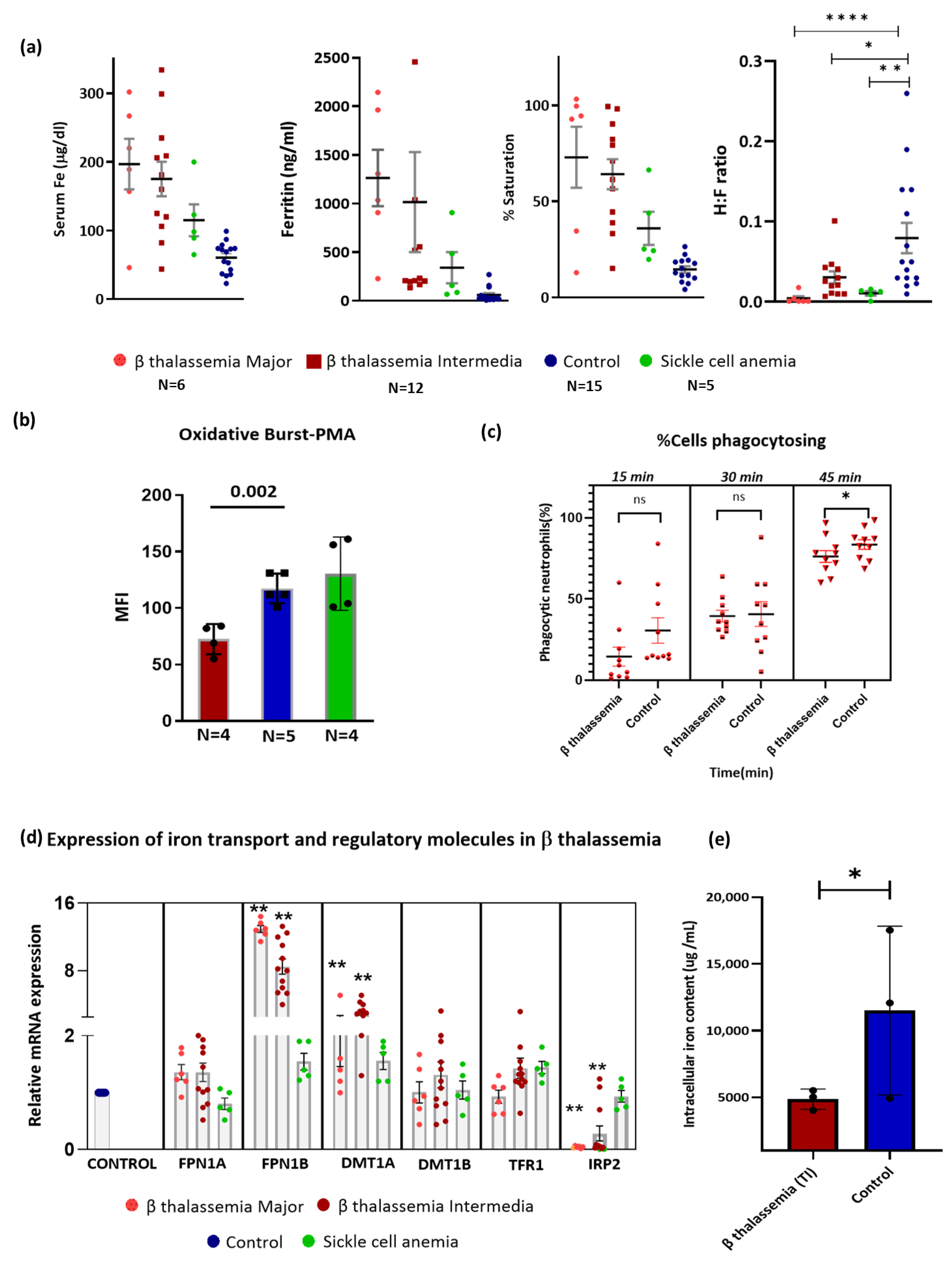
3.1. Hematological and Biochemical Parameters in the Study Participants

In this prospective study, we investigated a cohort of patients with β-thalassemia major (n = 6), β-thalassemia intermedia (n = 12), and sickle cell anemia (n = 5). The mean ages of the TM, TI, and SCA groups were 16 ± 7.8, 24.5 ± 9.5, and 19.6 ± 5 years, respectively. SCA patients had an average hemoglobin level of 10.1 ± 1.1 g/dL, and healthy controls had 13.2 ± 1.8 g/dL. The mean neutrophil counts in the TM, TI, SCA, and control were 3.37 × 10³ ± 2.11 cells/µL, 4.52 × 10³ ± 1.5 cells/µL, 5.58 × 10³ ± 2.4 cells/µL, and 5.48 × 10³ ± 1.3 cells/µL, respectively. Neutrophil counts and percentages in patients with β-thalassemia intermedia were significantly reduced compared to those in controls (Supplementary Figure S1). Demographic and biochemical parameters are listed in Table 1.
Beta-thalassemia patients were administered iron chelation therapy using Deferasirox, which is recommended for individuals with serum ferritin levels exceeding 1000 ng/mL. Deferasirox was started at 20 mg per kg per day and increased to 40 mg per kg per day based on the ferritin levels performed every 3 months. In this study, six patients with β-thalassemia major and five patients with β-thalassemia intermedia received iron chelation therapy.
Increased transferrin saturation (TSAT) levels were found in the TM, TI, and SCA as compared to healthy donors (p = 0.014, β thalassemia major vs. control; p = 0.001, β thalassemia intermedia vs. control; p = 0.011, SCA vs. control by the Mann–Whitney U rank-sum test) (Figure 1a). Notably, transferrin receptor (sTfR) levels were significantly different among the TM, TI, SCA, and control groups (p = 0.001, Kruskal–Wallis test).
Serum hepcidin levels were significantly lower in the TI group than in the controls (p = 0.019, TI vs. control by the Mann–Whitney U rank-sum test). We observed a significant difference in the hepcidin:ferritin ratio (H:F) among TM, TI, SCA, and control groups (p = 0.001) (Figure 1a). The details of all the patients with iron parameters are presented in Supplementary Table S2.

3.2. Neutrophils of β-Thalassemia Patients Exhibited Reduced Burst Activity and Phagocytosis

We further analyzed the oxidative burst capacity (OB) of neutrophils from patients with β-thalassemia major and intermedia. The mean fluorescence intensities (MFIs) of OB in the TM, TI, SCA, and control were 83 ± 1.4, 62 ± 9.5, 140 ± 31, and 117.4 ± 13.2, respectively. OB was significantly reduced in the β-thalassemia group (TM and TI) (p = 0.002) compared to that in healthy donors when exposed to the synthetic agent PMA, as shown in Figure 1b. However, there were no significant changes in the neutrophil oxidative burst activity in patients with SCA.
The neutrophil phagocytosis capacities assessed by the mean fluorescence intensity (MFI) of fluorescein-labeled S. aureus particles were 6.4 ± 2.2 in TM (N = 4), 6.0 ± 1.0 in TI (N = 6), and 8.6 ± 2.7 in controls (N = 10) after 15 min of incubation. At 30 min, the MFI increased to 14.4 ± 1.6 in TM, 12.8 ± 3.4 in TI, and 18.8 ± 8.4 in controls. By 45 min, the MFI further increased to 26.2 ± 3.1 in TM, 21.4 ± 7.1 in TI, and 32.7 ± 13.6 in controls, indicating a time-dependent increase in phagocytic activity across all groups. Phagocytosis capacity was significantly lower in the β-thalassemia group (TM and TI) compared to controls after 15 min of incubation (Supplementary Figure S2). The numbers of phagocytosed neutrophils after 15 min of incubation were 17.6 ± 1.74% in TM, 20 ± 3.5% in TI, and 30.5 ± 11.9% in the control group. After 30 min of incubation, the counts increased to 44 ± 10.6% in TM, 40 ± 14.3% in TI, and 40 ± 23% in controls. After 45 min, the numbers of phagocytosed neutrophils reached 70.8 ± 15.3% in TM, 72.2 ± 6.86% in TI, and 83.5 ± 8.9% in the control group. The recognition capacity of neutrophils toward bioparticles significantly decreased at 45 min incubation in the β-thalassemia group (p = 0.017) (Figure 1c).

3.3. Iron Metabolism in Neutrophils of β-Thalassemia

Gene expression was assessed for iron transporters, such as dimetal transporter (DMT1A) with iron-responsive element (IRE), DMT1B lacking IRE, ferroportin-FPN1A with IRE, FPN1B lacking IRE, transferrin receptor (TFRC), and cellular iron regulator IRP2, in the neutrophils of the β-thalassemia major (N = 6), β-thalassemia intermedia (N = 12), sickle cell anemia (N = 5), and controls (N = 15).
The mRNA expression of FPN1B, DMT1A, and IRP2 in neutrophils was differentially expressed in both β-thalassemia major (p = 0.001, p = 0.003, and p = 0.001, respectively) and β-thalassemia intermedia (p = 0.001, p = 0.001, and p = 0.001, respectively) compared to controls (Figure 1d). Furthermore, SCA patients did not demonstrate statistically significant differences in the expression of these genes compared to controls. There were no significant differences in mRNA expression among the TM, TI, and SCA groups, as determined by one-way ANOVA followed by Bonferroni post hoc tests.
Using atomic absorption spectrometry, we measured the neutrophil iron content in β-thalassemia intermedia patients. Our results revealed a significant decrease in intracellular iron content in β-thalassemia patients as compared to healthy donors (p = 0.018), as illustrated in Figure 1e.

3.4. Association of Systemic Iron Status with Neutrophil Function and Neutrophil Iron Metabolism

For statistical analysis, we combined β-thalassemia major and intermedia into a single cohort (N = 18) for correlation with mRNA expression and regression analysis because of the limited sample size. We correlated iron biomarkers with neutrophil function to determine whether iron status is associated with the modulation of neutrophil activity. We found a negative association of serum ferritin and transferrin saturation with neutrophil phagocytosis capacity (r = −0.714; p = 0.045 and r = −0.857; p = 0.014, respectively) (Supplementary Figure S3).
We observed a negative association between serum iron and hepcidin levels in the β-thalassemia cohort (r = −0.679, p = 0.022). Transferrin saturation was negatively associated with the percentage of neutrophils (n = 18) (r = −0.675, p = 0.002). In addition, positive associations of serum hepcidin levels and total iron binding capacity (TIBC) with neutrophils percentage were observed in the β-thalassemia cohort (r = 0.556, p = 0.031 and r = 0.770; p = 0.015, respectively) (Supplementary Figure S3).
In the β-thalassemia cohort, serum ferritin was negatively associated with mRNA expression of FPN1A (r = −0.690, p = 0.009) and positively associated with FPN1B (r = 0.564, p = 0.045). Soluble transferrin receptor was positively correlated with mRNA expression of FPN1B (r = 0.648, p = 0.017) (Supplementary Figure S3). Regression analysis depicted a negative association between neutrophils and serum iron levels (β = −0.017, p = 0.051).

4. Conclusions

Beta thalassemia and sickle cell anemia are life-threatening global health concerns, with India alone accounting for nearly 25% of β thalassemia cases and 14.5% of SCA cases worldwide [18,19]. Iron overload and severe anemia contribute to various complications, including immune dysfunction [7,20]. Iron is essential for neutrophil function. Iron stores must be tightly regulated because they are highly reactive [21]. However, iron regulation in neutrophils in patients with β-thalassemia has not been studied. Using neutrophils isolated from patients with β-thalassemia, we studied the impact of iron status on neutrophil iron metabolism and function.
In this study, we observed a reduced neutrophil percentage in β-thalassemia patients, similar to the findings of previous studies [8,22]. Several studies have reported impaired phagocytosis in β thalassemia patients [7,22]. Similarly, neutrophils from the β-thalassemia cohort showed a reduced phagocytic response to fluorescently labeled bacteria, as assessed using pHrodo™ S. aureus Bioparticle, compared to controls. This indicates a compromised ability to effectively engulf and control bacterial infections in patients with β-thalassemia.
Besides phagocytosis, another function used by neutrophils to fight pathogens is oxidative burst activity [23]. In our study, neutrophils isolated from β-thalassemia patients showed a significant reduction in ROS production compared to the controls when stimulated with the synthetic agent PMA. This finding confirms decreased neutrophil activity and microbial control. However, the oxidative burst activity remained unaffected in SCA patients. Additionally, our study found that β-thalassemia intermedia patients had lower intracellular iron content in neutrophils, which could be a contributing factor to the observed functional impairments. Iron deficiency has previously been linked to impaired ROS production in neutrophils [24], and our findings corroborate this, highlighting the pivotal role of iron in neutrophil function.
Our study is the first to characterize iron metabolism in neutrophils of β thalassemia patients. In response to cellular iron deficiency, DMT1A, FPN 1B, and IRP2 mRNA expressions were differentially expressed in patients with β-thalassemia. The FPN1B transcript, which lacks the IRE region, is not inhibited by neutrophil iron deficiency. Studies have shown that during iron deficiency, increased FPN1B mRNA expression and reduced IRP1/2 activity lead to higher ferroportin expression in enterocytes, facilitating iron transfer [25]. A similar mechanism in neutrophils can explain the observed iron depletion; however, investigation into the factors regulating ferroportin expression in these cells is required. Another possible explanation for the decreased iron content observed in beta-thalassemia patients can be the effect of chelation therapy, which targets intracellular iron levels.
Together, this study demonstrates that increased systemic iron status is associated with reduced neutrophil counts and aberrant neutrophil function in β thalassemia patients. In iron-deficient neutrophils, iron transporters were upregulated. These findings warrant further investigation in a larger cohort to understand how neutrophil iron homeostasis contributes to immune dysfunction. Exploring the role of iron chelation with neutrophils can lead to more effective therapies for β-thalassemia patients.

Supplementary Materials

The following supporting information can be downloaded at https://www.mdpi.com/article/10.3390/thalassrep15020004/s1, Table S1: qPCR Primers; Table S2: Detailed hematological and iron parameters for each patient. Red text indicates β thalassemia Major, blue text indicates β thalassemia intermedia, green text indicates sickle cell anemia and black text indicates controls; Supplementary Figure S1: Boxplot of neutrophil count and percentage in Beta thalassemia major, intermedia, sickle cell anemia and controls. p-value denoted for each group in comparison to control for both neutrophil count and neutrophil percentage. Statistical significance was calculated using Student’s t-test (two-tailed); Supplementary Figure S2: Evaluation of the phagocytosis capacity (number of phagocytised S aureus bioparticles) in neutrophils from beta thalassemia patients (red) vs. controls (blue); Supplementary Figure S3: Association of iron parameters with neutrophil function and neutrophil iron exporters.

Author Contributions

Conceptualization, E.S.E.; methodology, S.S. and L.S.; software, S.S.; validation, S.S. and E.S.E.; formal analysis, S.S. and A.B.; investigation, S.S.; resources, E.S.E.; data curation, S.S.; writing—original draft preparation, S.S.; writing—review and editing, S.S., B.G., U.K. and E.S.E.; supervision, E.S.E.; project administration, E.S.E.; funding acquisition, E.S.E. All authors have read and agreed to the published version of the manuscript.

Funding

This research was funded by the Department of Science and Technology, Government of India, grand number DST/CRG/2019/005494 to E.S.

Institutional Review Board Statement

The study was conducted in accordance with the Declaration of Helsinki and approved by the Institutional Review Board of CHRISTIAN MEDICAL COLLEGE (IRB Min No. 11480 and 22 August 2018).

Informed Consent Statement

Informed consent was obtained from all subjects involved in the study.

Data Availability Statement

The original contributions presented in this study are included in the article/Supplementary Materials. Further inquiries can be directed to the corresponding author.

Acknowledgments

We thank Arun Jose Nellickal for helping with the biochemical analysis.

Conflicts of Interest

The authors declare no conflicts of interest. The funders had no role in the design of the study; in the collection, analyses, or interpretation of data; in the writing of the manuscript; or in the decision to publish the results.

References

  1. Biswas, S.; Smrity, S.Z.; Bhuia, M.S.; Sonia, F.A.; Aktar, M.A.; Chowdhury, R.; Islam, T.; Islam, M.T.; Gonçalves Alencar, G.; Paulo, C.L.R.; et al. Beta-Thalassemia: A Pharmacological Drug-Based Treatment. Drugs Drug Candidates 2024, 3, 126–147. [Google Scholar] [CrossRef]
  2. Borgna-Pignatti, C.; Cappellini, M.D.; De Stefano, P.; Del Vecchio, G.C.; Forni, G.L.; Gamberini, M.R.; Ghilardi, R.; Origa, R.; Piga, A.; Romeo, M.A.; et al. Survival and Complications in Thalassemia. Ann. N. Y. Acad. Sci. 2005, 1054, 40–47. [Google Scholar] [CrossRef]
  3. Namazi Bayegi, S.; Ali Hamidieh, A.; Behfar, M.; Saghazadeh, A.; Bozorgmehr, M.; Tajik, N.; Delbandi, A.-A.; Delavari, S.; Shekarabi, M.; Rezaei, N. Unbalanced T-cell subsets in pediatric patients with beta-thalassemia. Hum. Immunol. 2023, 84, 224–234. [Google Scholar] [CrossRef] [PubMed]
  4. Vento, S.; Cainelli, F.; Cesario, F. Infections and thalassaemia. Lancet Infect. Dis. 2006, 6, 226–233. [Google Scholar] [CrossRef]
  5. Li, Y.; Wang, W.; Yang, F.; Xu, Y.; Feng, C.; Zhao, Y. The regulatory roles of neutrophils in adaptive immunity. Cell Commun. Signal. 2019, 17, 147. [Google Scholar] [CrossRef] [PubMed]
  6. Sae-Khow, K.; Charoensappakit, A.; Leelahavanichkul, A. Neutrophil Diversity (Immature, Aged, and Low-Density Neutrophils) and Functional Plasticity: Possible Impacts of Iron Overload in β-Thalassemia. Int. J. Mol. Sci. 2024, 25, 10651. [Google Scholar] [CrossRef]
  7. Thiengtavor, C.; Siriworadetkun, S.; Paiboonsukwong, K.; Fucharoen, S.; Pattanapanyasat, K.; Vadolas, J.; Svasti, S.; Chaichompoo, P. Increased ferritin levels in non-transfusion-dependent β°-thalassaemia/HbE are associated with reduced CXCR2 expression and neutrophil migration. Br. J. Haematol. 2020, 189, 187–198. [Google Scholar] [CrossRef] [PubMed]
  8. Cantinieaux, B.; Hariga, C.; Ferster, A.; De Maertelaere, E.; Toppet, M.; Fondu, P. Neutrophil dysfunctions in thalassaemia major: The role of cell iron overload. Eur. J. Haematol. 1987, 39, 28–34. [Google Scholar] [CrossRef]
  9. van Asbeck, B.S.; Marx, J.J.; Struyvenberg, A.; van Kats, J.H.; Verhoef, J. Effect of iron (III) in the presence of various ligands on the phagocytic and metabolic activity of human polymorphonuclear leukocytes. J. Immunol. 1984, 132, 851–856. [Google Scholar] [CrossRef]
  10. Lambeth, J.D.; Neish, A.S. Nox enzymes and new thinking on reactive oxygen: A double-edged sword revisited. Annu. Rev. Pathol. 2014, 9, 119–145. [Google Scholar] [CrossRef]
  11. Skoutelis, A.T.; Lianou, E.; Papavassiliou, T.; Karamerou, A.; Politi, K.; Bassaris, H.P. Defective phagocytic and bactericidal function of polymorphonuclear leucocytes in patients with β-thalassaemia major. J. Infect. 1984, 8, 118–122. [Google Scholar] [CrossRef]
  12. Cantinieaux, B.; Hariga, C.; Fondu, P.; Ferster, A.; Toppet, M. Desferrioxamine improves neutrophil phagocytosis in thalassemia major. Am. J. Hematol. 1990, 35, 13–17. [Google Scholar] [CrossRef]
  13. Kato, G.J.; Piel, F.B.; Reid, C.D.; Gaston, M.H.; Ohene-Frempong, K.; Krishnamurti, L.; Smith, W.R.; Panepinto, J.A.; Weatherall, D.J.; Costa, F.F.; et al. Sickle cell disease. Nat. Rev. Dis. Primers 2018, 4, 18010. [Google Scholar] [CrossRef] [PubMed]
  14. Mangla, A.; Ehsan, M.; Agarwal, N.; Maruvada, S. Sickle Cell Anemia. In StatPearls; StatPearls Publishing: Treasure Island, FL, USA, 2024. Available online: http://www.ncbi.nlm.nih.gov/books/NBK482164/ (accessed on 13 November 2024).
  15. de Azevedo, J.T.C.; Malmegrim, K.C.R. Immune mechanisms involved in sickle cell disease pathogenesis: Current knowledge and perspectives. Immunol. Lett. 2020, 224, 1–11. [Google Scholar] [CrossRef] [PubMed]
  16. Akinbo, D.B.; Ajayi, O.I.; Eluji, O.M.; Olatunji, I.; Okoroloko, T.M. Impaired phagocytosis and oxidative respiratory burst activity in sickle cell anemia leukocytes. J. Taibah Univ. Med. Sci. 2024, 19, 867. [Google Scholar] [CrossRef] [PubMed]
  17. Viprakasit, V.; Ekwattanakit, S. Clinical Classification, Screening and Diagnosis for Thalassemia. Hematol./Oncol. Clin. N. Am. 2018, 32, 193–211. [Google Scholar] [CrossRef]
  18. Singh, P.; Shaikh, S.; Parmar, S.; Gupta, R. Current Status of β-Thalassemic Burden in India. Hemoglobin 2023, 47, 181–190. [Google Scholar] [CrossRef]
  19. Rao, P.; Raj, E.A.; Natesan, S.; Gudi, N. Prevalence of Sickle cell disease, Sickle cell trait and HBS-beta-thalassemia in India: A systematic review and Meta-analysis. Clin. Epidemiol. Glob. Health 2024, 28, 101678. [Google Scholar] [CrossRef]
  20. Chuncharunee, S.; Teawtrakul, N.; Siritanaratkul, N.; Chueamuangphan, N. Review of disease-related complications and management in adult patients with thalassemia: A multi-center study in Thailand. PLoS ONE 2019, 14, e0214148. [Google Scholar] [CrossRef]
  21. Renassia, C.; Louis, S.; Cuvellier, S.; Boussetta, N.; Deschemin, J.-C.; Borderie, D.; Bailly, K.; Poupon, J.; Dang, P.M.-C.; El-Benna, J.; et al. Neutrophils from hereditary hemochromatosis patients are protected from iron excess and are primed. Blood Adv. 2020, 4, 3853–3863. [Google Scholar] [CrossRef]
  22. Siwaponanan, P.; Siegers, J.Y.; Ghazali, R.; Ng, T.; McColl, B.; Ng, G.Z.-W.; Sutton, P.; Wang, N.; Ooi, I.; Thiengtavor, C.; et al. Reduced PU.1 expression underlies aberrant neutrophil maturation and function in β-thalassemia mice and patients. Blood 2017, 129, 3087–3099. [Google Scholar] [CrossRef] [PubMed]
  23. Buttari, B.; Profumo, E.; Caprari, P.; Massimi, S.; Sorrentino, F.; Maffei, L.; Gabbianelli, M.; Riganò, R. Phenotypical and functional abnormalities of circulating neutrophils in patients with β-thalassemia. Ann. Hematol. 2020, 99, 2265–2277. [Google Scholar] [CrossRef] [PubMed]
  24. Kurtoglu, E.; Ugur, A.; Baltaci, A.K.; Mogolkoc, R.; Undar, L. Activity of neutrophil NADPH oxidase in iron-deficient anemia. Biol. Trace Elem. Res. 2003, 96, 109–115. [Google Scholar] [CrossRef] [PubMed]
  25. Drakesmith, H.; Nemeth, E.; Ganz, T. Ironing out ferroportin. Cell Metab. 2015, 22, 777–787. [Google Scholar] [CrossRef] [PubMed]
Figure 1. Systemic iron overload and neutrophil activity in β thalassemia patients. (a) Serum ferritin, hepcidin, iron, transferrin saturation, and hepcidin:ferritin ratio in β-thalassemia major, β-thalassemia intermedia, sickle cell anemia, and controls. (b) Oxidative burst assay for neutrophils incubated with DHR and stimulation with PMA on neutrophils isolated from beta-thalassemia patients (red) and controls (blue). (c) Evaluation of the phagocytosis capacity (number of phagocytized S. aureus bioparticles) in neutrophils from beta-thalassemia patients (red) vs. controls (blue). (d) Differentially expressed neutrophil mRNA expression of iron traffickers in beta-thalassemia major (N = 6), beta-thalassemia intermedia (N = 12), sickle cell anemia (N = 5), and control group (N = 15) quantified using real-time PCR. The expression level was normalized to β-actin by using the 2−ΔΔCt method. Statistical significance was calculated using Student’s t-test (two-tailed), and the p-values are denoted as ns (not significant), * p = 0.05, ** p = 0.001, and **** p = 0.00001. (e) Neutrophil intracellular iron content quantification by atomic absorption spectrometry (AAS), beta thalassemia (n = 3) versus control (n = 3). All data are presented as mean ± SD. ns, not significant. Statistical significance was calculated using Student’s t-test (two-tailed t-test) for normally distributed values and Mann–Whitney U rank-sum test for non-normally distributed values.
Thalassrep 15 00004 g001
Table 1. Laboratory parameters of the study cohort.
CohortAge
(Years)
GenderHb
(g/dL)
MCV
(fL)
Neutrophil
Count
(cells/µL)
Ferritin
(ng/mL)
Hepcidin
(ng/mL)
sTfR
(mg/dL)
TM
(N = 6)
16 ± 7.8M:1
F:5
8.3 ± 2.1 *84.1 ± 3.33.37 × 103 ± 2.111170.2
(229–2144.6)
18.1
(1–48.4)
16.7
(10.5–34.4)
TI
(N = 12)
24.5 ± 9.5M:8
F:4
8 ± 2.1 *75 ± 13.24.52 × 103 ± 1.5220
(167–2457.4)
3.7
(1–20)
16
(5.24–30)
SCA
(N = 5)
19.6 ± 5M:2
F:3
10.1 ± 1.175.6 ± 5.15.58 × 103 ± 2.4157.2
(68.3–2907.4)
2.1
(1–16.14)
12
(8.5–13.15)
Control
(N = 15)
28.5 ± 5M:6
F:9
13.2 ± 1.882.6 ± 6.15.48 × 103 ± 1.329.9
(16–269.4)
1.66
(1–8.2)
4.2
(2.3–7.3)
Values are mean ± SD for age, Hb level, and MCV. Values are the median (range) for ferritin, hepcidin, and sTfR. TM, β thalassemia Major; TI, β thalassemia intermedia; SCA, sickle cell anemia; M, Male; F: Female; Hb, hemoglobin; MCV, mean corpuscular volume; sTfR, soluble transferrin receptor; Transfused patients with β-thalassemia are denoted with an asterisk (*) after the Hb value.
Disclaimer/Publisher’s Note: The statements, opinions and data contained in all publications are solely those of the individual author(s) and contributor(s) and not of MDPI and/or the editor(s). MDPI and/or the editor(s) disclaim responsibility for any injury to people or property resulting from any ideas, methods, instructions or products referred to in the content.

