1. Introduction
Envenomation by hemotoxic enzymes continues to be a major cause of morbidity and mortality worldwide [
1], and these venoms are often complex, containing a myriad of diverse enzymes (metalloproteinases, serine proteases, phospholipase A
2), which have been investigated for medicinal and toxinological purposes [
2]. Over the past year, our laboratory has assessed a novel approach to modulating hemotoxic venom activity by directly exposing these substances to carboxyheme and metheme forming agents in insolation [
3,
4,
5,
6,
7,
8,
9,
10,
11,
12]. These assessments were performed with thrombelastography, and involved several vipers from Africa [
3,
4,
5], Asia [
6,
7,
8,
9], and Australia [
5,
8], as well as some from the Americas [
6,
7,
10,
11]. Importantly, by recently demonstrating that heme was bound to an isolated, purified fibrinogenolytic enzyme derived from
Crotalus atrox venom that was inhibited by carbon monoxide (CO), our hypothesis that snake venom enzymes may be modulated by heme groups was mechanistically supported [
12]. The pattern observed thus far has been that CO inhibits whole venom activity within a concentration range of 70–700 µM, whereas metheme induction by
O-phenylhydroxylamine (PHA) in the 10-30 mM range may inhibit, enhance, or not affect venom hemotoxic effects [
3,
4,
5,
6,
7,
8,
9,
10,
11]. In sum, several in vitro investigations have demonstrated the effects of heme modulation on hemotoxic venom activity in various regions of the world.
In order to generate a more international assessment of these matters, the present work sought to investigate thrombelastographically uncharacterized venoms obtained from vipers located in North, Central, and South America (Pan-American). For convenience, the various species studied are grouped as anticoagulant (
Table 1) or procoagulant (
Table 2) based on the indicated available literature [
13,
14,
15,
16,
17,
18,
19,
20,
21,
22,
23,
24,
25,
26,
27,
28,
29,
30,
31]. In cases where primary literature describing the venom was unavailable, the designation of procoagulant or anticoagulant activity was guided by the Clinical Toxinology Resources website (
www.toxinology.com), an internationally recognized site on envenomation that is maintained by the University of Adelaide in Australia. In terms of definitions,
anticoagulant venom enzymes have been thrombelastographically documented to exert their effects via fibrinogenolytic [
3,
6,
10,
12] or phospholipase [
32] mechanisms, which have not been differentiated with our methodology yet.
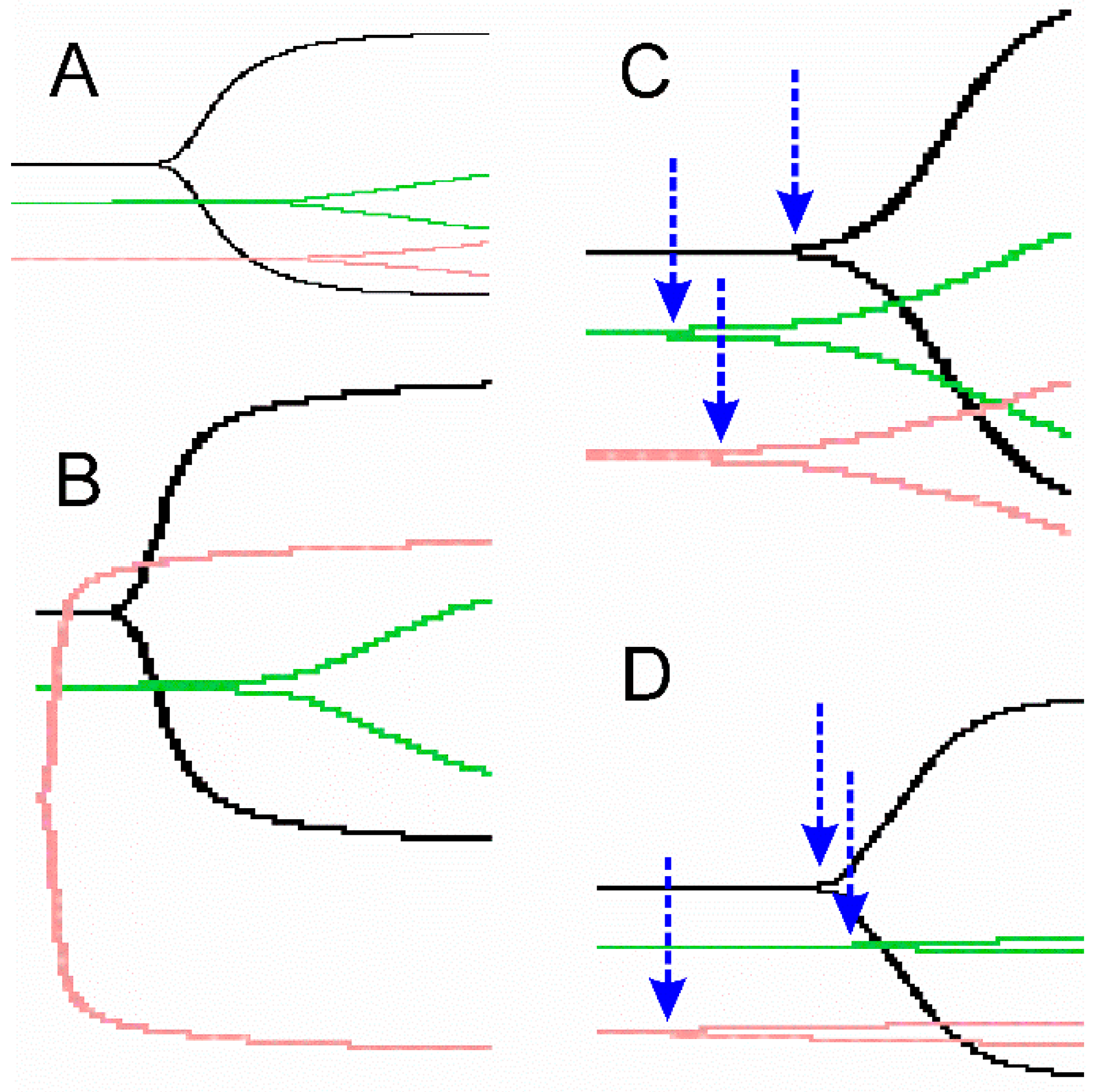
Procoagulant venoms with
thrombin-like activity cause rapid onset of coagulation, but with weak clot strength secondary to lack of engagement of Factor XIII (FXIII) [
7,
11,
31]. In contrast, venoms with what we term
thrombin-generating activity form thrombin from prothrombin and engage FXIII, which is critical for the development of normal clot strength [
33]. It is also important to realize that this kinetic approach has the important limitation of providing primarily functional data that do not discern the enzymatic composition of the proteome of any particular venom. As an example, if a venom is found to be procoagulant with thrombin-generating activity, the enzyme(s) responsible may be metalloproteinases, serine proteases, or another class of enzyme; thus, it may be possible to have two different venoms with identical thrombelastographic coagulation kinetic profiles generated by different proteomes. Conversely, similar venoms from closely related species may have subtle or significant coagulation kinetic profile differences that may potentially be explained by analyses of their venom proteomes. In summary, the present work proposed to use this functional, kinetomic approach to further define the similarities and diversity of Pan-American pit viper venoms.
Taking the aforementioned into consideration, the purposes of the present investigation were as follows: (1) thrombelastographically define in human plasma the effects on coagulation kinetics of these various Pan-American snake hemotoxic venom activities; (2) determine if CO and/or PHA directly inhibits these hemotoxic venom effects; and (3) combine these prospectively obtained data with thrombelastographic data collected with Pan-American venoms recently published [
10,
11,
34,
35,
36] to assess kinetomic patterns based on location.
3. Discussion
The present work has displayed some of the most distinct coagulation kinetic patterns in response to heme-based modulation seen yet, every bit as diverse as the African, Asian, and Australian venoms analyzed in recent works [
3,
4,
5,
6,
7,
8,
9]. These unique (e.g.,
Atropoides olmec) and similar, but different (e.g.,
Bothrops species) coagulation kinetic responses serve as identifying “fingerprints” of these venoms, and we have coined the term “venom kinetomics” to describe these datasets [
8,
9]. Rather than a static paradigm, this approach is intended to find new ways to describe the novel effects of toxins on coagulation, providing a quantitative and qualitative backdrop upon which new discoveries can be interpreted. Simple concepts, such as relative potency (e.g., µg/mL) of any particular venom to inflict its anticoagulant or procoagulant effect, are self-evident; however, the effects of heme modulation are more complex and, in a few cases, difficult to interpret. For example, two species,
Bothriechis schlegelii and
Crotalus organus abyssus, had significant attenuation of their anticoagulant activities after exposure to either CORM-2 or iRM, without any inhibition from PHA exposure. These two venoms are unique, and while there may very well be a heme attached to key enzymes, one cannot prove definitively that this is the case, as there is an equal CO-independent effect with iRM exposure and no effect with metheme formation with PHA. The use of other carbon monoxide releasing molecules (and iRMs) may be needed in such cases to detect heme modulation. There is also great variation in the amount of CO required to inhibit enzyme activity, with up to 700 µM required to inhibit
Bothrops moojeni and
Crotalus simus tzabcan venoms based on the amount of CO released by 1 mM CORM-2 [
39] as compared with all the other venoms inhibited by only 70 µM CO. There was also variation in the degree of CO mediated inhibition of procoagulant activity between the other three
Bothrops species venoms tested, and between the two
Bothrops jararaca venoms, which is likely secondary to their two distinct proteomes [
25]. Specifically,
B. jararaca S venom had 10.5% metalloproteinase and 28.6% serine protease, whereas
B. jararaca SE had 35.6% metalloproteinase and 13.7% serine protease in their proteomes [
25].
B. jararaca S venom had more robust activation of coagulation, less CO mediated inhibition of activity, and more PHA mediated inhibition of activity than
B. jararaca SE venom (
Table 4). Importantly, both metalloproteinase and serine protease enzymes have been demonstrated to have thrombin-generating activity [
25], so the variation in proteome almost certainly contributed to the differences in their kinetomic profiles. Lastly, exposure to PHA, a metheme forming agent, resulted in inhibition of venom activity, no effect on venom activity, or potential activation of latent enzymatic activity such as the apparent anticoagulant activity seen with
Agkistrodon bilineatus and
Bothrops colombiensis venoms. In summary, our selection of Pan-American venoms displayed a remarkable set of unique kinetomic patterns, many not previously documented with this thrombelastographic methodology.
Continuing refinements to this thrombelastographic methodology will include devising ways to further isolate the effects of specific enzyme types that may contribute to the overall apparent activity of a particular venom. Specifically, while metalloproteinases and serine proteases are well established fibrinogenolytic anticoagulant enzymes [
2], the impact of other classes of snake enzyme that anticoagulated by completely different mechanisms, such as phospholipase A
2 (PLA
2), have not been extensively quantitated by thrombelastography. Just recently, this laboratory characterized the kinetomic features of such a PLA
2 purified from
Crotalus adamanteus venom with thrombelastographic methods, and determined that this enzyme was CO-inhibitable [
32]. While of interest, it is important not to generalize this finding to all snake venom derived PLA
2. This finding is critical, given the diverse effects of snake venom PLA
2, which include myotoxicity and neurotoxicity [
2]. Thus, venoms containing metalloproteinases, serine proteases, and PLA
2 that display anticoagulant activity in vitro may exert this effect with one or all three enzyme classes; further, CO-mediated inhibition of anticoagulant activity may be secondary to inhibition of one or more of these classes of enzyme. Further development of differential inhibition of the various enzyme classes found in snake venom that is compatible with assessing coagulation with human plasma will be required in the future to increase mechanistic insight into these matters.
Our use of heme modulating agents to change enzymatic activity is new to this field, but it has a substantial scientific grounding. Heme has been found to be attached as an assumed post-translational modification of enzymes on a myriad of proteins [
38], and CO binds primarily to transition metals such as the Fe
+2 found in the center of the heme porphyrin ring [
40]. In the absence of an attached heme group, it would not be expected that CO would react with other portions of snake venom proteins as it is not a radical [
41], and if not bound to a heme attached to these proteins, then CO would be expected to rapidly diffuse away into the plasma sample. With regard to PHA, this compound is the fastest metheme forming agent available [
42], converting the central iron group of heme to an Fe
+3 state. The approach to identify fibrinogen as a heme modulated molecule included the use of CORM-2, PHA, and mass spectroscopy to explain the kinetic behaviors of fibrinogen in a thrombelastographic investigation [
43]. The present investigation involved isolated exposure of venom mixtures with carboxyheme and metheme forming agents at concentrations that, when diluted 100-fold, would not be expected to directly affect plasmatic coagulation, based on previous literature [
38]. This was verified with the experiments found in
Section 2.5 of Results. In further support of a post translationally bound heme group modulating snake venom enzymes, the kinetic modeling of inhibition of fibrinogenolytic activity of crude
Crotalus atrox venom [
6] and the inhibition of isolated
Crotalus adamanteus venom derived snake venom anticoagulant activity [
32] demonstrated a sigmoidal pattern, which is characteristic of heme-based protein modulation. As a practical matter, while the most direct scientific approach, it would be technically time-consuming and likely very expensive to isolate every protein of every venom individually and assess if heme is attached with mass spectroscopy or other heme detecting methods. It may also not be useful from a clinical or scientific standpoint, as one might need to assess dynamically with CO or PHA via thrombelastography if the predominant activity was modulated to address a given coagulopathy or hypothesis. Therefore, we have taken the approach that if isolated venom activity is affected by heme modulating agents, then there is very likely a heme present as the agents involved are not reacting with the enzyme directly, and the heme modulating agents will be so diluted in the plasma sample that no direct effect by them will be observed thrombelastographically.
While outside the scope of the present work, there has been concern expressed by reviewers over the years about the application of CO locally resulting in systemic CO poisoning. To allay these fears, we offer the following scenario for the readership to consider. If a viper inflicts a bite and releases several milliliters of venom into the muscle of a lower extremity, it dissects through the tissue planes and commences toxic effects. Then, within a few minutes, let it be assumed that some combination of rapid and slow CO releasing compounds with a vasoconstrictor in solution is injected near and within the bite site, resulting in as much as 100 mL of tissue to be exposed to up to 1000 µM CO as a pulse. Then, let it be assumed that this injection continues to release up to 100 µM CO per 100 mL of tissue for up to 4 h. The hope would be that the resident venom enzymes would have heme mediated inhibition, with CO bound to heme until a time occurred when either the enzyme was released into the circulation or the CO was released from the heme group. With regard to CO poisoning, if the entire initial burst of CO occurred within the systemic circulation of a 70 kg human being (estimated blood volume approximately 5 L), then the 100,000 µM of CO from the initial injection would be reduced to a concentration of 20 µM within the central circulation. To complete the analogy, a continuous release over 4 h of 10,000 µM of CO from the bite site would add 2 µM of CO per hour for a total of 8 µM of CO. To put these concentrations of CO into perspective, an addition of CO for a final concentration of 100 µM in whole human blood increased carboxyhemoglobin by only 0.6% [
44]; further, smoking two tobacco cigarettes results in an increase in carboxyhemoglobin concentration of 5% [
45]. Summated together, the data from the preceding scenario and associated literature demonstrate that the injection of a CORM-based solution to a snake bite would have as much danger of inflicting systemic CO poisoning as smoking one tobacco cigarette.
Another misconception that can be raised when investigating crude venoms is that they may contain iron as trace compounds, such as in hemoglobin or free iron from milking a snake, which may bind CO released from CORM-2 or any other CORM used in experimentation. Biochemically, this is not the case. First, free iron in aerobic solution is rapidly oxidized to the Fe+3 state, which does not bind with CO. Second, free iron would rapidly react with whatever anion was near it, and would not function as a modulatory entity of any snake venom enzyme. Similarly, free hemoglobin released into the venom from the trauma of milking the snake would be very small in concentration and would not be expected to somehow rapidly bind to the key enzymes without some sort of intermediary step, such as that associated with post-translational modification during enzyme synthesis within the venom gland cells. Taken as a whole, the use of carboxyheme and metheme forming agents to investigate crude venom is reasonable and not subject to the aforementioned misconception.
In conclusion, the present investigation characterized fourteen Pan-American viper venoms with a novel kinetomic method utilizing thrombelastography. This method has been recently used to characterize venoms derived from African, Asian, and Australian snakes [
3,
4,
5,
6,
7,
8,
9]. Using this paradigm, venoms are classified as anticoagulant (fibrinogenolytic or phospholipid digesting) or procoagulant (thrombin-generating, thrombin-like activity), and carboxyheme or metheme responsive [
3,
4,
5,
6,
7,
8,
9]. The ongoing determination of vulnerability of venoms to heme modulation is of interest, as it has recently been demonstrated that exposure of
Crotalus atrox venom to CO markedly inhibited its hemotoxic activity in vivo in a rabbit model [
46]. It may be possible that local application of CO via releasing molecules may serve as an adjunct therapy prior to antivenom administration. Finally, it is anticipated that our approach will assist in identifying clinically relevant enzymatic activity and allows assessment of the potential efficacy of antivenoms or heme modulating agents to treat envenomation by hemotoxic venoms.


