Article

The hokW-sokW Locus Encodes a Type I Toxin–Antitoxin System That Facilitates the Release of Lysogenic Sp5 Phage in Enterohemorrhagic Escherichia coli O157

Department of Biochemistry and Molecular Biology, Graduate School of Science and Engineering, Saitama University, 255 Shimo-Okubo, Sakura-Ku, Saitama 338-8570, Japan
*
Author to whom correspondence should be addressed.
Toxins 2021, 13(11), 796; https://doi.org/10.3390/toxins13110796
Submission received: 7 October 2021 / Revised: 5 November 2021 / Accepted: 9 November 2021 / Published: 11 November 2021
(This article belongs to the Section Bacterial Toxins)

Abstract

:
The toxin-antitoxin (TA) genetic modules control various bacterial events, such as plasmid maintenance, persister cell formation, and phage defense. They also exist in mobile genetic elements, including prophages; however, their physiological roles remain poorly understood. Here, we demonstrate that hokW-sokW, a putative TA locus encoded in Sakai prophage 5 (Sp5) in enterohemorrhagic Escherichia coli O157: H7 Sakai strain, functions as a type I TA system. Bacterial growth assays showed that the antitoxic activity of sokW RNA against HokW toxin partially requires an endoribonuclease, RNase III, and an RNA chaperone, Hfq. We also demonstrated that hokW-sokW assists Sp5-mediated lysis of E. coli cells when prophage induction is promoted by the DNA-damaging agent mitomycin C (MMC). We found that MMC treatment diminished sokW RNA and increased both the expression level and inner membrane localization of HokW in a RecA-dependent manner. Remarkably, the number of released Sp5 phages decreased by half in the absence of hokW-sokW. These results suggest that hokW-sokW plays a novel role as a TA system that facilitates the release of Sp5 phage progeny through E. coli lysis.
Key Contribution: The hokW-sokW type I toxin–antitoxin module encoded in the functional Sp5 prophage of Escherichia coli O157 facilitates the release of Sp5 phage progeny, which is a new physiological role of toxin–antitoxin systems.

1. Introduction

The toxin-antitoxin (TA) system is a genetic module composed of a toxin and an antitoxin. Genes encoding these elements are generally contiguous and have been discovered in almost all sequenced bacterial genomes [1]. While a toxin arrests cell growth, the antitoxin neutralizes its toxicity [2]. TA systems have been implicated in a wide range of bacterial events, such as plasmid maintenance [3], persister cell formation [4,5,6], and phage defense [7,8,9,10].
The first TA locus was discovered in the E. coli plasmid in 1983 [11,12]. Since then, TA loci have been found not only in plasmids but also on bacterial chromosomes [13,14,15]. TA loci are also closely linked to prophage genomes. For example, ~8% of the total TA loci have been overrepresented in the nine cryptic prophages of E. coli K-12 [16]. Cryptic prophages harbor mutations in genes required for virulence and phage formation, which render them trapped in the host genome. Although TA loci are also found in the genomes of functional prophages [17,18,19], the impact of their TA systems on the propagation of lysogenic phages remains poorly understood.
TA systems are currently classified into seven types according to the nature and function of antitoxins [20]. Type I antitoxins are small non-coding RNAs that bind to the cognate toxin mRNAs to inhibit translation or promote degradation [21,22]. The hok-sok locus encodes a type I TA system found in various plasmids and bacterial genomes. Hok (host killing) is a hydrophobic peptide toxin that depolarizes the plasma membrane and causes cell growth retardation and lysis [23,24]. sok (suppression of killing) is a small non-coding RNA that inhibits translation of mok (modulation of killing) mRNA that overlaps with hok mRNA. The base-pair RNAs formed between sok RNA and mok mRNA are degraded by RNase III that specifically cuts double-stranded RNA [25]. Since translation of hok and mok mRNAs is coupled, sok RNA indirectly inhibits the translation of hok mRNA [3]. While this TA system present in the plasmid has been implicated in plasmid maintenance [26] and phage defense [27], the roles of hok-sok loci encoded in prophage genomes remain unclear.
In this study, we aimed to clarify the role of a hok-sok locus encoded in a prophage of the enterohemorrhagic E. coli O157: H7 Sakai strain. Bacterial growth and survival assays and phage plaque formation experiments have provided evidence that supports a new role of the hok-sok TA system which facilitates the release of Sp5 phage progeny through E. coli lysis.

2. Results

2.1. hokW-sokW Functions as a TA System

E. coli O157: H7 Sakai strain has 18 prophages, three of which can infect non-pathogenic E. coli K-12 strains [28]. Among them, the lambdoid Sp5 (Sakai prophage 5) phage produces a major virulence factor called Shiga toxin 2. The genome of Sp5 phage encodes a gene pair named “hokW-sokW” that is predicted to be a member of the hok-sok family of type I TA system (Figure 1A) [29]. HokW (51 amino acids) shares 44% identity and 69% similarity with Hok (52 amino acids) in E. coli R1 plasmid (Figure 1B). We first examined whether hokW-sokW encoded in the Sp5 phage functions as a TA system. We constructed a plasmid pBAD24-hokW, in which a hokW is cloned under the control of an arabinose-inducible promoter. When HokW was expressed by arabinose in E. coli K-12 TY0807 cells, cell growth was immediately retarded (Figure 1C). Resumption of cell growth around 4 h after arabinose addition is probably due to the depletion of arabinose. Next, we tested whether sokW RNA suppressed the toxic activity of HokW. We cloned hokW-sokW in an arabinose-inducible plasmid, pBAD18-hokW-sokW, in which hokW mRNA is expressed from an arabinose-inducible promoter and sokW RNA from its own promoter. When TY0807 cells harboring this plasmid were treated with arabinose, no obvious growth retardation was observed (Figure 1D). To confirm the inhibitory effect of sokW RNA against HokW-mediated toxicity, we introduced mutations in the promoter sequence of sokW to repress its transcription (Figure 1E; pBAD18-hokW-sokW(pro-less)). TY0807 cells harboring pBAD18-hokW-sokW(pro-less) exhibited growth retardation for approximately 3 h after arabinose addition (Figure 1D). Importantly, the growth retardation observed in cells harboring pBAD18-hokW-sokW(pro-less) was partially restored by expressing sokW RNA using another plasmid, pACYC184-sokW (Figure 1F). These results strongly suggest that sokW RNA counteracts the toxic activity of HokW. Taken together, we concluded that HokW is a toxin and that sokW RNA functions as an antitoxin against HokW.

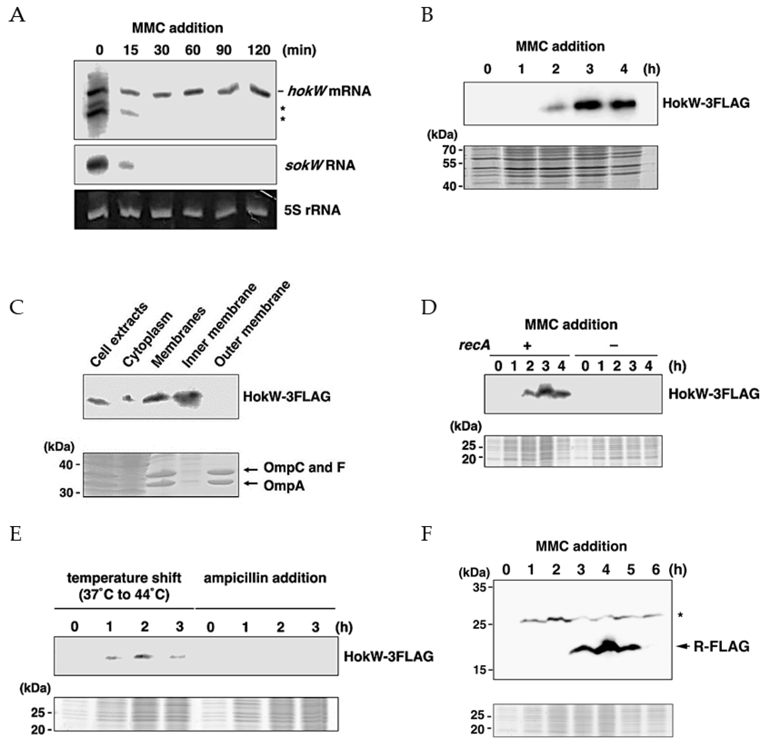
2.2. hokW-sokW Is a RNase III and Hfq Dependent Type I TA System

sokW RNA has a 71-nt complementary sequence to the 5′-UTR of hokW mRNA (Figure 1A). If sokW RNA forms a base pair in this region to repress hokW translation, it is likely that this process is RNase III-dependent [25]. To test this hypothesis, we performed cell growth assays with and without RNase III. While wild-type cells harboring pBAD18-hokW-sokW grew normally after arabinose addition, RNase III-deficient cells, ME5413, harboring the same plasmid exhibited growth retardation around 2 h after arabinose addition (Figure 2A). This result suggests the involvement of RNase III. RNase III-mediated double-strand RNA degradation is likely to play a crucial role in the antitoxic activity of sokW RNA. We next examined whether the antitoxic activity of sokW RNA requires Hfq, an RNA chaperone that facilitates base-pairing between the two RNAs [30]. When HokW expression was induced in E. coli cells lacking Hfq, cell growth was partially retarded despite the presence of sokW (Figure 2B). This result suggests that Hfq is involved in the antitoxic activity of sokW RNA and probably help base-pairing between sokW RNA and hokW mRNA. Together, these results suggest that hokW-sokW is a type I TA system whose antitoxin activity requires both RNase III and the Hfq chaperone.

2.3. hokW-sokW Assists Growth Retardation and Reduces Cell Viability after Mitomycin C Treatment

hokW-sokW is encoded in the lysogenic Sp5 prophage of the E. coli O157: H7 Sakai strain. To explore the potential role of hokW-sokW in Sp5-mediated cell growth retardation, we compared cell growth and viability after Sp5 prophage induction with or without this type I TA system. For the experiment, E. coli K-12 MG1655 cells with Sp5 prophage were used. We first confirmed that deletion of hokW-sokW did not affect cell growth under normal conditions (Figure 3A). The Sp5 prophage was induced in MG1655 cells using mitomycin C (MMC), a DNA-damaging agent that crosslinks two DNA bases and triggers prophage induction [31]. MMC is one of the most commonly used antibiotics for prophage induction. While MMC had no obvious effect on MG1655 cell growth, Sp5-containing cells (MG1655-Sp5) stopped growing around 2 h after MMC treatment and started to lyse around 3.5 h (Figure 3B). Remarkably, growth retardation started approximately 40 min later in the absence of hokW-sokW (MG1655-Sp5(∆hokW-sokW)), indicating that this TA system facilitates Sp5-mediated growth retardation. To assess whether hokW-sokW also affects cell survival, we measured the number of viable cells at different time points after MMC treatment. At 30 min after the treatment, the cell viability of MG1655-Sp5 was comparable to that of MG1655-Sp5(∆hokW-sokW). However, the viability of MG1655-Sp5 dropped to about 55% of that of MG1655-Sp5(∆hokW-sokW) for the cells harvested 120 min (Figure 3C). These results indicate that hokW-sokW not only facilitates growth retardation but also reduces cell survival.
HokW toxin is predicted to be a short peptide that destabilizes the plasma membrane. To examine whether hokW-sokW-mediated growth retardation is dependent on the lytic enzymes encoded in the Sp5 phage, we created a prophage that lacks both the R endolysin and the S holin (MG1655-Sp5(∆r-s)). The growth retardation of MG1655-Sp5(∆r-s) started approximately 3 h after MMC treatment, likely due to the production of Sp5 phages in the cells (Figure 3D). Notably, the retardation of cell growth was less severe in the absence of hokW-sokW (MG1655-Sp5(∆hokW-sokWr-s)). These results strongly suggest that hokW-sokW promotes growth retardation and possibly cell lysis independent of the lytic enzymes encoded in the Sp5 phage.

2.4. MMC Triggers HokW Toxin Production in a RecA Dependent Manner

These results suggest that MMC treatment induces HokW toxin expression. To examine this possibility, we first compared the amounts of hokW mRNA and sokW RNA before and after MMC treatment. As shown in Figure 4A, both hokW mRNA and sokW RNA were detected before MMC treatment, indicating that MMC does not promote hokW transcription. While hokW mRNA remained stable after MMC treatment, sokW RNA rapidly disappeared. These results suggest that HokW is produced after MMC treatment. Next, we measured the amount of HokW toxin using western blotting. Production of a FLAG-tagged HokW (HokW-3FLAG) started 2 h after MMC treatment and plateaued around 3 h (Figure 4B). Fractionated bacterial samples showed that HokW-3FLAG was enriched in the inner membrane, as seen with other members of the Hok family toxins (Figure 4C) [23]. These results suggest that MMC treatment promotes HokW production and localization to the inner membrane through the disappearance of sokW RNA.
It is well known that treating lysogenic E. coli with a DNA-damaging agent such as MMC activates RecA, which stimulates the self-cleavage of the CI repressor to initiate prophage induction [32]. RecA is a single-stranded DNA-binding protein that is involved in DNA recombination and repair [33]. To examine whether HokW production triggered by MMC requires RecA, we constructed a deletion construct of recA in MG1655-Sp5(hokW-3FLAG) and measured the level of HokW (Figure 4D). Western blotting revealed that HokW-3FLAG was not produced in ∆recA cells even 4 h after MMC treatment. This indicates that HokW production is RecA-dependent. HokW-3FLAG production was also observed when cells were shifted from 37 °C to 44 °C to trigger prophage induction (Figure 4E). Notably, HokW-3FLAG was not produced when cells were treated with ampicillin that does not trigger prophage induction. These results strongly suggest that RecA-mediated prophage induction causes HokW production.
Finally, we examined the expression timing of HokW and the R endolysin after MMC treatment. Western blotting showed that HokW production started approximately 1 h earlier than the R (Figure 4B,F). This result suggests that HokW-mediated growth retardation and possibly cell lysis precedes cell lysis mediated by lytic enzymes, the R endolysin and the S holin, of Sp5 phage.

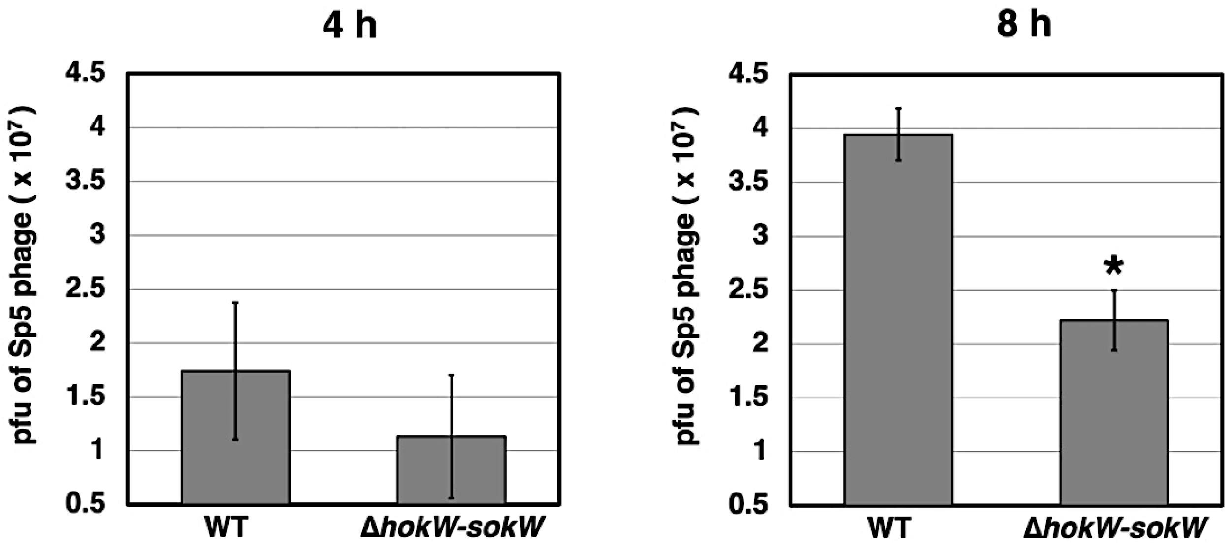
2.5. hokW-sokW Facilitates the Release of Sp5 Phage Progeny
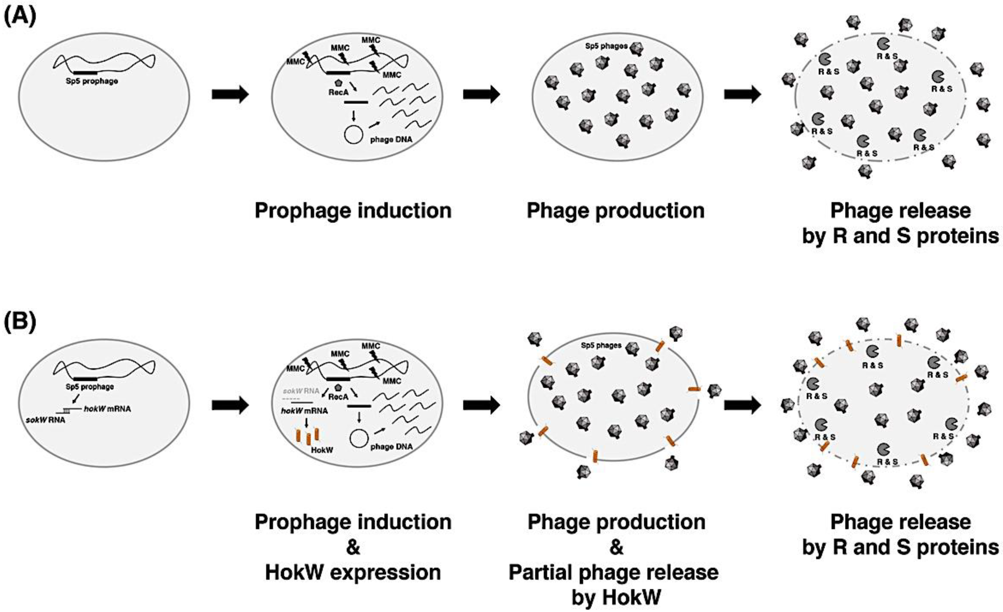
Finally, we assessed whether the hokW-sokW TA system plays a role in phage propagation. We measured the number of Sp5 phage progeny released from MG1655 cells. Compared to wild-type, Sp5 phage progenies released from ∆hokW-sokW cells at 8 h after MMC treatment were reduced by around half (Figure 5). This suggests that hokW-sokW facilitates the release of Sp5 phage progeny.

3. Discussion

TA loci are abundant in bacterial genomes and mobile genetic elements and are involved in various bacterial events, such as plasmid maintenance, persister cell formation, and phage defense. Here, we addressed the physiological role of a putative TA module encoded in the genome of a functional prophage in the E. coli O157: H7 Sakai strain.
We first demonstrated that hokW-sokW, encoded in the lysogenic Sp5 phage, functions as a type I TA system (Figure 1 and Figure 2). When cells are exposed to extracellular stresses such as phage infection, type II TA genes are transcriptionally repressed, and consequently, labile antitoxins disappear and the toxins are activated [9,10,34]. In the hokW-sokW TA system, sokW RNA disappeared immediately after MMC treatment, and the remaining hokW mRNA was translated (Figure 4A,B). Considering that MMC damages DNA, the disappearance of sokW RNA would be caused by blocking sokW transcription rather than augmenting mRNA degradation activity. MMC-mediated DNA damage results in the activation of RecA and initiation of prophage induction [31,32]. Intriguingly, HokW expression requires RecA (Figure 4D). Thus, prophage induction rather than DNA damage itself is likely to trigger the production of HokW toxin. This idea is also supported by the result of Figure 4E. Prophage induction involves many biological processes, such as excision, cyclization, replication of phage DNA, expression of phage proteins, and assembly of phage particles. Further studies are required to clarify which of these processes lead to the loss of sokW RNA and HokW production.
Previous reports demonstrated that hok mRNA from E. coli R1 plasmid is slowly processed at its 3′-end after the transcription is stopped by rifampicin [3]. This truncated hok RNA is translated more actively than full-length hok mRNA [35]. In Figure 4A, two smaller RNAs than full-length hokW mRNA were detected and rapidly disappeared after MMC addition. Whether these RNAs correspond to the truncated hok RNA from R1 plasmid needs to be investigated. Although RNase III and Hfq were suggested to be required for sokW RNA-mediated translational repression of hokW mRNA, this antitoxic activity was partially effective in the RNase III and Hfq defective constructs (Figure 2). Therefore, RNase III and Hfq are unlikely to play a major role in the sokW RNA-mediated translational repression. In line with this idea, neither RNase III nor Hfq mutants are lethal. Presumably, there is another mechanism for repressing HokW expression besides the cleavage of hokW mRNA by RNase III. And the base-pairing between hokW mRNA and sokW RNA would naturally occur in the absence of Hfq, albeit inefficiently.
The type I TA toxins are classified into two types: membrane-associated toxins and cytosolic toxins [36]. The membrane-associated toxins are small hydrophobic peptides that localize in the inner membrane, which affects the plasma membrane integrity and potential, and consequently leads to growth retardation and cell lysis [37,38]. Similarly, HokW are hydrophobic peptides (Figure 1B) and are localized in the inner membrane (Figure 4C). As expected, endogenous HokW production triggered by MMC facilitated growth retardation (Figure 3B,D) and decreased cell viability (Figure 3C). These results suggested that cell lysis would begin before the production of phage progeny is completed and that the number of phage progenies released would be decreased. However, contrary to this prediction, cells harboring hokW-sokW released phage progenies approximately 2-fold compared to the ∆hokW-sokW cells (Figure 5). Based on these results, we propose a working hypothesis in which hokW-sokW plays a positive role in Sp5 phage propagation. If the Sp5 prophage does not encode the hokW-sokW TA locus, MMC treatment triggers the initiation of Sp5 prophage induction through RecA activation. Lysis enzymes are expressed at the late stage after induction, which results in cell lysis and the release of phage progeny (Figure 6A). If the Sp5 prophage encodes hokW-sokW, MMC treatment triggers not only Sp5 prophage induction but also HokW production. HokW localizes to the inner membrane and partially causes cell lysis before lysis enzymes are fully expressed. Consequently, the release of Sp5 phage progeny starts earlier than usual (Figure 6B).
The RnlA-RnlB TA system present in the cryptic CP4-57 prophage of the E. coli K-12 strain inhibits the propagation of lytic bacteriophage T4 [9]. Other TA systems (antiQ-AbiQ, ToxIN, MazE-MazF, etc.) have also been reported to function in defense against lytic phages [7,8,34,39]. Therefore, TA loci in the bacterial genome are believed to function as a mechanism that inhibits phage propagation. However, this study demonstrates that a TA system encoded in a lysogenic prophage has a positive effect on phage propagation, which is a new physiological role of TA systems. To date, the hok-sok TA system present in the R1 plasmid has been reported to inhibit T4 phage propagation [27]. In E. coli K-12 strain harboring a high-copy plasmid containing hok-sok, the efficiency of plating of T4 phage was reduced by 42% and plaque size was decreased by ~85%. The molecular mechanism of phage inhibition by this hok-sok TA system remains unclear. Interestingly, TA systems belonging to the same family play opposite physiological roles in phage propagation depending on the type of mobile genetic element where the TA loci are located.

4. Materials and Methods

4.1. E. coli Strains

The E. coli strains and the primers (eurofins, Tokyo, Japan) used in this study are listed in Table 1 and Table 2. All E. coli strains used in this study belong to non-pathogenic E. coli K-12 strain. MG1655, MG1655-Sp5, and MG1655-Sp5(Kmr) were kindly gifted by Prof. Sekine at Rikkyo University [40]. MG1655-Sp5 and its derivatives do not produce Shiga toxin 2 because of the deletion of stx2A-stx2B. MG1655-Sp5(∆hokW-sokW::kmr), MG1655-Sp5(∆r-s::cat), and MG1655-Sp5(∆hokW-sokW::kmrr-s::cat) were constructed as described previously [41]. Briefly, a fragment containing a kanamycin-resistant or a chloramphenicol-resistant cassette flanked with the sequences upstream and downstream of the deleted genes was amplified by polymerase chain reaction (PCR) with pKD4 or pKD3 as a template. Primers YO-351 and YO-352 for hokW-sokW::kmr, or YO-635 and YO-636 for r-s::cat were used. The amplified fragment was inserted into MG1655-Sp5 or MG1655-Sp5(∆hokW-sokW::kmr) harboring pKD46 encoding λ phage Red recombinase, and kanamycin-resistant or chloramphenicol-resistant colonies were screened by PCR with primers YO-349 and YO-350, or YO-637 and YO-638. MG1655-Sp5(hokW-3FLAG-kmr) and MG1655-Sp5(r-FLAG-cat) were constructed as previously described [42]. Briefly, a fragment containing a kanamycin-resistant or a chloramphenicol-resistant cassette was amplified by PCR with the pSUP11 template and primers YO-560 and YO-561 for hokW-3FLAG-kmr or the pSU313 template and primers YO-644 and YO-657 for r-FLAG-cat. The amplified fragments were inserted into MG1655-Sp5 harboring pKD46 and kanamycin-resistant or chloramphenicol-resistant colonies were screened by PCR with primers YO-349 and YO-350, or YO-637 and YO-638. To eliminate the kanamycin-resistant cassette, MG1655-Sp5(hokW-3FLAG-kmr) was transformed with pCP20 that causes temperature-sensitive replication and thermal induction of FLP synthesis. MG1655-Sp5(hokW-3FLAG) was selected by PCR using primers YO-349 and YO-350. TY0807 ∆hfq::kmr and MG1655 ∆recA::kmr-Sp5(hokW-3FLAG) were constructed by GT7 phage-mediated transduction of a kanamycin-resistance cassette from JW4130 and JW2669, respectively [43].

4.2. Plasmid Construction

To generate pBAD24-hokW or pBAD18-hokW-sokW, a DNA fragment containing hokW or hokW-sokW was amplified by PCR using MG1655-Sp5 DNA as a template and primers YO-357 and YO-369, or YO-432 and YO-433, digested with EcoRI (NIPPON GENE, Tokyo, Japan) and PstI (NIPPON GENE, Tokyo, Japan) or EcoRI and HindIII (NIPPON GENE, Tokyo, Japan), and ligated into the corresponding sites of pBAD24 or pBAD18 [44]. The promotor mutant construct, pBAD18-hokW-sokW(pro-less), was created using the KOD-Plus-Mutagenesis kit (TOYOBO, Osaka, Japan) with primers YO-421 and YO-422 and pBAD18-hokW-sokW as a template. To construct pACYC184-sokW, a DNA fragment containing sokW was amplified by PCR using MG1655-Sp5 DNA as a template and primers YO-406 and YO-541, digested with BamHI (NIPPON GENE, Tokyo, Japan) and HindIII, and ligated into the corresponding sites of pACYC184. DNA sequences of the constructed plasmids were confirmed by sequencing.

4.3. E. coli Growth and CFU Assay

E. coli cells were grown in Luria–Bertani (LB) broth or LB broth supplemented with ampicillin (nacalai tesque, Kyoto, Japan) or chloramphenicol (nacalai tesque, Kyoto, Japan) at 37 °C. When the optical density at 660 nm (OD660) reached 0.3–0.5, 0.2% L-arabinose (L-ara) (FUJIFILM Wako Pure Chemical, Osaka, Japan) was added to express the protein from the pBAD plasmids or 2.0 µg/mL mitomycin C (MMC) (FUJIFILM Wako Pure Chemical, Osaka, Japan) was added to induce Sp5 prophage. Cell densities were monitored every 20 min using a biophotorecorder (ADVANTEC TVS062CA, Tokyo, Japan). At least triplicate measurements were performed, and similar results were obtained for each measurement. A representative result is shown in each figure. In colony-forming unit (CFU) assay, E. coli cells were grown in LB broth at 37 °C until the OD660 reached 0.4, and harvested at 0, 30, 60, 90, and 120 min after 2.0 µg/mL MMC addition. Cells were diluted with phosphate-buffered saline (PBS) (nacalai tesque, Kyoto, Japan), plated onto LB agar plates, and incubated at 37 °C overnight. The colonies that emerged were counted and the number of viable cells in 1 mL culture medium was calculated.

4.4. Northern Blotting Analysis

MG1655-Sp5 cells were grown in LB broth at 37 °C until the OD660 reached 0.4, and harvested at 0, 15, 30, 60, 90, and 120 min after 2.0 µg/mL MMC addition. Total RNA was isolated and purified as previously described [45]. Total RNA (2.0 µg) was electrophoresed on a 6% polyacrylamide gel (FUJIFILM Wako Pure Chemical, Osaka, Japan) containing 7 M urea (FUJIFILM Wako Pure Chemical, Osaka, Japan), followed by northern blotting. The digoxigenin (Dig)-labeled oligo-probe YO-597 or YO-592 (eurofins, Tokyo, Japan) was used for hokW mRNA or sokW RNA. After hybridization, the membranes were probed with anti-digoxigenin-AP Fab fragments (Roche, Basel, Switzerland), and RNAs were detected using CDP-Star chemiluminescent substrate (Roche, Basel, Switzerland) and the C-DiGit Blot Scanner (LI-COR, Lincoln, NE, USA).

4.5. Fractionation of Cell Extracts and Western Blotting Analysis

E. coli cells were grown in LB broth at 37 °C until the OD660 reached 0.4, harvested at appropriate times after 2.0 µg/mL MMC addition, and resuspended in PBS. For fractionation, MG1655-Sp5(hokW-3FLAG-kmr) cells were grown in 20 mL LB broth supplemented with kanamycin (nacalai tesque, Kyoto, Japan) at 37 °C and harvested at 2 h after 2.0 µg/mL MMC addition. Cells were resuspended in 0.8 mL PBS and lysed by sonication. After cell debris was removed, the resulting supernatant (0.7 mL) was centrifuged at 20,000× g for 45 min at 4 °C to separate the cytoplasmic and membrane fractions. To separate the inner and outer membranes, the pellet containing membrane fraction was resuspended in 0.35 mL PBS containing 0.4% Sarcosyl (nacalai tesque, Kyoto, Japan) and incubated for 30 min at room temperature [46]. This mixture was centrifuged at 20,000× g for 30 min at 4 °C, and the supernatant was used as the inner membrane fraction. The pellet was resuspended in 0.35 mL PBS and used as the outer membrane fraction. Proteins were separated on 12.5% or 18% polyacrylamide gels (FUJIFILM Wako Pure Chemical, Osaka, Japan) containing sodium dodecyl sulfate (FUJIFILM Wako Pure Chemical, Osaka, Japan) for western blotting and Coomassie Brilliant Blue (CBB) (nacalai tesque, Kyoto, Japan) staining. For detection of FLAG-tagged proteins, proteins were electroblotted onto an Amersham Hybond P PVDF 0.2 membrane (Cytiva, Tokyo, Japan), probed with a mouse anti-FLAG M2 monoclonal antibody (Sigma-Aldrich, St. Louis, MO, USA) and with a horseradish peroxidase-conjugated sheep anti-mouse IgG (GE Healthcare, Chicago, IL, USA). Chemi-Lumi One Ultra (nacalai tesque, Kyoto, Japan) or Amersham ECL Prime (GE Healthcare, Chicago, IL, USA) were used as substrates for detection with the C-DiGit Blot Scanner (LI-COR).

4.6. Plaque Formation Assay of Sp5 Phages

MG1655-Sp5(kmr) or MG1655-Sp5(∆hokW-sokW) cells were grown in LB broth supplemented with kanamycin at 37 °C until the OD660 reached 0.4 and treated with 2.0 µg/mL MMC for 4 or 8 h. Cell cultures were centrifuged at 8000× g for 3 min and the supernatant was used for plaque formation assay. The phage solution was diluted and mixed with MG1655 as an indicator cell in soft agar containing LB broth, 0.3% agar (FUJIFILM Wako Pure Chemical, Osaka, Japan), 1.5 µg/mL MMC and 10 mM CaCl2 (FUJIFILM Wako Pure Chemical, Osaka, Japan). The mixture was poured onto LB-agar plate and incubated at 37 °C overnight. The plaques that emerged on the plate were counted and the number of phages in 1 mL phage solution was calculated for the plaque-forming units (pfu).

Author Contributions

Conceptualization, Y.O.; Investigation, K.T., K.H., T.S. and Y.O.; Writing–original draft preparation, Y.O., Funding acquisition, Y.O. All authors have read and agreed to the published version of the manuscript.

Funding

This work was supported by JSPS KAKENHI Grant Number JP20K07493.

Institutional Review Board Statement

Not applicable.

Informed Consent Statement

Not applicable.

Data Availability Statement

All data for this study are available within the article.

Acknowledgments

We thank Toshimitsu Kawate at Cornell University and Tetsuro Yonesaki at Osaka University for their invaluable help with the experiments and the manuscript.

Conflicts of Interest

The authors declare no conflict of interest.

References

  1. Pandey, D.P. Toxin-antitoxin loci are highly abundant in free-living but lost from host-associated prokaryotes. Nucleic Acids Res. 2005, 33, 966–976. [Google Scholar] [CrossRef]
  2. Harms, A.; Brodersen, D.E.; Mitarai, N.; Gerdes, K. Toxins, targets, and triggers: An overview of toxin-antitoxin biology. Mol. Cell 2018, 70, 768–784. [Google Scholar] [CrossRef] [Green Version]
  3. Thisted, T.; Gerdes, K. Mechanism of post-segregational killing by the hok/sok system of plasmid R1: Sok antisense RNA regulates hok gene expression indirectly through the overlapping mok gene. J. Mol. Biol. 1992, 223, 41–54. [Google Scholar] [CrossRef]
  4. Germain, E.; Castro-Roa, D.; Zenkin, N.; Gerdes, K. Molecular mechanism of bacterial persistence by HipA. Mol. Cell 2013, 52, 248–254. [Google Scholar] [CrossRef]
  5. Harms, A.; Maisonneuve, E.; Gerdes, K. Mechanisms of bacterial persistence during stress and antibiotic exposure. Science 2016, 354, aaf4268. [Google Scholar] [CrossRef]
  6. Page, R.; Peti, W. Toxin-antitoxin systems in bacterial growth arrest and persistence. Nat. Chem. Biol. 2016, 12, 208–214. [Google Scholar] [CrossRef]
  7. Hazan, R.; Engelberg-Kulka, H. Escherichia coli mazEF-mediated cell death as a defense mechanism that inhibits the spread of phage P1. Mol. Genet. Genom. 2004, 272, 227–234. [Google Scholar] [CrossRef] [PubMed]
  8. Fineran, P.; Blower, T.R.; Foulds, I.J.; Humphreys, D.P.; Lilley, K.S.; Salmond, G.P.C. The phage abortive infection system, ToxIN, functions as a protein–RNA toxin–antitoxin pair. Proc. Natl. Acad. Sci. USA 2009, 106, 894–899. [Google Scholar] [CrossRef] [PubMed] [Green Version]
  9. Koga, M.; Otsuka, Y.; Lemire, S.; Yonesaki, T. Escherichia coli rnlA and rnlB Compose a Novel Toxin–Antitoxin System. Genetics 2011, 187, 123–130. [Google Scholar] [CrossRef] [PubMed] [Green Version]
  10. Guegler, C.K.; Laub, M.T. Shutoff of host transcription triggers a toxin-antitoxin system to cleave phage RNA and abort infection. Mol. Cell 2021, 81, 2361–2373.e9. [Google Scholar] [CrossRef]
  11. Karoui, H.; Bex, F.; Drèze, P.; Couturier, M. Ham22, a mini-F mutation which is lethal to host cell and promotes recA-dependent induction of lambdoid prophage. EMBO J. 1983, 2, 1863–1868. [Google Scholar] [CrossRef]
  12. Ogura, T.; Hiraga, S. Mini-F plasmid genes that couple host cell division to plasmid proliferation. Proc. Natl. Acad. Sci. USA 1983, 80, 4784–4788. [Google Scholar] [CrossRef] [Green Version]
  13. Fozo, E.; Makarova, K.S.; Shabalina, S.A.; Yutin, N.; Koonin, E.V.; Storz, G. Abundance of type I toxin–antitoxin systems in bacteria: Searches for new candidates and discovery of novel families. Nucleic Acids Res. 2010, 38, 3743–3759. [Google Scholar] [CrossRef] [PubMed]
  14. Lehnherr, H.; Yarmolinsky, M.B. Addiction protein Phd of plasmid prophage P1 is a substrate of the ClpXP serine protease of Escherichia coli. Proc. Natl. Acad. Sci. USA 1995, 92, 3274–3277. [Google Scholar] [CrossRef] [Green Version]
  15. Goeders, N.; Chai, R.; Chen, B.; Day, A.; Salmond, G.P. Structure, evolution, and functions of bacterial type III toxin-antitoxin systems. Toxins 2016, 8, 282. [Google Scholar] [CrossRef] [PubMed] [Green Version]
  16. Wang, X.; Wood, T.K. Cryptic prophages as targets for drug development. Drug Resist. Updat. 2016, 27, 30–38. [Google Scholar] [CrossRef] [Green Version]
  17. DeShazer, D. Genomic diversity of Burkholderia pseudomallei clinical isolates: Subtractive hybridization reveals a Burkholderia mallei-specific prophage in B. pseudomallei 1026b. J. Bacteriol. 2004, 186, 3938–3950. [Google Scholar] [CrossRef] [PubMed] [Green Version]
  18. Decewicz, P.; Dziewit, L.; Golec, P.; Kozlowska, P.; Bartosik, D.; Radlinska, M. Characterization of the virome of Paracoccus spp. (Alphaproteobacteria) by combined in silico and in vivo approaches. Sci. Rep. 2019, 9, 7899. [Google Scholar] [CrossRef]
  19. Fraikin, N.; Goormaghtigh, F.; Van Melderen, L. type II toxin-antitoxin systems: Evolution and revolutions. J. Bacteriol. 2020, 202, 00763–19. [Google Scholar] [CrossRef] [Green Version]
  20. Wang, X.; Yao, J.; Sun, Y.-C.; Wood, T.K. Type VII toxin/antitoxin classification system for antitoxins that enzymatically neutralize toxins. Trends Microbiol. 2021, 29, 388–393. [Google Scholar] [CrossRef]
  21. Gerdes, K.; Wagner, E.G.H. RNA antitoxins. Curr. Opin. Microbiol. 2007, 10, 117–124. [Google Scholar] [CrossRef]
  22. Fozo, E.M.; Hemm, M.R.; Storz, G. Small toxic proteins and the antisense RNAs that repress them. Microbiol. Mol. Biol. Rev. 2008, 72, 579–589. [Google Scholar] [CrossRef] [Green Version]
  23. Gerdes, K.; Bech, F.; Jørgensen, S.; Løbner-Olesen, A.; Rasmussen, P.; Atlung, T.; Boe, L.; Karlstrom, O.; Molin, S.; von Meyenburg, K. Mechanism of postsegregational killing by the hok gene product of the parB system of plasmid R1 and its homology with the relF gene product of the E. coli relB operon. EMBO J. 1986, 5, 2023–2029. [Google Scholar] [CrossRef] [PubMed]
  24. Pecota, D.C.; Osapay, G.; Selsted, M.E.; Wood, T.K. Antimicrobial properties of the Escherichia coli R1 plasmid host killing peptide. J. Biotechnol. 2003, 100, 1–12. [Google Scholar] [CrossRef]
  25. Gerdes, K.; Nielsen, A.; Thorsted, P.; Wagner, E.H. Mechanism of killer gene activation. Antisense RNA-dependent RNase III cleavage ensures rapid turn-over of the stable Hok, SrnB and PndA effector messenger RNAs. J. Mol. Biol. 1992, 226, 637–649. [Google Scholar] [CrossRef]
  26. Gerdes, K.; Rasmussen, P.B.; Molin, S. Unique type of plasmid maintenance function: Postsegregational killing of plasmid-free cells. Proc. Natl. Acad. Sci. USA 1986, 83, 3116–3120. [Google Scholar] [CrossRef] [PubMed] [Green Version]
  27. Pecota, D.C.; Wood, T.K. Exclusion of T4 phage by the hok/sok killer locus from plasmid R1. J. Bacteriol. 1996, 178, 2044–2050. [Google Scholar] [CrossRef] [Green Version]
  28. Ohnishi, M.; Kurokawa, K.; Hayashi, T. Diversification of Escherichia coli genomes: Are bacteriophages the major contributors? Trends Microbiol. 2001, 9, 481–485. [Google Scholar] [CrossRef]
  29. Ogura, Y.; Mondal, S.I.; Islam, M.S.; Mako, T.; Arisawa, K.; Katsura, K.; Ooka, T.; Gotoh, Y.; Murase, K.; Ohnishi, M.; et al. The Shiga toxin 2 production level in enterohemorrhagic Escherichia coli O157: H7 is correlated with the subtypes of toxin-encoding phage. Sci. Rep. 2015, 5, 16663. [Google Scholar] [CrossRef] [Green Version]
  30. Kavita, K.; De Mets, F.; Gottesman, S. New aspects of RNA-based regulation by Hfq and its partner sRNAs. Curr. Opin. Microbiol. 2018, 42, 53–61. [Google Scholar] [CrossRef]
  31. Fuchs, S.; Mühldorfer, I.; Donohue-Rolfe, A.; Kerényi, M.; Emődy, L.; Alexiev, R.; Nenkov, P.; Hacker, J. Influence of RecA on in vivo virulence and Shiga toxin 2 production in Escherichia coli pathogens. Microb. Pathog. 1999, 27, 13–23. [Google Scholar] [CrossRef] [PubMed]
  32. Ennis, D.G.; Fisher, B.; Edmiston, S.; Mount, D.W. Dual role for Escherichia coli RecA protein in SOS mutagenesis. Proc. Natl. Acad. Sci. USA 1985, 82, 3325–3329. [Google Scholar] [CrossRef] [Green Version]
  33. Bell, J.; Kowalczykowski, S.C. RecA: Regulation and mechanism of a molecular search engine. Trends Biochem. Sci. 2016, 41, 491–507. [Google Scholar] [CrossRef] [Green Version]
  34. Alawneh, A.M.; Qi, D.; Yonesaki, T.; Otsuka, Y. An ADP-ribosyltransferase Alt of bacteriophage T4 negatively regulates the Escherichia coli MazF toxin of a toxin-antitoxin module. Mol. Microbiol. 2015, 99, 188–198. [Google Scholar] [CrossRef] [PubMed]
  35. Franch, T.; Gerdes, K. Programmed cell death in bacteria: Translational repression by mRNA end-pairing. Mol. Microbiol. 1996, 21, 1049–1060. [Google Scholar] [CrossRef] [PubMed]
  36. Brielle, R.; Pinel-Marie, M.-L.; Felden, B. Linking bacterial type I toxins with their actions. Curr. Opin. Microbiol. 2016, 30, 114–121. [Google Scholar] [CrossRef] [PubMed]
  37. Otsuka, Y.; Ishikawa, T.; Takahashi, C.; Masuda, M. A short peptide derived from the ZorO Toxin Functions as an effective antimicrobial. Toxins 2019, 11, 392. [Google Scholar] [CrossRef] [PubMed] [Green Version]
  38. Nonin-Lecomte, S.; Fermon, L.; Felden, B.; Pinel-Marie, M.-L. Bacterial type I toxins: Folding and membrane interactions. Toxins 2021, 13, 490. [Google Scholar] [CrossRef]
  39. Emond, E.; Dion, E.; Walker, S.A.; Vedamuthu, E.R.; Kondo, J.K.; Moineau, S. AbiQ, an abortive infection mechanism from Lactococcus lactis. Appl. Environ. Microbiol. 1998, 64, 4748–4756. [Google Scholar] [CrossRef] [PubMed] [Green Version]
  40. Mitsunaka, S.; Sudo, N.; Sekine, Y. Lysogenisation of Shiga toxin-encoding bacteriophage represses cell motility. J. Gen. Appl. Microbiol. 2018, 64, 34–41. [Google Scholar] [CrossRef] [PubMed] [Green Version]
  41. Datsenko, K.A.; Wanner, B.L. One-step inactivation of chromosomal genes in Escherichia coli K-12 using PCR products. Proc. Natl. Acad. Sci. USA 2000, 97, 6640–6645. [Google Scholar] [CrossRef] [PubMed] [Green Version]
  42. Uzzau, S.; Figueroa-Bossi, N.; Rubino, S.; Bossi, L. Epitope tagging of chromosomal genes in Salmonella. Proc. Natl. Acad. Sci. USA 2001, 98, 15264–15269. [Google Scholar] [CrossRef] [PubMed] [Green Version]
  43. Wilson, G.G.; Young, K.K.Y.; Edlin, G.J.; Konigsberg, W. High-frequency generalised transduction by bacteriophage T4. Nat. Cell Biol. 1979, 280, 80–82. [Google Scholar] [CrossRef] [PubMed]
  44. Guzman, L.M.; Belin, D.; Carson, M.J.; Beckwith, J. Tight regulation, modulation, and high-level expression by vectors containing the arabinose PBAD promoter. J. Bacteriol. 1995, 177, 4121–4130. [Google Scholar] [CrossRef] [PubMed] [Green Version]
  45. Kai, T.; Selick, H.E.; Yonesaki, T. Destabilization of bacteriophage T4 mRNAs by a mutation of gene 61.5. Genetics 1996, 144, 7–14. [Google Scholar] [CrossRef]
  46. Fontaine, F.; Fuchs, R.T.; Storz, G. Membrane localization of small proteins in Escherichia coli. J. Biol. Chem. 2011, 286, 32464–32474. [Google Scholar] [CrossRef] [Green Version]
Figure 1. hokW-sokW functions as a TA system. (A) Genetic organization of hokW-sokW locus is shown. (B) Amino acid alignments of HokW from Sakai prophage 5 (Sp5) and Hok from E. coli R1 plasmid are shown. Red and blue letters indicate identical and similar residues, respectively. (C) TY0807 cells harboring pBAD24 or pBAD24-hokW were treated with or without L-arabinose (L-ara) when the OD660 reached 0.4 (indicated by the arrow) (D) TY0807 cells harboring pBAD18-hokW-sokW or pBAD18-hokW-sokW(pro-less) were treated with or without L-ara when the OD660 reached 0.4–0.5. (E) Nucleotide sequence around the –10 promoter element of sokW is shown. An asterisk is the transcription start site and arrows show nucleotides substituted in pBAD18-hokW-sokW(pro-less). (F) TY0807 cells harboring pBAD18-hokW-sokW(pro-less) plus either pACYC184-sokW or its vector pACYC184 were treated with or without L-ara when the OD660 reached 0.4.
Toxins 13 00796 g001
Figure 2. hokW-sokW is a RNase III and Hfq dependent type I TA system. (A) TY0807 (WT) or ME5413 (rnc-105) cells harboring pBAD18-hokW-sokW or (B) TY0807 (WT) or TY0807∆hfq (∆hfq) cells harboring pBAD18-hokW-sokW were treated with or without L-ara when the OD660 reached 0.4 (indicated by the arrow).
Toxins 13 00796 g002
Figure 3. hokW-sokW assists the growth retardation and reduces cell viability after MMC treatment. (A) MG1655, MG1655-Sp5 or MG1655-Sp5(∆hokW-sokW) cells were grown in LB broth at 37 °C. (B) MG1655, MG1655-Sp5 or MG1655-Sp5(∆hokW-sokW) cells were treated with mitomycin C (MMC) when the OD660 reached to 0.5 (indicated by the arrow). (C) MG1655-Sp5 (WT) or MG1655-Sp5(∆hokW-sokW) (∆hokW-sokW) cells were grown until the OD660 reached to 0.4 and then treated with MMC for 0, 30, 60, 90, and 120 min. The colony-forming unit (CFU) data represents the mean ± standard deviation (SD) of at least triplicate measurements. * p < 0.05 versus WT cells. (D) MG1655-Sp5(∆r-s) (∆r-s) or MG1655-Sp5(∆hokW-sokWr-s) (∆hokW-sokW ∆r-s) cells were treated with or without MMC when the OD660 reached to 0.4–0.5.
Toxins 13 00796 g003
Figure 4. MMC triggers HokW toxin production in an RecA-dependent manner. (A) Total RNA was extracted from MG1655-Sp5 cells at the indicated times after MMC addition and subjected to northern blotting with oligo-probes for hokW mRNA and sokW RNA. Ethidium bromide-stained 5S rRNA was used as a loading control. Asterisks indicate bands corresponding to truncated hokW RNAs or non-specific bands. (B) Total proteins extracted from MG1655-Sp5(hokW-3FLAG) cells at the indicated times after MMC addition were subjected to western blotting with an anti-FLAG antibody (upper panel). Coomassie Brilliant Blue (CBB)-stained proteins were used as loading controls (bottom panel). (C) MG1655-Sp5(hokW-3FLAG) cells were treated with MMC for 2 h and cell lysates were fractionated as described in Materials and Methods. The upper and lower panels show the western blot or the CBB staining. OmpA, C, and F are the outer membrane proteins. Many proteins were detected in the CBB staining of cell extracts and cytoplasm fraction because of concentration to get a signal in the western blotting. (D) Total proteins extracted from MG1655-Sp5(hokW-3FLAG) (+) or MG1655 ∆recA-Sp5(hokW-3FLAG) (–) cells were subjected to western blotting and CBB staining. (E) MG1655-Sp5(hokW-3FLAG) cells were shifted from 37 °C to 44 °C or treated with 1 µg/mL ampicillin, and total proteins were subjected to western blotting and CBB staining. (F) Total proteins extracted from MG1655-Sp5(r-FLAG) cells were subjected to western blotting and CBB staining. The asterisk indicates a non-specific band.
Toxins 13 00796 g004
Figure 5. hokW-sokW facilitates the release of Sp5 phage progeny. MG1655-Sp5(kmr) (WT) or MG1655-Sp5(∆hokW-sokW) (∆hokW-sokW) cells were grown and then treated with MMC for 4 h or 8 h. Sp5 phages released were plated with MG1655 cells to measure plaque-forming units (pfu). The pfu data represent the mean ± SD of at least triplicate measurements. * p < 0.05, versus WT cells.
Toxins 13 00796 g005
Figure 6. Working hypothesis for the function of hokW-sokW in Sp5 phage propagation. (A) When Sp5 prophage does not encode the hokW-sokW TA locus, MMC treatment triggers the initiation of Sp5 prophage induction through RecA activation. After Sp5 phage DNA is excised from the host genome, cyclized, and amplified, Sp5 phage particles are produced. Lytic enzymes (Holin; S and Endolysin; R) are expressed at the late stage after induction, which results in cell lysis and the release of phage progeny. (B) When Sp5 prophage encodes hokW-sokW, MMC treatment triggers not only Sp5 prophage induction but also HokW production. HokW localizes to the inner membrane and partially causes cell lysis before lytic enzymes are fully expressed. Consequently, Sp5 phage progenies are released earlier than usual.
Toxins 13 00796 g006
Table 1. Escherichia coli strains used in this study.
Strains GenotypeSource/Reference
TY0807sup0 araD139 hsdRlacX74 rpsL araD+[9]
TY0807 ∆hfqTY0807 ∆hfq::kmrThis study
BW25113rrnB3lacZ4787 hsdR514(araBAD)567(rhaBAD)568 rph-1NBRP-E. coli at NIG
JW2669BW25113 ∆recA::kmrNBRP-E. coli at NIG
JW4130BW25113 ∆hfq::kmrNBRP-E. coli at NIG
ME5413metB1 his rnc-105 ranA2074NBRP-E. coli at NIG
MG1655λ- ilvG- rfb-50 rph-1[40]
MG1655-Sp5MG1655-Sp5(∆stx2AB)[40]
MG1655-Sp5(kmr)MG1655-Sp5(∆stx2AB::kmr)[40]
MG1655-Sp5(∆hokW-sokW)MG1655-Sp5(∆stx2ABhokW-sokW::kmr)This study
MG1655-Sp5(∆r-s)MG1655-Sp5(∆stx2ABr-s::cat)This study
MG1655-Sp5(∆hokW-sokWr-s)MG1655-Sp5(∆stx2ABhokW-sokW::kmrr-s::cat)This study
MG1655-Sp5(hokW-3FLAG-kmr)MG1655-Sp5(∆stx2AB hokW-3FLAG-kmr)This study
MG1655-Sp5(hokW-3FLAG)MG1655-Sp5(∆stx2AB hokW-3FLAG)This study
MG1655 ∆recA-Sp5(hokW-3FLAG)MG1655 ∆recA::kmr-Sp5(∆stx2AB hokW-3FLAG) This study
MG1655-Sp5(r-FLAG)MG1655-Sp5(∆stx2AB r-FLAG-cat)This study
Table 2. The oligonucleotides used in this study.
Primer NameSequence (5′–3′)
YO-349CTTGAGGCTATCTGCCTCGGGCATG
YO-350GCGTTGAGGATGCCTGACACATCAG
YO-351GCGGGTGCTTGAGGCTATCTGCCTCGGGCATGAACACCAACGGCAGATAGCATATGAATATCCTCCTTAG
YO-352CACATCAGAGGTGGCGGGAGATTACTCCCCCGCTTGGTCTCTTACTTCTCGTGTAGGCTGGAGCTGCTTC
YO-357AGGAATTCACCATGAAGCAGCAAAAGGCGATG
YO-369TCCTGCAGTTACTTCTCAGATTCGTAGTC
YO-406TCAAGCTTTCATAGCCTGCTTCTCCTTGCC
YO-421 CCGGCTCGCCTCTTACGTGCCGAAAG
YO-422 CATGTGTTCAGCATGGATTGAGCCTC
YO-432 AGGAATTCGAATCAATGACCTGGCCTGAAGC
YO-433 AGAAGCTTGAAATAAGTGCTGCAATCAATAC
YO-541 AGGGATCCGCCTCGGGCATGAACACCAAC
YO-560 GAACCGGTCAGACGGAGGTCGCTGTCTTCGTAGACTACGAATCTGAGAAGGACTACAAAGACCATGACGG
YO-561 TCAGAGATGAACATTCAAACAGCATTTTCAGTATGGTAAAGCGCGGGTGCCATATGAATATCCTCCTTAG
YO-592 [Dig]-CATTAATCTGAGGCTCAATCCATGCTGAAC
YO-597 [Dig]-CCTTGCCTTTCGGCACGTAAGAGGCTAACC
YO-635 ACAGCTGCTGGCCTTTTTCATGTTGTGAGCTTCCGGATTGCGGGAGACGGGTGTAGGCTGGAGCTGCTTC
YO-636 TTCGTGTTATCCGTCCATGTAAGCAAACCTCATTTT TCAGCAAAATATTCCATATGAATATCCTCCTTAG
YO-637 CCCGAATCGGTCATGATGCTGTAAC
YO-638 AACTGTTTTGACTTTATTCACTTAC
YO-644 TTTTGACTTTATTCACTTACATTTTGCCAATTTGCAGGATTTCGTGTTATCATATGAATATCCTCCTTAG
YO-657 GTATCCCGTCGTGACCAGGAGAGCGCGCTGGCGTGCTGGGGAATCGACAGAGACTACAAAGATGACGACG
Publisher’s Note: MDPI stays neutral with regard to jurisdictional claims in published maps and institutional affiliations.

