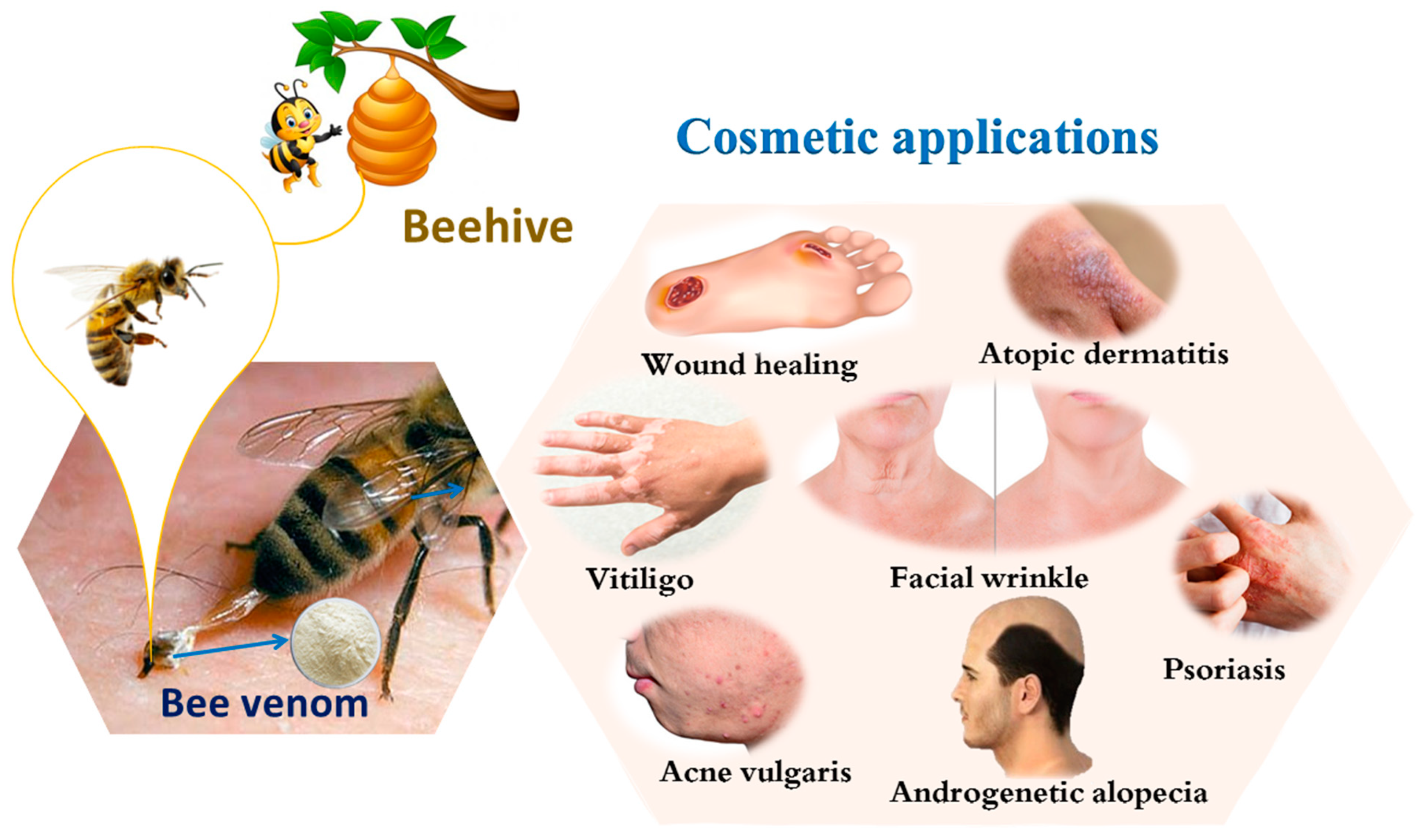
Cosmetic Applications of Bee Venom

 ,
,  , , , ,
  and
and 
Abstract
:1. Introduction
2. Cosmetic Applications of Bee Venom
2.1. Atopic Dermatitis (AD)
2.2. Acne Vulgaris
2.3. Androgenetic Alopecia (AGA)
2.4. Wound Healing
2.5. Facial Wrinkles
2.6. Vitiligo
2.7. Psoriasis

| Skin Diseases | Model | Dose Used | Mechanism | References |
|---|---|---|---|---|
| Atopic dermatitis (AD) | Male HR-1 mice | 0.1, 0.25, and 0.5 µg of BV | Inhibits the activation of NF-κB and resulting in the reduction in pro- inflammatory cytokines TNF-α, IL-1β, and IL-6. The inhibition of iNOS and COX- 2 expression in a PA-induced AD animal model in a dose-dependent manner. | [21] |
| Male BALB/c mice | 0.3 mg/kg daily at BL40 acupuncture points for 5 days from BV | Inhibits the proliferation and infiltration of T cells, the production of Th1 and Th2 cytokines, and the synthesis of IL-4 and IgE—typical allergic Th2 responses in blood. | [22] | |
| Male BALB/c mice | 0.01 and 0.1 mg/kg of BV | Inhibits the mast cell degranulation and pro-inflammatory cytokine expression via NF-κB activation. | [24] | |
| A human keratinocyte cell line, HaCaT cells | 0.1 µg/mL of apamin | Inhibits TNF-α- and IFN-γ-induced pro-inflammatory cytokines and Th2 lymphocyte chemokines via down-regulation of NF-κB signaling pathway and STAT. | ||
| Acne Vulgaris | HaCaT and THP-1 cells | 1, 10, and 100 ng/mL of BV | Has anti-inflammatory properities against Propionibacterium acnes. Blocked TLR2 expression and suppressed the production of IFN-γ, IL-1β, IL-8, and TNF-α induced by P. acnes. | [38] |
| Androgenetic alopecia (AGA) | C57BL/6 mice | 0.005% and 0.01% BV | Stimulate the expression levels of growth FGF-2, IGF-1R, and VEG. | [46] |
| Wound healing | Sprague-Dawley rats | 6% w/w of BV into chitosan film | In comparison to chitosan-free films, the combination had a better anti-inflammatory impact. | [48] |
| Adult male Wistar albino | 4 % BV incorporated in hydrogel prepared from 10% PVA and 0.6% chitosan | In comparison to the control, had increased hydroxyproline and glutathione levels and lower IL-6 levels. | [50] | |
| Male BALB/c mice | 50 µL equivalent to 200 µg/kg of BV/wounded area/day for 15 days | Significantly restored ATF-3- and iNOS-mediated oxidative stress and MMP-9 expression, as well as enhanced CXCL12-mediated migration EPCs to damaged tissues. | [51] | |
| Male mice | HPCS-BV nanofibers at different time intervals (3, 5, 7, 10, and 12 days) | Improved collagen deposition and the overall wound-healing process by preventing the inflammatory phase from extending. | [52] | |
| Facial Wrinkles | Twenty-two mature Korean women | BV facial serum at a concentration of 0.006%. | The total wrinkle area, total wrinkle count, and average wrinkle depth were all reduced in clinical trials; however, the exact mechanism is uncertain. | [5] |
| Human keratinocyte (HaCaT) and human dermal fibroblast (HDF) cells | 1 μg/mL of bvPlA2-free BV | Under UVB exposure, repair cell damage and collagen formation while inhibiting MMP-1 and -13 in HaCaT cells and MMP-1, -2, and -3 in HDF cells. Activation of ERK1/2 and p38 exerts anti-wrinkle properties. | [55] | |
| Vitiligo | Normal human epidermal melanocyte | 10 µg/mL BV for 1, 3, 5 or 7 days | Induces cAMP production in melanocytes. Proliferation of melanocytes increased. | [58] |
| Psoriasis | Twenty-five patients | The doses starting 0.05–0.1 mL/once for a week, and then increased gradually by 0.05 mL every session to a dose of 1 mL for every injection was reached. The total treatment period was 3 months. | Compared to the control group, there was a statistically significant decrease in TNF-α. | [63] |
3. Concluding Remarks
Author Contributions
Funding
Institutional Review Board Statement
Informed Consent Statement
Data Availability Statement
Acknowledgments
Conflicts of Interest
References
- Aufschnaiter, A.; Kohler, V.; Khalifa, S.; El-Wahed, A.; Du, M.; El-Seedi, H.; Büttner, S. Apitoxin and its components against cancer, neurodegeneration and rheumatoid arthritis: Limitations and possibilities. Toxins 2020, 12, 66. [Google Scholar] [CrossRef] [PubMed] [Green Version]
- Al-Safar, M.A.; Obied, H.N.; Ghaleb, R.A.; Kashkol, A.S. In-vitro cytotoxic anticancer effects of honeybee venom fractions on different cell lines. Int. J. Drug Deliv. Technol. 2020, 10, 141–144. [Google Scholar] [CrossRef]
- Mohamed, D.; Tedawy, E.L.; Mahmoud, M.; Alhaseeb, A.B.D.; Helmy, M.W.; Ghoneim, A.I. Systemic bee venom exerts anti-arthritic and anti-inflammatory properties in a rat model of arthritis. Biomed. Rep. 2020, 13, 20–28. [Google Scholar] [CrossRef]
- Flávia, A.; Pereira, M.; Albano, M.; Cristina, F.; Alves, B.; Fernanda, B.; Teles, M.; Furlanetto, A.; Mores, V.L. Influence of apitoxin and melittin from Apis mellifera bee on Staphylococcus aureus strains. Microb. Pathog. 2020, 141, 104011. [Google Scholar] [CrossRef]
- Han, S.M.; Hong, I.P.; Woo, S.O.; Chun, S.N.; Park, K.K.; Nicholls, Y.M.; Pak, S.C. The beneficial effects of honeybee-venom serum on facial wrinkles in humans. Clin. Interv. Aging 2015, 10, 1587–1592. [Google Scholar] [CrossRef] [Green Version]
- Shah, S.; Gupta, A.; Karne, S.P.; Kamble, S.; Shinde, B. Anti-inflammatory activity of sting protein from Apis mellifera. Int. J. Life Sci. Sci. Res. 2017, 3, 914–919. [Google Scholar] [CrossRef]
- Han, S.M.; Kim, J.M.; Hong, I.P.; Woo, S.O.; Kim, S.G.; Jang, H.R.; Pak, S.C. Antibacterial activity and antibiotic-enhancing effects of honeybee venom against methicillin-resistant Staphylococcus aureus. Molecules 2016, 21, 79. [Google Scholar] [CrossRef] [Green Version]
- Han, S.M.; Lee, G.G.; Park, K.K. Skin sensitization study of bee venom (Apis mellifera L.) in guinea pigs. Toxicol. Res. 2012, 28, 1–4. [Google Scholar] [CrossRef]
- Matysiak, J.; Schmelzer, C.E.H.; Neubert, R.H.H.; Kokot, Z.J. Characterization of honeybee venom by MALDI-TOF and nanoESI-QqTOF mass spectrometry. J. Pharm. Biomed. Anal. 2011, 54, 273–278. [Google Scholar] [CrossRef]
- Bonifazi, F.; Jutel, M.; Biló, B.M.; Birnbaum, J.; Muller, U. Prevention and treatment of hymenoptera venom allergy: Guidelines for clinical practice. Allergy 2005, 60, 1459–1470. [Google Scholar] [CrossRef]
- Müller, U.R.; Haeberli, G. Use of β-blockers during immunotherapy for Hymenoptera venom allergy. J. Allergy Clin. Immunol. 2005, 115, 606–610. [Google Scholar] [CrossRef]
- Diwakar, L.; Ewan, P.; Huber, P.A.J.; Clark, A.; Nasser, S.; Krishna, M.T. The impact of national guidelines on venom immunotherapy practice in the United Kingdom. Clin. Exp. Allergy 2016, 46, 749–753. [Google Scholar] [CrossRef] [Green Version]
- Čerpes, U.; Arzt-Gradwohl, L.; Schrautzer, C.; Koch, L.; Bokanovic, D.; Laipold, K.; Tripolt, P.; Binder, B.; Sturm, G.J. Simultaneous up-dosing of bee and vespid venom immunotherapy is safe. Allergy Eur. J. Allergy Clin. Immunol. 2020, 75, 721–723. [Google Scholar] [CrossRef] [Green Version]
- Tanei, R. Atopic dermatitis in the elderly. Inflamm. Allergy Drug Targets 2009, 8, 398–404. [Google Scholar] [CrossRef]
- Bin, L.; Leung, D.Y.M. Genetic and epigenetic studies of atopic dermatitis. Allergy Asthma Clin. Immunol. 2016, 12, 52–65. [Google Scholar] [CrossRef] [Green Version]
- Giavina-Bianchi, M.; Giavina-Bianchi, P. Systemic treatment for severe atopic dermatitis. Arch. Immunol. Ther. Exp. 2019, 67, 69–78. [Google Scholar] [CrossRef] [PubMed]
- Ferrucci, S.; Tavecchio, S.; Berti, E.; Angileri, L. Dupilumab and prurigo nodularis-like phenotype in atopic dermatitis: Our experience of efficacy. J. Dermatolog. Treat. 2021, 32, 453–454. [Google Scholar] [CrossRef] [PubMed]
- Singh, R.; Heron, C.E.; Ghamrawi, R.I.; Strowd, L.C.; Feldman, S.R. Emerging role of janus kinase inhibitors for the treatment of atopic dermatitis. ImmunoTargets Ther. 2020, 9, 255–272. [Google Scholar] [CrossRef] [PubMed]
- Wegner, J.; Weinmann-menke, J.; von Stebut, E. Immunoadsorption for treatment of severe atopic dermatitis. Atheroscler. Suppl. 2017, 30, 264–270. [Google Scholar] [CrossRef] [PubMed]
- Jin, Y.; Myung, L.; Oh, J.; Hun, D.; Yong, L.; Lee, S.; Lee, J.; Hyun, D.; Cheol, K.; Choi, H. Anti-inflammatory effect of bee venom in phthalic anhydride—Induced atopic dermatitis animal model. Inflammopharmacology 2020, 28, 253–263. [Google Scholar] [CrossRef]
- Oh, M.J.; Song, H.-S. Anti-Inflammatory effects of bee venom on phthalic anhydride-induced atopic dermatitis. J. Acupunct. Res. 2020, 37, 42–48. [Google Scholar] [CrossRef] [Green Version]
- Sur, B.; Lee, B.; Yeom, M.; Hong, J.-H.; Kwon, S.; Kim, S.-T.; Lee, H.S.; Park, H.-J.; Lee, H.; Hahm, D.-H. Bee venom acupuncture alleviates trimellitic anhydride-induced atopic dermatitis-like skin lesions in mice. BMC Complement. Altern. Med. 2016, 16, 38–50. [Google Scholar] [CrossRef] [Green Version]
- Kim, W.; An, H.; Kim, J.; Gwon, M.; Gu, H.; Sung, W.J.; Han, S.M.; Pak, S.C.; Kim, M.; Park, K. Beneficial effects of melittin on ovalbumin-induced atopic dermatitis in mouse. Sci. Rep. 2017, 7, 17679–17690. [Google Scholar] [CrossRef] [Green Version]
- Kim, K.; Lee, W.; An, H.; Kim, J.; Chung, H.; Han, S.; Lee, K.; Pak, S.C.; Park, K. Bee venom ameliorates compound 48 / 80-induced atopic dermatitis-related symptoms. Int. J. Clin. Exp. Pathol. 2013, 6, 2896–2903. [Google Scholar]
- Kim, W.-H.; An, H.-J.; Kim, J.-Y.; Gwon, M.-G.; Gu, H.; Lee, S.-J.; Park, J.Y.; Park, K.-D.; Han, S.-M.; Kim, M.-K.; et al. Apamin inhibits TNF-α- and IFN-γ-induced inflammatory cytokines and chemokines via suppressions of NF-κB signaling pathway and STAT in human keratinocytes. Pharmacol. Rep. 2017, 69, 1030–1035. [Google Scholar] [CrossRef]
- You, C.E.; Moon, S.H.; Lee, K.H.; Kim, K.H.; Park, C.W.; Seo, S.J.; Cho, S.H. Effects of emollient containing bee venom on atopic dermatitis: A double-blinded, randomized, base-controlled, multicenter study of 136 patients. Ann. Dermatol. 2016, 28, 593–599. [Google Scholar] [CrossRef] [Green Version]
- An, H.J.; Kim, J.Y.; Kim, W.H.; Gwon, M.G.; Gu, H.M.; Jeon, M.J.; Han, S.M.; Pak, S.C.; Lee, C.K.; Park, I.S.; et al. Therapeutic effects of bee venom and its major component, melittin, on atopic dermatitis in vivo and in vitro. Br. J. Pharmacol. 2018, 175, 4310–4324. [Google Scholar] [CrossRef] [PubMed] [Green Version]
- Jung, K.H.; Baek, H.; Kang, M.; Kim, N.; Lee, S.Y.; Bae, H. Bee venom phospholipase A2 ameliorates house dust mite extract induced atopic dermatitis like skin Lesions in mice. Toxins 2017, 9, 68. [Google Scholar] [CrossRef] [PubMed]
- Williams, H.C.; Dellavalle, R.P.; Garner, S. Acne vulgaris. Lancet 2012, 379, 361–372. [Google Scholar] [CrossRef]
- Zhou, M.; Xie, H.; Cheng, L.; Li, J. Clinical characteristics and epidermal barrier function of papulopustular rosacea: A comparison study with acne vulgaris. Pak. J. Med. Sci. 2016, 32, 1344–1348. [Google Scholar] [CrossRef] [PubMed]
- Jappe, U. Pathological mechanisms of acne with special emphasis on Propionibacterium acnes and related therapy. Acta Derm. Venereol. 2003, 83, 241–248. [Google Scholar] [CrossRef] [Green Version]
- Leccia, M.T.; Auffret, N.; Poli, F.; Claudel, J.P.; Corvec, S.; Dreno, B. Topical acne treatments in Europe and the issue of antimicrobial resistance. J. Eur. Acad. Dermatol. Venereol. 2015, 29, 1485–1492. [Google Scholar] [CrossRef] [PubMed]
- Nakase, K.; Nakaminami, H.; Takenaka, Y.; Hayashi, N.; Kawashima, M.; Noguchi, N. Relationship between the severity of acne vulgaris and antimicrobial resistance of bacteria isolated from acne lesions in a hospital in Japan. J. Med. Microbiol. 2014, 63, 721–728. [Google Scholar] [CrossRef] [PubMed]
- Dessinioti, C.; Katsambas, A.D. The role of Propionibacterium acnes in acne pathogenesis: Facts and controversies. Clin. Dermatol. 2010, 28, 2–7. [Google Scholar] [CrossRef] [PubMed]
- Qin, M.; Pirouz, A.; Kim, M.-H.; Krutzik, S.R.; Garbán, H.J.; Kim, J. Propionibacterium acnes induces IL-1β secretion via the NLRP3 inflammasome in human monocytes. J. Investig. Dermatol. 2014, 134, 381–388. [Google Scholar] [CrossRef] [PubMed] [Green Version]
- Jugeau, S.; Tenaud, I.; Knol, A.C.; Jarrousse, V.; Quereux, G.; Khammari, A.; Dreno, B. Induction of toll-like receptors by Propionibacterium acnes. Br. J. Dermatol. 2005, 153, 1105–1113. [Google Scholar] [CrossRef] [PubMed]
- Jahns, A.C.; Lundskog, B.; Ganceviciene, R.; Palmer, R.H.; Golovleva, I.; Zouboulis, C.C.; McDowell, A.; Patrick, S.; Alexeyev, O.A. An increased incidence of Propionibacterium acnes biofilms in acne vulgaris: A case-control study. Br. J. Dermatol. 2012, 167, 50–58. [Google Scholar] [CrossRef]
- Kim, J.Y.; Lee, W.R.; Kim, K.H.; An, H.J.; Chang, Y.C.; Han, S.M.; Park, Y.Y.; Pak, S.C.; Park, K.K. Effects of bee venom against Propionibacterium acnes-induced inflammation in human keratinocytes and monocytes. Int. J. Mol. Med. 2015, 35, 1651–1656. [Google Scholar] [CrossRef] [PubMed] [Green Version]
- An, H.J.; Lee, W.R.; Kim, K.H.; Kim, J.Y.; Lee, S.J.; Han, S.M.; Lee, K.G.; Lee, C.K.; Park, K.K. Inhibitory effects of bee venom on Propionibacterium acnes-induced inflammatory skin disease in an animal model. Int. J. Mol. Med. 2014, 34, 1341–1348. [Google Scholar] [CrossRef] [PubMed] [Green Version]
- Han, S.M.; Pak, S.C.; Nicholls, Y.M.; Macfarlane, N. Evaluation of anti-acne property of purified bee venom serum in humans. J. Cosmet. Dermatol. 2016, 15, 324–329. [Google Scholar] [CrossRef] [Green Version]
- Han, S.M.; Lee, K.G.; Pak, S.C. Effects of cosmetics containing purified honeybee (Apis mellifera L.) venom on acne vulgaris. J. Integr. Med. 2013, 11, 320–326. [Google Scholar] [CrossRef] [PubMed] [Green Version]
- Adil, A.; Godwin, M. The effectiveness of treatments for androgenetic alopecia: A systematic review and meta-analysis. J. Am. Acad. Dermatol. 2017, 77, 136–141. [Google Scholar] [CrossRef] [PubMed]
- Gan, D.C.C.; Sinclair, R.D. Prevalence of male and female pattern hair loss in Maryborough. J. Investig. Dermatol. Symp. Proc. 2005, 10, 184–189. [Google Scholar] [CrossRef] [PubMed]
- Varothai, S.; Bergfeld, W.F. Androgenetic alopecia: An evidence-based treatment update. Am. J. Clin. Dermatol. 2014, 15, 217–230. [Google Scholar] [CrossRef]
- Jain, R.; De-Eknamkul, W. Potential targets in the discovery of new hair growth promoters for androgenic alopecia. Expert Opin. Ther. Targets 2014, 18, 787–806. [Google Scholar] [CrossRef]
- Park, S.; Erdogan, S.; Hwang, D.; Hwang, S.; Han, E.H.; Lim, Y.-H. Bee venom promotes hair growth in association with inhibiting 5α-reductase expression. Biol. Pharm. Bull. 2016, 39, 1060–1068. [Google Scholar] [CrossRef] [PubMed]
- Han, S.-M.; Lee, K.-G.; Yeo, J.-H.; Kim, W.-T.; Park, K.-K. Biological effects of treatment of an animal skin wound with honeybee (Apis melifera L.) venom. J. Plast. Reconstr. Aesthetic Surg. 2011, 64, e67–e72. [Google Scholar] [CrossRef]
- Amin, M.A.; Madkor, H.R. Wound healing and anti-inflammatory activities of bee venom-chitosan blend films. J. Drug Deliv. Sci. Technol. 2008, 18, 424–430. [Google Scholar] [CrossRef]
- Hozzein, W.N.; Badr, G.; Badr, B.M.; Allam, A.; Al, A.; Al-wadaan, M.A.; Al-waili, N.S. Bee venom improves diabetic wound healing by protecting functional macrophages from apoptosis and enhancing Nrf2, Ang-1 and Tie-2 signaling. Mol. Immunol. 2018, 103, 322–335. [Google Scholar] [CrossRef]
- Amin, M.A.; Abd-Raheem, I. Accelerated wound healing and anti-inflammatory effects of physically cross linked polyvinyl alcohol-chitosan hydrogel containing honey bee venom in diabetic rats. Arch. Pharm. Res. 2014, 37, 1016–1031. [Google Scholar] [CrossRef]
- Badr, G.; Hozzein, W.N.; Badr, B.M.; Al Ghamdi, A.; Saad Eldien, H.M.; Garraud, O. Bee venom accelerates wound healing in diabetic mice by suppressing activating transcription factor-3 (ATF-3) and inducible nitric oxide synthase (iNOS)-mediated oxidative stress and recruiting bone marrow-derived endothelial progenitor cells. J. Cell. Physiol. 2016, 231, 2159–2171. [Google Scholar] [CrossRef]
- Sarhan, W.A. Apitherapeutics and phage-loaded nanofibers as wound dressings with enhanced wound healing and antibacterial activity. Nanomedicine 2017, 12, 2055–2067. [Google Scholar] [CrossRef]
- Kezic, S.; Novak, N.; Jakasa, I.; Jungersted, J.M.; Simon, M.; Brandner, J.M. Skin barrier in atopic dermatitis. Front. Biosci. 2014, 19, 542–556. [Google Scholar] [CrossRef] [PubMed] [Green Version]
- Hord, N.G.; Fenton, J.I. Context is everything: Mining the normal and preneoplastic microenvironment for insights into the diet and cancer risk conundrum. Mol. Nutr. Food Res. 2007, 51, 100–106. [Google Scholar] [CrossRef] [PubMed]
- Lee, H.; Kyeong, S.; Pyo, B.M.; Heo, Y.; Goo, C. Anti-wrinkle effect of PLA 2 -free bee venom against UVB-irradiated human skincells. J. Agric. Life Sci. 2015, 49, 125–135. [Google Scholar] [CrossRef] [Green Version]
- Bastonini, E.; Bellei, B.; Filoni, A.; Kovacs, D.; Iacovelli, P.; Picardo, M. Involvement of non-melanocytic skin cells in vitiligo. Exp. Dermatol. 2019, 28, 667–673. [Google Scholar] [CrossRef] [Green Version]
- Roberts, G.H.L.; Santorico, S.A.; Spritz, R.A. The genetic architecture of vitiligo. Pigment. Cell Melanoma Res. 2019, 33, 8–15. [Google Scholar] [CrossRef] [Green Version]
- Jeon, S.; Kim, N.H.; Koo, B.S.; Lee, H.J.; Lee, A.Y. Bee venom stimulates human melanocyte proliferation, melanogenesis, dendricity and migration. Exp. Mol. Med. 2007, 39, 603–613. [Google Scholar] [CrossRef] [PubMed] [Green Version]
- Maeda, K.; Tomita, Y.; Naganuma, M.; Tagami, H. Phospholipases induce melanogenesis in organ-cultured skin. Photochem. Photobiol. 1996, 64, 220–223. [Google Scholar] [CrossRef] [PubMed]
- Kim, N.H.; Lee, A.Y. Histamine effect on melanocyte proliferation and vitiliginous keratinocyte survival. Exp. Dermatol. 2010, 19, 1073–1079. [Google Scholar] [CrossRef]
- Takeshita, J.; Grewal, S.; Langan, S.M.; Mehta, N.N.; Ogdie, A.; Van Voorhees, A.S.; Gelfand, J.M. Psoriasis and comorbid diseases: Epidemiology. J. Am. Acad. Dermatol. 2017, 76, 377–390. [Google Scholar] [CrossRef] [PubMed] [Green Version]
- El-Wahed, A.A.A.; Khalifa, S.A.; Sheikh, B.Y.; Farag, M.A.; Saeed, A.; Larik, F.A.; Koca-Caliskan, U.; AlAjmi, M.F.; Hassan, M.; Wahabi, H.A. et al. Bee venom composition: From chemistry to biological activity. In Studies in Natural Products Chemistry; Elsevier: Amsterdam, The Netherlands, 2019; Volume 60, pp. 459–484. ISBN 9780444641816. [Google Scholar]
- Eltaher, S.; Mohammed, G.F.; Younes, S.; Elakhras, A. Efficacy of the apitherapy in the treatment of recalcitrant localized plaque psoriasis and evaluation of tumor necrosis factor-alpha (TNF-α) serum level: A double-blind randomized clinical trial. J. Dermatol. Treat. 2015, 26, 335–339. [Google Scholar] [CrossRef]
- Kokot, Z.J.; Matysiak, J.; Urbaniak, B.; Dereziński, P. New CZE-DAD method for honeybee venom analysis and standardization of the product. Anal. Bioanal. Chem. 2011, 399, 2487–2494. [Google Scholar] [CrossRef] [PubMed] [Green Version]
- Ridolo, E.; Pellicelli, I.; Kihlgren, P.; Nizi, M.C.; Pucciarini, F.; Senna, G.; Incorvaia, C. Immunotherapy and biologicals for the treatment of allergy to Hymenoptera stings. Expert Opin. Biol. Ther. 2019, 19, 919–925. [Google Scholar] [CrossRef] [PubMed]
- Van Vaerenbergh, M.; Cardoen, D.; Formesyn, E.M.; Brunain, M.; Van Driessche, G.; Blank, S.; Spillner, E.; Verleyen, P.; Wenseleers, T.; Schoofs, L.; et al. Extending the honey bee venome with the antimicrobial peptide apidaecin and a protein resembling wasp antigen 5. Insect Mol. Biol. 2013, 22, 199–210. [Google Scholar] [CrossRef]
- King, T.P.; Spangfort, M.D. Structure and biology of stinging insect venom allergens. Int. Arch. Allergy Immunol. 2000, 123, 99–106. [Google Scholar] [CrossRef]
- Francese, S.; Turillazzi, S.; Moneti, G.; Clench, M.; Barber, D.; Kingdom, U. In situ imaging of honeybee (Apis mellifera) venom components from aqueous and aluminum hydroxide-adsorbed venom immunotherapy preparations. J. Allergy Clin. Immunol. 2011, 129, 1314–1320. [Google Scholar] [CrossRef]
- Silva, T.C.; De Paula Moura, S.; Ramos, H.R.; De Araujo, P.S.; Bueno Da Costa, M.H. Design of a modern liposome and bee venom formulation for the traditional VIT-venom immunotherapy. J. Liposome Res. 2008, 18, 353–368. [Google Scholar] [CrossRef]
- Ahn, Y.J.; Shin, J.S.; Lee, J.; Lee, Y.J.; Kim, M.R.; Shin, Y.S.; Park, K.B.; Kim, E.J.; Kim, M.J.; Lee, J.W.; et al. Safety of essential bee venom pharmacopuncture as assessed in a randomized controlled double-blind trial. J. Ethnopharmacol. 2016, 194, 774–780. [Google Scholar] [CrossRef]

Publisher’s Note: MDPI stays neutral with regard to jurisdictional claims in published maps and institutional affiliations. |
© 2021 by the authors. Licensee MDPI, Basel, Switzerland. This article is an open access article distributed under the terms and conditions of the Creative Commons Attribution (CC BY) license (https://creativecommons.org/licenses/by/4.0/).
Share and Cite
El-Wahed, A.A.A.; Khalifa, S.A.M.; Elashal, M.H.; Musharraf, S.G.; Saeed, A.; Khatib, A.; Tahir, H.E.; Zou, X.; Naggar, Y.A.; Mehmood, A.; et al. Cosmetic Applications of Bee Venom. Toxins 2021, 13, 810. https://doi.org/10.3390/toxins13110810
El-Wahed AAA, Khalifa SAM, Elashal MH, Musharraf SG, Saeed A, Khatib A, Tahir HE, Zou X, Naggar YA, Mehmood A, et al. Cosmetic Applications of Bee Venom. Toxins. 2021; 13(11):810. https://doi.org/10.3390/toxins13110810
Chicago/Turabian StyleEl-Wahed, Aida A. Abd, Shaden A. M. Khalifa, Mohamed H. Elashal, Syed G. Musharraf, Aamer Saeed, Alfi Khatib, Haroon Elrasheid Tahir, Xiaobo Zou, Yahya Al Naggar, Arshad Mehmood, and et al. 2021. "Cosmetic Applications of Bee Venom" Toxins 13, no. 11: 810. https://doi.org/10.3390/toxins13110810
APA StyleEl-Wahed, A. A. A., Khalifa, S. A. M., Elashal, M. H., Musharraf, S. G., Saeed, A., Khatib, A., Tahir, H. E., Zou, X., Naggar, Y. A., Mehmood, A., Wang, K., & El-Seedi, H. R. (2021). Cosmetic Applications of Bee Venom. Toxins, 13(11), 810. https://doi.org/10.3390/toxins13110810