Article

Heterosigma akashiwo in Patagonian Fjords: Genetics, Growth, Pigment Signature and Role of PUFA and ROS in Ichthyotoxicity

by
Ana Flores-Leñero
1,*,
Valentina Vargas-Torres
2,
Javier Paredes-Mella
1,3,
Luis Norambuena
1,
Gonzalo Fuenzalida
1,4,
Kim Lee-Chang
5 and
Jorge I. Mardones
1,3,6,*
1
Centro de Estudios de Algas Nocivas (CREAN), Instituto de Fomento Pesquero (IFOP), Puerto Montt 5501679, Chile
2
Instituto de Ingeniería Biológica y Médica (IIBM), Pontificia Universidad Católica de Chile, Santiago 8331150, Chile
3
CAICAI Foundation, Puerto Varas 5550000, Chile
4
Departamento de Ciencias Básicas, Facultad de Ciencias, Universidad Santo Tomás, Buena Vecindad #91, Puerto Montt 5504749, Chile
5
CSIRO Ocean and Atmosphere, G.P.O. Box 1538, Hobart, TAS 7001, Australia
6
Centro de Investigación en Dinámica de Ecosistemas Marinos de Altas Latitudes (IDEAL), Valdivia 5110566, Chile
*
Authors to whom correspondence should be addressed.
Toxins 2022, 14(9), 577; https://doi.org/10.3390/toxins14090577
Submission received: 19 July 2022 / Revised: 4 August 2022 / Accepted: 9 August 2022 / Published: 23 August 2022
(This article belongs to the Special Issue Effects of Harmful Algal Blooms on Aquatic Organisms)

Abstract

:
Heterosigma akashiwo is the only raphidophyte described for Chilean waters. A recent 2021 fish-killing bloom event of this raphidophyte ignited scientific research, but the ichthyotoxic mechanism and environmental conditions that promote its growth are still unclear. This is the first study confirming the occurrence of H. akashiwo in Chilean waters on the basis of the region D1/D2 of the 28S ribosomal gene. The pigment signature of the CREAN_HA03 strain revealed chlorophyll-a, fucoxanthin, and violaxanthin as the most abundant pigments, but profiles were variable depending on culture and field conditions. A factorial temperature–salinity growth experiment showed a maximal growth rate of 0.48 d−1 at 17 °C and 35 in salinity, but reached a maximal cell abundance of ~50,000 cells mL−1 at 12 °C and 25 in salinity. The fatty acid profile included high levels of saturated (16:0) and polyunsaturated (18:4 ω3; 20:5 ω3) fatty acids, but superoxide production in this strain was low (~0.3 pmol O2– cell−1 h−1). The RTgill-W1 bioassay showed that the H. akashiwo strain was cytotoxic only at high cell concentrations (>47,000 cells mL−1) and after cell rupture. In conclusion, salmon mortality during H. akashiwo bloom events in Patagonian fjords is likely explained by the high production of long-chain PUFAs at high cell densities, but only in the presence of high ROS production.
Key Contribution: The first genetic sequence, ROS estimation, and lipid profile from a Chilean H. akashiwo. Cultured Chilean H. akashiwo is toxic only at high cell densities and after cell rupture. Chilean H. akashiwo is a high PUFA but low ROS producer.

1. Introduction

Heterosigma akashiwo is a small raphidophyte that is widely distributed in coastal ecosystems and is well-known as a driver of brown tides in many parts of the world. This species is a marine phytoflagellate that is described as an eurythermal and euryhaline organism, meaning that it can tolerate broad ranges of temperatures from 11 to 24 °C [1,2,3] and salinity from 5 to 35 [2,4]. Along with this, Heterosigma akashiwo has the ability to form resting cysts [5] when exposed to adverse conditions that coincide with seasonal differences in light, temperature, and nutrients present in temperate climates [6,7]. Another adaptation mechanism is the large quantity of pigments with photoprotective and antioxidant functions, enabling this microalga to live under high irradiance. Heterosigma akashiwo’s pigment profile contains Chl a, Chl c1, Chl c2, beta-carotene, and carotenoids fucoxanthin and violaxanthin as its pigment markers [2]. These characteristics possibly give H. akashiwo a competitive advantage over other organisms, allowing for it to colonize coastal ecosystems, such as bays and estuaries [4].
Despite the wide range of abiotic conditions in which this species can live, strains of H. akashiwo extracted from blooms in different locations have shown surprisingly low genetic variability. Engesmo et al. [8] demonstrated that the rDNA of H. akashiwo shows signs of segregation consistent with geographical origin, differentiating at least two polymorphic positions within small-subunit (18S) rDNA, one specific for strains extracted from Australia or New Zealand, and the other from the Atlantic coast of North America. However, in the same study, H. akashiwo strains displayed low genetic variability in the ITS region, which is considered to be the most variable rDNA region.
The blooms of H. akashiwo have been implicated in fish-killing events in several coastal areas, including Canada, Japan, New Zealand and Chile [9], and have been associated with economic losses to the aquacultural industry in those countries [10]. In Chile, the first H. akashiwo salmon-killing event was reported in September 1988 in the Los Lagos region (Reloncavi sound, Hornopiren and Chiloe Island), where the alga’s maximal cell density reached 100,000 cells mL−1 [3]. In April 2021, a massive H. akashiwo bloom occurred in the Comau fjord, Northern Patagonia, with cell abundances that ranged between 70,000 and 210,000 cells mL−1, causing a mass mortality of farmed salmon [11]. The historical monitoring of the Chilean H. akashiwo has shown its occurrence at temperatures ranging from 11.2 to 17.3 °C, and salinities from 11.4 to 33.3 [3], and ichthyotoxicity linked to high cell densities. A recently performed cell line bioassay analysis on the Chilean H. akashiwo has supported this hypothesis, with its ichthyotoxicity found to be low at concentrations of 2000 cells mL−1 [12,13].
The Raphidophyceae class is well-known for being responsible for massive fish mortalities, and while the cytotoxic compounds produced by H. akashiwo are still not well-understood, some possible ichthyotoxic mechanisms have been hypothesized. One mechanism involves the production of a putative neurotoxin with a similar effect to brevetoxins; however, this compound is not structurally related to the brevetoxin group, so the mode of action on fish remains unknown [14,15]. Another potential mechanism is the production of haemolytic compounds that can affect different tissues and organs in fish. These metabolites, along with the production of reactive oxygen species (ROS) such as hydrogen peroxide, hydroxyl radicals, and superoxide can produce severe injuries to fish gill tissues such as cell necrosis, epithelial lifting, and the alteration of chloride cells [16]. These injuries produce excessive mucus secretion that can cover the fish gills and cause asphyxiation [17]. However, ROS on their own may not be the main cause of ichthyotoxicity in this species [16]. Some authors proposed that the toxicity could be related to the presence of high concentrations of polyunsaturated fatty acids (PUFAs) [18]. The production of mucus or extracellular polymeric substances, such as polysaccharide–protein complexes (A–PCs), was also proposed as a toxic mechanism in H. akashiwo [19].
The main objective of this study is to define the phylogenetic position, pigment signature, superoxide production, fatty acid profile, and potential toxic effects of a Chilean H. akashiwo strain, and the environmental conditions that might promote its growth in the Patagonian fjords.

2. Results

2.1. Molecular Identification

The Bayesian phylogenetic and maximal likelihood analysis of the LSU D1–D2 region showed that the Chilean H. akashiwo (CREAN_HA03) sequence is in a monophyletic clade that comprises only H. akashiwo sequences, supported by one probability value (Figure 1). The monophyletic clade shows that this sequence of Heterosigma has a genetic distance of 0.00 against the sequences reported in Japan, Australia, EEUU, Demark, China, and New Zealand. In addition, the genetic distance with strains from Brazil, Canada, Korea, and New Zealand was 0.001, and the distance with H. minor from the USA resulted in a value of 0.064.

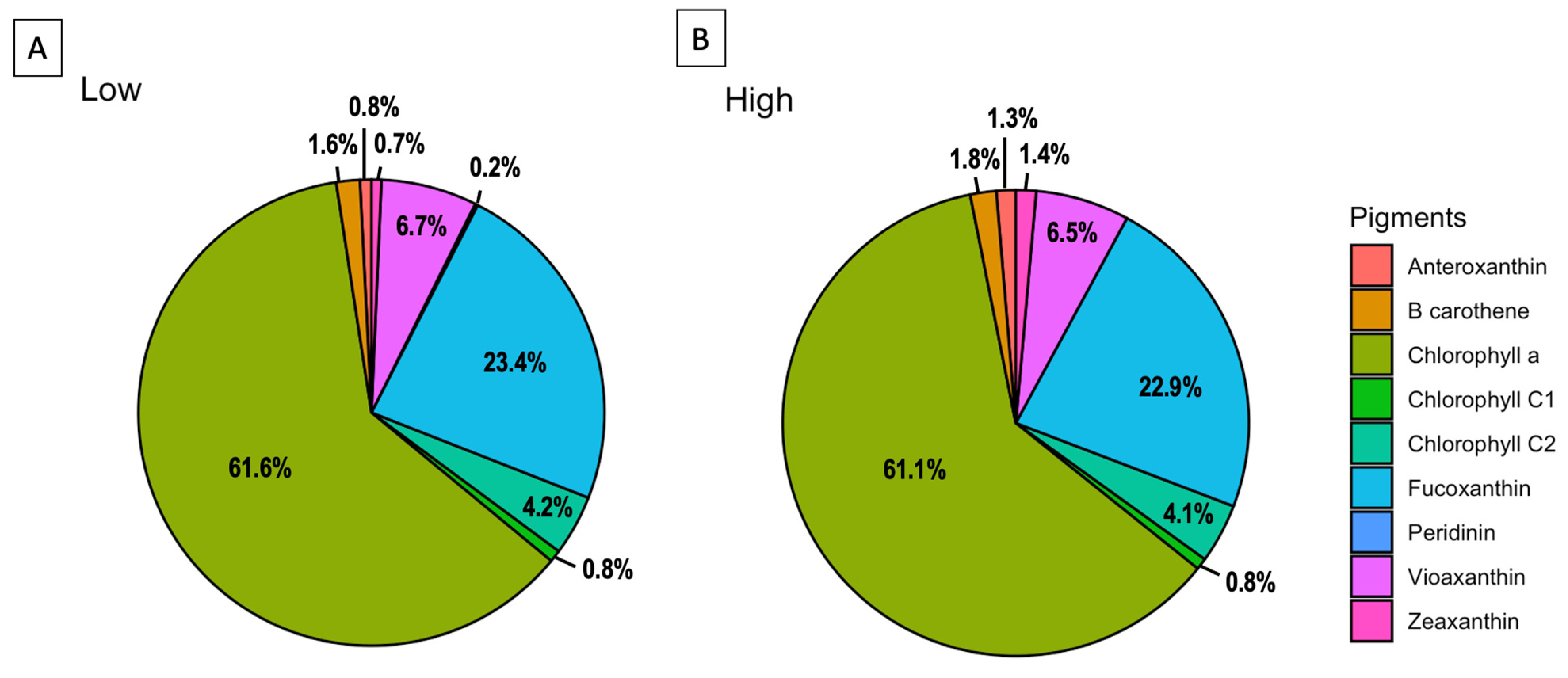
2.2. Pigments Signature

The pigment analysis of the H. akashiwo culture (CREAN_HA03), grown under the two light conditions of 50 (low light, LL) and 150 μmol photon m−2 s−1 (high light, HL), shows a similar pattern in pigment concentrations. The most abundant pigment was chlorophyll-a with a concentration of 8.9 at LL and 7.16 pg cell−1 at HL exposure (62% and 61% of the total pigments, respectively). The most abundant carotenoids at the LL and HL treatments were as follows: fucoxanthin with 3.38 and 2.68 pg cell−1 (23% of the total pigments in both samples), and violaxanthin with 0.96 and 0.76 pg cell−1 (7% of the total pigments in both samples), followed by chlorophyll c2, β-carotene, antheraxanthin, zeaxanthin, chlorophyll c1 and peridinin (Figure 2).
The HPLC pigment profiles of a bloom sample collected during the 2021 H. akashiwo event, and three profiles from a cultured strain (CREAN_HA03) measured at two different periods of time (one in 2020 at 100 μmol photon m−2 s−1, and two in 2022 at 50 and 150 μmol photon m−2 s−1) are presented in Table 1. The pigment distribution of the 2021 bloom sample shows a similar pattern compared with the CREAN_03 strain measured in 2020 but differed in pigment content (%) with the same strain measured in 2022. Ratios of Fuco:Chl a for the 2021 field sample and CREAN_HA03 strain measured in 2020 were 4.98 and 3.29, respectively. The Fuco:Chl a ratios of the CREAN_HA03 strain measured in 2022 showed lower values of 0.38 and 0.37 (50 and 150 μmol photon m−2 s−1 light, respectively).

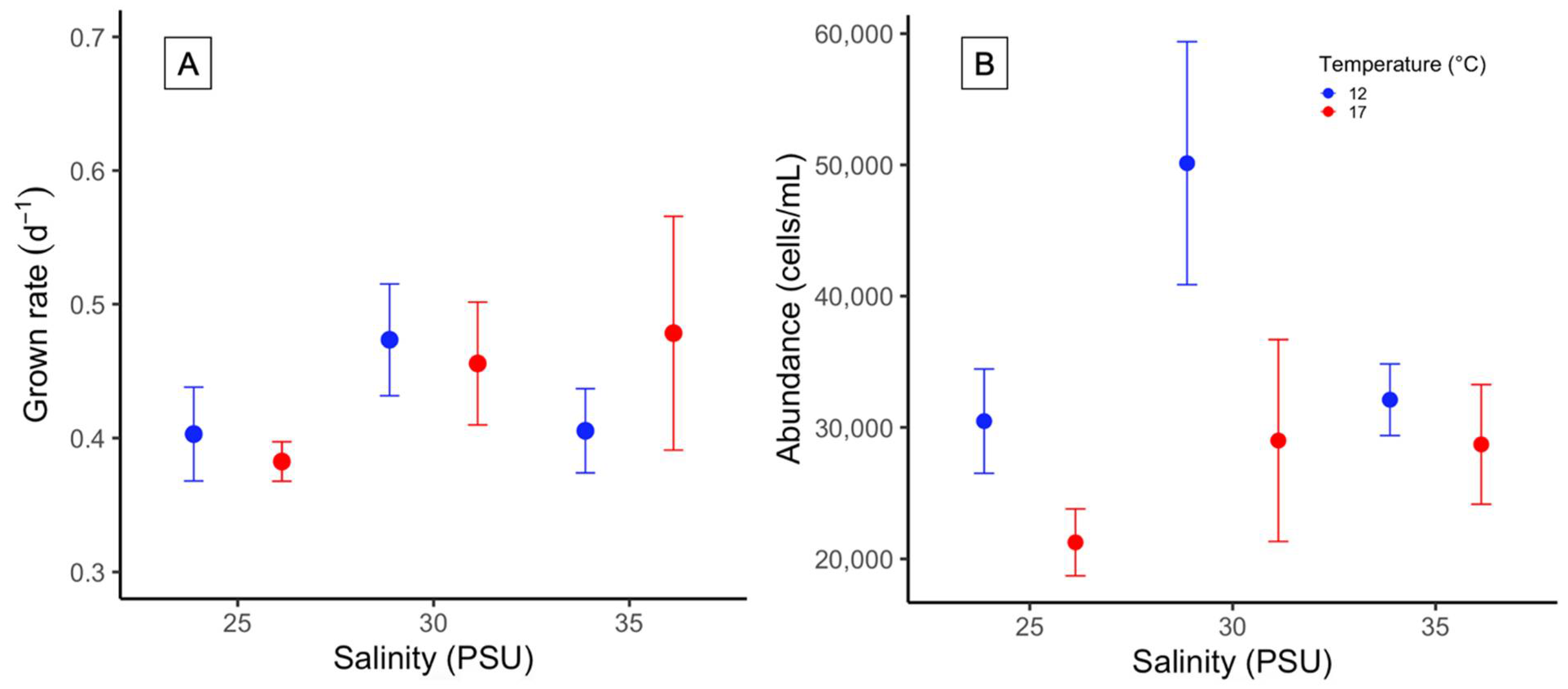
2.3. Growth Rate and Cell Density

The response of the Chilean H. akashiwo (CREAN_HA03) to different salinity and temperature conditions was not significantly different in terms of growth rate (μ; ANOVA, p > 0.05). The μ ranged between 0.38 ± 0.03 d1 and 0.48 ± 0.15 d1. The lowest µmax was reached at 25 in salinity at 17 °C, and the highest at 35 in salinity at 17 °C, respectively (Figure 3A). Temperature significantly affected the maximal cell abundance on Day 19, reaching a density of 50,000 cells mL1 at 12 °C, and 30 in salinity (ANOVA, p = 0.03, Figure 3B).

2.4. Fatty Acid Profile and Superoxide Production

The fatty acid composition of H. akashiwo strain CREAN_HA03 was dominated by polyunsaturated fatty acids (PUFAs) with a 40.4%, followed by saturated fatty acids (SFAs), and monounsaturated fatty acids (MUFAs) with 30.7% and 22.2%, respectively. The main fatty acids in relative abundance were, in decreasing order, palmitic acid (16:0 PA; 20.94%), EPA (20:5ω3; 13.04%), and stearidonic acid (18:4ω3; 10,44%) (Table 2).
Whole cells of H. akashiwo CREAN_HA03 showed similar rates of superoxide anion production (0.3075 ± 0.0299 pmol O2− cell−1 h−1) compared to those of lysed cells (0.2925 ± 0.0126 pmol O2− cell−1 h−1) (Table 3).

2.5. Intra- and Extracellular Ichthyotoxicity

Lysed (intracellular) and supernatant (extracellular) treatments at different cell abundances of H. akashiwo exposed against the RTgill-W1 cell line showed distinct effects only at high cell abundances. The highest cytotoxic effect was measured in the intracellular treatment at 47,000 cells mL1, significantly decreasing gill cell viability down to 61% of control (ANOVA, p < 0.05, Figure 4). Extracellular treatment at 47,000 cells mL1 only reduced the viability down to 90% of control. In either treatment, low densities did not have a significant effect against the Rtgill-W1 cell line (Figure 4).

3. Discussion

Heterosigma akashiwo is the only raphidophyte species that has been reported in Chilean waters producing deleterious effects on salmon farming activities. The massive bloom event of this species that occurred in austral summer–fall 2021 in the Comau fjord, ignited new research attention on this poorly studied species in the Chilean coast. In this study, we assessed the phylogenetic position and in vitro characteristics of a Chilean H. akashiwo strain with special emphasis on its ichthyotoxic potency.
To our knowledge, this is the first formal genetic identification of H. akashiwo in Chilean waters. Previous studies carried out in Chile only identified this raphidophyte on the basis of morphological characteristics [3,12,13,17]. In terms of genetic variability, the Chilean 28S rDNA sequence showed low intraspecific genetic variability against worldwide distributed H. akashiwo genetic sequences [20]. Heterosigma akashiwo sequences formed a monophyletic clade [8] expressing low genetic divergence regardless of geographical patterns [21,22]. As no further studies on Chilean H. akashiwo have focused on the intraspecific variation in the physiological response of local strains, the presence of different ecotypes is still unknown. A suggested direct association between chloroplast gene signatures and ecophysiological diversity allows, for instance, for an avenue for tracking different H. akashiwo populations from the west coast of North America [23]. The examination of selected regions within the chloroplast genome of additional Chilean strains may allow for the detection of strain-specific sequences and ecotypes that explain the ability of this species to form persistent blooms, as observed in 2021 in the highly variable Patagonian fjords ecosystem.
This study shows that the Chilean H. akashiwo has a flexible pigment signature in response to environmental conditions. This is in line with the well-known capacity of phytoplankton cells to modify their pigment content and ratios under different light and nutrient conditions [24]. Our data show that the most abundant secondary pigments in the CREAN_HA03 strain were fucoxanthin and violaxanthin. Other authors also found fucoxanthin to be the most abundant carotenoid in H. akashiwo strains from other coastal areas [2] and among others Chilean strains [17]. Under high irradiance conditions, microalgal cells increase photoprotective carotenoids, such as fucoxanthin, violaxanthin, and antheraxanthin, to prevent photo-oxidative damage [25,26]. In fact, a high fucoxanthin concentration was detected in field samples from the 2021 H. akashiwo bloom event at irradiances as high as 1430 μmol photon m−2 s−1, with a Fuco/Chl-a ratio of 4.98 (Table 1). However, a similar pigment proportion was found in the CREAN_HA03 strain one year after its isolation (harvested in 2020) at a low irradiance of 100 μmol photon m−2 s−1 with a Fuco/Chl-a ratio of 3.39, suggesting that pigment signatures could be conservative after cell isolation and culture establishment. This assertion was further denied after assessing the pigment signature of the same H. akashiwo strain cultured at 50 and 150 μmol photon m−2 s−1, which decreased fucoxanthin and increased Chl-a concentrations, reaching Fuco/Chl-a ratios of 0.38 and 0.37, respectively. Some authors suggested that Fuco/Chl-a ratios do not always dependent on light intensities [2], as factors such as the availability of nutrients or external stressor conditions can induce carotenoid production or degradation by microalgae [26,27]. For instance, (i) the mixotrophic behaviour observed in microalgal species (e.g., H. akashiwo) induces fucoxanthin accumulation [28,29], and (ii) the diel vertical migration (DVM) observed in H. akashiwo may also produce variations of pigment/Chl-a ratios with depth in the water column as an ecophysiological response of photoprotective and light harvesting functions. Thus, caution must be taken, for instance, when using pigment signatures for chemotaxonomic purposes during H. akashiwo blooms.
In this study, growth rates obtained from the Chilean H. akashiwo strain that ranged between 0.38 ± 0.03 and 0.48 d1 were consistent with growth rate values reported from experiments carried out under similar environmental conditions (values between 0.319 and 0.455 d1) [2,4]. In contrast, recent studies have reported higher growth rate values and enormous cell abundances from other Chilean H. akashiwo strains (1,000,000 cells mL1) that did not relate with natural field cell densities reported from bloom events that occurred in southern Chile (max. 100,000 cells mL1) [13,17]. Despite differing results being able to respond to the intraspecific variation of the strains, attention on cell count methods used in these studies must be taken. As H. akashiwo cells are quite small and extremely fragile, the Uthermöhl and Sedgewick-Rafter chambers, strong cell fixatives (e.g., Lugol), and long sample storage periods are not recommended. The use of haematocytometers (e.g., Fuchs-Rosenthal and Neubauer chambers), low concentrations of glutaraldehyde (1% final concentration), and immediate sample analysis may improve cell count results. On the other hand, no differences were observed in the H. akashiwo (CREAN_HA03 strain) growth response under different salinity and temperature treatments, stressing its euryhaline and eurythermal ability to grow in the environmentally complex Patagonian waters. These results are in line with most of the global studies reporting the positive growth of this raphidophyte species at salinities between 5 and 35 [2,27], and with the detection of this species in Patagonian estuarine waters in a range of salinity between 11 and 33 [3]. In vitro cultures of the Chilean CCM-UdeC 225 strain coincided with better growth performance at low salinity levels of 15 and 20 [13,17]. The main differences observed in growth rate and maximal cell abundances between the CCM-UdeC 225 and the CREAN_HA03 strains might correspond to their contrasting origin of offshore vs. fjord environments, respectively.
This study shows that the Chilean CREAN_HA03 strain is lightly toxic only at high cell densities. This finding goes against previous studies that state that H. akashiwo is cytotoxic at low algal concentrations [13,17]. This fact might be due to the different methods used to determine the in vitro cytotoxic potency of the Chilean H. akashiwo. For instance, Gómez et al. [17] studied the toxic effects produced by a strain (CCM-UdeC 225) of this raphidophyte on Artemia salina larvae, reporting a maximal effective concentration (EC50) of 1937 cells mL1. Sandoval-Sanhueza et al. [13], using the same H. akashiwo strain, observed 100% reduction in the viability of cell line CHSE-214 derived from embryos of Chinook salmon. Other studies using Chilean and Australian H. akashiwo strains [12,18] reported mildly toxic effects against the RTgill-W1 fish gill cell line. Low toxic effects are more in line with measurements carried out in the field during the 2021 H. akashiwo bloom event, where salmon mortality was low (6000 tons) compared with the extreme raphidophyte cell abundances reported during weeks (>70,000 cell mL1; IFOP, unpublished data). A plausible explanation for the differences in the cytotoxic potency reported for the Chilean H. akashiwo strains is their geographical origin. While the CCM-UdeC 225 strain was isolated from an estuarine area with strong oceanic influence, the CREAN_HA03 strain was isolated from the Comau fjord, which is characterized by intensive salmon farming activities. On the other hand, it is also possible that the use of different methods for assessing ichthyotoxicity among the Chilean strains might have led to different conclusions. It is important to mention that an international colloquium of HAB experts held in Chile in 2019 resolved that the RTgill-W1 bioassay is the most suitable methodology to determine ichthyotoxicity so far.
Gill cell assay experiments using the CREAN_HA03 strain showed that exudates released into the media were less toxic than intracellular compounds. This observation is in contrast with results obtained by Sandoval-Sanhueza et al. [13], and Gómez et al. [17], who observed that intracellular and extracellular compounds produced by CCM-UdeC 225 exerted similar toxic effects. The production of toxic extracellular compounds might be related with prey capture when H. akashiwo switches to a mixotrophic behaviour [30].
This study assesses the first lipid composition and ROS production (as superoxide anion) of a Chilean H. akashiwo strain. The CREAN_HA03 strain showed high amounts of palmitic acid (PA 16:0; 20.94%), followed by the PUFA eicosapentaenoic acid (EPA 20:5ω3; 13.04%), and stearidonic acid (18:4ω3; 10,44%). Despite saturated fatty acids such as palmitic acid, and PUFAs with large carbon chains and double bounds that are toxic to gill cells [18], toxicity is exerted when present only at high concentrations. The high concentration of PUFAs in the Chilean CREAN_HA03 strain (40% of all FA) is in line with the lipid profile of an Australian H. akashiwo strain (HABG01) (PUFAs 52%) [18]. On the other hand, superoxide production by the CREAN_HA03 strain was low (~0.3 pmol O2- cell1 h1) compared with that of other ichthyotoxic species such as K. selliformis and A. catenella that are associated with fish-kill events in Chilean waters [31,32]. The low O2 production by the Chilean CREAN_HA03 suggests that this reactive molecule does not play an important role in enhancing the toxicity of PUFAs during H. akashiwo bloom events in southern Chile. This result supports the low ichthyotoxicity observed during the 2021 bloom event at the Comau fjord, Los Lagos region. Further studies should explore other toxic mechanisms, such as the production of mucus or extracellular polymeric substances, for the advanced understanding of fish-killing events attributed to H. akashiwo in the south of Chile.
In summary, this is the first study (1) genetically confirming the presence of H. akashiwo in Chilean waters, and reporting (2) ROS production and (3) a lipid profile from a Chilean strain. The Chilean CREAN_HA03 strain showed euryhaline and eurythermal behaviour, a characteristic that is commonly described for other globally distributed strains. The high variability in fucoxanthin observed in the Chilean H. akashiwo was not directly associated with light intensity, suggesting an important role of other chemical (e.g., nutrients), and biological (e.g., mixotrophy) factors in its synthesis. The high production of saturated and polyunsaturated fatty acids by Chilean H. akashiwo may contribute to some extent to the observed ichthyotoxicity during bloom events. However, the low superoxide anion production suggests further research on other ichthyotoxic mechanism in this raphidophyte besides the ROS/PUFA synergistic reaction.

4. Materials and Methods

4.1. Microalgal Culture Conditions

One monoclonal culture of H. akashiwo (CREAN_HA03 strain) was isolated from the Comau fjord in 2019 (42°22.166′ S, 72°27.30′ W; Los Lagos region). The CREAN_HA03 strain was maintained in culture at the CREAN-IFOP algal collection in Puerto Montt, Chile. Nonaxenic cultures were grown in L1 medium at 15 °C in sterile filtered (0.22 μm), in seawater at 33 of salinity at 100 μmol photon m−2 s−1 (cool white fluorescent lamps) under an 18:6 h light:dark cycle.

4.2. DNA Extraction, Amplification, Sequencing, and Phylogeny

The CREAN_HA03 culture was centrifugated for 10 min at 1500× g at room temperature, and the supernatant was discarded. The cell pellets were then incubated in 1 mL cetyltrimethylammonium bromide (CTAB) and 10 μL proteinase K (10 mg mL−1) for 1 h at 65 °C. The region D1/D2 of the 28S ribosomal gene was amplified using D1–D2 primers [33,34]. The PCR protocol started with an initial denaturation step for 1 min at 95 °C, and then 35 cycles for 1 min at 95 °C, 56 °C for 1 min, 72 °C for 1 min, followed by a 7 min extension at 72 °C. The visualisation of PCR products was carried out on a 1.5% agarose gel, then a purification step was performed using the Illustra GFX PCR DNA and gel band purification kit (GE Healthcare Chicago, Illinois, USA), and the sequencing of ribosomal regions was carried out in Macrogen Sequencing Facility (Macrogen®, South Korea). Phylogenetic reconstruction was carried out on the basis of available sequences in GenBank using an alignment of 920 bp. Maximal likelihood analysis (ML) was assessed using PhyML Version 3.0 software [35], and appropriate models for sequence evolution (LSU: GTR + I) were identified using jModeltest [36]. The likelihood ratio test (aLRT) was used to estimate the node reliability [37] and bootstrap analysis (1000 replicates). Lastly, Bayesian analyses were conducted using MrBayes V3.1.2 under the appropriate model (GTR + I) [38].

4.3. HPLC Pigment Analysis

A field sample (200 mL) collected from the 2021 H. akashiwo bloom event at the Comau fjord, and three cultures (40 mL) of the CREAN_HA03 strain grown at three light levels (50, 100, and 150 μmol photon m−2 s−1) were used for pigment analysis. The field sample and the H. akashiwo cultures (in the exponential growth phase) were centrifuged at 3000 rpm for 20 min, and the cells collected on these pellets were subsequently extracted in 1.5 mL acetone (100%) after probe sonication (60 s) and soaking for 24 h at −20 °C. Photosynthetic pigments were measured using a Shimadzu high-performance liquid chromatograph (HPLC) with a quaternary LC-10AT pump, a Sil-10AF autosampler, DGU-14A degasser, and CBM-20A system controller as described by Sanz et al. [39]. Chromatographic separation was carried out using an ACE C18 PFP column of 150 × 4.6 mm, 3 μm particle size (Advanced Chromatography Technologies, Aberdeen, UK) at 40 °C. Mobile phase B was arranged with ethanol, and mobile phase A with methanol: 225 mM ammonium acetate (82:12 m v/v). The gradient was programmed at a flow rate of 1.0 mL min−1. Certified reference standards for chlorophyll a, peridinin, chlorophyllide a, chlorophyll c2, chlorophyll c1, fucoxanthin, violaxanthin, antheraxanthin, zeaxanthin, antheraxanthin, B-carotene were obtained from DHI (DHI Laboratory Products, Hoersholm, Denmark). Ammonium acetate, ethanol, and methanol were of HPLC gradient grade (Merck; Darmstadt, Germany).

4.4. Lipid Extraction and Analysis

H. akashiwo (CREAN_HA03 strain) samples were extracted using the modified Bligh and Dyer method with dichloromethane/methanol/water (1:2:0.8, v/v/v) as described by Mooney et al. [40]. The detailed description of lipid extraction and analysis was provided by Mardones et al. [31]. Briefly, the extracted lipids were transmethylated, concentrated using nitrogen gas, and analysed by gas chromatography using an Agilent Technologies 7890 N GC (Palo Alto, Santa Clara, CA, USA) equipped with an Equity-1 crosslinked methyl silicone-fused silica capillary column (15 m × 0.1 mm i.d.), and an FID. The FAME identification was assessed by comparing with retention times of laboratory standards. Gas chromatography–mass spectrometry (GC–MS) analyses were carried out using a Finnigan Thermoquest GCQ GC–MS, and using Thermoquest Xcalibur software (Austin, TX, USA).

4.5. Production of Superoxide Anion by H. akashiwo

The lysed cultures and intact algal cells of the CREAN_HA03 strain were assessed for superoxide anion production using the method described by Godrant et al. [41]. Briefly, the CREAN_HA03 strain cultured at 17 °C at 100 mmol photon m2 s−1 (cool white fluorescent lamps) under a 12:12 h light:dark cycle was harvested at exponential growth. From the harvested sample, two treatments of intact and lysed cells were set up. The lysed cell suspension was prepared with the sonication of samples for 2 min at an amplitude of 10 μm peak to peak at 17 °C. Using a 96-well microplate, 270 mL of the H. akashiwo culture was mixed with 3 mL of xanthine (X7375, Sigma, St. Louis, MO, USA) at 5 mM. The blank correction was set using 3 mL of superoxide dismutase (S7571, Sigma) at 5 kU L−1. A standard curve was established using xanthine oxidase (X1875, Sigma) at 0.7, 0.4, and 0.1 U L−1. After adding 5 mL of MCLA, 6-(4-methoxyphenyl)-2-methyl-3,7dihydroimidazo [1,2-a]pyrazin-3(7H)-one hydrochloride (87787, Sigma) at 125 mM, luminescence was monitored for 20 min using a microplate reader (FLUOstar OPTIMA, BMG Labtech, 413-3350 Ortenberg, Germany).

4.6. RTgill-W1 Cell Line Maintenance

Cell line RTgill-W1 [42] was obtained from the American Type Culture Collection (CRL-2523, ATCC, Manassas, VA, USA). For culturing, gill cells were maintained in the dark at 19 °C in Leibovitz’s L-15 medium (L1518 Sigma) supplemented with an antibiotic–antimycotic solution (A5955, Sigma) containing amphotericin B (25 mg mL−1), streptomycin (10 mg mL−1), and penicillin (10,000 units mL−1), and 10% (v/v) fetal bovine serum (FBS, 12003C, Sigma), in 25 cm2 culture-treated flasks. TrypLE™ Express (Gibco™) was used to remove cells from the bottom of the flask. Subcultures were routinely set twice per week at a ratio of 1:2 with L-15 medium renewal.

4.7. Gill Cell Assay with H. akashiwo

Gill cell viability was assessed using conventional 96-well microplates according to Dorantes-Aranda et al. [43]. The CREAN_HA03 strain cultured at 15 °C and salinity of 27 was prepared at 5 different cell abundances (47,000; 4700; 470; 47; 5 cells mL−1) for experimental treatments. Cultures with confluent gill cells were trypsinized for detachment, counted using a haemocytometer, and adjusted to a concentration of 1–2 × 10⁵ cells mL−1. Subsequently, gill cells were seeded in quadruplicate in 96-well flat-bottom microplates (3860-096, Iwaki, Shizuoka, Japan), using a volume of 100 μL per well. After 48 h at 19 °C in the dark for gill cell attachment, the L-15 medium was discarded and exposed to 100 μL of the experimental dinoflagellate treatments. The extracellular treatment was set with the centrifugation of the H. akashiwo culture at 3500 rpm for 10 min and then diluted as needed. The intracellular treatment was prepared through the sonication of diluted samples for 2 min at an amplitude of 10 μm peak to peak at 17 °C and filtered using a syringe with a nylon filter (0.22 μm). The exposure of H. akashiwo extra- and intracellular compounds was performed for 2 h at 19 °C in the dark.
Gill cell viability was determined using an L-15/ex medium, a modified version of the L-15 medium, containing 5% of the indicator dye alamarBlue (DAL1025, Invitrogen, Waltham, MA, USA) [44]. The medium was added to all cell-seeded wells and incubated for 1 h in the dark [43]. Using a microplate reader (FLUOstar Omega, BMG Labtech 415-2871), the fluorescence of alamarBlue was detected with excitation and emission filters of 540 and 590 nm, respectively. The viability of gill cells was expressed as the response percentage of the treatments relative to the controls (% of control).

4.8. H. akashiwo Growth under Different Temperatures and Salinities

The H. akashiwo (CREAN_HA03 strain) experiments were carried out in triplicate using sterile flasks, each containing 45 mL of L1 culture medium inoculated with 300 cells mL−1. The growth rate and culture yield were studied using a crossed factorial design with 6 different conditions, obtained by combining three salinities (25, 30, and 35) and two temperatures (12 and 17 °C). These different salinities and temperature ranges are commonly found within the inner Patagonian fjords. To prevent modifications in the physiological response of H. akashiwo due to fluctuations in salinity and temperature, cultures were preacclimated to the required experimental conditions for a period of >20 days. The experiment was set for 19 days, and every 2–3 days, samples were collected and fixed with buffered glutaraldehyde. Cell abundance was assessed instantly after sampling under an inverted light microscope using a Fuch-Rosenthal chamber. The mean cell number obtained from the three replicates was used to estimate the growth rate μ (d−1) as follows:
μ = ln ( c 1 / c 0 ) t 1 t 1
where c0 and c1 are the cell densities (cells mL−1) at the beginning (t0) and end (t1) of the incubation period (days), respectively.

4.9. Statistical Analysis

To assess the effect of environmental variables on growth and ichthyotoxicity of the Chilean H. akashiwo strain, analysis of variance (ANOVA) was performed. The normality and homogeneity of variances were assessed with the Shapiro–Wilk and Levene tests, respectively. An a posteriori Tukey test was performed to identify the differences between the treatments. All analyses were carried out using the software R v. 3.0.1 [45].

Author Contributions

Conceptualization, A.F.-L. and J.I.M.; methodology, A.F.-L., V.V.-T., J.P.-M., L.N., G.F., K.L.-C. and J.I.M.; formal analysis, A.F.-L., V.V.-T., J.P.-M., L.N., G.F., K.L.-C. and J.I.M.; investigation, A.F.-L., V.V.-T., J.P.-M. and J.I.M.; writing—original draft preparation, A.F.-L., V.V.-T. and J.I.M.; writing—review and editing, A.F.-L., V.V.-T. and J.I.M.; supervision, J.I.M. All authors have read and agreed to the published version of the manuscript.

Funding

FIPA 2020-08 grant (SUBPESCA) and Instituto de Fomento Pesquero (IFOP).

Institutional Review Board Statement

Not applicable.

Informed Consent Statement

Not applicable.

Data Availability Statement

The data presented in this study are available in this article.

Acknowledgments

We thank our colleagues from the CREAN laboratory for microalgae culture support and Carlos Flores for technical assistance.

Conflicts of Interest

The authors declare no conflict of interest.

References

  1. Yamochi, S. Mechanisms for outbreak of Heterosigma akashiwo red tide in Osaka Bay, Japan. Bull. Osaka Pref. Fish. Exp. Stat. 1989, 8, 1–110. [Google Scholar]
  2. Martínez, R.; Orive, E.; Laza-Martínez, A.; Seoane, S. Growth response of six strains of Heterosigma akashiwo to varying temperature, salinity and irradiance conditions. J. Plankton Res. 2010, 32, 529–538. [Google Scholar] [CrossRef]
  3. Mardones, J.; Clément, A.; Rojas, X. Monitoring potentially ichthyotoxic phytoflagellates in southern fjords of Chile. Harmful Algae News 2012, 45, 6–7. [Google Scholar]
  4. Strom, S.L.; Harvey, E.L.; Fredrickson, K.A.; Menden-Deuer, S. Broad salinity tolerance as a refuge from predation in the harmful raphidophyte alga Heterosigma akashiwo (Raphidophyceae). J. Phycol. 2013, 49, 20–31. [Google Scholar] [CrossRef] [PubMed]
  5. Imai, I.; Itakura, S. Importance of cysts in the population dynamics of the red tide flagellate Heterosigma akashiwo (Raphidophyceae). Mar. Biol. 1999, 133, 755–762. [Google Scholar] [CrossRef]
  6. Itakura, S.; Nagasaki, K.; Yamaguchi, M.; Imai, I. Cyst formation in the red tide flagellate Heterosigma akashiwo (Raphidophyceae). J. Plankton Res. 1996, 18, 1975–1979. [Google Scholar] [CrossRef]
  7. Branco, S.; Menezes, M.; Alves-de-Souza, C.; Domingos, P.; Schramm, M.A.; Proença, L.A.O. Recurrent blooms of Heterosigma akashiwo (Raphidophyceae) in the Piraquê Channel, rodrigo de freitas Lagoon, southeast Brazil. Braz. J. Biol. 2014, 74, 529–537. [Google Scholar] [CrossRef] [Green Version]
  8. Engesmo, A.; Eikrem, W.; Seoane, S.; Smith, K.; Edvardsen, B.; Hofgaard, A.; Tomas, C.R. New insights into the morphology and phylogeny of Heterosigma akashiwo (Raphidophyceae), with the description of Heterosigma minor sp. nov. Phycologia 2016, 55, 279–294. [Google Scholar] [CrossRef]
  9. Hallegraeff, G.M.; Hara, Y. Taxonomy of harmful marine raphidophytes. In Manual on Harmful Marine Microalgae; UNESCO Digital Library: Paris, France, 2003; pp. 511–522. [Google Scholar]
  10. Powers, L.; Creed, I.F.; Trick, C.G. Sinking of Heterosigma akashiwo results in increased toxicity of this harmful algal bloom species. Harmful Algae 2012, 13, 95–104. [Google Scholar] [CrossRef]
  11. Salmon Expert. Heterosigma Akashiwo Y Karenia: Las Dos Microalgas Detectadas En Fiordo Comau. Available online: https://www.salmonexpert.cl/article/heterosigma-akashiwo-y-karenia-las-dos-microalgas-detectadas-en-fiordo-comau/ (accessed on 29 June 2022).
  12. Mardones, J. Screening of Chilean fish-killing microalgae using a gill cell-based assay. Lat. Am. J. Aquat. Res. 2020, 48, 329–335. [Google Scholar] [CrossRef]
  13. Sandoval-Sanhueza, A.; Aguilera-Belmonte, A.; Basti, L.; Figueroa, R.I.; Molinet, C.; Álvarez, G.; Oyanedel, S.; Riobó, P.; Mancilla-Gutiérrez, G.; Díaz, P.A. Interactive effects of temperature and salinity on the growth and cytotoxicity of the fish-killing microalgal species Heterosigma akashiwo and Pseudochattonella verruculosa. Mar. Pollut. Bull. 2022, 174, 113234. [Google Scholar] [CrossRef] [PubMed]
  14. Astuya, A.; Ramírez, A.E.; Aballay, A.; Araya, J.; Silva, J.; Ulloa, V.; Fuentealba, J. Neurotoxin-like compounds from the ichthyotoxic red tide alga Heterosigma akashiwo induce a TTX-like synaptic silencing in mammalian neurons. Harmful Algae 2015, 47, 1–8. [Google Scholar] [CrossRef]
  15. Gallardo-Rodríguez, J.; Astuya-Villalón, A.; Avello, V.; Llanos-Rivera, A.; Krock, B.; Agurto-Muñoz, C.; Sánchez-Mirón, A.; García-Camacho, F. Production of extracts with anaesthetic activity from the culture of Heterosigma akashiwo in pilot-scale photobioreactors. Algal Res. 2020, 45, 101760. [Google Scholar] [CrossRef]
  16. Astuya, A.; Rivera, A.; Vega-Drake, K.; Aburto, C.; Cruzat, F.; Ulloa, V.; Caprile, T.; Gallardo-Rodríguez, J.J. Study of the ichthyotoxic microalga Heterosigma akashiwo by transcriptional activation of sublethal marker Hsp70b in Transwell co-culture assays. PLoS ONE 2018, 13, e0201438. [Google Scholar]
  17. Gómez, P.I.; Inostroza, I.; Castro-Varela, P.; Silva, J.; Clément, A.; Rojas, G.; Belmonte, A.A. Comparison of a Chilean strain of the ichthyotoxic phytoflagellate Heterosigma akashiwo (Raphidophyceae) with strains from France, Spain and New Zealand. Phycologia 2022, 61, 7–15. [Google Scholar] [CrossRef]
  18. Dorantes-Aranda, J.J.; Seger, A.; Mardones, J.I.; Nichols, P.D.; Hallegraeff, G.M. Progress in understanding algal bloom-mediated fish kills: The role of superoxide radicals, phycotoxins and fatty acids. PLoS ONE 2015, 10, e0133549. [Google Scholar] [CrossRef]
  19. Yamasaki, Y.; Shikata, T.; Nukata, A.; Ichiki, S.; Nagasoe, S.; Matsubara, T.; Shimasaki, Y.; Nakao, M.; Yamaguchi, K.; Oshima, Y.; et al. Extracellular polysaccharide-protein complexes of a harmful alga mediate the allelopathic control it exerts within the phytoplankton community. ISME J. 2009, 3, 808–817. [Google Scholar] [CrossRef] [Green Version]
  20. Branco, S.; Almeida, L.L.; Alves-De-Souza, C.; Oliveira, M.M.; Proença, L.A.; Menezes, M. Morphological and genetic characterization of bloom-forming Raphidophyceae from Brazilian coast. Phycol. Res. 2019, 67, 279–290. [Google Scholar] [CrossRef]
  21. Connell, L.B. Nuclear ITS region of the alga Heterosigma akashiwo (Chromophyta: Raphidophyceae) is identical in isolates from Atlantic and Pacific basins. Mar. Biol. 2000, 136, 953–960. [Google Scholar] [CrossRef]
  22. Bowers, H.A.; Tomas, C.; Tengs, T.; Kempton, J.W.; Lewitus, A.J.; Oldach, D.W. Raphidophyceae [Chaedefaud ex silva] Systematics and rapid identification: Sequence analyses and real-time PCR assays. J. Phycol. 2006, 42, 1333–1348. [Google Scholar] [CrossRef] [Green Version]
  23. Fredrickson, K.A.; Strom, S.L.; Crim, R.; Coyne, K.J. Interstrain variability in physiology and genetics of Heterosigma akashiwo (Raphidophyceae) from the west coast of north America. J. Phycol. 2011, 47, 25–35. [Google Scholar] [CrossRef] [PubMed]
  24. Marzetz, V.; Spijkerman, E.; Striebel, M.; Wacker, A. Phytoplankton community responses to interactions between light intensity, light variations, and phosphorus supply. Front. Environ. Sci. 2020, 8, 539733. [Google Scholar] [CrossRef]
  25. Demmig-Adams, B.; Adams, W.W.; Ebbert, V.; Logan, B.A. Ecophysiology of the xanthophyll cycle. In The Photochemistry of Carotenoids; Springer: Dordrecht, The Netherlands, 1999; Volume 8, Chapter 14; pp. 245–269. [Google Scholar]
  26. Jacob-Lopes, E.; Queiroz, M.I.; Zepka, L.Q. Pigments from Microalgae Handbook; Springer: Cham, Switzerland, 2020. [Google Scholar]
  27. Butrón, A.; Madariaga, I.; Orive, E. Tolerance to high irradiance levels as a determinant of the bloom-forming Heterosigma akashiwo success in estuarine waters in summer. Estuar. Coast. Shelf Sci. 2012, 107, 141–149. [Google Scholar] [CrossRef]
  28. Jeong, H.J. Mixotrophy in red tide algae raphidophytes. J. Eukaryot. Microbiol. 2011, 58, 215–222. [Google Scholar] [CrossRef] [PubMed]
  29. Villanova, V.; Spetea, C. Mixotrophy in diatoms: Molecular mechanism and industrial potential. Physiol. Plant. 2021, 173, 603–611. [Google Scholar] [CrossRef] [PubMed]
  30. Seong, K.; Jeong, H.J.; Kim, S.; Kim, G.; Kang, J. Bacterivory by co-occurring red-tide algae, heterotrophic nanoflagellates, and ciliates. Mar. Ecol. Prog. Ser. 2006, 322, 85–97. [Google Scholar] [CrossRef]
  31. Mardones, J.I.; Norambuena, L.; Paredes, J.; Fuenzalida, G.; Dorantes-Aranda, J.J.; Chang, K.J.L.; Guzmán, L.; Krock, B.; Hallegraeff, G. Unraveling the Karenia selliformis complex with the description of a non-gymnodimine producing Patagonian phylo-type. Harmful Algae 2020, 98, 101892. [Google Scholar] [CrossRef]
  32. Mardones, J.I.; Dorantes-Aranda, J.J.; Nichols, P.D.; Hallegraeff, G.M. Fish gill damage by the dinoflagellate Alexandrium catenella from Chilean fjords: Synergistic action of ROS and PUFA. Harmful Algae 2015, 49, 40–49. [Google Scholar] [CrossRef]
  33. Marshall, J.A.; Nichols, P.D.; Hamilton, B.; Lewis, R.J.; Hallegraeff, G.M. Ichthyotoxicity of Chattonella marina (Raphidophyceae) to damselfish (Acanthochromis polycanthus): The synergistic role of reactive oxygen species and free fatty acids. Harmful Algae 2003, 2, 273–281. [Google Scholar] [CrossRef]
  34. Scholin, C.A.; Herzog, M.; Sogin, M.; Anderson, D.M. Identification of group-and strain-specific genetic markers for globally distributed Alexandrium (Dinophyceae). ii. sequence analysis of a fragment of the LSU rRNA gene. J. Phycol. 1994, 30, 999–1011. [Google Scholar] [CrossRef]
  35. Edvardsen, B.; Shalchian-Tabrizi, K.; Jakobsen, K.S.; Medlin, L.K.; Dahl, E.; Brubak, S.; Paasche, E. Genetic variability and molecular phylogeny of Dinophysis species (DINOPHYCEAE) from Norwegian waters inferred from single cell analyses of rDNA. J. Phycol. 2003, 39, 395–408. [Google Scholar] [CrossRef]
  36. Guindon, S.; Dufayard, J.-F.; Lefort, V.; Anisimova, M.; Hordijk, W.; Gascuel, O. New algorithms and methods to estimate maximum-likelihood phylogenies: Assessing the performance of PhyML 3.0. Syst. Biol. 2010, 59, 307–321. [Google Scholar] [CrossRef] [PubMed] [Green Version]
  37. Posada, D. jModelTest: Phylogenetic model averaging. Mol. Biol. Evol. 2008, 25, 1253–1256. [Google Scholar] [CrossRef] [PubMed]
  38. Anisimova, M.; Gascuel, O. Approximate likelihood-ratio test for branches: A fast, accurate, and powerful alternative. Syst. Biol. 2006, 55, 539–552. [Google Scholar] [CrossRef]
  39. Sanz, N.; García-Blanco, A.; Gavalás-Olea, A.; Loures, P.; Garrido, J.L. Phytoplankton pigment biomarkers: HPLC separation using a pentafluorophenyloctadecyl silica column. Methods Ecol. Evol. 2015, 6, 1199–1209. [Google Scholar] [CrossRef] [Green Version]
  40. Mooney, B.D.; Nichols, P.D.; de Salas, M.F.; Hallegraeff, G.M. Lipid, fatty acid and sterol composition of eight species of Kareniaceae (Dinophyta): Chemotaxonomy and putative lipid phycotoxins. J. Phycol. 2007, 43, 101–111. [Google Scholar] [CrossRef]
  41. Godrant, A.; Rose, A.L.; Sarthou, G.; Waite, T.D. New method for the determination of extracellular production of superoxide by marine phytoplankton using the chemiluminescence probes MCLA and red-CLA. Limnol. Oceanogr. Methods 2009, 7, 682–692. [Google Scholar] [CrossRef]
  42. Bols, N.C.; Barlian, A.; Chirino-Trejo, M.; Caldwell, S.J.; Goegan, P.; Lee, L.E.J. Development of a cell line from primary cultures of rainbow trout, Oncorhynchus mykiss (Walbaum), gills. J. Fish Dis. 1994, 17, 601–611. [Google Scholar] [CrossRef]
  43. Dorantes-Aranda, J.J.; Waite, T.D.; Godrant, A.; Rose, A.L.; Tovar, C.D.; Woods, G.M.; Hallegraeff, G.M. Novel application of a fish gill cell line assay to assess ichthyotoxicity of harmful marine microalgae. Harmful Algae 2011, 10, 366–373. [Google Scholar] [CrossRef]
  44. Page, B.; Page, M.; Noel, C. A new fluorometric assay for cytotoxicity measurements in vitro. Int. J. Oncol. 1993, 3, 473–476. [Google Scholar] [CrossRef]
  45. Ihaka, R.; Gentleman, R. R: A language for data analysis and graphics. J. Comput. Graph. Stat. 1996, 5, 299–314. [Google Scholar]
Figure 1. Phylogeny of the large-subunit (LSU rDNA) sequence of the Chilean H. akashiwo strain and related species. In bold is the Chilean CREAN sequence analysed in this study.
Toxins 14 00577 g001
Figure 2. Pigment profiles of the CREAN_HA03 strain cultured at two different light conditions. (A) Low-light (LL) treatment at 50 μmol photon m−2 s−1; (B) high-light (HL) treatment at 150 μmol photon m−2 s−1. The chart shows the percentage of each pigment over the total pigment content.
Toxins 14 00577 g002
Figure 3. In vitro (A) growth rate (d−1) and (B) maximal cell abundance (cells mL−1) of H. akashiwo at two temperatures (12 and 17 °C), and at three different salinities (25, 30 and 35). Symbols represent the mean and error bars the standard error from triplicate measurements.
Toxins 14 00577 g003
Figure 4. Effect of H. akashiwo intracellular content (blue) and extracellular exudates (red) at five different cell abundances (47,000; 4700; 470; 47; 5 cells mL−1) on gill cell viability (% of control). Circles represent the mean and error bars the standard error of cell viability from quadruplicate measurements.
Toxins 14 00577 g004
Table 1. Comparison of pigment profiles measured in field and cultured Heterosigma akashiwo samples.
PigmentsSampleBloom
H. akashiwo
CREAN_ HA03
Year202120202022
Light Intensity143010050150
Peridinin NDND0.20ND
Chlorophyll C1 7.242.730.840.80
Chlorophyll C2 15.0918.324.194.11
Fucoxanthin 48.7848.4323.4122.84
Violaxanthin 14.739.776.676.77
Antheraxanthin 1.783.800.801.34
Zeaxanthin 1.602.250.681.34
Chlorophyll a 9.8014.7061.5960.95
β-Carotene NDND1.631.84
Chlorophyllide a 0.980.00NDND
Ratio Fuco/Chl a 4.983.290.380.37
Abbreviations: ND, not detected. Note: light intensity in μmol photon m−2 s−1.
Table 2. Fatty acid profile of the Chilean Heterosigma akashiwo (as % of total fatty acids).
Fatty AcidMean SD
SFA
13:00.02±0.02
14:05.47±0.08
15:00.83±0.04
16:0 PA20.94±0.14
17:00.25±0.01
18:02.77±0.09
20:00.07±0
22:00.16±0.13
24:00.02±0.02
Hexadecenal0.04±0.05
Octadecenal0.15±0
Branched
a15:00.01±0.01
i14:00.02±0.02
i15:00.63±0.01
i16:00.22±0.01
i17:00.03±0
i18:00.16±0.02
MUFA
17:1ω8c + a17:00.15±0.03
14:010.06±0.02
16:1ω9c0.33±0.29
16:1ω7c8.60±0.09
16:1ω7t0.02±0.03
16:1ω5c2.54±0.02
16:1ω13t1.05±0.91
18:1ω9c1.36±0.04
18:1ω7c7.09±0.09
18:1ω7t0.05±0.05
18:1 a0.10±0.08
18:1 b0.04±0.07
18:1 c0.04±0.04
19:1 a0.01±0.01
19:1 b0.02±0.02
20:1ω11c0.40±0.01
20:1ω9c0.12±0
20:1ω7c0.14±0
22:1ω11c0.05±0.04
22:1ω9c0.10±0.07
PUFA
18:3ω6 + 18:5ω33.63±0.05
18:4ω310.44±0.13
18:2ω63.73±0.06
18:3ω32.54±0.05
20:4ω61.09±0.01
20:5ω313.04±0.15
20:3ω60.66±0.02
20:4ω32.93±0.03
20:2ω60.11±0.01
21:5ω30.04±0.03
22:5ω60.16±0.02
22:6ω3 DHA1.70±0.02
22:5ω30.34±0.06
FALD
16:0 FALD5.11±0.06
18:0 FALD0.26±0.04
CLA
CLAa0.04±0.07
CLAb0.01±0.02
CLAc0.12±0.1
Sum SFA30.73
Sum Branched1.06
Sum MUFA22.27
Sum PUFA40.41
Sum FALD5.37
Sum CLA0.17
Abbreviations: SFA, saturated fatty acid; Branched—branched SFA; MUFA, monounsaturated fatty acid; PUFA, polyunsaturated fatty acid; PA, palmitic acid (16:0); DHA, docosahexaenoic acid (22:6ω3); FALD, fatty aldehyde; CLA, conjugated linoleic acid.
Table 3. Production of superoxide anion (pmol O2− cell−1 h−1) in whole and lysed cells of Chilean H. akashiwo. Superoxide from one strain of the Chilean dinoflagellate Karenia selliformis was included for comparative purposes.
SpeciesStrainIsolatorOriginWhole Cells (pmol O2– cell−1 h−1)Lysed Cells
(pmol O2– cell−1 h−1)
MeanSDMeanSD
H. akashiwoCREAN_HA03J.I.
Mardones, 2019
Chile, Los Lagos0.3080.030.2930.013
K.
selliformis
CREAN_KS02J.I.
Mardones, 2018
Chile, Aysén0.3110.4670.8610.535
Publisher’s Note: MDPI stays neutral with regard to jurisdictional claims in published maps and institutional affiliations.

