Review

Recent Advances on Biological Activities and Structural Modifications of Dehydroabietic Acid

1
College of Plant Protection, Northwest A&F University, Yangling 712100, China
2
Key Laboratory of Vector Biology and Pathogen Control of Zhejiang Province, College of Life Science, Huzhou University, Huzhou 313000, China
*
Authors to whom correspondence should be addressed.
Toxins 2022, 14(9), 632; https://doi.org/10.3390/toxins14090632
Submission received: 1 July 2022 / Revised: 1 September 2022 / Accepted: 2 September 2022 / Published: 12 September 2022

Abstract

:
Dehydroabietic acid is a tricyclic diterpenoid resin acid isolated from rosin. Dehydroabietic acid and its derivatives showed lots of medical and agricultural bioactivities, such as anticancer, antibacterial, antiviral, antiulcer, insecticidal, and herbicidal activities. This review summarized the research advances on the structural modification and total synthesis of dehydroabietic acid and its derivatives from 2015 to 2021, and analyzed the biotransformation and structure-activity relationships in order to provide a reference for the development and utilization of dehydroabietic acid and its derivatives as drugs and pesticides.
Key Contribution: This review presents an overview on the bioactivities, structural modifications, total synthesis, biotransformation, and structure-activity relationships of dehydroabietic acid and its derivatives.

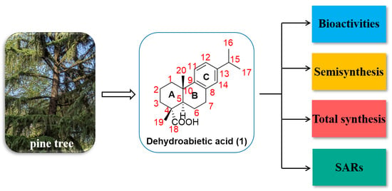
Graphical Abstract

1. Introduction

Terpenoids containing a variety of biological activities, are the promising lead structures for the development of drugs and pesticides [1,2,3,4,5,6]. Rosin is an important natural renewable resource in pine trees [7,8]. Dehydroabietic acid (1, Figure 1), a tricyclic diterpenoid, is a natural resin acid isolated from rosin and shows a wide range of biological activities [9]. The content of dehydroabietic acid in rosin was low, and it was not suitable for direct extraction of dehydroabietic acid. Storage-stable dehydroabietic acid can be purified by disproportionating abietic acid or rosin. In the industry, disproportionated rosin was obtained by disproportionation reaction of rosin as raw material, and then dehydroabietic acid can be obtained by organic amine salt and solvent recrystallization methods. Therefore, the research focus of purification was mainly on the preparation of disproportionated rosin, and the most effective catalyst for the disproportionation process of rosin was the expensive Pd/C catalyst, so it was very important to seek an efficient and economical purification condition [10]. In addition, dehydroabietic acid can also be isolated from two cyanobacteria strains [11].
The molecular skeleton of dehydroabietic acid contains one carboxyl group, one aromatic ring, and two alicyclic rings, with a total of twenty carbons. Dehydroabietic acid and its derivatives showed a variety of biological activities such as antiviral [12], antitumor [13], wound-healing [14], antiulcer [15], gastroprotective [16], anxiolytic [17], herbicidal [18] and antibacterial properties [19]. Furthermore, Xie et al. found that dehydroabietic acid had insecticidal activity against Peridroma saucia (Lepidoptera: Noctuidae) [20]. It demonstrated that larch sawfly exposure to dehydroabietic acid resulted in reduced feeding and slowed growth [21]. Dehydroabietic acid acted as an antagonist of insect juvenile hormone, interfering with the endocrine regulation of insects [22], and its derivatives also had an attracting effect on Spodoptera litura [23]. Recently, Xin et al. reported that novel multifunctional nanomedicines assembled from chitosan oligosaccharide-melanin complexes and dehydroabietic acid hexamers can achieve efficient and precise treatment of tumors [24]. Huang et al. found that biomass-based carbon dots prepared from dehydroabietic acid by hydrothermal reaction not only can sensitively and selectively detect heavy metal ions, but also can be used for cell imaging with low cytotoxicity [25]. However, while its biological activities were diverse, it had certain toxic and side effects. It reported that resin acids may be toxic to fish [26,27]. Based on the above studies, with the characteristics of good stability (due to the aromatic ring) and wide biological activities, dehydroabietic acid was a candidate of interest in the fields of medicine and agriculture. Moreover, to solve the disadvantages of its toxic and side effects, it was necessary to develop new dehydroabietic acid derivatives with high bioactivity and low toxicity. Therefore, this review summarized the structural modifications and biological activities of dehydroabietic acid and its derivatives from 2015 to 2021.

2. Bioactivities of Dehydroabietic Acid and Its Derivatives

2.1. Antitumor Activity

Dehydroabietic acid derivatives exhibited anti-cancer activity through various mechanisms including inhibiting tumor cell migration and inducing tumor apoptosis [7]. Lee et al. found that dehydroabietic acid induced apoptosis of human lung cells by inducing the division of caspase-3 and PARP in these cells and interfering with mitochondria [28]. Moreover, Luo et al. suggested that a derivative of dehydroabietic acid (2, Figure 2), maybe a therapeutic drug for gastric cancer because it induced damage to cell membranes and organelles, and ultimately led to apoptosis of gastric cancer cells [29]. Through flow cytometry and cell cycle analysis, it was found that compound 3 (Figure 2) can induce HepG2 cell apoptosis and block the HepG2 cell line in the G1 phase to exert anti-cancer effects [30]. Dehydroabietic oxime was a possible method for the treatment of pancreatic cancer and its related inflammation. It can up-regulate the level of p27, and it can also down-regulate the expression of cyclin D1, thereby preventing the growth of pancreatic cancer cells in the G1 phase [31]. Recently, some dehydroabietic acid derivatives were potential drugs targeting the kinase domain of EGFR, which showed anticancer activity against HepG2 cancer cell lines [32].

2.2. Anti-Inflammatory Activity

Kim et al. evaluated the anti-inflammatory effects of dehydroabietic acid and found that not only the production of nitric oxide (NO) in the macrophage cell line was reduced under the action of dehydroabietic acid, but also the expression of inflammatory genes was descended. In addition, through the treatment of dehydroabietic acid, both the activity of kinases in the NF-κB cascade and TAK1 (transforming growth factor β-activated kinase 1) in the AP-1 cascade was inhibited [33]. A study conducted by Kang et al. proved that the treatment of dehydroabietic acid was an effective way to ameliorate inflammatory changes related to obesity-related diabetes. It demonstrated that dehydroabietic acid was an activator of PPARα and PPARγ and can inhibit the production of MCP-1, TNF-α, and NO (pro-inflammatory mediators) [34].

2.3. Antibacterial and Antifungal Activities

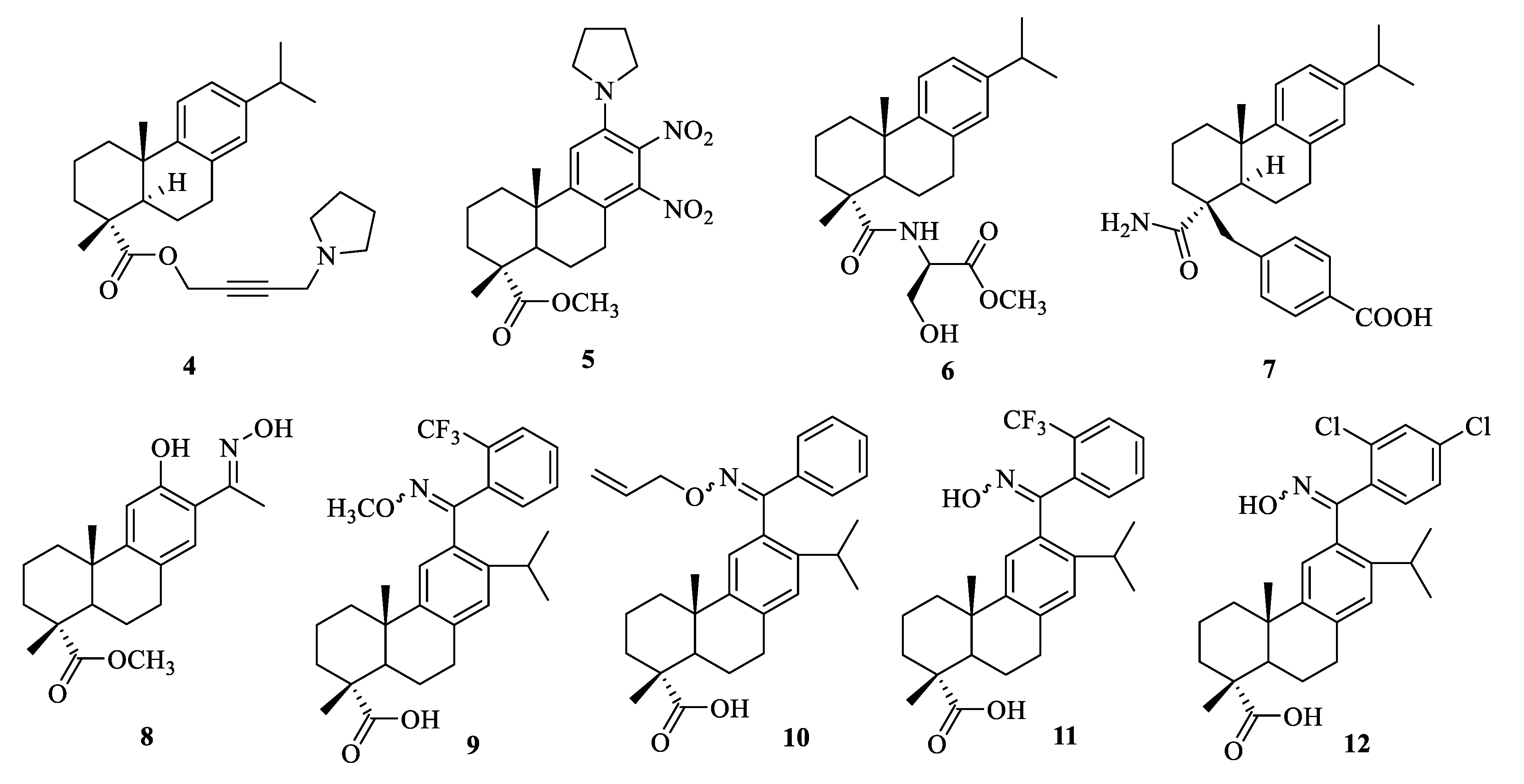
Experiments by Tretyakova et al. proved that pyrrolidine-containing dehydroabietic acid acetylene derivatives (4, Figure 3) showed growth inhibitory effects on the fungi Candida albicans and Cryptococcus neoformans, and had low hemolytic capacity [35]. In addition, Chen et al. reported that the derivative of dehydroabietic acid (5, Figure 3) exhibited excellent antibacterial activity against Gram-positive bacteria such as Bacillus subtilis and Staphylococcus aureus with the MIC (minimum inhibitory concentration) value of 4 and 2 μg/mL, respectively [36]. Dehydroabietic acid-containing serine derivatives had excellent antibacterial activity against Gram-positive bacteria. For example, the MIC90 values of compound 6 (Figure 3) against methicillin resistant S. aureus, Staphylococcus epidermidis, and Streptococcus mitis were 8 μg/mL [19]. Furthermore, through the broth micro-dilution method, it was found that dehydroabietic acid showed antibacterial activity against four kinds of Streptococcus mutans (including S. mutans ATCC 12175, S. mutans NRPC 801, S. mutans NRPC 804, and S. mutans DMST 18777) [37]. Hassan et al. used dehydroabietic acid derivatives to prepare antibacterial nanocellulose membranes to solve the inevitable toxicity of silver and cationic antimicrobial agents. By simulating the physiological environment of chronic wounds, they found that it prevented the colonization of bacteria on the surface [38]. It was revealed that the dehydroabietic acid analog (7, Figure 3) exhibited antimicrobial activity against methicillin resistant S. aureus (MIC: 32 μg/mL) [39]. It is urgent to develop new antibacterial agents against methicillin-resistant and methicillin-sensitive S. aureus infections (MRSA and MSSA). Compound 8 (Figure 3) had effective inhibitory activity against the above-mentioned bacteria, and its MIC values were from 3.9 to 15.6 μg/mL. Moreover, it had no cytotoxicity and hemolytic activity in mammalian cells [40]. In addition, Liu et al. elucidated that some 12-oxime and O-oxime ether derivatives of dehydroabietic acid had strong antistaphylococcal activity. Compound 9 showed high inhibitory activity against S. aureus Newman with MIC value of 0.39–0.78 μg/mL, while compounds 1012 had MIC values of 1.25–3.13 μg/mL for multidrug-resistant S. aureus. [41]. Additionally, some MIC values of dehydroabietic acid derivatives were listed in Table 1.

2.4. Insecticidal Activity

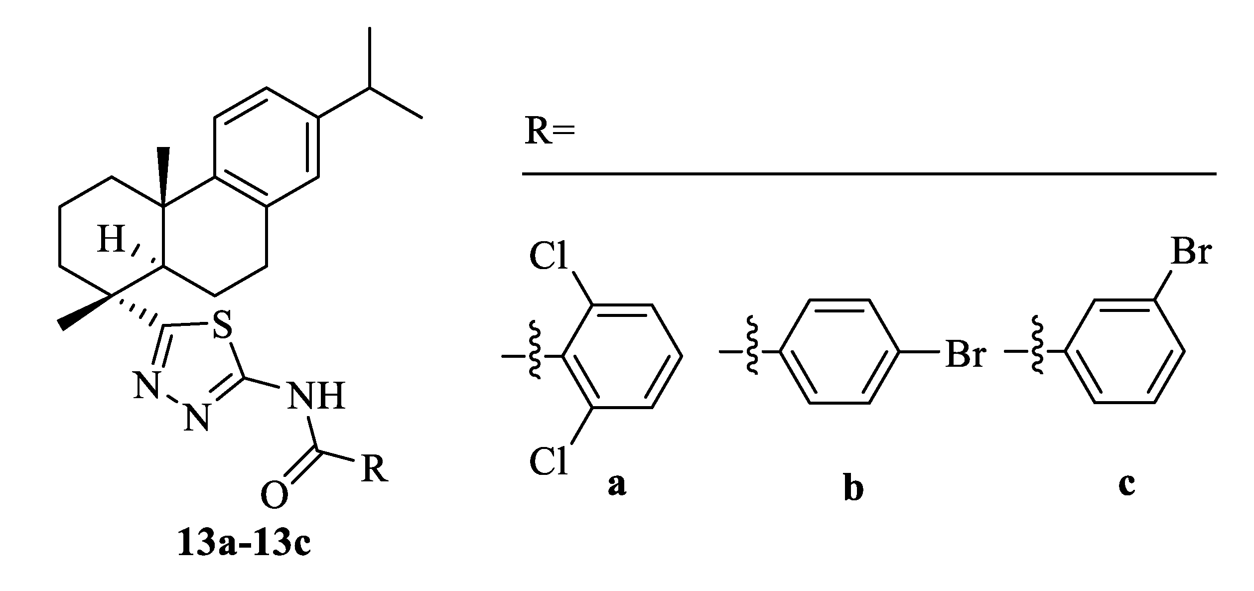
Dehydroabietic acid displayed antifeedant activity against Indian meal moth Plodia interpunctella, but had no obvious effect on the growth of larvae [22]. Additionally, dehydroabietic acid had certain insecticidal activity against Aedes aegypti larvae, with a lethality rate of 65% at 10 ppm [42]. Gao et al. found that dehydroabietic acid amide derivatives containing the thiadiazole fragment (13a13c, Figure 4) exhibited remarkable insecticidal activities against diamondback moth (Plutella xylostella) with the LC50 of 0.222–0.224 μg/mL [43]. Dehydroabietic acid had good antifeedant activity and a certain toxic effect on Mythimna separata larvae [44]. Moreover, the mountain pine beetle was often affected by the chemical defense of host oleoresin secretions which contained dehydroabietic acid [45].

2.5. Antiprotozoal Activity

Dehydroabietic acid not only inhibited the proliferation of the promastigote form, but also induced the production and output of ROS (reactive oxygen species) and downregulated Nrf2 (nuclear factor erythroid 2-related factor 2) to exert its anti-Leishmania activity, mainly due to its antioxidant properties [46]. Pertino et al. reported that dehydroabietic acid had certain antiprotozoal activity against Trypanosoma cruzi, Leishmaniabraziliensis, and Leishmania infantum [47]. Moreover, the dehydroabietic acid derivative containing amino acid structure was a new and effective antiprotozoal drug with good selectivity to Leishmania donovani and T. cruzi [48].

2.6. Other Activities

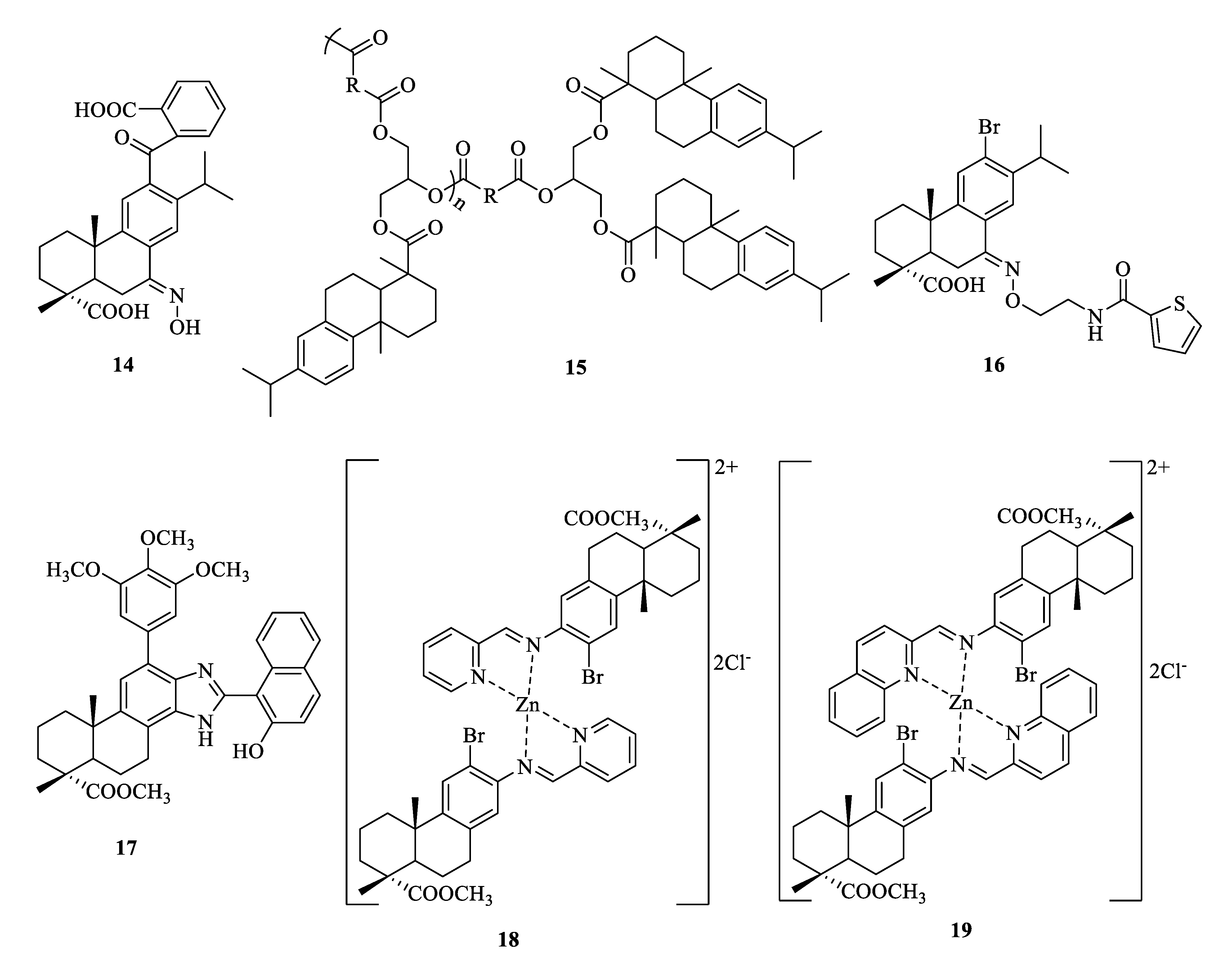
The treatment of dehydroabietic acid alleviated insulin resistance and liver steatosis caused by a high-fat diet in mice as a dual agonist of PPAR-α/γ [49]. Moreover, the anti-obesity effect of dehydroabietic acid may be exerted by improving the levels of plasma glucose and insulin via PPARα/γ-dependent pathways [34,50,51]. Dehydroabietic acid reduced hypertrophy by activating the Keap1/Nrf2-ARE signaling pathway, increasing the expression of the ferroptosis suppressor protein 1 (FSP1) gene, and inhibiting the accumulation of ROS to ameliorate non-alcoholic fatty liver disease [52]. Dehydroabietic acid inhibited angiotensin converting enzyme in human umbilical vein endothelial cells and induced p-Akt, which could reduce the systolic blood pressure of spontaneously hypertensive rats, thereby exerting an anti-hypertensive effect [53]. Dehydroabietic acid as an anti-aging agent could directly bind to SIRT1 protein and activated SIRT1 [54]. Park et al. suggested that dehydroabietic acid protected and regenerated the collagen fibers in the skin irradiated by ultraviolet B [55]. In addition, through in vivo and in vitro experiments, it was found that the assembled dehydroabietic acid derivative (14, Figure 5)/ZnAlTi-LDH (layered double hydroxide) composite material can promote wound healing, kill Gram-negative and positive bacteria infecting the wound, and block ultraviolet rays to protect the skin [56]. Since dehydroabietic acid can promote osteogenic differentiation, it may be a new idea for the treatment of osteoporosis in the elderly [57]. Through in vitro experimental studies, Nachar et al. found that dehydroabietic acid may be an effective drug for the treatment of type 2 diabetes. It not only reduced the activity of G6Pase to descend glucose production but also stimulated GS activity to ascend glucose storage [58]. Additionally, a dehydroabietic acid polymer (15, Figure 5) can be used as a toner for xerography [59]. BK channels, K+ channels activated by large-conductance calcium, had vital physiological functions such as the release of neurotransmitters. A thiophene derivative of dehydroabietic acid (16, Figure 5) showed BK channel opening activity [60,61]. A dehydroabietic acid derivative (17, Figure 5) can be used as fluorescent probes and can sensitively detect Fe3+ and Hg2+ ions in cells [62]. Moreover, dehydroabietic acid derivatives (18 and 19, Figure 5) as natural resources provided a new method for the synthesis of fluorescent materials [63].

3. Structural Modification of Dehydroabietic Acid and Its Derivatives

3.1. Structural Modification at C-18 of Dehydroabietic Acid

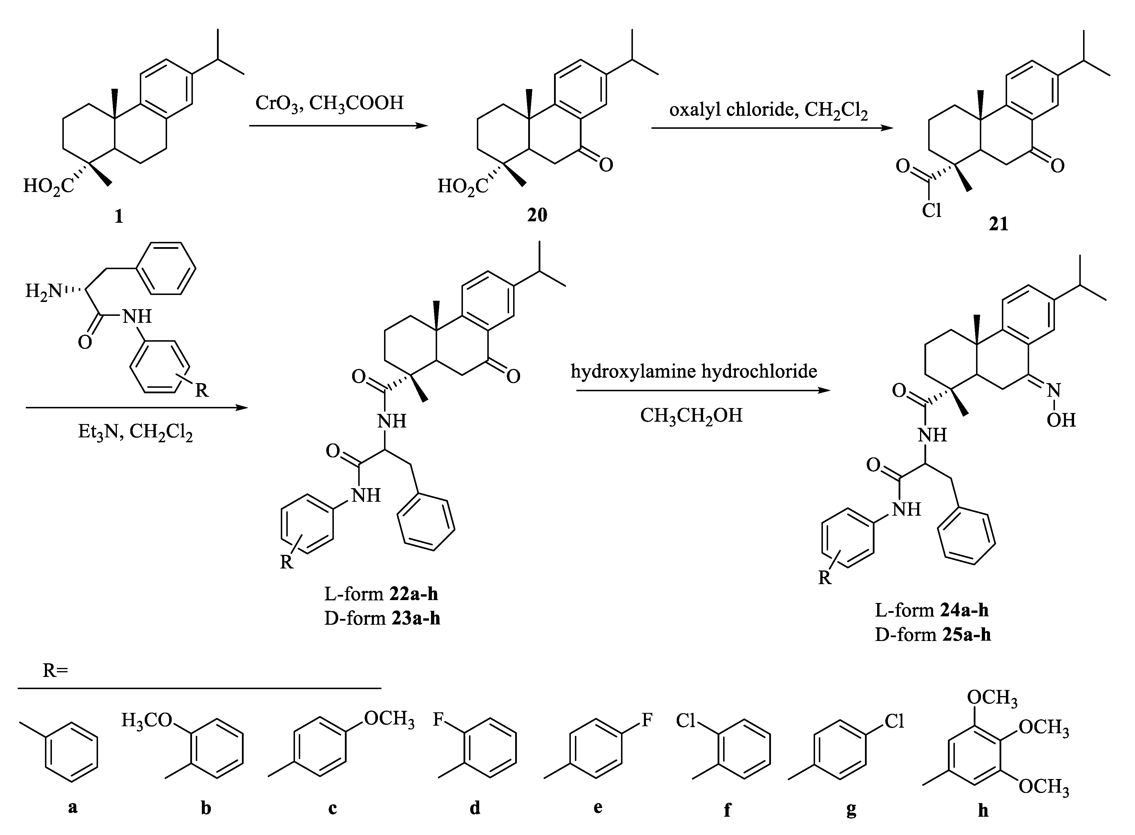
As depicted in Scheme 1, Huang et al. synthesized a series of chiral dipeptide derivatives (22(a-h)−25(a-h)) of dehydroabietic acid and tested their anticancer activities by the MTT method against HeLa, NCI-H460, and MGC-803 tumor cell lines. Among them, compound 22f had the strongest inhibitory effect on the HeLa cancer cell line, with an IC50 value of 7.76 ± 0.98 μM, and induced cell apoptosis through the mitochondrial pathway [64].
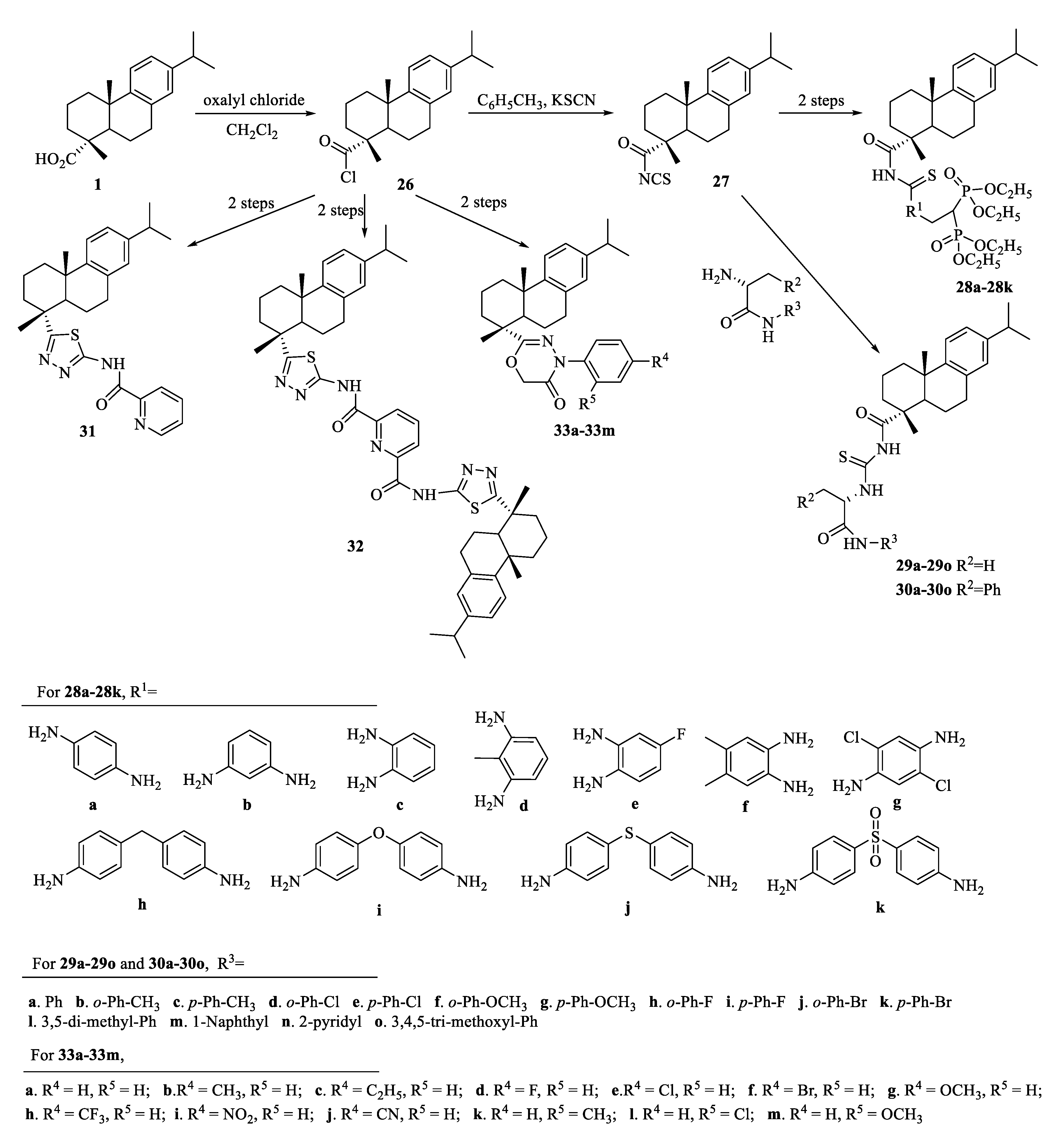
Subsequently, compounds containing thiourea and bisphosphonates (28a28k) were synthesized as effective antitumor agents (Scheme 2). Compound 28e showed the best anticancer activity against the SK-OV-3 cell line with an IC50 value of 1.79 ± 0.43 μM, which arrested the cell cycle in the G1 phase and induced apoptosis [65]. As shown in Scheme 2, a novel class of dehydroabietic acid acyl-thiourea derivatives (29(a-o)−30(a-o)) were obtained and their antitumor activity was evaluated against HeLa, SK-OV-3, HL-7702, and MGC-803 cell lines. Most of these compounds demonstrated cytotoxicity, especially compound 30n (IC50: 6.58 ± 1.11 μM against HeLa) exhibited better inhibitory effect than 5-Fu (a commercial antitumor drug, IC50: 36.58 ± 1.55 μM). Moreover, compound 30n could block HeLa cells in the S phase and induce HeLa cell apoptosis via the mitochondrial pathway [66]. In 2020, Li et al. designed and synthesized compounds 31 and 32 (Scheme 2). Compared with cisplatin and oxaliplatin, compound 31 had better cytotoxic activity on A431 cells and a stronger ability to bind with DNA [67]. Moreover, compounds 33a33m (Scheme 2) were synthesized as antitumor and antimicrobial agents. Compound 33e, a MIC value of 1.9 μg/mL, demonstrated the most potent inhibitory activity against B. subtilis. Meanwhile, compound 33m showed anticancer activity comparable to the positive control etoposide [68].
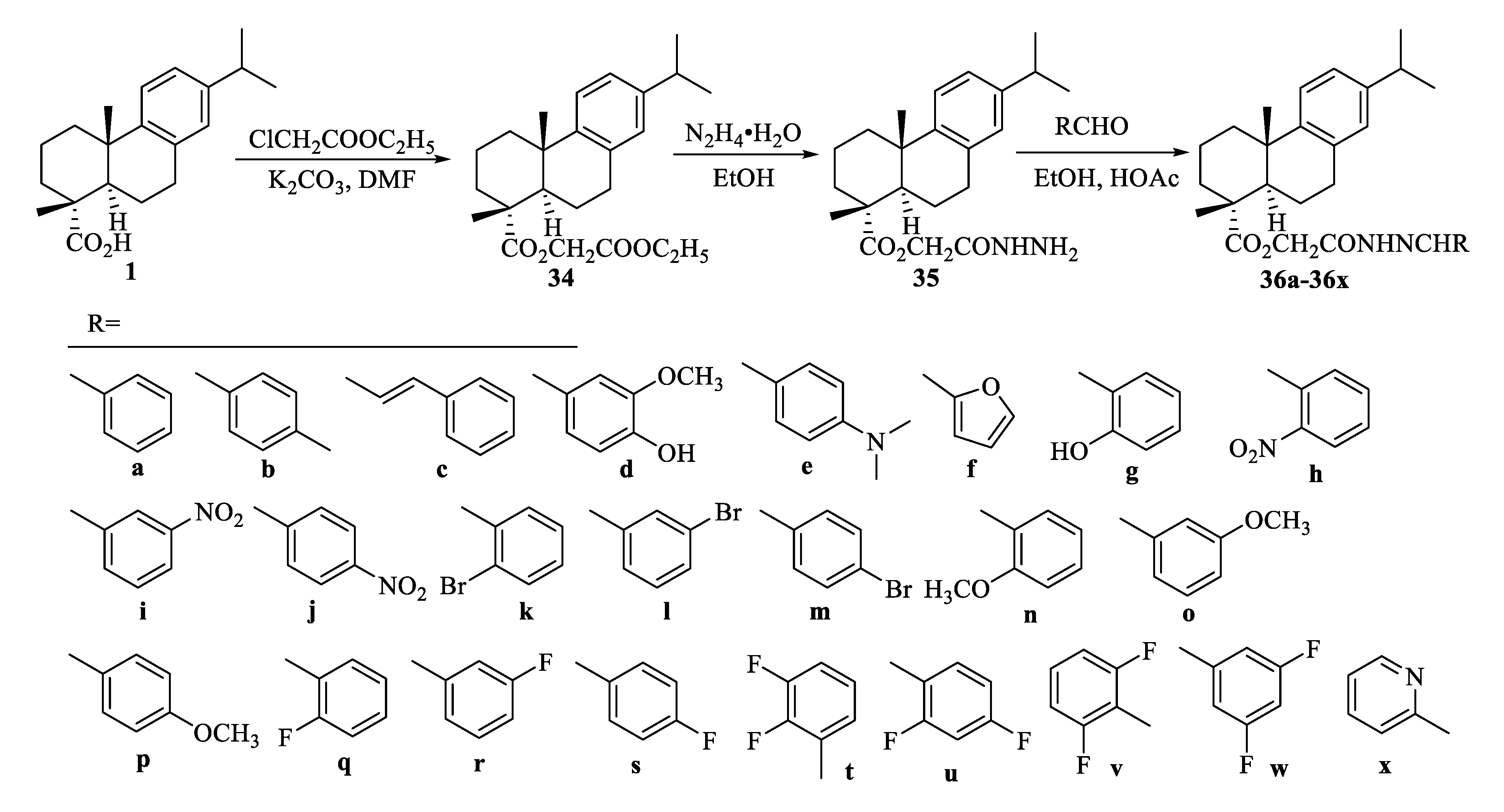
First, dehydroabietic acid (1) reacted with ethyl chloroacetate to obtain intermediate compound 34. Aminolysis of 34 gave hydrazide 35, which reacted with different aromatic aldehydes to obtain the target products 36a36x (Scheme 3). Compounds 36i, 36k, 36l, 36w, and 36x displayed potent anticancer activity. Especially compound 36w exhibited the promising inhibitory effects against HeLa and BEL-7402 cells with IC50 values of 2.21 and 14.46 µM, respectively [69].
As described in Scheme 4, Wang et al. designed and synthesized a novel class of dehydroabietic acid derivatives containing oxazolidinone moiety (39a39o). Compound 39j not only induced apoptosis of MGC-803 cells (IC50 = 3.82 ± 0.18 µM), but also arrested the cell cycle in the G1 phase [70].
Additionally, dehydroabietic acid containing 1,2,3-triazole derivatives (41a41p, Scheme 5) were prepared. The most potential compounds 41c (5.90 ± 0.41 µM) and 41k (6.25 ± 0.37 µM) exhibited better antiproliferation activity against HepG2 cells than that of cisplatin [71].
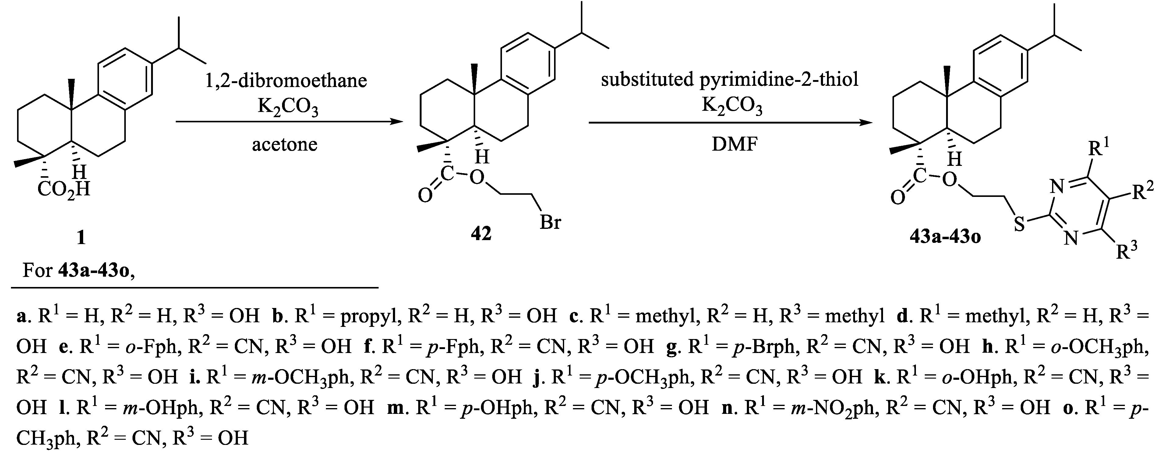
As depicted in Scheme 6, in the presence of anhydrous potassium carbonate, dehydroabietic acid (1) reacted with 1,2-dibromoethane to give intermediate 42. Target compounds 43a43o were obtained by reacting compound 42 with substituted pyrimidine-2-thiol. Compound 43b showed good inhibitory activity on the tested cells (IC50: 7.00–11.93 μM), but it had a little toxic effect on normal cells. The mechanism of action of compound 43b was to block MCF-7 cells in the S phase and induced apoptosis [72].
As described in Scheme 7, Li et al. synthesized a class of dehydroabietic acid-nitrate conjugates (47a47r) as antitumor agents. Compound 47n showed the most promising activity against the BEL-7402 cell line (IC50: 11.23 ± 0.21 μM). Compound 47j exhibited the most potent cytotoxicity against the CNE-2 cell line (IC50: 8.36 ± 0.14 μM) and evaluation of NO release indicated that the cytotoxic activity improved with the increase of the amount of NO produced in the CNE-2 cell line [73].
L-/D-amino acids and unusual amino acids were used as side chains to prepare new dehydroabietic acid derivatives (49a49f, Scheme 8). Compounds 49b and 49f were the most effective anti-biofilm agents, which can quickly and effectively destroy membrane integrity [74].
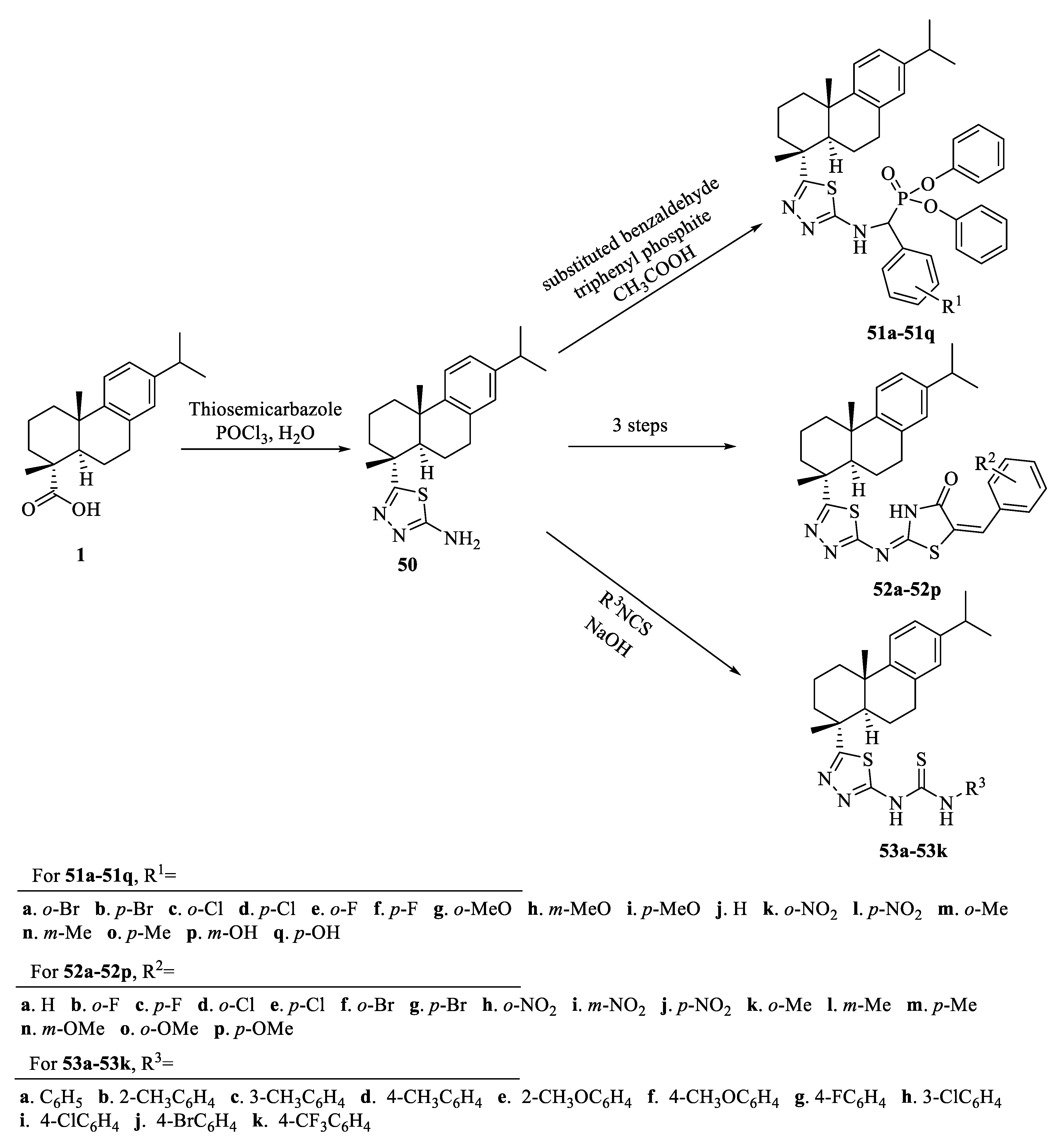
As illustrated in Scheme 9, compounds 51a51q were smoothly synthesized by cyclization and Mannich-type reactions. At 50 µg/mL, compounds 51e, 51f, 51h, and 51i had better antifungal activity than azoxystrobin (a commercial antifungal drug) [75]. A series of dehydroabietic acid derivatives containing 1,3,4-thiadiazole-thiazolidinone (52a52p) were synthesized as antifungal agents. They showed excellent antifungal activities against Gibberella zeae at a concentration of 50 µg/mL. Additionally, the antifungal activity of compounds 52c, 52f, and 52n (inhibition rate: 91.3%) was comparable to that of azoxystrobin (positive control) [76]. Mo et al. designed and synthesized a series of novel dehydroabietic acid derivatives containing 1,3,4-thiadiazole thiourea (53a53k) to determine their insecticidal activity against Helicoverpa armigera, P. xylostella, and Ostrinia mubilalis. Compounds 53a and 53c showed excellent insecticidal activity against H. armigera at 200 mg/L with mortality rates of 93.3% and 83.3%, respectively. The control effects of compounds 53b, 53c, 53d, and 53i against corn borer were 66.7%, 66.7%, 73.3%, and 60%, respectively [77].

3.2. Structural Modification at B Ring of Dehydroabietic Acid

A series of 7-N-acylaminopropyloxime derivatives of dehydroabietic acid (57a57s, Scheme 10) were synthesized, and their antibacterial activities against S. aureus Newman strain, NRS-1, NRS-70, NRS-100, NRS-108, and NRS-271 were studied. Compound 57j showed high antibacterial activities against five kinds of multi-drug resistant S. aureus with MIC values of 1.56–3.13 μg/mL [78].
In 2018, Zhang et al. synthesized a series of N-sulfonaminoethyloxime derivatives of dehydroabietic acid from compound 55 (Scheme 10). The MIC value of analog 59w against S. aureus Newman was 0.39–0.78 μg/mL, exhibiting the highest activity. In addition, the remaining analogs also showed better antibacterial activities against five multi-drug resistant S. aureus, with MIC values ranging from 0.78 to 1.56 μg/mL [79].
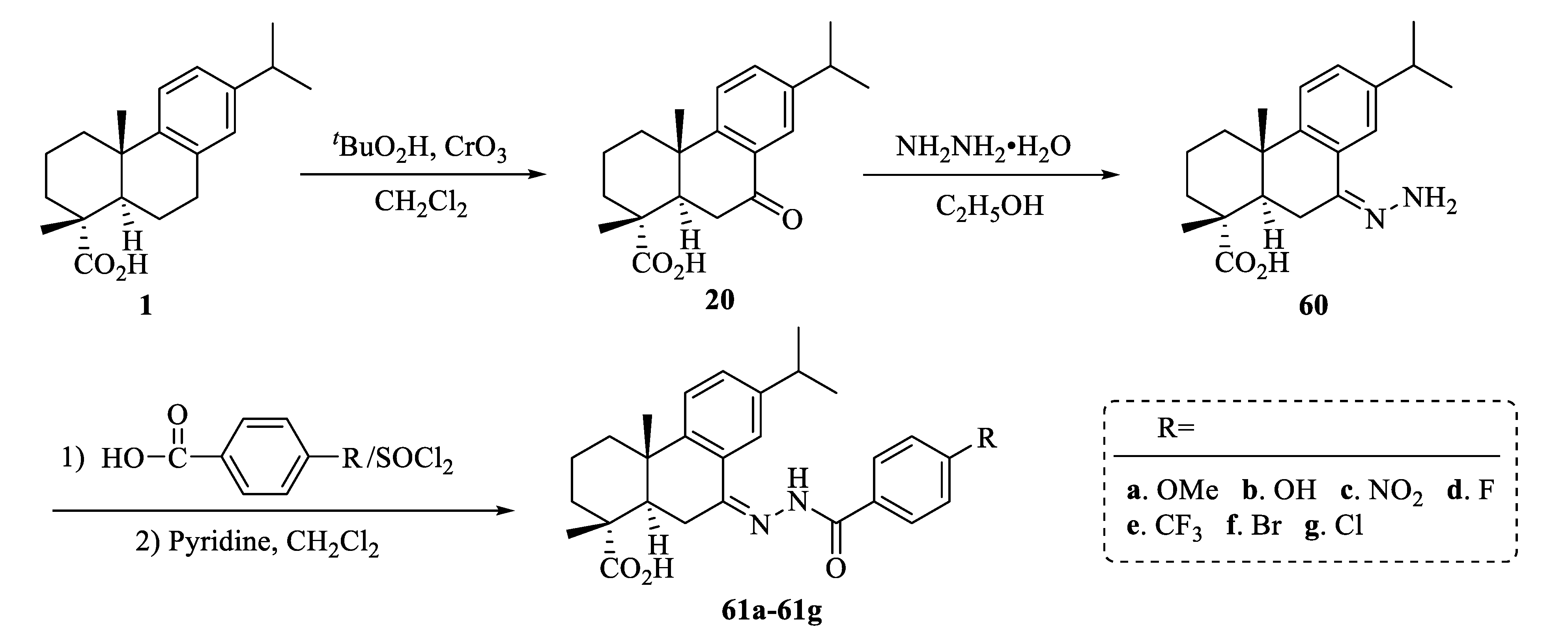
A series of hydrazone derivatives of dehydroabietic acid (61a61g, Scheme 11) were synthesized. All compounds showed antibacterial activities against Escherichia coli, S. aureus, and B. subtilis. Especially compound 61d was the potent antimicrobial agent against B. subtilis and S. aureus [80].
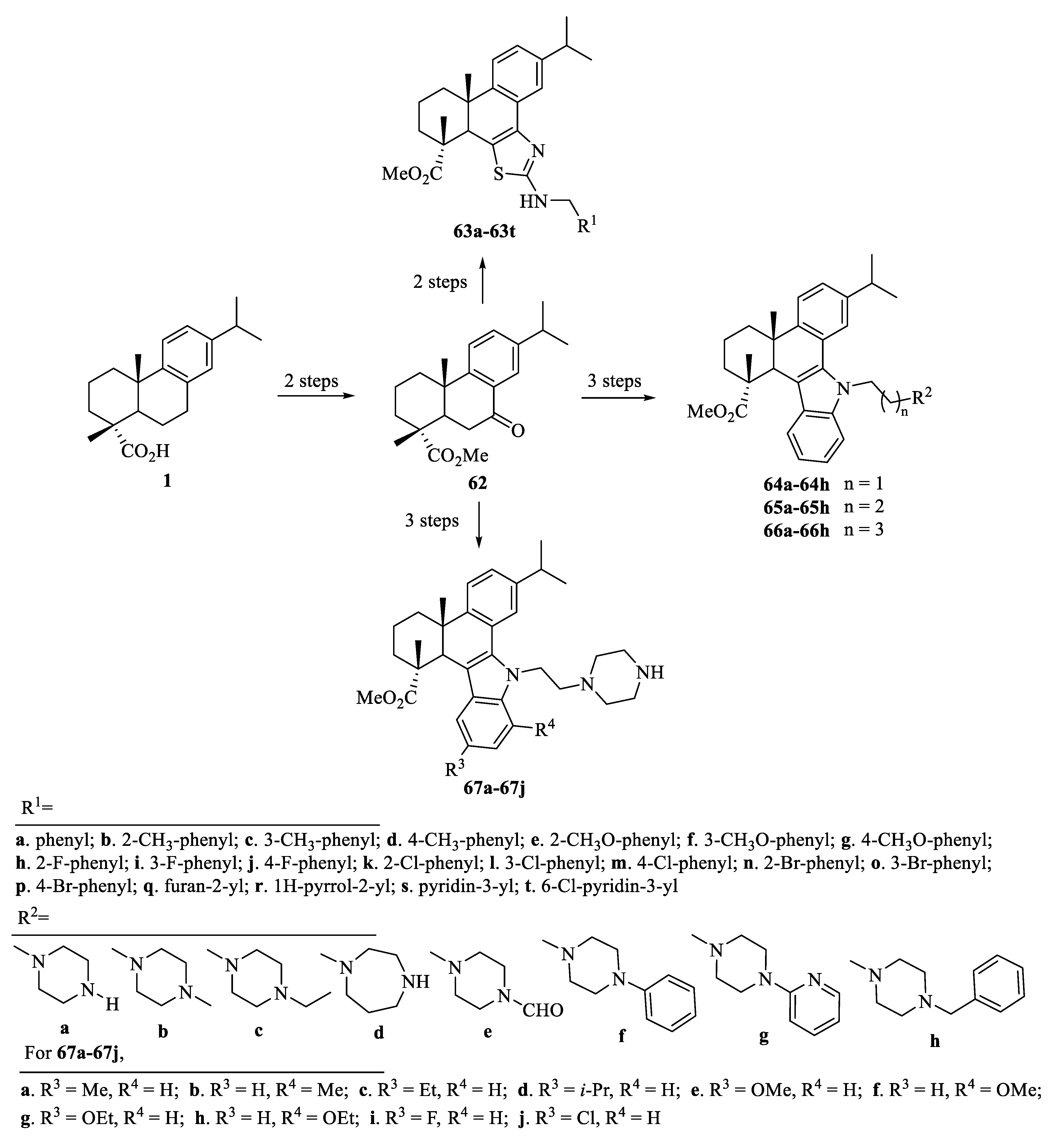
Chen et al. synthesized a series of dehydroabietic acid derivatives (63a63t, Scheme 12) to find novel and effective antitumor agents, and tested in vitro cytotoxic activity against HepG2, SCC9, and 293T by CCK-8 method. Compounds 63r and 63s had a certain cytotoxic activity to cancer cells, but were weak to normal cells. Two compounds could inhibit the PI3K/AKT/mTOR signaling pathway to exert anti-cancer effects. The results of molecular docking studies indicated that the compounds may inhibit the pathway through ATP competition [81].
As depicted in Scheme 12, compounds 64(a-h)66(a-h) and 67a67j were synthesized smoothly from dehydroabietic acid. Among these compounds, compound 67g had excellent anti-proliferative activity against three liver cancer cell lines (SMMC-7721, HepG2, and Hep3B), with IC50 values of 0.51–1.39 μM. In addition, compound 67g may inhibit MEK1 kinase activity, increase intracellular ROS levels, destroy cell membranes, and thus cause HepG2 cell apoptosis [82].

3.3. Structural Modification at C Ring of Dehydroabietic Acid

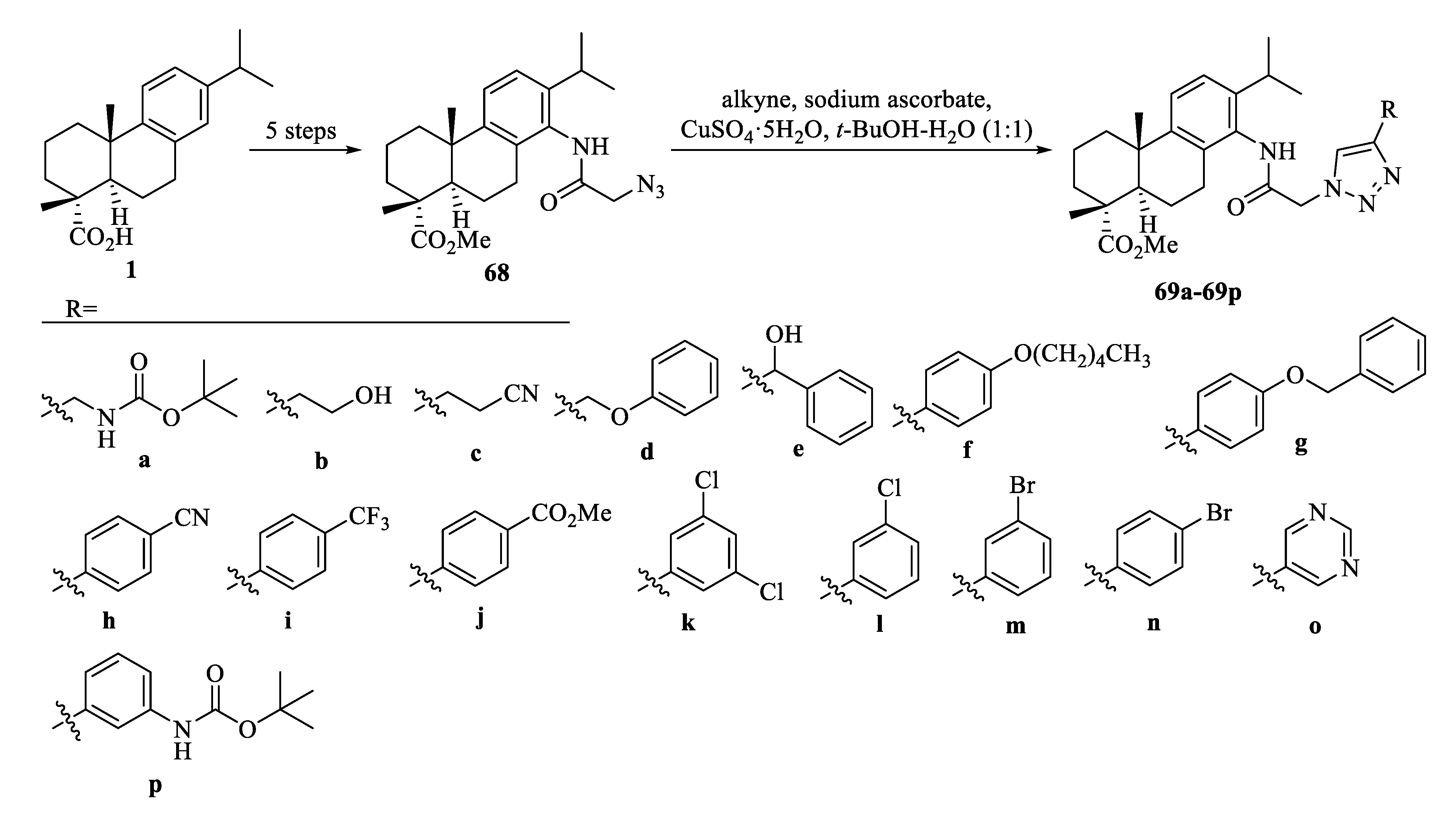
In order to find effective new antimicrobial agents, a class of new derivatives of dehydroabietic acid (69a69o, Scheme 13) was obtained. Compound 69o showed excellent antibacterial activity against both Gram-negative and positive bacteria (MIC: 1.6–3.1 μg/mL) but had no obvious toxicity to mammalian cells. In addition, compound 69o (containing 1,2,3-triazole moiety at the C-14 position) had good drug-like properties [83]. Additionally, compound 69p (IC50 values from 0.7 to 1.2 μM) not only induced apoptosis of MDA-MB-231 cells but also had weak toxicity to normal cells. And its anti-proliferative activity was better than 5-Fu (average IC50: 16.1 μM) [84].
Several derivatives of dehydroabietic acid containing 12-thiazole moiety (71a71e) were designed and synthesized as shown in Scheme 14. In order to understand the importance of hABHD16A inhibition in vivo, the feasibility of these derivatives as a starting point for the design of selective ABHD16A (a new target for inflammation-mediated pain) inhibitors was investigated. Compound 71d had an IC50 value of 3.4 ± 0.2 µM with good selectivity [85].
Gu et al. studied the cytotoxicity of a series of newly synthesized dehydroabietic acid derivatives (74(a-k)−75(a-k), Scheme 15) on liver cancer cells and found that most of the compounds had obvious cytotoxic activity on SMMC-7721 and HepG2 liver cancer cells with low toxicity on normal human liver cells. Among them, compounds 74b and 74e showed the best cytotoxicity against SMMC-7721 and HepG2 cells (IC50 values: 0.36 ± 0.13 and 0.12 ± 0.03 μM, respectively). In addition, compound 74b not only caused SMMC-7721 cells’ cell cycle arrest in the G2/M phase, but also induced apoptosis [86].
Subsequently, novel quinoxaline derivatives of dehydroabietic acid (77a77o, Scheme 15) were synthesized and tested against MCF-7, SMMC-7721, and HeLa cancer cell lines. Compound 77b caused SMMC-7721 cells to arrest in the G0/G1 phase of the cell cycle and induced their apoptosis in a dose-dependent manner. Its IC50 values against three different cancer cells were 0.72–1.78 μM, which had the best anti-cancer effect, while its toxicity to LO2 cells was reduced [87]. In 2020, a series of N-(1H-benzo[d]imidazol-2-yl)benzamide/benzenesulfonamide derivatives of dehydroabietic acid (78(a-h)−79(a-h), Scheme 15) were prepared. Compounds 78a, 78g, 78h, 79g, and 79h showed good anticancer effects on at least one cancer cell line (IC50 <10 μM), while compounds 1, 78b, 78c, 78e, and 78f were inactive (IC50 > 50 μM). In addition, benzenesulfonamide derivatives (79a79h) had stronger inhibitory activity than benzamide ones (78a78h). The most cytotoxic compound 79h (IC50: 0.87–9.39 μM) can arrest MCF-7 cells in the S phase through ROS-mediated mitochondrial pathway, and finally induce MCF-7 cell apoptosis [88].
Miao et al. synthesized a series of novel 2-aryl-benzimidazole derivatives of dehydroabietic acid (80(a-k)−81(a-k), Scheme 16). Compound 80j exhibited the strongest cytotoxic activity with the IC50 value of 0.08–0.42 μM. In addition, further in-depth exploration elucidated that compound 80j significantly inhibited the migration ability of SMMC-7721 cells, and induced cell cycle arrest and apoptosis of the cells in the G2/M phase. Through tubulin polymerization and immunofluorescence assays, it was found that compound 80j not only inhibited tubulin polymerization but also destroyed the intracellular microtubule network [89].
As shown in Scheme 17, a series of sulfonylurea derivatives of dehydroabietic acid (84a84k) were prepared. Compounds 84a, 84c, 84e, and 84i had higher anti-HCT-116 cell activity than 5-Fu. In particular, compound 84a, an IC50 value of which was 1.18 ± 0.52 μM, showed the best anti-tumor proliferation activity [90].
Some IC50 values and MIC values of the most active derivatives were listed in Table 2 and Table 3.

3.4. Biotransformation of Dehydroabietic Acid

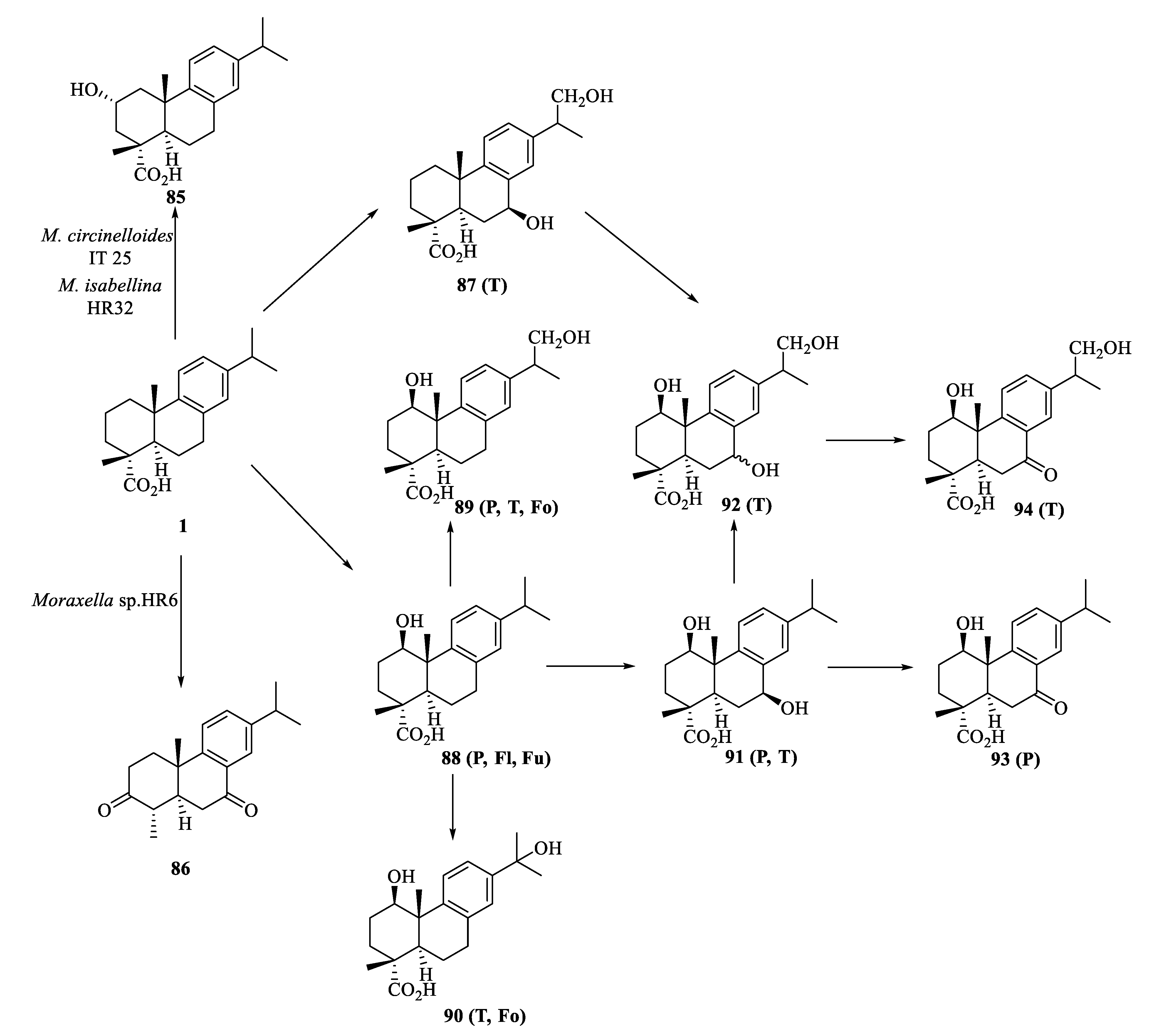
Possible pathways for the biotransformation of dehydroabietic acid by Mucor circinelloides IT25, Mortierella isabellina HR32, Moraxella sp. HR6, Trametes versicolor, Phlebiopsis gigantea, Flavobacterium resinovorum, Fusarium oxyosporum/F. moniliforme and Fomes annosum were shown in Scheme 18. These microorganisms were some of the previously reported microorganisms with hydroxylated metabolites. Two molds (M. circinelloides IT 25 and M. isabellina HR32) regioselectively and stereoselectively hydroxylated dehydroabietic acid to 2α-hydroxydehydroabietic acid (85). Dehydroabietic acid was oxidized at the C-3 and C-7 positions, decarboxylated at the C-4 position by the action of bacteria Moraxella sp. HR6, and finally converted to 3,7-dioxodehydroabietin (86) [91]. The first step in the degradation of dehydroabietic acid was caused by two fungi, T. versicolor and P. gigantea, and a stereoselective hydroxylation was at the C-1 position, followed by further hydroxylation at the C-7 or C-16 position to form dihydroxylated compounds 89 and 91. Compound 87 or 91 was further hydroxylated at the C-1 or C-7 position, resulting in trihydroxylated compound 92. The hydroxyl group at the C-7 position of compounds 91 and 92 can be further oxidized to a carbonyl functional group to afford compounds 93 and 94 [92,93].

4. Total Synthesis of Dehydroabietic Acid

Dehydroabietic acid has attracted the attention of researchers because of its diverse biological activities. However, separation and purification of dehydroabietic acid usually required expensive catalysts, and the organic amine salt method was more toxic and polluted the environment [10]. Therefore, it was particularly important to find other methods for synthesis of dehydroabietic acid. Some total synthesis processes of dehydroabietic acid were as follows.
In 1956, the first synthesis of dehydroabietic acid was begun with 2-isopropylnaphthalene (95) as the starting material (Scheme 19). The most important step was to define the C-4 center, that is, to alkylate phenanthrone (96) with ethyl bromoacetate. The enone (96) was then alkylated to obtain the keto ester (97), which reacted with HSCH2CH2SH to form the thioketal (98). The thioketal (98) was converted into its methyl ester (99) by ester hydrolysis and diazotization reactions. After Raney nickel desulfurization, ester hydrolysis, and Pd/C hydrogenation, intermediate 100 was obtained, which reacted with CH2N2 to produce methyl ester compound 101. Compound 103 was obtained by Barbiere Wieland degradation of 101. Finally, dehydroabietic acid (1) was obtained by hydrogenation of 103 on Pd/C [94].
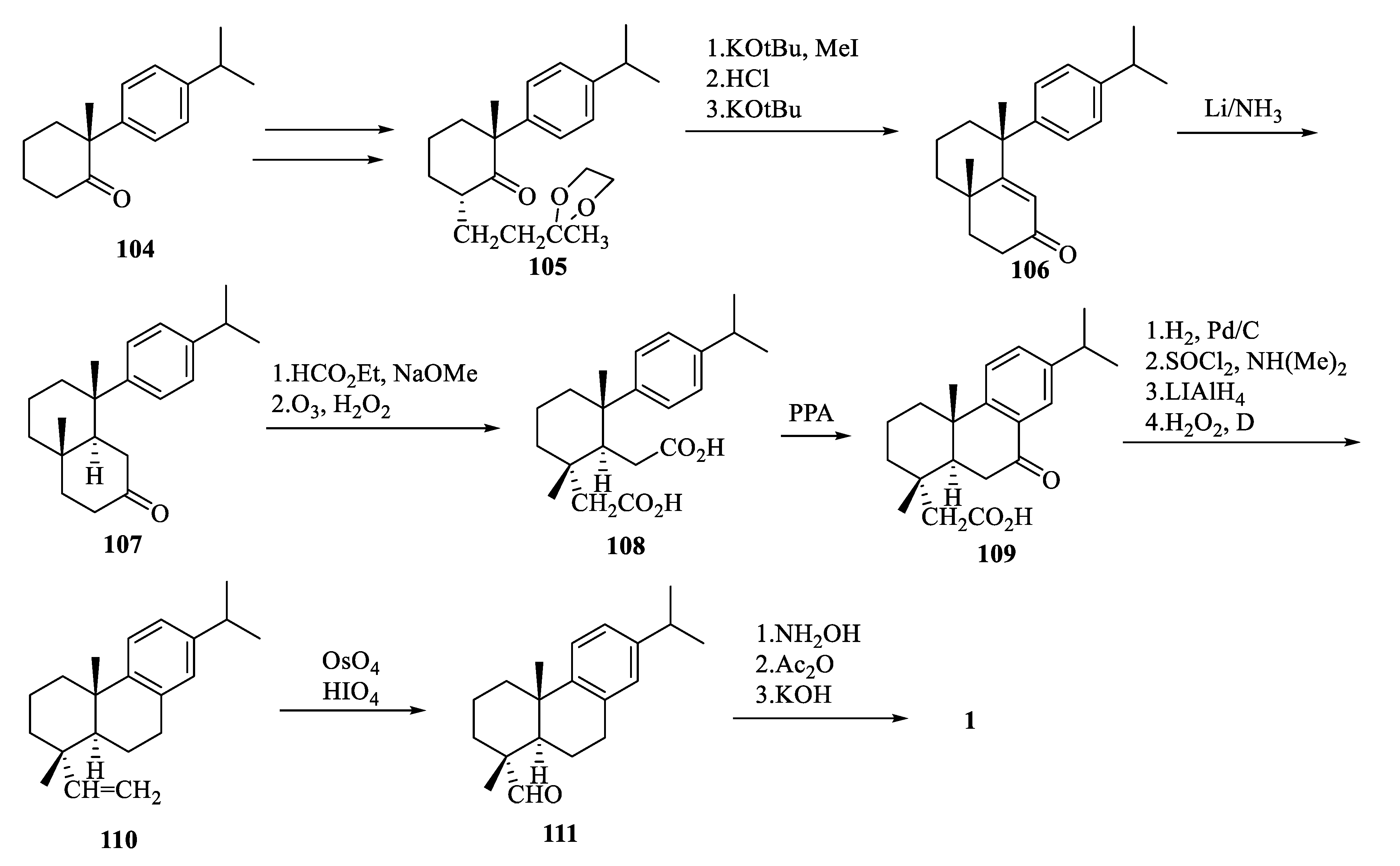
The second method started with 2-methyl-2-(p-isopropylphenyl)cyclohexanone (104) was described in Scheme 20. The key steps were stereoselective alkylation of 105 with KOt-Bu and MeI, and reduction with Li in liquid ammonia to give decalone 107, which reacted with HCO2Et, O3, and polyphosphoric acid (PPA) to give tricyclic keto acid 109. After the hydrogenolysis reaction, the amine oxide pyrolyzed to afford the vinylic compound 110, which reacted with OsO4 and HIO4 to give the corresponding aldehyde (111). Finally, dehydroabietic acid (1) was obtained by reaction of 111 with NH2OH and KOH [94].
The third method was a short enantioselective synthesis starting from the geranyl acetate derivative (112) as shown in Scheme 21. Compound 112 was converted to alkene 113 by the Wittig reaction, which was further catalyzed by Li2CuCl4 to give compound 114. The enantioselective cyclization of 114 and 115/SbCl5 complex afforded compound 116, which was a key step in the reaction. Finally, compound 116 reacted with KMnO4 and NaIO4 to obtain dehydroabietic acid (1) [94].

5. Structure-Activity Relationships of Dehydroabietic Acid and Its Derivatives

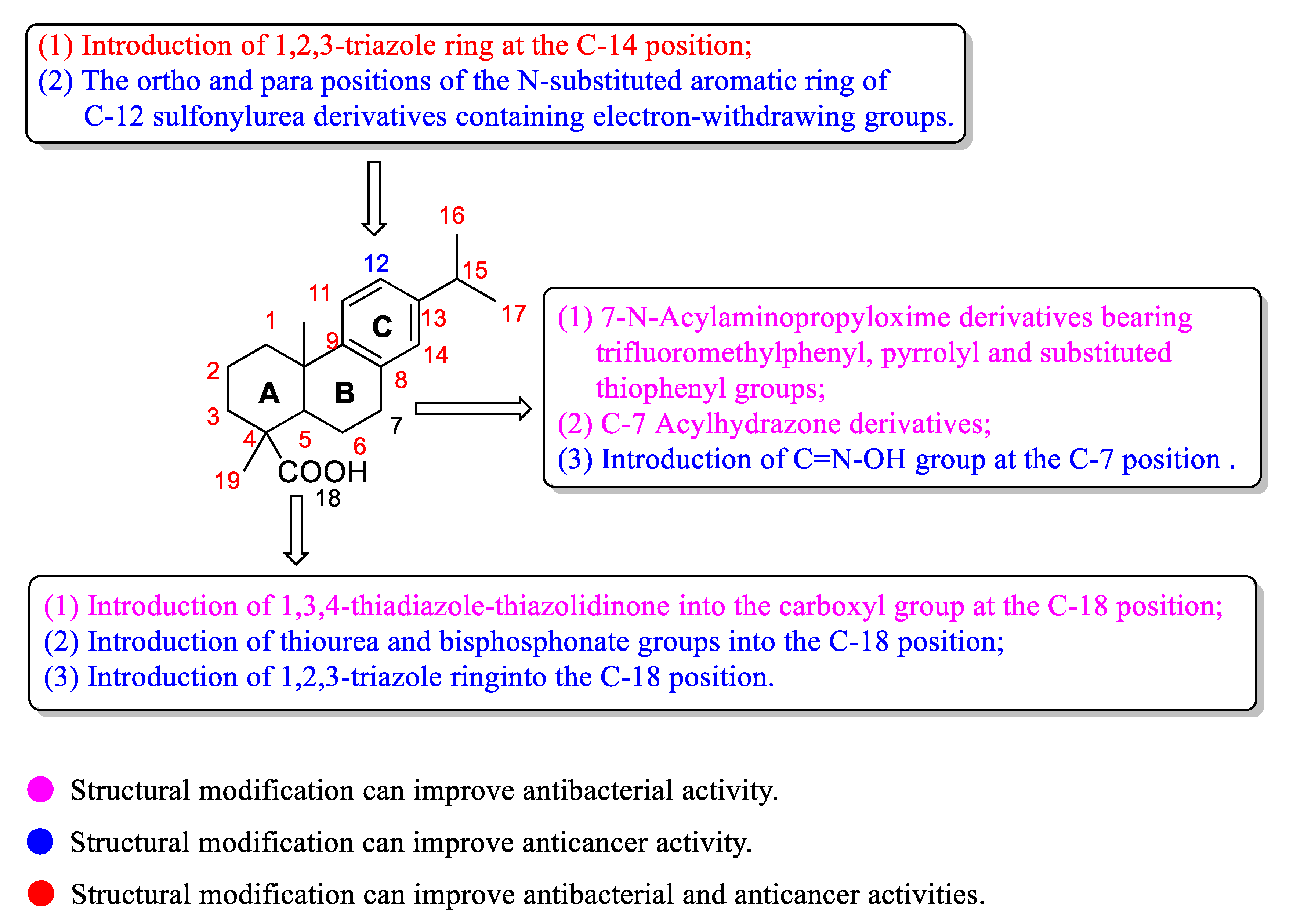
The SARs analysis of dehydroabietic acid and its derivatives about the antibacterial and anticancer activities (Figure 6) were as follows:
(1) For antibacterial activity: (a) Introduction of trifluoromethyl phenyl, pyrrolyl, or substituted thiophenyl groups (57h57j and 57p57s) into the oxime derivatives at the C-7 position of dehydroabietic acid was beneficial to the antibacterial activity [78]. C-7 Acylhydrazone derivatives of dehydroabietic acid (especially p-fluorobenzoyl hydrazone) can increase the antibacterial activity [80]. (b) Introduction of 1,3,4-thiadiazole-thiazolidinone into the C-18 carboxyl group of dehydroabietic acid can effectively improve the antibacterial activity [76]. (c) Introduction of 1,2,3-triazole ring at the C-14 position of dehydroabietic acid can improve the antibacterial activity [83].
(2) For anticancer activity: (a) Introduction of the C=N-OH group at the C-7 position can improve anti-tumor activity [64]. (b) Introduction of thiourea and bisphosphonate groups into the C-18 position of dehydroabietic acid was beneficial to the anticancer activity [65]. (c) 1,2,3-Triazole ring at the C-18 position played a key role for dehydroabietic acid derivatives showing the anti-cancer effect [71,84]. (d) C-12 sulfonylurea derivatives of dehydroabietic acid bearing electron-withdrawing groups at the ortho or para position on the N-substituted aromatic ring, can improve the antiproliferative activity [90].
Researchers expected to synthesize high bioactive compounds with low toxicity. Herein, we summarized some IC50 values of the most active derivatives against human normal cell lines (Table 4). Most of these derivatives exhibited more potent activity than dehydroabietic acid against cancer cell lines, and they showed low cytotoxicity against human normal cells. The IC50 value of compound 36w against HeLa cells (2.21 ± 0.04 μM) was about 17-folds higher than that of the precursor (37.40 ± 0.64 μM), while the cytotoxicity of compound 36w against normal HL-7702 human liver cell was lower (66.08 ± 1.84 μM) [69]. Compound 39j exhibited good cytotoxicity against the tested cancer cell lines (IC50 values:.82–17.76 μM) compared to the lead compound, with no increase in cytotoxicity against normal cells (IC50 > 100 μM) [70]. In addition, the anticancer activity of compounds 41c, 41k, 47j, and 47n was significantly improved, but their toxicity against human normal cells was not increased compared to that of dehydroabietic acid; that is, they exhibited excellent selectivity between normal and cancer cells [71,73]. The cytotoxicity of compound 77b against normal human liver cell LO2 (IC50: 11.09 ± 0.57 µM) was much lower than that of compound 77b against MCF-7, SMMC-7721, and HeLa cancer cells (IC50 values: 0.72–1.78 µM). However, compared with that of dehydroabietic acid (IC50 > 50 μM), it was more toxic to normal cells [87]. Compound 79h was also more cytotoxic against normal human LO2 cell (IC50: 42.83 ± 3.18 μM) than dehydroabietic acid (IC50 > 50 μM) [88]. Based on the above results, when compared with the precursor, the bioactivity of some compounds was significantly improved after structural modifications, but their toxicity against normal human cells was also increased. Fortunately, compounds 39j, 41c, 41k, 47j, and 47n not only showed promising biological activity, but also had good selectivity between normal and malignant cells; so they can be further studied and developed as potential anticancer drugs.

6. Conclusions

Dehydroabietic acid, a tricyclic diterpenoid resin acid, showed a wide range of biological activities. The structural characteristics suggested that dehydroabietic acid had multiple structural modification sites, and using it as a lead compound can improve its application value. This review gave an overview of the biological activities, structural modifications, biotransformation, total synthesis, and structure-activity relationships of dehydroabietic acid and its derivatives. Although several structural modifications of dehydroabietic acid and its derivatives were summarized, their properties were mainly focused on anticancer and antibacterial activities. Obviously, their agricultural and other activities needed to be expanded in the future. Therefore, we hope that this review can provide references for further research and development of dehydroabietic acid and its derivatives, as well as their applications as drugs and pesticides in the medical and agricultural fields.

Author Contributions

All persons who have made substantial contributions to present work are named in the manuscript. M.H., H.X. and M.L. designed the work, analyzed the data, and wrote and revised the paper. All authors have read and agreed to the published version of the manuscript.

Funding

This research was funded by the National Natural Science Foundation of China (Project No. 31872013).

Acknowledgments

M.L. want to thank the National Natural Science Foundation of China (No. 31872013), and State Key Laboratory of Elemento-Organic Chemistry, Nankai University (No. 202109).

Conflicts of Interest

The authors declare no conflict of interest.

Abbreviations

AP-1Activating protein-1
5-Fu5-Fluorouracil
G6PaseGlucose-6-Phosphatase
GSGlycogen Synthase
IC50Half-inhibitory concentration
MCP-1Monocyte chemoattractant protein-1
MICMinimum inhibitory concentration
NF-κBNuclear factor-κB
Nrf2Nuclear factor erythroid 2-related factor 2
NONitric oxide
PARPPoly (ADP-ribose) polymerase
PPAPolyphosphoric acid
PPARPeroxisome proliferator-activated receptors
ROSReactive oxygen species
TAK1Transforming growth factor β-activated kinase 1
TNF-αTumor necrosis factor-α

References

  1. Zhang, Y.Y.; Xu, H. Recent progress in the chemistry and biology of limonoids. RSC Adv. 2017, 7, 35191–35220. [Google Scholar] [CrossRef]
  2. Li, Q.; Huang, X.; Li, S.; Ma, J.; Lv, M.; Xu, H. Semisynthesis of esters of fraxinellone C4/10-oxime and their pesticidal activities. J. Agric. Food Chem. 2016, 64, 5472–5478. [Google Scholar] [CrossRef]
  3. Nagini, S.; Nivetha, R.; Palrasu, M.; Mishra, R. Nimbolide, a neem limonoid, is a promising candidate for the anticancer drug arsenal. J. Med. Chem. 2021, 64, 3560–3577. [Google Scholar] [CrossRef] [PubMed]
  4. Huang, X.B.; Li, T.; Shan, X.; Lu, R.; Hao, M.; Lv, M.; Sun, Z.; Xu, H. High value-added use of citrus industrial wastes in agriculture: Semisynthesis and anti-tobacco mosaic virus/insecticidal activities of ester derivatives of limonin modified in the B ring. J. Agric. Food Chem. 2020, 68, 12241–12251. [Google Scholar] [CrossRef] [PubMed]
  5. Huang, X.B.; Lv, M.; Ma, Q.; Zhang, Y.; Xu, H. High value-added application of natural products in crop protection: Semisynthesis and acaricidal activity of limonoid-type derivatives and investigation of their biocompatible O/W nanoemulsions as agronanopesticide candidates. J. Agric. Food Chem. 2021, 69, 14488–14500. [Google Scholar] [CrossRef]
  6. Sun, Y.; Yin, Y.; Sun, Y.; Li, Q.; Cui, L.; Xu, W.; Kong, L.; Luo, J. Aglatestine A, a rearranged limonoid with a 3/6/6 tricarbocyclic framework from the fruits of Aglaia edulis. J. Org. Chem. 2021, 86, 11263–11268. [Google Scholar] [CrossRef] [PubMed]
  7. Huang, X.C.; Wang, M.; Pan, Y.M.; Yao, G.Y.; Wang, H.S.; Tian, X.Y.; Qin, J.K.; Zhang, Y. Synthesis and antitumor activities of novel thiourea α-aminophosphonates from dehydroabietic acid. Eur. J. Med. Chem. 2013, 69, 508–520. [Google Scholar] [CrossRef]
  8. Xu, H.; Liu, L.; Fan, X.; Zhang, G.; Li, Y.; Jiang, B. Identification of a diverse synthetic abietane diterpenoid library for anticancer activity. Bioorganic Med. Chem. Lett. 2017, 27, 505–510. [Google Scholar] [CrossRef] [PubMed]
  9. Gonzalez, M.A.; Perez-Guaita, D.; Correa-Royero, J.; Zapata, B.; Agudelo, L.; Mesa-Arango, A.; Betancur-Galvis, L. Synthesis and biological evaluation of dehydroabietic acid derivatives. Eur. J. Med. Chem. 2010, 45, 811–816. [Google Scholar] [CrossRef] [PubMed]
  10. Han, C.R.; Song, Z.Q.; Shang, S.B. Research progress on abietic acid, dehydroabietic acid and their bioactive derivatives. Chem. Ind. Eng. Prog. 2007, 26, 490–495. [Google Scholar] [CrossRef]
  11. Costa, M.S.; Rego, A.; Ramos, V.; Afonso, T.B.; Freitas, S.; Preto, M.; Lopes, V.; Vasconcelos, V.; Magalhaes, C.; Leao, P.N. The conifer biomarkers dehydroabietic and abietic acids are widespread in Cyanobacteria. Sci. Rep. 2016, 6, 23436. [Google Scholar] [CrossRef]
  12. Tagat, J.R.; Nazareno, D.V.; Puar, M.S.; McCombie, S.W.; Ganguly, A.K. Synthesis and anti-herpes activity of some A-ring functionalized dehydroabietane derivatives. Bioorganic Med. Chem. Lett. 1994, 4, 1101–1104. [Google Scholar] [CrossRef]
  13. Kim, W.-J.; Kang, H.-G.; Kim, S.-J. Dehydroabietic acid inhibits the gastric cancer cell growth via induced apoptosis and cell cycle arrest. Mol. Cell. Toxicol. 2021, 17, 133–139. [Google Scholar] [CrossRef]
  14. Jokinen, J.J.; Sipponen, A. Refined spruce resin to treat chronic wounds: Rebirth of an old folkloristic therapy. Adv. Wound Care 2016, 5, 198–207. [Google Scholar] [CrossRef]
  15. Gonzalez, M.A. Aromatic abietane diterpenoids: Their biological activity and synthesis. Nat. Prod. Rep. 2015, 32, 684–704. [Google Scholar] [CrossRef]
  16. Sepulveda, B.; Astudillo, L.; Rodriguez, J.A.; Yanez, T.; Theoduloz, C.; Schmeda-Hirschmann, G. Gastroprotective and cytotoxic effect of dehydroabietic acid derivatives. Pharmacol. Res. 2005, 52, 429–437. [Google Scholar] [CrossRef] [PubMed]
  17. Tolmacheva, I.A.; Taranfin, A.V.; Boteva, A.A.; Anikina, L.V.; Vikharev, Y.B.; Grishko, V.V.; Tolstikov, A.G. Synthesis and biological activity of nitrogen-containing derivatives of methyl dehydroabietate. Pharm. Chem. J. 2006, 40, 489–493. [Google Scholar] [CrossRef]
  18. Duan, W.G.; Li, X.R.; Mo, Q.J.; Huang, J.X.; Cen, B.; Xu, X.T.; Lei, F.H. Synthesis and herbicidal activity of 5-dehydroabietyl-1,3,4-oxadiazole derivatives. Holzforschung 2011, 65, 191–197. [Google Scholar] [CrossRef]
  19. Helfenstein, A.; Vahermo, M.; Nawrot, D.A.; Demirci, F.; Iscan, G.; Krogerus, S.; Yli-Kauhaluoma, J.; Moreira, V.M.; Tammela, P. Antibacterial profiling of abietane-type diterpenoids. Bioorganic Med. Chem. 2017, 25, 132–137. [Google Scholar] [CrossRef] [PubMed]
  20. Xie, Y.S.; Isman, M.B.; Yi, F.; Wong, A. Diterpene resin acids: Major active principles in tall oil against Variegated cutworm, Peridroma saucia (Lepidoptera, Noctuidae). J. Chem. Ecol. 1993, 19, 1075–1084. [Google Scholar] [CrossRef] [PubMed]
  21. Robert, J.A.; Madilao, L.L.; White, R.; Yanchuk, A.; King, J.; Bohlmann, J. Terpenoid metabolite profiling in Sitka spruce identifies association of dehydroabietic acid, (+)-3-carene, and terpinolene with resistance against white pine weevil. Botany 2010, 88, 810–820. [Google Scholar] [CrossRef]
  22. Oh, H.W.; Yun, C.S.; Jeon, J.H.; Kim, J.A.; Park, D.S.; Ryu, H.W.; Oh, S.R.; Song, H.H.; Shin, Y.; Jung, C.S.; et al. Conifer diterpene resin acids disrupt juvenile hormone-mediated endocrine regulation in the Indian meal moth Plodia interpunctella. J. Chem. Ecol. 2017, 43, 703–711. [Google Scholar] [CrossRef]
  23. Rao, X.; Song, Z.; Han, Z.; Jiang, Z. Synthesis and insect attractant activity of fluorine-containing Pinus diterpenic amides and imines. Nat. Prod. Res. 2009, 23, 851–860. [Google Scholar] [CrossRef] [PubMed]
  24. Xin, C.; Zhang, Y.; Bao, M.; Yu, C.; Hou, K.; Wang, Z. Novel carrier-free, charge-reversal and DNA-affinity nanodrugs for synergistic cascade cancer chemo-chemodynamic therapy. J. Colloid Interface Sci. 2022, 606, 1488–1508. [Google Scholar] [CrossRef] [PubMed]
  25. Huang, X.; Liu, J.; Zhao, B.; Bai, Y.; Peng, Z.; Zhou, J.; Wang, C.; Zhao, X.; Han, S.; Zhang, C. One-step synthesis of biomass-based carbon dots for detection of metal ions and cell imaging. Front. Energy Res. 2022, 10, 871617. [Google Scholar] [CrossRef]
  26. Singh, A.K.; Chandra, R. Pollutants released from the pulp paper industry: Aquatic toxicity and their health hazards. Aquat. Toxicol. 2019, 211, 202–216. [Google Scholar] [CrossRef] [PubMed]
  27. Pandelides, Z.; Guchardi, J.; Holdway, D. Dehydroabietic acid (DHAA) alters metabolic enzyme activity and the effects of 17β-estradiol in rainbow trout (Oncorhynchus mykiss). Ecotoxicol. Environ. Saf. 2014, 101, 168–176. [Google Scholar] [CrossRef]
  28. Lee, S.; Lee, S.; Roh, H.S.; Song, S.S.; Ryoo, R.; Pang, C.; Baek, K.H.; Kim, K.H. Cytotoxic constituents from the sclerotia of Poria cocos against human lung adenocarcinoma cells by inducing mitochondrial apoptosis. Cells 2018, 7, 116. [Google Scholar] [CrossRef]
  29. Luo, D.; Ni, Q.; Ji, A.; Gu, W.; Wu, J.; Jiang, C. Dehydroabietic acid derivative QC4 induces gastric cancer cell death via oncosis and apoptosis. Biomed. Res. Int. 2016, 2016, 2581061. [Google Scholar] [CrossRef]
  30. Huang, R.Z.; Liang, G.B.; Huang, X.C.; Zhang, B.; Zhou, M.M.; Liao, Z.X.; Wang, H.S. Discovery of dehydroabietic acid sulfonamide based derivatives as selective matrix metalloproteinases inactivators that inhibit cell migration and proliferation. Eur. J. Med. Chem. 2017, 138, 979–992. [Google Scholar] [CrossRef]
  31. Kolsi, L.E.; Leal, A.S.; Yli-Kauhaluoma, J.; Liby, K.T.; Moreira, V.M. Dehydroabietic oximes halt pancreatic cancer cell growth in the G1 phase through induction of p27 and downregulation of cyclin D1. Sci. Rep. 2018, 8, 15923. [Google Scholar] [CrossRef] [PubMed]
  32. Zaki, H.; Belhassan, A.; Benlyas, M.; Lakhlifi, T.; Bouachrine, M. New dehydroabietic acid (DHA) derivatives with anticancer activity against HepG2 cancer cell lines as a potential drug targeting EGFR kinase domain. CoMFA study and virtual ligand-based screening. J. Biomol. Struct. Dyn. 2021, 39, 2993–3003. [Google Scholar] [CrossRef] [PubMed]
  33. Kim, E.; Kang, Y.G.; Kim, Y.J.; Lee, T.R.; Yoo, B.C.; Jo, M.; Kim, J.H.; Kim, J.H.; Kim, D.; Cho, J.Y. Dehydroabietic acid suppresses inflammatory response via suppression of Src-, Syk-, and TAK1-mediated pathways. Int. J. Mol. Sci. 2019, 20, 1593. [Google Scholar] [CrossRef] [PubMed]
  34. Kang, M.S.; Hirai, S.; Goto, T.; Kuroyanagi, K.; Lee, J.Y.; Uemura, T.; Ezaki, Y.; Takahashi, N.; Kawada, T. Dehydroabietic acid, a phytochemical, acts as ligand for PPARs in macrophages and adipocytes to regulate inflammation. Biochem. Biophys. Res. Commun. 2008, 369, 333–338. [Google Scholar] [CrossRef] [PubMed]
  35. Tretyakova, E.V.; Salimova, E.V.; Parfenova, L.V. Synthesis and antimicrobial and antifungal activity of resin acid acetylene derivatives. Russ. J. Bioorganic Chem. 2020, 45, 545–551. [Google Scholar] [CrossRef]
  36. Chen, H.; Geng, Y.; Wang, S.F.; Gu, W. Syntheses, crystal structures and antibacterial activities of two new methyl 12-alkylamino-13,14-dinitrodeisopropyl-dehydroabietates. Chin. J. Struct. Chem. 2019, 38, 257–262. [Google Scholar] [CrossRef]
  37. Sakunpak, A.; Sueree, L. Thin-layer chromatography–contact bioautography as a tool for bioassay-guided isolation of anti-Streptococcus mutans compounds from Pinus merkusii heartwood. JPC-J. Planar Chromat. 2018, 31, 355–359. [Google Scholar] [CrossRef]
  38. Hassan, G.; Forsman, N.; Wan, X.; Keurulainen, L.; Bimbo, L.M.; Stehl, S.; van Charante, F.; Chrubasik, M.; Prakash, A.S.; Johansson, L.-S.; et al. Non-leaching, highly biocompatible nanocellulose surfaces that efficiently resist fouling by bacteria in an artificial dermis model. ACS Appl. Bio Mater. 2020, 3, 4095–4108. [Google Scholar] [CrossRef]
  39. Berger, M.; Roller, A.; Maulide, N. Synthesis and antimicrobial evaluation of novel analogues of dehydroabietic acid prepared by C-H-Activation. Eur. J. Med. Chem. 2017, 126, 937–943. [Google Scholar] [CrossRef] [PubMed]
  40. Chaban, M.F.; Antoniou, A.I.; Karagianni, C.; Toumpa, D.; Joray, M.B.; Bocco, J.L.; Sola, C.; Athanassopoulos, C.M.; Carpinella, M.C. Synthesis and structure-activity relationships of novel abietane diterpenoids with activity against Staphylococcus aureus. Future Med. Chem. 2019, 11, 3109–3124. [Google Scholar] [CrossRef] [PubMed]
  41. Liu, M.L.; Pan, X.Y.; Yang, T.; Zhang, W.M.; Wang, T.Q.; Wang, H.Y.; Lin, H.X.; Yang, C.G.; Cui, Y.M. The synthesis and antistaphylococcal activity of dehydroabietic acid derivatives: Modifications at C-12. Bioorganic Med. Chem. Lett. 2016, 26, 5492–5496. [Google Scholar] [CrossRef]
  42. Touré, S.; Dusfour, I.; Stien, D.; Eparvier, V. Two new tetrahydrofuran derivatives from the fungus Mucor spp. SNB-VECD11D isolated from the Chrysomelidae Acalymma bivittula. Tetrahedron Lett. 2017, 58, 3727–3729. [Google Scholar] [CrossRef]
  43. Gao, Y.Q.; Li, J.; Song, Z.Q.; Song, J.; Shang, S.B.; Xiao, G.M.; Wang, Z.D.; Rao, X.P. Turning renewable resources into value-added products: Development of rosin-based insecticide candidates. Ind. Crops Prod. 2015, 76, 660–671. [Google Scholar] [CrossRef]
  44. Liu, L.; Yan, X.Y.; Gao, Y.Q.; Rao, X.P. Synthesis and antifeedant activities of rosin-based esters against armyworm. Comb. Chem. High Throughput Screen. 2016, 19, 193–199. [Google Scholar] [CrossRef] [PubMed]
  45. Chiu, C.C.; Keeling, C.I.; Henderson, H.M.; Bohlmann, J. Functions of mountain pine beetle cytochromes P450 CYP6DJ1, CYP6BW1 and CYP6BW3 in the oxidation of pine monoterpenes and diterpene resin acids. PLoS ONE 2019, 14, e0216753. [Google Scholar] [CrossRef]
  46. Goncalves, M.D.; Bortoleti, B.T.S.; Tomiotto-Pellissier, F.; Miranda-Sapla, M.M.; Assolini, J.P.; Carloto, A.C.M.; Carvalho, P.G.C.; Tudisco, E.T.; Urbano, A.; Ambrosio, S.R.; et al. Dehydroabietic acid isolated from Pinus elliottii exerts in vitro antileishmanial action by pro-oxidant effect, inducing ROS production in promastigote and downregulating Nrf2/ferritin expression in amastigote forms of Leishmania amazonensis. Fitoterapia 2018, 128, 224–232. [Google Scholar] [CrossRef]
  47. Pertino, M.W.; Vega, C.; Rolon, M.; Coronel, C.; Rojas de Arias, A.; Schmeda-Hirschmann, G. Antiprotozoal activity of triazole derivatives of dehydroabietic acid and oleanolic acid. Molecules 2017, 22, 369. [Google Scholar] [CrossRef] [PubMed]
  48. Vahermo, M.; Krogerus, S.; Nasereddin, A.; Kaiser, M.; Brun, R.; Jaffe, C.L.; Yli-Kauhaluoma, J.; Moreira, V.M. Antiprotozoal activity of dehydroabietic acid derivatives against Leishmania donovani and Trypanosoma cruzi. MedChemComm 2016, 7, 457–463. [Google Scholar] [CrossRef]
  49. Xie, Z.; Gao, G.; Wang, H.; Li, E.; Yuan, Y.; Xu, J.; Zhang, Z.; Wang, P.; Fu, Y.; Zeng, H.; et al. Dehydroabietic acid alleviates high fat diet-induced insulin resistance and hepatic steatosis through dual activation of PPAR-γ and PPAR-α. Biomed. Pharmacother. 2020, 127, 110155. [Google Scholar] [CrossRef] [PubMed]
  50. Kang, M.S.; Hirai, S.; Goto, T.; Kuroyanagi, K.; Kim, Y.I.; Ohyama, K.; Uemura, T.; Lee, J.Y.; Sakamoto, T.; Ezaki, Y.; et al. Dehydroabietic acid, a diterpene, improves diabetes and hyperlipidemia in obese diabetic KK-Ay mice. Biofactors 2009, 35, 442–448. [Google Scholar] [CrossRef]
  51. Islam, M.T.; Ali, E.S.; Mubarak, M.S. Anti-obesity effect of plant diterpenes and their derivatives: A review. Phytother. Res. 2020, 34, 1216–1225. [Google Scholar] [CrossRef]
  52. Gao, G.; Xie, Z.; Li, E.W.; Yuan, Y.; Fu, Y.; Wang, P.; Zhang, X.; Qiao, Y.; Xu, J.; Holscher, C.; et al. Dehydroabietic acid improves nonalcoholic fatty liver disease through activating the Keap1/Nrf2-ARE signaling pathway to reduce ferroptosis. J. Nat. Med. 2021, 75, 540–552. [Google Scholar] [CrossRef]
  53. Park, J.; Kim, W.J.; Kim, W.; Park, C.; Choi, C.Y.; Cho, J.H.; Kim, S.J.; Cheong, H. Antihypertensive effects of dehydroabietic and 4-epi-trans-communic acid isolated from Pinus densiflora. J. Med. Food 2021, 24, 50–58. [Google Scholar] [CrossRef] [PubMed]
  54. Kim, J.; Kang, Y.G.; Lee, J.Y.; Choi, D.H.; Cho, Y.U.; Shin, J.M.; Park, J.S.; Lee, J.H.; Kim, W.G.; Seo, D.B.; et al. The natural phytochemical dehydroabietic acid is an anti-aging reagent that mediates the direct activation of SIRT1. Mol. Cell. Endocrinol. 2015, 412, 216–225. [Google Scholar] [CrossRef]
  55. Park, N.H.; Kang, Y.G.; Kim, S.H.; Bae, I.H.; Lee, S.H.; Kim, D.Y.; Hwang, J.S.; Kim, Y.J.; Lee, T.R.; Lee, E.S. Dehydroabietic acid induces regeneration of collagen fibers in ultraviolet B-irradiated human dermal fibroblasts and skin equivalents. Ski. Pharmacol. Physiol. 2019, 32, 109–116. [Google Scholar] [CrossRef]
  56. Liu, X.; Hu, T.; Lin, G.; Wang, X.; Zhu, Y.; Liang, R.; Duan, W.; Wei, M. The synthesis of a DHAD/ZnAlTi-LDH composite with advanced UV blocking and antibacterial activity for skin protection. RSC Adv. 2020, 10, 9786–9790. [Google Scholar] [CrossRef]
  57. Lee, S.; Choi, E.; Yang, S.M.; Ryoo, R.; Moon, E.; Kim, S.H.; Kim, K.H. Bioactive compounds from sclerotia extract of Poria cocos that control adipocyte and osteoblast differentiation. Bioorganic Chem. 2018, 81, 27–34. [Google Scholar] [CrossRef]
  58. Nachar, A.; Saleem, A.; Arnason, J.T.; Haddad, P.S. Regulation of liver cell glucose homeostasis by dehydroabietic acid, abietic acid and squalene isolated from balsam fir (Abies balsamea (L.) Mill.) a plant of the Eastern James Bay Cree traditional pharmacopeia. Phytochemistry 2015, 117, 373–379. [Google Scholar] [CrossRef]
  59. Sacripante, G.G.; Zhou, K.; Farooque, M. Sustainable polyester resins derived from rosins. Macromolecules 2015, 48, 6876–6881. [Google Scholar] [CrossRef]
  60. Ohwada, T.; Nonomura, T.; Maki, K.; Sakamoto, K.; Ohya, S.; Muraki, K.; Imaizumi, Y. Dehydroabietic acid derivatives as a novel scaffold for large-conductance calcium-activated K+ channel openers. Bioorganic Med. Chem. Lett. 2003, 13, 3971–3974. [Google Scholar] [CrossRef] [PubMed]
  61. Cui, Y.M.; Liu, X.L.; Zhang, W.M.; Lin, H.X.; Ohwada, T.; Ido, K.; Sawada, K. The synthesis and BK channel-opening activity of N-acylaminoalkyloxime derivatives of dehydroabietic acid. Bioorganic Med. Chem. Lett. 2016, 26, 283–287. [Google Scholar] [CrossRef]
  62. Li, A.L.; Wang, Z.L.; Wang, W.Y.; Liu, Q.S.; Sun, Y.; Wang, S.F.; Gu, W. A novel dehydroabietic acid-based fluorescent probe for detection of Fe3+ and Hg2+ ions and its application in live-cell imaging. Microchem. J. 2021, 160, 105682. [Google Scholar] [CrossRef]
  63. Cai, X.M.; Mu, T.Q.; Lin, Y.T.; Zhang, X.D.; Tang, Z.G.; Huang, S.L. Syntheses and photophysical properties of natural dehydroabietic acid-based ligands and their zinc complexes. J. Mol. Struct. 2021, 1229, 129793. [Google Scholar] [CrossRef]
  64. Huang, X.C.; Jin, L.; Wang, M.; Liang, D.; Chen, Z.F.; Zhang, Y.; Pan, Y.M.; Wang, H.S. Design, synthesis and in vitro evaluation of novel dehydroabietic acid derivatives containing a dipeptide moiety as potential anticancer agents. Eur. J. Med. Chem. 2015, 89, 370–385. [Google Scholar] [CrossRef]
  65. Huang, X.; Huang, R.; Liao, Z.; Pan, Y.; Gou, S.; Wang, H. Synthesis and pharmacological evaluation of dehydroabietic acid thiourea derivatives containing bisphosphonate moiety as an inducer of apoptosis. Eur. J. Med. Chem. 2016, 108, 381–391. [Google Scholar] [CrossRef] [PubMed]
  66. Jin, L.; Qu, H.E.; Huang, X.C.; Pan, Y.M.; Liang, D.; Chen, Z.F.; Wang, H.S.; Zhang, Y. Synthesis and biological evaluation of novel dehydroabietic acid derivatives conjugated with acyl-thiourea peptide moiety as antitumor agents. Int. J. Mol. Sci. 2015, 16, 14571–14593. [Google Scholar] [CrossRef]
  67. Li, L.Y.; Fei, B.L.; Wang, P.; Kong, L.Y.; Long, J.Y. Discovery of novel dehydroabietic acid derivatives as DNA/BSA binding and anticancer agents. Spectrochim. Acta A Mol. Biomol. Spectrosc. 2021, 246, 118944. [Google Scholar] [CrossRef] [PubMed]
  68. Jin, X.Y.; Zhang, K.P.; Chen, H.; Miao, T.T.; Wang, S.F.; Gu, W. Synthesis, in vitro antimicrobial, and cytotoxic activities of new 1,3,4-oxadiazin-5(6H)-one derivatives from dehydroabietic acid. J. Chin. Chem. Soc. 2018, 65, 538–547. [Google Scholar] [CrossRef]
  69. Li, F.Y.; Wang, X.; Duan, W.G.; Lin, G.S. Synthesis and in vitro anticancer activity of novel dehydroabietic acid-based acylhydrazones. Molecules 2017, 22, 1087. [Google Scholar] [CrossRef]
  70. Wang, X.; Pang, F.H.; Huang, L.; Yang, X.P.; Ma, X.L.; Jiang, C.N.; Li, F.Y.; Lei, F.H. Synthesis and biological evaluation of novel dehydroabietic acid-oxazolidinone hybrids for antitumor properties. Int. J. Mol. Sci. 2018, 19, 3116. [Google Scholar] [CrossRef] [Green Version]
  71. Li, F.Y.; Huang, L.; Li, Q.; Wang, X.; Ma, X.L.; Jiang, C.N.; Zhou, X.Q.; Duan, W.G.; Lei, F.H. Synthesis and antiproliferative evaluation of novel hybrids of dehydroabietic acid bearing 1,2,3-triazole moiety. Molecules 2019, 24, 4191. [Google Scholar] [CrossRef]
  72. Huang, L.; Huang, R.; Pang, F.; Li, A.; Huang, G.; Zhou, X.; Li, Q.; Li, F.; Ma, X. Synthesis and biological evaluation of dehydroabietic acid-pyrimidine hybrids as antitumor agents. RSC Adv. 2020, 10, 18008–18015. [Google Scholar] [CrossRef]
  73. Li, F.; Huang, L.; Zhou, X.; Li, Q.; Ma, X.; Duan, W.; Wang, X. Synthesis and cytotoxicity evaluation of dehydroabietic acid derivatives bearing nitrate moiety. Chin. J. Org. Chem. 2020, 40, 2845–2854. [Google Scholar] [CrossRef]
  74. Manner, S.; Vahermo, M.; Skogman, M.E.; Krogerus, S.; Vuorela, P.M.; Yli-Kauhaluoma, J.; Fallarero, A.; Moreira, V.M. New derivatives of dehydroabietic acid target planktonic and biofilm bacteria in Staphylococcus aureus and effectively disrupt bacterial membrane integrity. Eur. J. Med. Chem. 2015, 102, 68–79. [Google Scholar] [CrossRef]
  75. Chen, N.-Y.; Duan, W.-G.; Liu, L.-Z.; Li, F.-Y.; Lu, M.-P.; Liu, B.-M. Synthesis and antifungal activity of dehydroabietic acid-based thiadiazole-phosphonates. Holzforschung 2015, 69, 1069–1075. [Google Scholar] [CrossRef]
  76. Chen, N.; Duan, W.; Lin, G.; Liu, L.; Zhang, R.; Li, D. Synthesis and antifungal activity of dehydroabietic acid-based 1,3,4-thiadiazole-thiazolidinone compounds. Mol. Divers. 2016, 20, 897–905. [Google Scholar] [CrossRef]
  77. Mo, Q.J.; Liu, L.Z.; Duan, W.G.; Cen, B.; Lin, G.S.; Chen, N.Y.; Huang, Y.; Liu, B.M. Synthesis and insecticidal activities of N-(5-dehydroabietyl-1,3,4-thiadiazol-2-yl)-N’-substituted thioureas. Chem. Ind. Forest Prod. 2015, 35, 8–16. [Google Scholar] [CrossRef]
  78. Zhang, W.M.; Yang, T.; Pan, X.Y.; Liu, X.L.; Lin, H.X.; Gao, Z.B.; Yang, C.G.; Cui, Y.M. The synthesis and antistaphylococcal activity of dehydroabietic acid derivatives: Modifications at C12 and C7. Eur. J. Med. Chem. 2017, 127, 917–927. [Google Scholar] [CrossRef]
  79. Zhang, W.M.; Yao, Y.; Yang, T.; Wang, X.Y.; Zhu, Z.Y.; Xu, W.T.; Lin, H.X.; Gao, Z.B.; Zhou, H.; Yang, C.G.; et al. The synthesis and antistaphylococcal activity of N-sulfonaminoethyloxime derivatives of dehydroabietic acid. Bioorganic Med. Chem. Lett. 2018, 28, 1943–1948. [Google Scholar] [CrossRef]
  80. Zhou, Z.; Zhou, T.T. Synthesis and antibacterial activity of C-7 acylhydrazone derivatives of dehydroabietic acid. J. Chem. Res. 2018, 8, 405–407. [Google Scholar] [CrossRef]
  81. Chen, N.Y.; Xie, Y.L.; Lu, G.D.; Ye, F.; Li, X.Y.; Huang, Y.W.; Huang, M.L.; Chen, T.Y.; Li, C.P. Synthesis and antitumor evaluation of (aryl)methyl-amine derivatives of dehydroabietic acid-based B ring-fused-thiazole as potential PI3K/AKT/mTOR signaling pathway inhibitors. Mol. Divers. 2021, 25, 967–979. [Google Scholar] [CrossRef]
  82. Chen, H.; Qiao, C.; Miao, T.T.; Li, A.L.; Wang, W.Y.; Gu, W. Synthesis and biological evaluation of novel N-(piperazin-1-yl)alkyl-1H-dibenzo[a,c]carbazole derivatives of dehydroabietic acid as potential MEK inhibitors. J. Enzym. Inhib. Med. Chem. 2019, 34, 1544–1561. [Google Scholar] [CrossRef] [PubMed]
  83. Hou, W.; Zhang, G.; Luo, Z.; Li, D.; Ruan, H.; Ruan, B.H.; Su, L.; Xu, H. Identification of a diverse synthetic abietane diterpenoid library and insight into the structure-activity relationships for antibacterial activity. Bioorg. Med. Chem. Lett. 2017, 27, 5382–5386. [Google Scholar] [CrossRef] [PubMed]
  84. Hou, W.; Luo, Z.; Zhang, G.; Cao, D.; Li, D.; Ruan, H.; Ruan, B.H.; Su, L.; Xu, H. Click chemistry-based synthesis and anticancer activity evaluation of novel C-14 1,2,3-triazole dehydroabietic acid hybrids. Eur. J. Med. Chem. 2017, 138, 1042–1052. [Google Scholar] [CrossRef]
  85. Ahonen, T.J.; Savinainen, J.R.; Yli-Kauhaluoma, J.; Kalso, E.; Laitinen, J.T.; Moreira, V.M. Discovery of 12-thiazole abietanes as selective inhibitors of the human metabolic serine hydrolase hABHD16A. ACS Med. Chem. Lett. 2018, 9, 1269–1273. [Google Scholar] [CrossRef] [PubMed]
  86. Gu, W.; Miao, T.T.; Hua, D.W.; Jin, X.Y.; Tao, X.B.; Huang, C.B.; Wang, S.F. Synthesis and in vitro cytotoxic evaluation of new 1H-benzo[d]imidazole derivatives of dehydroabietic acid. Bioorganic Med. Chem. Lett. 2017, 27, 1296–1300. [Google Scholar] [CrossRef]
  87. Gu, W.; Wang, S.; Jin, X.; Zhang, Y.; Hua, D.; Miao, T.; Tao, X.; Wang, S. Synthesis and evaluation of new quinoxaline derivatives of dehydroabietic acid as potential antitumor agents. Molecules 2017, 22, 1154. [Google Scholar] [CrossRef]
  88. Li, A.L.; Yang, Y.Q.; Wang, W.Y.; Liu, Q.S.; Sun, Y.; Gu, W. Synthesis, cytotoxicity and apoptosis-inducing activity of novel 1H-benzo[d]imidazole derivatives of dehydroabietic acid. J. Chin. Chem. Soc. 2020, 67, 1668–1678. [Google Scholar] [CrossRef]
  89. Miao, T.-T.; Tao, X.-B.; Li, D.-D.; Chen, H.; Jin, X.-Y.; Geng, Y.; Wang, S.-F.; Gu, W. Synthesis and biological evaluation of 2-aryl-benzimidazole derivatives of dehydroabietic acid as novel tubulin polymerization inhibitors. RSC Adv. 2018, 8, 17511–17526. [Google Scholar] [CrossRef] [PubMed]
  90. Jun, L.; Ming, L.Y.; Qun, S.L.; Qun, W.A. Synthesis and antitumor activity evaluation of dehydroabietic acid sulfonylureas derivatives. Chin. J. New Drugs 2019, 28, 1778–1783. [Google Scholar]
  91. Mitsukura, K.; Imoto, T.; Nagaoka, H.; Yoshida, T.; Nagasawa, T. Regio- and stereo-selective hydroxylation of abietic acid derivatives by Mucor circinelloides and Mortierella isabellina. Biotechnol. Lett. 2005, 27, 1305–1310. [Google Scholar] [CrossRef] [PubMed]
  92. Rico-Martínez, M.; Medina, F.G.; Marrero, J.G.; Osegueda-Robles, S. Biotransformation of diterpenes. RSC Adv. 2014, 4, 10627–10647. [Google Scholar] [CrossRef]
  93. van Beek, T.A.; Claassen, F.W.; Dorado, J.; Godejohann, M.; Sierra-Alvarez, R.; Wijnberg, J.B.P.A. Fungal biotransformation products of dehydroabietic acid. J. Nat. Prod. 2007, 70, 154–159. [Google Scholar] [CrossRef] [PubMed]
  94. González, M.A. Aromatic abietane diterpenoids: Total syntheses and synthetic studies. Tetrahedron 2015, 71, 1883–1908. [Google Scholar] [CrossRef]
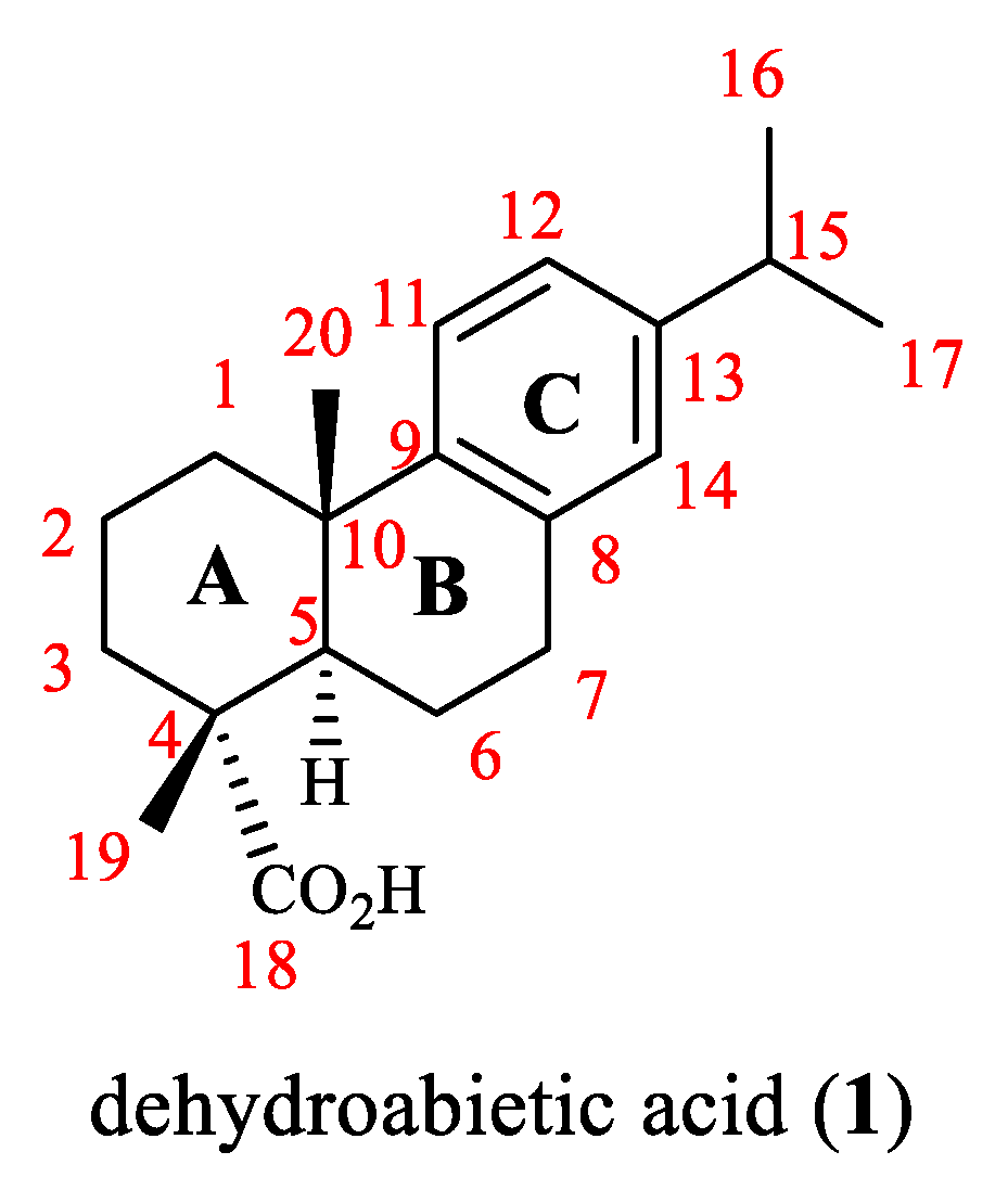
Figure 1. Chemical structure of dehydroabietic acid (1).
Toxins 14 00632 g001
Figure 2. Chemical structures of compounds 2 and 3.
Toxins 14 00632 g002
Figure 3. Chemical structures of compounds 412.
Toxins 14 00632 g003
Figure 4. Chemical structures of compounds 13a13c.
Toxins 14 00632 g004
Figure 5. Chemical structures of compounds 1419.
Toxins 14 00632 g005
Scheme 1. Synthesis of compounds 22(a-h)−25(a-h).
Toxins 14 00632 sch001
Scheme 2. Synthesis of compounds 28a28k, 29a29o, 30a30o, 31, 32 and 33a33m.
Toxins 14 00632 sch002
Scheme 3. Synthesis of compounds 36a36x.
Toxins 14 00632 sch003
Scheme 4. Synthesis of compounds 39a39o.
Toxins 14 00632 sch004
Scheme 5. Synthesis of compounds 41a41p.
Toxins 14 00632 sch005
Scheme 6. Synthesis of compounds 43a43o.
Toxins 14 00632 sch006
Scheme 7. Synthesis of compounds 47a47r.
Toxins 14 00632 sch007
Scheme 8. Synthesis of compounds 49a49f.
Toxins 14 00632 sch008
Scheme 9. Synthesis of compounds 51a51q, 52a52p and 53a53k.
Toxins 14 00632 sch009
Scheme 10. Synthesis of compounds 57a57s and 59a59ai.
Toxins 14 00632 sch010
Scheme 11. Synthesis of compounds 61a61g.
Toxins 14 00632 sch011
Scheme 12. Synthesis of compounds 63a63t, 64(a-h)−66(a-h) and 67a67j.
Toxins 14 00632 sch012
Scheme 13. Synthesis of compounds 69a69p.
Toxins 14 00632 sch013
Scheme 14. Synthesis of compounds 71a71e.
Toxins 14 00632 sch014
Scheme 15. Synthesis of compounds 74(a-k)−75(a-k), 77a77o and 78(a-h)−79(a-h).
Toxins 14 00632 sch015
Scheme 16. Synthesis of compounds 80(a-k)81(a-k).
Toxins 14 00632 sch016
Scheme 17. Synthesis of compounds 84a84k.
Toxins 14 00632 sch017
Scheme 18. Possible pathways for the biotransformation of dehydroabietic acid by Mucor circinelloides IT25, Mortierella isabellina HR32, Moraxella sp. HR6, Trametes versicolor (T), Phlebiopsis gigantea (P), Flavobacterium resinovorum (Fl), Fusarium oxyosporum/F. moniliforme (Fu) and Fomes annosum (Fo).
Toxins 14 00632 sch018
Scheme 19. Total synthesis of dehydroabietic acid from 2-isopropylnaphthalene.
Toxins 14 00632 sch019
Scheme 20. Total synthesis of dehydroabietic acid from 2-methyl-2-(p-isopropylphenyl)cyclohexanone.
Toxins 14 00632 sch020
Scheme 21. Total synthesis of dehydroabietic acid from the geranyl acetate derivative.
Toxins 14 00632 sch021
Figure 6. The overall SARs of dehydroabietic acid and its derivatives for antibacterial and anticancer activities.
Toxins 14 00632 g006
Table 1. Some MIC values of dehydroabietic acid derivatives against different bacteria.
CompoundBacteriaMIC Value
5B. subtilis4 μg/mL [36]
5S. aureus2 μg/mL [36]
7methicillin resistant S. aureus32 μg/mL [39]
8LA-MRSA LGA251(ST425-XI)3.9 μg/mL [40]
9S. aureus Newman0.39–0.78 μg/mL [41]
10NRS-701.25–1.56 μg/mL [41]
11S. aureus Newman, NRS-1, NRS-70, NRS-100, NRS-108, and NRS-2711.56–3.13 μg/mL [41]
12S. aureus Newman, NRS-70, NRS-108, and NRS-2711.56–3.13 μg/mL [41]
Table 2. Some IC50 values of the most active derivatives against different cancer cell lines.
CompoundCancer Cell LineIC50 Value
22fHeLa7.76 ± 0.98 μM [64]
28eSK-OV-31.79 ± 0.43 μM [65]
30nHeLa6.58 ± 1.11 μM [66]
36wHeLa2.21 ± 0.04 μM [69]
36wBEL-740214.46 ± 0.22 μM [69]
39jMGC-8033.82 ± 0.18 μM [70]
41cHepG25.90 ± 0.41 μM [71]
41kHepG26.25 ± 0.37μM [71]
43bMCF-77.00 ± 0.96 μM [72]
47nBEL-740211.23 ± 0.21 μM [73]
47jCNE-28.36 ± 0.14 μM [73]
63rHepG224.41 ± 0.26 μM [81]
63sHepG222.92 ± 0.24 μM [81]
67gSMMC-77211.39 ± 0.13 μM [82]
67gHepG20.51 ± 0.09 μM [82]
67gHep3B0.73 ± 0.08 μM [82]
69pMDA-MB-2310.7 ± 0.1 μM [84]
74bSMMC-77210.36 ± 0.13 μM [86]
74eHepG20.12 ± 0.03 μM [86]
77bSMMC-77210.72 ± 0.09 μM [87]
79hMCF-70.87 ± 0.18 μM [88]
80jSMMC-77210.08 ± 0.01 μM [89]
84aHCT-1161.18 ± 0.52 μM [90]
Table 3. Some MIC values of the most active compounds against different bacteria.
CompoundBacteriaMIC Value
33eB. subtilis1.9 μg/mL [68]
57jS. aureus NRS-70, NRS-100, NRS-108 and NRS-2711.56–2.5 μg/mL [78]
69oS. aureus, E. coli and P. fluorescens1.6 μg/mL [83]
Table 4. Some IC50 values of the most active derivatives against human normal cell lines.
CompoundHuman Normal Cell LineIC50 Value
22fHL-770253.78 ± 1.4 μM [64]
36wHL-770266.08 ± 1.84 μM [69]
39jLO2>100 μM [70]
41c, 41kHL-7702>100 μM [71]
47jHL-7702, NP69>100 μM [73]
47nNP69>100 μM [73]
47nHL-770288.18± 0.23 μM [73]
67gQSG-770112.52 ± 0.58 μM [80]
74bLO23.89 ± 0.29 μM [86]
74eLO23.02 ± 0.21 μM [86]
77bLO211.09 ± 0.57 μM [87]
79hLO242.83 ± 3.18 μM [88]
80jQSG-77015.82 ± 0.38 μM [89]
Publisher’s Note: MDPI stays neutral with regard to jurisdictional claims in published maps and institutional affiliations.