Review

Antimicrobial Compounds from Skin Secretions of Species That Belong to the Bufonidae Family

by
Rodrigo Ibarra-Vega
1,
Alan Roberto Galván-Hernández
1,
Hermenegildo Salazar-Monge
2,
Rocio Zataraín-Palacios
1,
Patricia Elizabeth García-Villalvazo
1,
Diana Itzel Zavalza-Galvez
1,
Laura Leticia Valdez-Velazquez
1 and
Juana María Jiménez-Vargas
1,3,*
1
Facultad de Ciencias Químicas, Universidad de Colima, Coquimatlán 28040, Mexico
2
Facultad de Medicina, Universidad de Colima, Coquimatlán 28040, Mexico
3
Consejo Nacional de Ciencia y Tecnología (CONACYT), Mexico City 03940, Mexico
*
Author to whom correspondence should be addressed.
Toxins 2023, 15(2), 145; https://doi.org/10.3390/toxins15020145
Submission received: 31 December 2022 / Revised: 30 January 2023 / Accepted: 7 February 2023 / Published: 10 February 2023
(This article belongs to the Special Issue Biotechnological Potential of Animal Venom and Toxins)

Abstract

:
Skin secretions of toads are a complex mixture of molecules. The substances secreted comprise more than 80 different compounds that show diverse pharmacological activities. The compounds secreted through skin pores and parotid glands are of particular interest because they help toads to endure in habitats full of pathogenic microbes, i.e., bacteria, fungi, viruses, and protozoa, due to their content of components such as bufadienolides, alkaloids, and antimicrobial peptides. We carried out an extensive literature review of relevant articles published until November 2022 in ACS Publications, Google Scholar, PubMed, and ScienceDirect. It was centered on research addressing the biological characterization of the compounds identified in the species of genera Atelopus, Bufo, Duttaphrynus, Melanophryniscus, Peltopryne, Phrynoidis, Rhaebo, and Rhinella, with antibacterial, antifungal, antiviral, and antiparasitic activities; as well as studies performed with analogous compounds and skin secretions of toads that also showed these activities. This review shows that the compounds in the secretions of toads could be candidates for new drugs to treat infectious diseases or be used to develop new molecules with better properties from existing ones. Some compounds in this review showed activity against microorganisms of medical interest such as Staphylococcus aureus, Escherichia coli, Bacillus subtilis, Coronavirus varieties, HIV, Trypanosoma cruzi, Leishmania chagasi, Plasmodium falciparum, and against different kinds of fungi that affect plants of economic interest.
Key Contribution: In this review, we showed that skin secretions of toads are a source of biomolecules with diverse antimicrobial activities. Buforin II, Dehydrobufotenine analogs like Compound 12 and 17, and Telocinobufagin are candidates for developing new antibiotics, antifungal, and antiviral.

1. Introduction

The true toads are members of the family Bufonidae, which is formed by 52 genera and 644 species distributed worldwide [1,2]. These amphibians produce cutaneous secretions by two exocrine glands: mucous, and parotoid or granular. The first type is associated with respiration and hydration, while the parotoid glands secrete components related to defense, providing protection against predators and microorganisms [3,4,5,6]. These secretions consist of a complex composition of biomolecules containing alkaloids (dehydrobufotenine, atelopidtoxin, bufothionine, pumiliotoxin), biogenic amines (adrenaline, dopamine, serotonin), steroids (arenobufagin, Gamma-bufotalin, resibufogenin, telocinobufagin), peptides (alyteserin 1C, Bv8, cathelicidin), proteins, and others [7,8,9,10,11,12,13]. These components have various biological activities: antibacterial, anticancer, antifungal, anti-inflammatory, antiprotozoal, antitumoral, antiviral, insecticidal, and immunomodulatory [14,15,16,17,18,19,20,21]. In the data repository of antimicrobial peptides (DRAMP) it is reported that 1223 sequences were identified in amphibians. Still, few peptides have the potential to be characterized in clinical trials and to be used in humans as therapeutic agents [22]. Four antimicrobial peptides (AMPs) have been isolated from bufonid toads, but only one reached pre-clinical studies, as will be seen later. This review describes the diverse compounds of cutaneous secretions with antimicrobial activities identified in toads of the genera Atelopus, Bufo, Duttaphrynus, Melanophryniscus, Peltopryne, Phrynoidis, Rhaebo, and Rhinella, as well as of some chemically synthesized analogs that showed higher antimicrobial activity than the parent molecule dehydrobufotenine, and possible applications as antimicrobials. These compounds could be alternative drugs to control infections and antimicrobial resistance (AMR). With the outbreak of SARS-CoV2, 70% of the patients that presented with COVID-19 received antibiotics, which can lead to emergence of bacterial resistance [23,24,25]. Likewise, some molecules from toad skin secretions show antiviral activity. These compounds may be used against coronavirus variants that still are a worldwide problem, or can be used against some other viruses responsible for causing diseases [14,15,16,17,18]. Furthermore, compounds of secretions from toads may be used in agriculture as fungicides and pesticides [26,27]. Additionally, secretions from the parotoid glands of toads contain substances that have shown activity against intracellular parasites, so they can be an alternative for treating diseases caused by the genera Plasmodium spp., Leishmania spp., and Trypanosome spp. [11,28].

2. Methodology

2.1. Literature Review

All papers published until November 2022 on compounds from cutaneous toad secretions of the family Bufonidae with antimicrobial activities were considered for the literary review. The information search was carried out in the journals from ACS Publications, Google, Google Scholar, PubMed, and ScienceDirect, and the keywords used were “toad skin secretions”, “toad antimicrobials”, “antimicrobial”, “antibacterial”, “antiviral”, “antiparasitic”, and “antiprotozoal”.

2.2. Chemical Structure of Compounds and Determination of Molecular Weight

The determination of molecular weight, chemical formulas, and the chemical structure of the bufadienolides and alkaloids were performed with ChemBioDraw Ultra v12.0 software (PerkinElmer Inc., Waltham, MA, USA) [29]. In contrast, the secondary structure of the peptides was generated using the Maestro v.13.3.121 program (Schrodinger Inc., New York, NY, USA), and their molecular weight was determined with peptide mass calculator tools [30].

3. Antimicrobial Activities of Compounds from Toads’ Cutaneous Secretions

This review focuses on the bufadienolides, alkaloids, and antimicrobial peptides identified in toads’ cutaneous secretions with activity against bacteria, fungi, viruses, and parasites.

3.1. Compounds with Antibacterial Activity

After antibiotic misuse or overuse, it is almost inevitable that bacteria will become resistant to antibiotics. This kind of situation demands the development of new anti-infective drugs. The pharmaceutical industry attempts to meet this need through the timely redesign and modification of existing antibiotics and the development of new ones [31].
Research on the therapeutic potential of extracts and compounds isolated from true toads has intensified recently. Some studies support using secretions from bufonids in common treatments for bacterial diseases [32]. The water-soluble secretion of the parotid gland and the methanolic extract of the skin of the Sudanese toad Duttaphrynus melanostictus showed antibiotic activity against Bacillus cereus, Escherichia coli, Klebsiella pneumoniae, Salmonella typhimurium, Staphylococcus aureus, and Staphylococcus epidermidis; however, no action was found against methicillin-resistant S. aureus [33]. Sales et al. [34] observed that combining antibiotics of the aminoglycoside and β-lactam groups with the glandular secretion of Rhinella jimi led to a significant reduction in the minimal inhibitory concentration (MIC) value, mainly in strains of E. coli, Pseudomonas aeruginosa, and S. aureus. This indicated that components of the venom of R. jimi have significant pharmaceutical value in combination with available antibiotics. Other studies evaluated the antibiotic capacity of secretions from the dorsal skin of Phrynoidis asper extracted with deionized water, and demonstrated their action against the Gram-positive pathogens S. aureus and Bacillus subtilis with MIC of 12.25 ± 0.4 and 25 ± 1.3 μg/mL, respectively [35]. On the other hand, Thirupathi et al. [36] observed antibacterial activity from skin secretions from the Indian toad Bufo melanostictus on E. coli, S. aureus, P. vulgaris, and K. pneumoniae through the Kirby-Bauer Test.
In the Cundinamarca forest region of Colombia, Rhinella toads are used to treat erysipelas, an infection caused by the multi-drug-resistant bacterium Streptococcus pyogenes [37]. The Rhinella marina toad from the Brazilian Amazon has also been used to treat patients with erysipelas. Additionally, analysis of the crude extract and fractions of secretions from R. marina showed biological activity against S. aureus , P. aeruginosa, and E. coli [32]. In northeastern Brazil, the skin and fatty tissue of Rhinella jimi are used to treat some infections, such as, sore throat, flu, cough, and earache [13]. Bufadienolides are the most abundant compounds in the toads’ cutaneous secretions [38,39]. Their basic structure consists of a steroid nucleus of 17 carbon atoms arranged in four rings, and an α-pyrone ring at the C17-position, like in marinobufagin and telocinobufagin. It is important to mention that chemical modifications at various steroid molecule positions can generate remarkable differences in their antimicrobial activity (Figure 1). Cunha Filho et al. [40] used extraction with chloroform to isolate two bufadienolides, marinobufagin and telocinobufagin, from skin secretions of the Brazilian toad Bufo rubescens. The antibacterial capacity of both molecules was evaluated against E. coli ATCC 25922 and S. aureus ATCC 25213 and showed activities better than amoxicillin, imipenem, and trimethoprim antibiotics, especially against the Gram-positive bacteria (Table 1). Further, these authors determined that the two molecules can promote and increase cardiac contraction force. Both bufadienolides have been identified in toads of the genera Atelopus [41], Rhinella, Rhaebo [42], and Peltopryne [43].
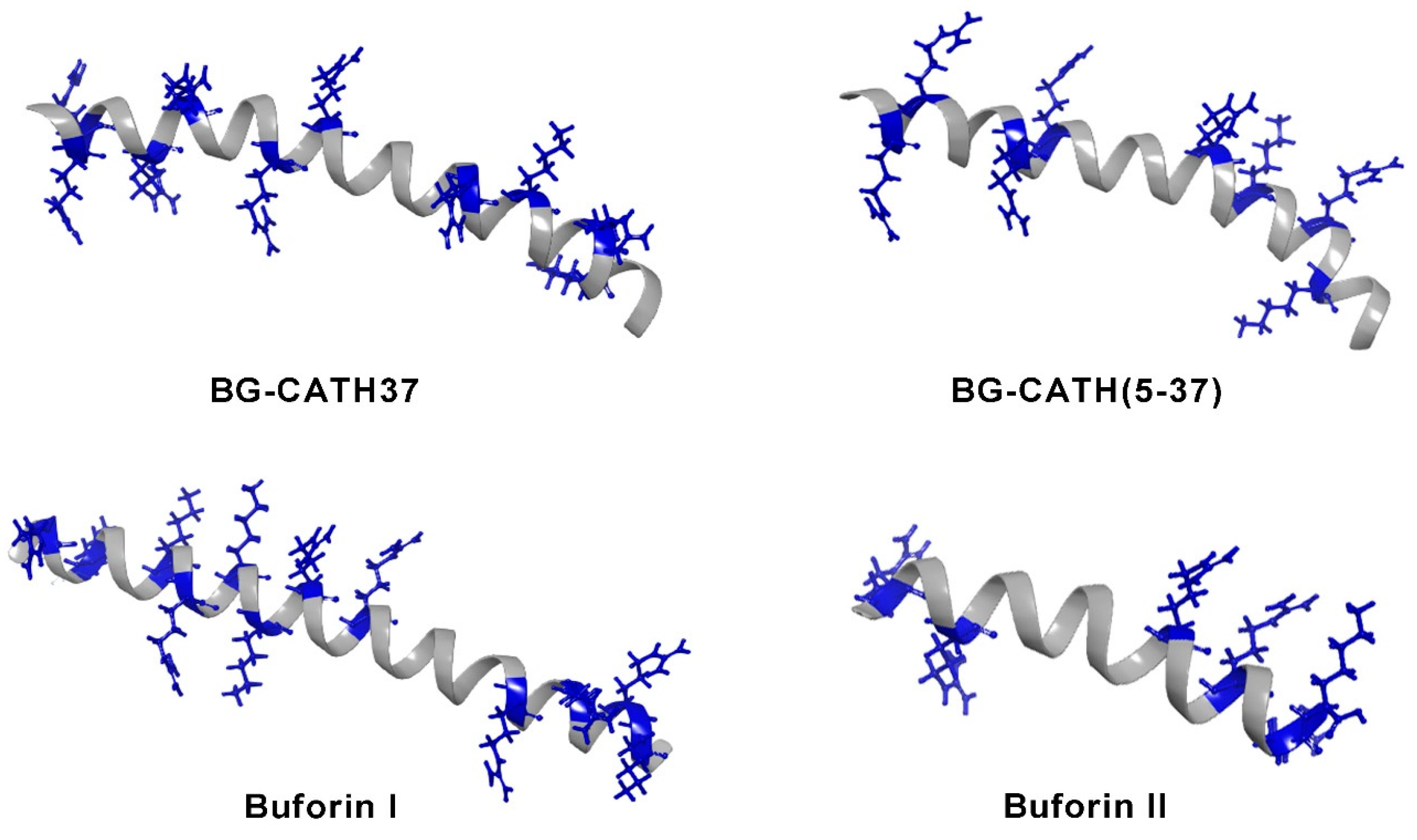
The increasing spread of resistant bacteria has led to the constant search for bioactive compounds. The study of amphibian antimicrobial peptides (AMPs) has been employed as a folk remedy to treat bacterial infectious diseases. One group of these AMPs are cathelicidins, identified in the skin secretion of the Asian toad Bufo bufo gargarizans. Two sequences coding for these peptides were found through a skin transcriptomic analysis; they were chemically synthesized and named BG-CATH37 and BG-CATH(5-37). The first is formed by 37 amino acids, the second by 32, and both are rich in glycine and arginine residues. The antibacterial activity of these peptides was evaluated against E. coli, S. aureus, P. aeruginosa, and against aquatic bacteria (Aeromorus hydrophia, Vibrio harveyi, Vibrio vulnificus, Vibrio splendidus, Lysogenic bacterium, and Streptococcus iniae), these cathelicidins showed weak antimicrobial activity against human pathogenic bacteria (MIC > 200 μg/mL), but they showed strong antimicrobial activities against aquatic bacteria (MIC 3.125–40 μg/mL) from their habitat [44]. Other antibacterial peptides isolated from this toad were buforin I and buforin II (Table 1) [45]. These AMPs are from the histone family are composed of 39 and 21 amino acid residues, respectively, and can adopt a secondary structure of an alpha helix (Figure 2). The parent peptide buforin I was found to have a high degree of sequence similarity to the N-terminal segment of the histone H2A DNA-binding protein, leading to the assumption that this AMP is structurally similar to histone H2A and would have similar antibacterial effects to buforin II [46]. Buforin I showed antimicrobial activity against a wide range of microorganisms, including the Gram-positive, Bacillus subtilis, S. aureus, Streptococcus mutans, Streptococcus pneumoniae, Pseudomonas putida; the Gram-negative bacteria E. coli, S. typhimurium, Serratia sp., and the fungi Candida albicans, Cryptococcus neoformans, and Saccharomyces cerevisiae [45]. Subsequently, Roshanak et al. [47] demonstrated that this peptide has an antibacterial effect on the most resistant and sensitive microbial strains, Staphylococcus salivarius and Clostridium perfringens, with a minimum inhibitory concentration (MIC) of 4 and 16 μg/mL, respectively. Furthermore, buforin I inhibited the formation of biofilms. Additionally, the thermal stability of the peptide in plasma at different temperatures was evaluated, and hemolytic and cytotoxic assays were performed, indicating that this peptide has the pharmaceutical potential to be used as an antibiotic in infectious diseases (Table 1).
Meanwhile, buforin II, a peptide derived from the digestive system with endoproteinase Lys-C, retains the residues from Thr16 to Lys 36 of buforin I, and is more than 12 times more potent than buforin I [48]. A proline hinge region in buforin II is essential for inducing membrane permeation in E. coli [46,49]. It has been suggested that buforin II inhibits cellular processes by interfering with DNA and RNA metabolism [50].

3.2. Compounds with Antifungal Activity

Skin secretions of many toads contain peptides with antifungal activity. These peptides are stored in a granular gland located mainly in the skin of the dorsal region. Most amphibian species produce one or more peptides with potent activity against fungi [7].
The skin secretions of Leptophryne cruentata show antifungal activity against Trichophyton mentagrophytes; forty microliters caused an inhibition halo of 14.5 ± 2.9 mm diameter [51]. Furthermore, the skin secretions of Rhinella icterica showed antifungal activity against Candida krusei [28]. The bufadienolides arenobufagin, gamabufotalin, and telocinobufagin were identified in the skin secretions of the toad Anaxyrus boreas. These compounds inhibited the growth of the Batrachochytrium dendrobatidis strain, and their MIC were 12.9, 50.0, and 14.3 μM, respectively [7]. These bufadienolides were also identified in Rhinella horribilis [52]. The discovery of these compounds, effective against these fungal strains, will allow the development of therapeutic agents that treat infections caused in humans and other animals [53]. One alkaloid called dehydrobufotenine was found in the skin secretions of the toad Bufo arenarum. It showed fungicidal activity against phytopathogenic fungi that affect plants of economic interest. Further, forty-five analogs of dehydrobufotenine were synthesized and evaluated for their activity. Compounds 6, 16c, 16d, 16h, 16j, and 19 showed similar or higher activity than the control carbendazim at 50 μg/mL. Dehydrobufotenine and most of its analogues showed higher activity against Alternaria solani (5–400%) than the controls chlorothalonil and carbendazim at 50 μg/mL. Their fungicidal activity is shown in Table 2.

3.3. Compounds with Antiviral Activity

Infectious diseases caused by viruses have impacted human life throughout evolutionary history and certainly in the adaptation of many civilizations. Molecular phylogenies support the hypothesis that some “modern” viruses, such as herpes or human papillomavirus (HPV), have co-evolved in association with some human ethnic groups and other vertebrate lineages and that these were dispersed around the world due to migration. The emergence or re-emergence of new diseases caused by viruses such as influenza, dengue, or hepatitis can be explained by the accelerated growth of populations and the density of human settlements, where the viruses were able to establish and persist, giving rise to the first epidemics [54].
In the last two years, the COVID-19 pandemic caused by the SARS-CoV-2 outbreak resulted in more than 50 million official cases and countless deaths. Due to this, many researchers focused on searching for alternatives to diagnose and treat this virus and others, such as influenza A (H1N1 and H5N1), HIV, Chikungunya, Zika, and Hepatitis C and B [55].
The importance of research on disease-causing viruses lies in the global public health problem and economic burden caused by the ability of viruses to transmit from nature to humans, triggering unpredictable outbreaks. The current strategy for managing viral diseases consists, on the one hand, of vaccines, whose development is usually slow, long, laborious, and costly; and on the other hand, of broad-spectrum antiviral treatments, an effective option to reduce human-to-human transmission [56]. For this reason, the bioactive components of toad skin secretions represent an important source for the development of new therapeutic agents that help in the treatment of various viral diseases.
Bufalin and cinobufagin are bufadienolides isolated from the toad skin secretion of the Bufo bufo gargarizan. Both are pharmacologically active components of huachansu or cinobufacini, a preparation of traditional Chinese medicine, which has been used as a treatment against hepatitis B virus (HBV). The activity of cinobufacini, bufalin, and cinobufagin was examined against HBV. The three of them were tested in the human cell line HepG2.2.15 at different concentrations, where all showed HBV secretion antigen inhibition activity at high concentrations (Table 3). Unlike bufalin and cinobufagin, the results demonstrated that cinobufacini exhibited higher activity against HBV at low concentrations. This was attributed to the specific inhibition of HBV messenger RNA expression [17,57,58].
In another study, bufalin and resibufogenin were studied for their effects against Epstein–Barr virus (EBV) or human herpesvirus 4, one of the most common viruses in humans. Both bufadienolides showed inhibitory activity on early activation of EBV antigen. In addition, bufalin showed in vivo inhibitory activity in two-stage carcinogenesis tests on mouse cutaneous papilloma caused by human papillomavirus (HPV) [59] and inhibition of rhinovirus [60].
Bufotenine is an alkaloid isolated from the secretions of Rhinella jimi. This compound was tested against rabies virus, which is responsible for a deadly zoonotic disease that affects more than 150 countries causing about 55,000 deaths annually. The study showed that bufotenine could inhibit the penetration of the rabies virus into mammalian cells through an apparent mechanism of competition for the nicotinic acetylcholine receptor. However, bufotenine is not presented as a proposal for drug development due to its hallucinogenic, cytotoxic, and psychotropic effects [61].
In 2021, Jin and coworkers tried to find broad-spectrum antiviral drugs to treat coronavirus infections. They assessed the antiviral activity of some cardiotonic steroids (bufadienolides) found in skin secretions of the toad species Bufo bufo gargarizans. The activity was tested against MERS-CoV, SARS-CoV, and SARS-CoV2 coronavirus varieties (Table 3). In the study, the authors identified antiviral activity. The IC50 values against coronavirus varieties were bufalin (0.016–0.544 μM), cinobufagin (0.017–0.616 μM), telocinobufagin (0.027–0.465 μM), bufotalin (0.027–1.630 μM), cinobufotalin (0.023–3.958 μM), and resinobufogenin (1.364–15.970 μM) (Table 3). Resinobufogenin showed low activity, and, sadly, bufalin and cinobufagin showed lethal activity in mice after intraperitoneal administration of 10 mg/kg/day in a 5-day repeated dose toxicity test. Cinobufotalin showed lethal activity in rats after 1mg/kg IV injection during pharmacokinetic studies. Telocinobufagin was the best candidate as a therapeutic drug for COVID-19 among the bufadienolides tested [16].
Further, the components of toad skin secretions can act against viruses that affect plants, as in the case of dehydrobufotenine (Figure 1). This alkaloid was found in skin secretions from Bufo arenarum. It was selected by Tian et al. [27] who designed and synthesized dehydrobufotenine analogs, with the goal of evaluating their biological activity. Of the forty-five synthesized analogs, compounds 12 and 17 showed ~50% higher antiviral activity against Tobacco mosaic virus (TMV) than dehydrobufotenine. Remarkably, the potency of these molecules was higher than ningnanmycin, which is perhaps the most effective anti-plant-virus agent (ningnanmycin inhibitory effect of 50–60% over TMV at 500 μg/mL). Compound 12 presented evidence of virus-assembly inhibition (Table 3) [27]. So, these two analogs could be considered new antiviral candidates.

3.4. Compounds with Antiparasitic Activity

A parasite is an “organism that lives on or in a host organism and gets its food from or at the expense of its” host [62]. The parasites use vectors such as mosquitos to reach their host. Some of these organisms cause diseases that have been associated with poverty [63] and tropical conditions [64]. Therefore warm weather in developing countries favors an increase in the case number, leading to deaths and other health problems [65].
Death due to parasitic diseases went down from 2000 to 2019. Still, in 2020, due to the SARS-CoV-2 pandemic, only malaria was determined to increase by 12%, estimating 627,000 deaths caused by the Plasmodium vivax parasite [64].
Chagas disease is caused by the parasite Trypanosoma cruzi, which is widespread in America (mainly rural zones of Latin America), affecting approximately 6–7 million people. The symptoms are nonspecific or asymptomatic in the acute phase of the infection. Patients suffer heart or digestive diseases only when the chronic stage is reached [28].
Drugs used in parasite-infection treatment are mainly formed by nitrogen rings, such as pyrimidine and imidazole, other substituents with nitrogen, and sulfhydryl groups are also common. There are antiparasitic drugs with broad spectra, such as albendazole and mebendazole; the majority are indicated against special cases, such as the use of mefloquine, which is a drug used against malaria produced by P. falciparum, a chloroquine-resistant strain, or suramin which is used on Trypanosoma brucei rhodesiense infection treatment [66].
For T. cruzi, benznidazole is a drug indicated for treating Chagas disease, especially in patients in the acute phase when the parasite is still in the blood. On the other hand, nifurtimox can be used in the acute phase or the chronic phase [67].
Toad venom comprises chemical compounds such as proteins, peptides, steroidal bufadienolides, alkaloids, and others [68]. The genus Bufo was divided into two genera in South America, Rhinella and Rhaebo. The secretions from nine different species (eight of Rhinella and one of Rhaebo) were analyzed biochemically, founding molecules in common. But, the authors realized that related species living in different habitats secrete distinct molecules, so they proposed that biological features influence skin secretion [68].
Telocinobufagin and hellebrigenin were isolated from Rhinella jimi and tested against Leishmania chagasi and T. cruzi (Table 4). Both bufadienolides inhibited the growth of the promastigotes from Leishmania, while hellebrigenin showed activity against trypomastigotes of T. cruzi. These molecules did not show cytotoxicity at 200 μg/mL, so that result is a promising for developing antiparasitic agents [28].
The skin secretion of the toads R. marina and R. guttatus were evaluated for their antiplasmodial activity against the Plasmodium falciparum W2 strain and showed a mortality above 70% against the parasite. Banfi et al. [69] isolated the compounds bufalin, dehydrobufotenin, marinobufotoxin, and marinobufagin of R. marina toads and evaluated their action in vitro on P. falciparum viability, showed values of IC50 of 1.329, 3.88, 5.31, and 1.557 μg/mL, respectively (Table 4). Although the bufalin was more active, only dehydrobufotenin demonstrated high selectivity for the parasites when analyzed in a cytotoxicity assay. Hence, it is a good candidate for the design of new therapeutic agents. On the other hand, secretions of Rhinella marina showed biological activity against Leishmania guyanensis and Leishmania braziliensis [32].
Meanwhile, ten bufadienolides isolated from Rhinella alata exhibited anti-trypanosomal activity, where the ranges of IC50 were between 1.329 and 14.343 μg/mL on Trypanosoma cruzi in an in vitro assay (Table 4). The anti-trypanosomal activity revealed that an arginyl-diacid at C-3 and an OH group at C-14 in the structure of bufadienolides were essential for the bioactivity observed.
Bufotalin and cinobufagin (IC50 8.713 and 12.833 μg/mL against T. cruzi) presented low cytotoxicity (LD50 20.446 and 23.897 μg/mL) in epithelial kidney monkey Vero cells (ATCC) [11].

4. Discussion

The skin secretions of the toads Bufo bufo gargarizans and Bufo melanostictus have been used in Chinese traditional medicine to treat bacterial infections, abdominal pain, and diarrhea [70]. These pharmacological properties are due to substances the secretions contain: bufadienolides, alkaloids, toxins, peptides, and proteins. In this work, we performed a review of the compounds with antimicrobial activity. The bufadienolides such as marinobufagin and telocinobufagin from Bufo rubescens presented lower antibacterial activity than the peptides buforins, especially against medically important bacteria such as E. coli, S. aureus, S. pneumoniae, and S. typhimurium. Buforin II showed better activity than buforin I. Although both peptides are rich in positive residues, the distribution of these in the alpha helix is crucial to induce bacterial killing [71]. In the case of buforin II, these positive residues are mainly distributed on the hydrophilic side. In this, the presence of arginine residues provides capabilities to interact with the negatively charged phospholipid headgroups of bacterial membranes. The studies with buforin II only included preclinical assays despite showing antibacterial activities similar to the derived peptide of magainin, called MSI-78, which was evaluated in Phase IIbAII clinical trials and demonstrated effectiveness in the treatment of impetigo, a skin infection caused by group A Streptococcus and S. aureus [72]. Buforins and cathelicidins could be used as parent peptides to design some analogs to increase their antimicrobial activity and prepare formulations of use to treat skin infections caused by bacteria.
Bufadienolides, also called cardiotonic steroids, contain a 6-membered lactone ring in the C17 position of the steroid nucleus (Figure 1) and have a broad spectrum of antimicrobial activities. Telocinobufagin and marinobufagin showed moderate antibiotic activity against E. coli (64 and 16 μg/mL) and low activity against S. aureus (128 μg/mL). Arenobufagin, gamabufotalin and marinobufagin showed medium antifungal activity (MIC 12.9, 50, 14.3 μg/mL, respectively). Dehydrobufotenine and some of its analogs showed better antifungal activity against Alternaria solani than the controls chlorothalonil and carbendazim.
The bufadienolides secreted from the skin of Anaxyrus boreas present an important opportunity for biology, since they present activity against the Batrachochytrium dendrobatidis strain, which affects the skin of many amphibians and has shown resistance to conventional treatment. This fungus has caused the extinction or near extinction of more than 200 species of amphibians, radically reprogramming ecosystems around the planet [7]. Alkaloids synthesized from the secretion of the toad Bufo arenarum showed potent fungicidal activity, surpassing fungicides such as chlorothalonil and carbendazim. Of all the synthesized components, it was found that compound 16h was the main candidate to be a broad-spectrum fungicide [27].
On the other hand, telocinobufagin is one of the most interesting bufadienolides, it showed high activity against the MERS-CoV and SARS-CoV-2 coronavirus variants, and against L. chagasi, and, most importantly, it caused low cytotoxicity. Telocinobufagin is one of the crucial bufadienolides to consider a new medicine. In this review, we revised that slight differences in the molecules can cause significant changes in biological activities. There are still different compounds that have not been analyzed yet.
Steroidal compounds show high activity against parasites; 15 bufadienolides obtained from toad secretions demonstrated good activity (IC50 126.2 to 1.32 μg/mL), but only 7 demonstrated viability in the therapeutic window. Commercial drugs used in treating parasites usually have hydrophobic areas that help in transportation across lipid barriers and contain amino hetero-substituted structures such as triazole and benzimidazole compounds. SAR-guided studies of aryl methylamino steroids showed steroid moieties as essential in biological activity to cross membrane barriers, and 3-OH and 17-OH substitution as essential to disable side effects of steroid hormones [73] like those observed in skin secretions of toads in this review.

5. Conclusions

In this review, we showed that skin secretions of toads are a source of biomolecules with diverse antimicrobial activities. In the secretions, bufadienolides are a diverse group of molecules. Some possess high antimicrobial activity but were reported to have toxic effects in mice, such as bufalin and cinobufagin. Telocinobufagin had moderate antifungal activity, high antiviral activity (against MERS-CoV, SARS-CoV, and SARS-CoV-2), and low toxicity. This molecule is a candidate for a new antiviral drug or could be a base for developing better antiviral agents.
Buforin II had high antibacterial activity killing Gram-negative and Gram-positive bacteria. This AMP is a clear candidate for a new antibiotic and could be used to treat skin and stomach infections. On the other hand, Buforin II could be used as a food preservative.
The study on dehydrobufotenine analogs was a clear example of designing new molecules with better properties; compounds 6, 16c, 16d, 16h, 16j, and 19 increased their antifungal activity compared to dehydrobufotenine. They exhibited similar or higher activity than the commercial antifungals chlorothalonil and carbendazim. Furthermore, compounds 12 and 17 achieved higher activity that ningnanmycin, which may be the most effective anti-plant virus agent. Compounds 12 and 17 are therefore new antiviral candidates.
Up to date, 644 species in the Bufonidae family have been described, but the secretion of many of them has not been studied yet. We think there are still many antimicrobial compounds that can help us fight multiple diseases caused by microorganisms.

Author Contributions

Conceptualization: R.I-V. and J.M.J.-V., Review and editing: R.I.-V., J.M.J.-V., A.R.G.-H., H.S.-M., R.Z.-P., P.E.G.-V., D.I.Z.-G. and L.L.V.-V., Funding acquisition: J.M.J.-V. and L.L.V.-V. All authors have read and agreed to the published version of the manuscript.

Funding

This was partially supported by a grand of Fortalecimiento de Cuerpos Académicos PRODEP 2021-ICOL-CA 117 “Bioquímica aplicada” (L.L.V.-V. and J.M.J.-V.).

Institutional Review Board Statement

Not applicable.

Informed Consent Statement

Not applicable.

Data Availability Statement

Not applicable.

Acknowledgments

This work was also partially supported by the CONACyT research program by Mexico obtained by J-VJM; grant PRODEP 2021-ICOL-CA 117.

Conflicts of Interest

The authors declare no conflict of interest.

References

  1. AmphibiaWeb AmphibiaWeb<https://Amphibiaweb.Org> University of California, Berkeley, CA, USA. Available online: https://amphibiaweb.org/lists/Bufonidae.shtml (accessed on 1 November 2022).
  2. Frost D Amphibian Species of the World: An Online Reference (Version 6). Available online: https://amphibiansoftheworld.amnh.org/Amphibia/Anura/Bufonidae (accessed on 1 November 2022).
  3. Stewart, M.M. Biology of Amphibians. William E. Duellman, Linda Trueb. Q. Rev. Biol. 1994, 69, 670. [Google Scholar] [CrossRef]
  4. Jared, C.; Antoniazzi, M.M.; Jordão, A.E.C.; Silva, J.R.M.C.; Greven, H.; Rodrigues, M.T. Parotoid Macroglands in Toad (Rhinella Jimi): Their Structure and Functioning in Passive Defence. Toxicon 2009, 54, 197–207. [Google Scholar] [CrossRef]
  5. Regueira, E.; Dávila, C.; Sassone, A.G.; O’Donohoe, M.E.A.; Hermida, G.N. Post-Metamorphic Development of Skin Glands in a True Toad: Parotoids versus Dorsal Skin. J. Morphol. 2017, 278, 652–664. [Google Scholar] [CrossRef] [PubMed]
  6. Regueira, E.; Dávila, C.; Hermida, G.N. Morphological Changes in Skin Glands During Development in Rhinella Arenarum (Anura: Bufonidae). Anat. Rec. 2016, 299, 141–156. [Google Scholar] [CrossRef] [PubMed]
  7. Barnhart, K.; Forman, M.E.; Umile, T.P.; Kueneman, J.; McKenzie, V.; Salinas, I.; Minbiole, K.P.C.; Woodhams, D.C. Identification of Bufadienolides from the Boreal Toad, Anaxyrus Boreas, Active Against a Fungal Pathogen. Microb. Ecol. 2017, 74, 990–1000. [Google Scholar] [CrossRef]
  8. Clarke, B.T. The Natural History of Amphibian Skin Secretions, Their Normal Functioning and Potential Medical Applications. Biol. Rev. 1997, 72, 365–379. [Google Scholar] [CrossRef] [PubMed]
  9. Li, F.J.; Hu, J.H.; Ren, X.; Zhou, C.M.; Liu, Q.; Zhang, Y.Q. Toad Venom: A Comprehensive Review of Chemical Constituents, Anticancer Activities, and Mechanisms. Arch. Pharm. 2021, 354, 2100060. [Google Scholar] [CrossRef]
  10. Mailho-Fontana, P.L.; Antoniazzi, M.M.; Toledo, L.F.; Verdade, V.K.; Sciani, J.M.; Barbaro, K.C.; Pimenta, D.C.; Rodrigues, M.T.; Jared, C. Passive and Active Defense in Toads: The Parotoid Macroglands in Rhinella Marina and Rhaebo Guttatus. J. Exp. Zool. Part A Ecol. Genet. Physiol. 2014, 321, 65–77. [Google Scholar] [CrossRef]
  11. Rodriguez, C.; Ibáñez, R.; Mojica, L.; Ng, M.; Spadafora, C.; Durant-Archibold, A.A.; Gutiérrez, M. Bufadienolides from the Skin Secretions of the Neotropical Toad Rhinella Alata (Anura: Bufonidae): Antiprotozoal Activity against Trypanosoma Cruzi. Molecules 2021, 26, 4217. [Google Scholar] [CrossRef]
  12. Rodríguez, C.; Rollins-Smith, L.; Ibáñez, R.; Durant-Archibold, A.A.; Gutiérrez, M. Toxins and Pharmacologically Active Compounds from Species of the Family Bufonidae (Amphibia, Anura). J. Ethnopharmacol. 2017, 198, 235–254. [Google Scholar] [CrossRef] [Green Version]
  13. Rodriguez, C.; Ibáñez, R.; Rollins-Smith, L.A.; Gutiérrez, M.; Durant-Archibold, A.A. Antimicrobial Secretions of Toads (Anura, Bufonidae): Bioactive Extracts and Isolated Compounds against Human Pathogens. Antibiotics 2020, 9, 843. [Google Scholar] [CrossRef]
  14. Chen, J.; Xu, L.; Sun, S.; Zhang, H.; Ma, T.; Su, W.; Jiang, C. Identification of Cinobufagin and Resibufogenin as Inhibitors of Enterovirus 71 Infection. Chem. Res. Chin. Univ. 2014, 30, 953–958. [Google Scholar] [CrossRef]
  15. El-Seedi, H.; Yosri, N.; El-Aarag, B.; Mahmoud, S.; Zayed, A.; Du, M.; Saeed, A.; Musharraf, S.; El-Garawani, I.; Habib, M.; et al. Chemistry and the Potential Antiviral, Anticancer, and Anti-Inflammatory Activities of Cardiotonic Steroids Derived from Toads. Molecules 2022, 27, 6586. [Google Scholar] [CrossRef]
  16. Jin, Y.H.; Jeon, S.; Lee, J.; Kim, S.; Jang, M.S.; Park, C.M.; Song, J.H.; Kim, H.R.; Kwon, S. Broad Spectrum Antiviral Properties of Cardiotonic Steroids Used as Potential Therapeutics for Emerging Coronavirus Infections. Pharmaceutics 2021, 13, 1839. [Google Scholar] [CrossRef] [PubMed]
  17. Cui, X.; Inagaki, Y.; Xu, H.; Wang, D.; Qi, F.; Kokudo, N.; Fang, D.; Tang, W. Anti-Hepatitis B Virus Activities of Cinobufacini and Its Active Components Bufalin and Cinobufagin in HepG2.2.15 Cells. Biol. Pharm. Bull. 2010, 33, 1728–1732. [Google Scholar] [CrossRef]
  18. Wong, R.W.; Lingwood, C.A.; Ostrowski, M.A.; Cabral, T.; Cochrane, A. Cardiac Glycoside/Aglycones Inhibit HIV-1 Gene Expression by a Mechanism Requiring MEK1/2-ERK1/2 Signaling. Sci. Rep. 2018, 8, 850. [Google Scholar] [CrossRef] [PubMed]
  19. Kamboj, A.; Rathour, A.; Kaur, M. Bufadienolides and Their Medicinal Utility: A Review. Int. J. Pharm. Pharm. Sci. 2013, 5, 20–27. [Google Scholar]
  20. Baldo, E.C.F.; Anjolette, F.A.P.; Arantes, E.C.; Baldo, M.A. Toad poison and drug discovery. In Toxins and Drug Discovery; Springer: Berlin/Heidelberg, Germany, 2017. [Google Scholar]
  21. Dos Filho, E.S.M.; Chaves, M.H.; Ferreira, P.M.P.; Pessoa, C.; Lima, D.J.B.; Maranhão, S.S.A.; de Jesus Rodrigues, D.; Vieira Júnior, G.M. Cytotoxicity Potential of Chemical Constituents Isolated and Derivatised from Rhinella Marina Venom. Toxicon 2021, 194, 37–43. [Google Scholar] [CrossRef]
  22. Shi, G.; Kang, X.; Dong, F.; Liu, Y.; Zhu, N.; Hu, Y.; Xu, H.; Lao, X.; Zheng, H. DRAMP 3.0: An Enhanced Comprehensive Data Repository of Antimicrobial Peptides. Nucleic Acids Res. 2022, 50, D488–D496. [Google Scholar] [CrossRef]
  23. Knight, G.M.; Glover, R.E.; McQuaid, C.F.; Olaru, I.D.; Gallandat, K.; Leclerc, Q.J.; Fuller, N.M.; Willcocks, S.J.; Hasan, R.; van Kleef, E.; et al. Antimicrobial Resistance and Covid-19: Intersections and Implications. Elife 2021, 10, e64139. [Google Scholar] [CrossRef]
  24. Pritchard, M.; Dankwa, E.A.; Hall, M.; Baillie, J.K.; Carson, G.; Docherty, A.; Donnelly, C.A.; Dunning, J.; Fraser, C.; Hardwick, H.; et al. ISARIC Clinical Data Report 4 October 2020 International Severe Acute Respiratory and Emerging Infections Consortium. medRxiv 2020. [Google Scholar] [CrossRef]
  25. Langford, B.J.; So, M.; Raybardhan, S.; Leung, V.; Soucy, J.P.R.; Westwood, D.; Daneman, N.; MacFadden, D.R. Antibiotic Prescribing in Patients with COVID-19: Rapid Review and Meta-Analysis. Clin. Microbiol. Infect. 2021, 27, 520–531. [Google Scholar] [CrossRef]
  26. Raasch-Fernandes, L.D.; Bonaldo, S.M.; De Jesus Rodrigues, D.; Vieira-Junior, G.M.; Schwan-Estrada, K.R.F.; Da Silva, C.R.; Verçosa, A.G.A.; De Oliveira, D.L.; Debiasi, B.W. Induction of Phytoalexins and Proteins Related to Pathogenesis in Plants Treated with Extracts of Cutaneous Secretions of Southern Amazonian Bufonidae Amphibians. PLoS ONE 2019, 14, e0211020. [Google Scholar] [CrossRef]
  27. Tian, Z.; Liao, A.; Kang, J.; Gao, Y.; Lu, A.; Wang, Z.; Wang, Q. Toad Alkaloid for Pesticide Discovery: Dehydrobufotenine Derivatives as Novel Agents against Plant Virus and Fungi. J. Agric. Food Chem. 2021, 69, 9754–9763. [Google Scholar] [CrossRef]
  28. Tempone, A.G.; Pimenta, D.C.; Lebrun, I.; Sartorelli, P.; Taniwaki, N.N.; de Andrade, H.F.; Antoniazzi, M.M.; Jared, C. Antileishmanial and Antitrypanosomal Activity of Bufadienolides Isolated from the Toad Rhinella Jimi Parotoid Macrogland Secretion. Toxicon 2008, 52, 13–21. [Google Scholar] [CrossRef] [PubMed]
  29. Cousins, K.R. Computer Review of ChemDraw Ultra 12.0. J. Am. Chem. Soc. 2011, 133, 8388. [Google Scholar] [CrossRef] [PubMed]
  30. Beavis, R.; Winsor, D.; Council, C. Peptide Mass Calculator. Available online: https://www.peptidesynthetics.co.uk/tools/ (accessed on 1 November 2022).
  31. Gordon, Y.J.; Romanowski, E.G.; McDermott, A.M. Mini Review: A Review of Antimicrobial Peptides and Their Therapeutic Potential as Anti-Infective Drugs. Curr. Eye Res. 2005, 30, 505–515. [Google Scholar] [CrossRef]
  32. de Medeiros, D.S.S.; Rego, T.B.; dos Santos, A.P.d.A.; Pontes, A.S.; Moreira-Dill, L.S.; Matos, N.B.; Zuliani, J.P.; Stábeli, R.G.; Teles, C.B.G.; Soares, A.M.; et al. Biochemical and Biological Profile of Parotoid Secretion of the Amazonian Rhinella Marina (Anura: Bufonidae). Biomed Res. Int. 2019, 2019, 4209743. [Google Scholar] [CrossRef] [PubMed]
  33. Zahari, M.; Darnis, D.; Hamid, T.H.T.A. Protein Profiles and Antimicrobial Activity of Common Sunda Toad, Duttaphrynus Melanostictus Paratoid Secretions. In Proceedings of the 3rd International Conference on Biological, Chemical & Environmental Sciences (BCES-2015), Kuala Lumpur, Malaysia, 21–22 September 2015. [Google Scholar]
  34. Sales, D.L.; Morais-Braga, M.F.B.; dos Santos, A.T.L.; Machado, A.J.T.; de Araujo Filho, J.A.; de Dias, D.Q.; da Cunha, F.A.B.; Saraiva, R.d.A.; Menezes, I.R.A.d.; Coutinho, H.D.M.; et al. Antibacterial, Modulatory Activity of Antibiotics and Toxicity from Rhinella Jimi (Stevaux, 2002) (Anura: Bufonidae) Glandular Secretions. Biomed. Pharmacother. 2017, 92, 554–561. [Google Scholar] [CrossRef]
  35. Dahham, S.S.; Hew, C.S.; Jaafar, I.; Gam, L.H. The Protein Profiling of Asian Giant Toad Skin Secretions and Their Antimicrobial Activity. Int. J. Pharm. Pharm. Sci. 2016, 8, 88–95. [Google Scholar]
  36. Thirupathi, K.; Shankar, C.; Chandrakala, G.; Krishna, I.; Venkaiah, Y. The Antifungal Activity of Skin Secretion and Its Extract of Indian Toad Bufo Melanostictus. Int. J. Environ. Ecol. Fam. urban Stud. 2019, 9, 7–12. [Google Scholar] [CrossRef]
  37. Riós-Orjuela, J.C.; Falcón-Espitia, N.; Arias-Escobar, A.; Espejo-Uribe, M.J.; Chamorro-Vargas, C.T. Conocimiento E Interacciones De La Comunidad Local Con La Herpetofauna En La Forestal De Quininí (Tibacuy-Cundinamarca, Colombia). J. Ethnobiol. Ethnomed. 2020, 16, 17. [Google Scholar] [CrossRef] [PubMed] [Green Version]
  38. Garraffo, H.M.; Gros, E.G. Biosynthesis of Bufadienolides in Toads. VI. Experiments with [1,2-3H]Cholesterol, [21-14C]Coprostanol, and 5β-[21-14C]Pregnanolone in the Toad Bufo Arenarum. Steroids 1986, 48, 251–257. [Google Scholar] [CrossRef]
  39. Krenn, L.; Kopp, B. Bufadienolides from Animal and Plant Sources. Phytochemistry 1998, 48, 1–29. [Google Scholar] [CrossRef]
  40. Cunha Filho, G.A.; Schwartz, C.A.; Resck, I.S.; Murta, M.M.; Lemos, S.S.; Castro, M.S.; Kyaw, C.; Pires, O.R.; Leite, J.R.S.; Bloch, C.; et al. Antimicrobial Activity of the Bufadienolides Marinobufagin and Telocinobufagin Isolated as Major Components from Skin Secretion of the Toad Bufo Rubescens. Toxicon 2005, 45, 777–782. [Google Scholar] [CrossRef] [PubMed]
  41. Pearson, K.C.; Tarvin, R.D. A Review of Chemical Defense in Harlequin Toads (Bufonidae: Atelopus). Toxicon X 2022, 13, 100092. [Google Scholar] [CrossRef] [PubMed]
  42. Ferreira, P.M.P.; Lima, D.J.B.; Debiasi, B.W.; Soares, B.M.; da Machado, K.C.; da Noronha, J.C.; Rodrigues, D.d.J.; Sinhorin, A.P.; Pessoa, C.; Vieira Júnior, G.M. Antiproliferative Activity of Rhinella Marina and Rhaebo Guttatus Venom Extracts from Southern Amazon. Toxicon 2013, 72, 43–51. [Google Scholar] [CrossRef]
  43. Perera Córdova, W.H.; Leitão, S.G.; Cunha-Filho, G.; Bosch, R.A.; Alonso, I.P.; Pereda-Miranda, R.; Gervou, R.; Touza, N.A.; Quintas, L.E.M.; Noël, F. Bufadienolides from Parotoid Gland Secretions of Cuban Toad Peltophryne Fustiger (Bufonidae): Inhibition of Human Kidney Na+/K+-ATPase Activity. Toxicon 2016, 110, 27–34. [Google Scholar] [CrossRef]
  44. Sun, T.; Zhan, B.; Gao, Y. A Novel Cathelicidin from Bufo Bufo Gargarizans Cantor Showed Specific Activity to Its Habitat Bacteria. Gene 2015, 571, 172–177. [Google Scholar] [CrossRef]
  45. Park, C.B.; Kim, M.S.; Kim, S.C. A Novel Antimicrobial Peptide from Bufo Bufo Gargarizans. Biochem. Biophys. Res. Commun. 1996, 218, 408–413. [Google Scholar] [CrossRef]
  46. Park, C.B.; Yi, K.S.; Matsuzaki, K.; Kim, M.S.; Kim, S.C. Structure-Activity Analysis of Buforin II, a Histone H2A-Derived Antimicrobial Peptide: The Proline Hinge Is Responsible for the Cell-Penetrating Ability of Buforin II. Proc. Natl. Acad. Sci. USA 2000, 97, 8245–8250. [Google Scholar] [CrossRef] [PubMed]
  47. Roshanak, S.; Shahidi, F.; Yazdi, F.T.; Javadmanesh, A.; Movaffagh, J. Evaluation of Antimicrobial Activity of Buforin I and Nisin and the Synergistic Effect of Their Combination as a Novel Antimicrobial Preservative. J. Food Prot. 2020, 83, 2018–2025. [Google Scholar] [CrossRef]
  48. Cho, J.H.; Sung, B.H.; Kim, S.C. Buforins: Histone H2A-Derived Antimicrobial Peptides from Toad Stomach. Biochim. Biophys. Acta-Biomembr. 2009, 1788, 1564–1569. [Google Scholar] [CrossRef]
  49. Kobayashi, S.; Takeshima, K.; Park, C.B.; Kim, S.C.; Matsuzaki, K. Interactions of the Novel Anfimicrobial Peptide Buforin 2 with Lipid Bilayers: Proline as a Translocation Promoting Factor. Biochemistry 2000, 39, 8648–8654. [Google Scholar] [CrossRef] [PubMed]
  50. Park, C.B.; Kim, H.S.; Kim, S.C. Mechanism of Action of the Antimicrobial Peptide Buforin II: Buforin II Kills Microorganisms by Penetrating the Cell Membrane and Inhibiting Cellular Functions. Biochem. Biophys. Res. Commun. 1998, 244, 253–257. [Google Scholar] [CrossRef] [PubMed]
  51. Made Artika, I.; Pinontoan, S.; Kusrini, M.D. Antifungal Activity of Skin Secretion of Bleeding Toad Leptophryne Cruentata and Javan Tree Frog Rhacophorus Margaritifer. Am. J. Biochem. Biotechnol. 2015, 11, 5–10. [Google Scholar] [CrossRef]
  52. Schmeda-Hirschmann, G.; de Andrade, J.P.; Soto-Vasquez, M.R.; Alvarado-García, P.A.A.; Palominos, C.; Fuentes-Retamal, S.; Mellado, M.; Correa, P.; Urra, F.A. The Parotoid Gland Secretion from Peruvian Toad Rhinella Horribilis (Wiegmann, 1833): Chemical Composition and Effect on the Proliferation and Migration of Lung Cancer Cells. Toxins 2020, 12, 608. [Google Scholar] [CrossRef]
  53. Scheele, B.C.; Pasmans, F.; Skerratt, L.F.; Berger, L.; Martel, A.; Beukema, W.; Acevedo, A.A.; Burrowes, P.A.; Carvalho, T.; Catenazzi, A.; et al. Amphibian Fungal Panzootic Causes Catastrophic and Ongoing Loss of Biodiversity. Science 2019, 363, 1459–1463. [Google Scholar] [CrossRef]
  54. De Souza Leal, É.; De Andrade Zanotto, P.M. Viral Diseases and Human Evolution. Mem. Inst. Oswaldo Cruz 2000, 95, 193–200. [Google Scholar] [CrossRef]
  55. Abate, C.; Carnamucio, F.; Giuffrè, O.; Foti, C. Metal-Based Compounds in Antiviral Therapy. Biomolecules 2022, 12, 933. [Google Scholar] [CrossRef]
  56. Oksenych, V.; Kainov, D.E. Broad-Spectrum Antivirals and Antiviral Drug Combinations. Viruses 2022, 14, 301. [Google Scholar] [CrossRef] [PubMed]
  57. Wang, X.-L.; Zhao, G.-H.; Zhang, J.; Shi, Q.-Y.; Guo, W.-X.; Tian, X.-L.; Qiu, J.-Z.; Yin, L.-Z.; Deng, X.-M.; Song, Y. Immunomodulatory Effects of Cinobufagin Isolated from Chan Su on Activation and Cytokines Secretion of Immunocyte in Vitro. J. Asian Nat. Prod. Res. 2011, 13, 383–392. [Google Scholar] [CrossRef] [PubMed]
  58. Qi, J.; Tan, C.K.; Hashimi, S.M.; Zulfiker, A.H.M.; Good, D.; Wei, M.Q. Toad Glandular Secretions and Skin Extractions as Anti-Inflammatory and Anticancer Agents. Evid.-Based Complement. Altern. Med. 2014, 2014, 312684. [Google Scholar] [CrossRef] [PubMed]
  59. Inada, A.; Nakanishi, T.; Nishino, H.; Ichiishi, E.; Mukainaka, T.; Okuda, M.; Tokuda, H. Inhibitory Effects of Bufadienolides on Epstein-Barr Virus Early Antigen Activation and on Growth of Mouse Skin and Mouse Pulmonary Tumors. Nat. Med. 1999, 53, 324–328. [Google Scholar]
  60. Kamano, Y.; Pettit, G.R.; Smith, C.R.; Satoh, N.; Nakayoshi, H. Rhinovirus Inhibition by Bufadienolides1a). Chem. Pharm. Bull. 1988, 36, 326–332. [Google Scholar] [CrossRef] [PubMed]
  61. Vigerelli, H.; Sciani, J.M.; Jared, C.; Antoniazzi, M.M.; Caporale, G.M.M.; da Silva, A.d.C.R.; Pimenta, D.C. Bufotenine Is Able to Block Rabies Virus Infection in BHK-21 Cells. J. Venom. Anim. Toxins Incl. Trop. Dis. 2014, 20, 45. [Google Scholar] [CrossRef]
  62. CDC CDC—Parasites—About Parasites. Available online: https://www.cdc.gov/parasites/about.html (accessed on 1 November 2022).
  63. Fouque, F.; Knox, T. Special Programme for Research and Training in Tropical Diseases-Coordinated Multicountry Study to Determine the Burden and Causes of Residual Malaria across Different Regions. J. Infect. Dis. 2021, 223, S91–S98. [Google Scholar] [CrossRef]
  64. Geneva: World Health Organization World Malaria Report 2021. Angew. Chemie Int. Ed. 2021, 6, 951–952.
  65. Theel, E.S.; Pritt, B.S. Parasites. Microbiol. Spectr. 2016, 4, 411–466. [Google Scholar] [CrossRef]
  66. Pérez, J.L.; Carranza, C.; Mateos, F. Antiparasitic Drugs. Review of the Useful Drugs in the Treatment of Classic and Emergent Parasitic Diseases. Rev. Esp. Quimioter. 2009, 22, 93–105. [Google Scholar]
  67. Rojo-Medina, J.; Ruiz-Matus, C.; Salazar-Schettino, P.M.; González-Roldán, J.F. Enfermedad de Chagas En México. Gac. Mexico 2018, 154, 6–16. [Google Scholar] [CrossRef] [PubMed]
  68. Sciani, J.M.; Angeli, C.B.; Antoniazzi, M.M.; Jared, C.; Pimenta, D.C. Differences and Similarities among Parotoid Macrogland Secretions in South American Toads: A Preliminary Biochemical Delineation. Sci. World J. 2013, 2013, 937407. [Google Scholar] [CrossRef]
  69. Banfi, F.F.; Krombauer, G.C.; da Fonseca, A.L.; Nunes, R.R.; Andrade, S.N.; de Rezende, M.A.; Chaves, M.H.; dos Santos Monção Filho, E.; Taranto, A.G.; de Jesus Rodrigues, D.; et al. Dehydrobufotenin Extracted from the Amazonian Toad Rhinella Marina (Anura: Bufonidae) as a Prototype Molecule for the Development of Antiplasmodial Drugs. J. Venom. Anim. Toxins Incl. Trop. Dis. 2021, 27. [Google Scholar] [CrossRef]
  70. Chinese Pharmacopoeia Commission. In Pharmacopoeia of the People’s Republic of China; People’s Medical Publishing House, Co., Ltd.: Beijing, China, 2020; Volume 1, p. 402.
  71. Jiang, Z.; Vasil, A.I.; Hale, J.D.; Hancock, R.E.W.; Vasil, M.L.; Hodges, R.S. Effects of Net Charge and the Number of Positively Charged Residues on the Biological Activity of Amphipathic α-Helical Cationic Antimicrobial Peptides. Biopolymers 2008, 90, 369–383. [Google Scholar] [CrossRef] [PubMed]
  72. Ge, Y.; MacDonald, D.L.; Holroyd, K.J.; Thornsberry, C.; Wexler, H.; Zasloff, M. In Vitro Antibacterial Properties of Pexiganan, an Analog of Magainin. Antimicrob. Agents Chemother. 1999, 43, 369–383. [Google Scholar] [CrossRef] [Green Version]
  73. Krieg, R.; Jortzik, E.; Goetz, A.A.; Blandin, S.; Wittlin, S.; Elhabiri, M.; Rahbari, M.; Nuryyeva, S.; Voigt, K.; Dahse, H.M.; et al. Arylmethylamino Steroids as Antiparasitic Agents. Nat. Commun. 2017, 8, 14478. [Google Scholar] [CrossRef] [PubMed] [Green Version]
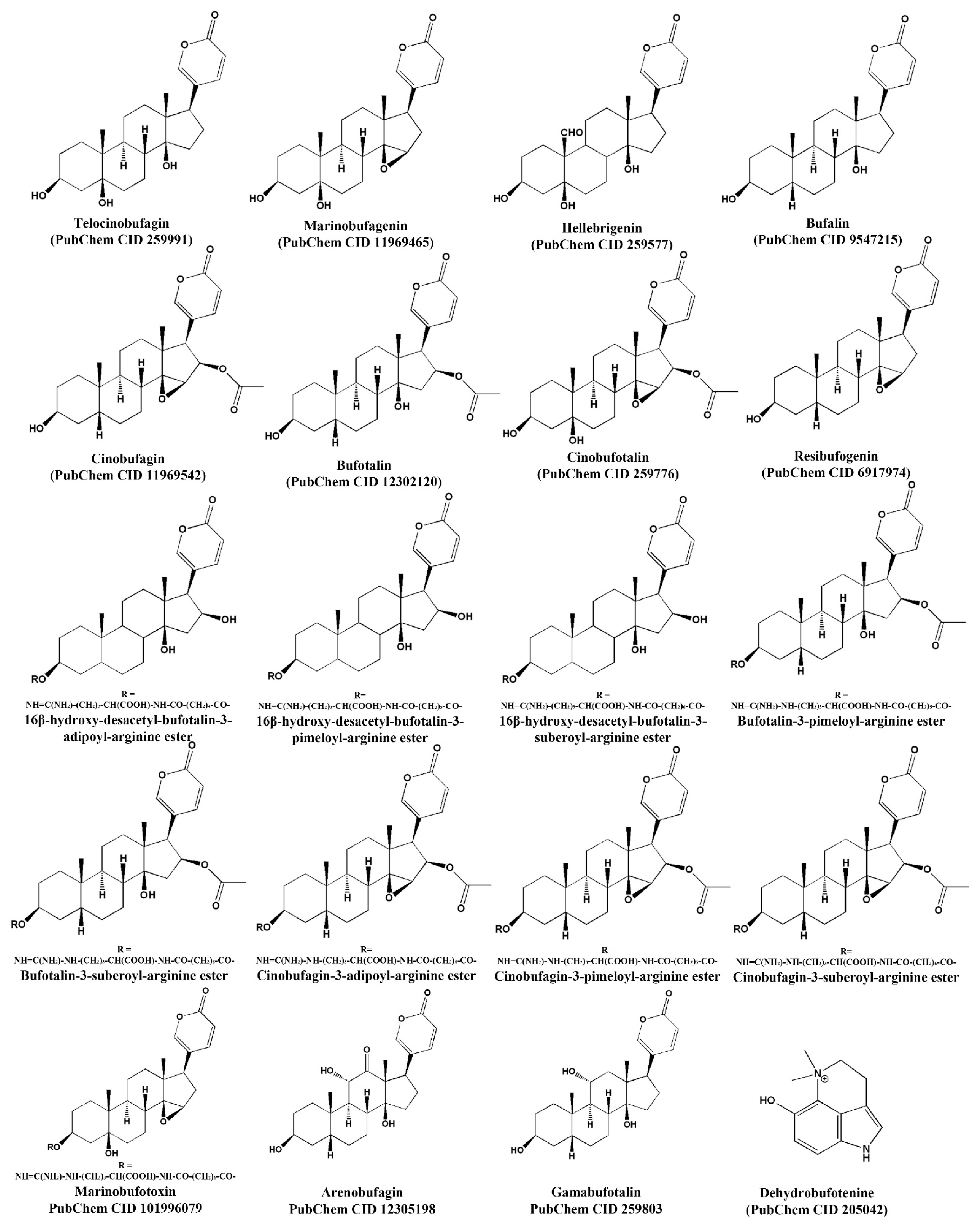
Figure 1. Chemical structure of molecules with antimicrobial activity found in toads’ skin secretions. The first 19 structures belong to the group of bufadienolides, and the last one is an alkaloid.
Toxins 15 00145 g001
Figure 2. Secondary structure of antimicrobial peptides found in toads. These peptides adopt an alpha helical conformation. They are rich in lysine residues (represented in blue) and the first amino acids interacting with bacterial membranes. Component names are in bold.
Toxins 15 00145 g002
Table 1. Compounds with antibacterial activity found in toads.
SpeciesComponentChem. Form.
M. Weight (Theo./Exp.)
Activity AgainstRef.
Bufo rubescensTelocinobufagin
(bufadienolide)
C 24 H 34 O 5
 
402.52 Da/402.1609 Da
E. coli ATCC 25922 (64 μg/mL),
S. aureus ATCC 25213 (128 μg/mL).
[40]
Marinobufagin
(bufadienolide)
C 24 H 32 O 5
 
400.51 Da/400.1515 Da
E. coli ATCC 25922 (16 μg/mL),
S. aureus ATCC 25213 (128 μg/mL).
[40]
Bufo bufo gargarizansBG-CATH37
(peptide)
Amino acid sequence:
 
SSRRPCRGRSCGPRLRGGYTLIGRPVKNQNRPKYMWV
 
4299.25 Da
Streptococcus iniae (12.5 μg/mL),
S. aureus (>200 μg/mL),
Vibrio splendidus (6.25 μg/mL),
Aeromonas hydrophila (40 μg/mL),
P. aeruginosa (200 μg/mL),
E. coli (>200 μg/mL).
[44]
BG-CATH(5-37)
(peptide)
Amino acid sequence:
 
PCRGRSCGPRLRGGYTLIGRPVKNQNRPKYMWV
 
3812.99 Da
S. iniae (12.5 μg/mL),
S. aureus (>200 μg/mL),
V. splendidus (3.125 μg/mL),
Aeromonas hydrophila (12.5 μg/mL),
P. aeruginosa (200 μg/mL),
E. coli (>200 μg/mL).
[44]
Buforin I
(peptide)
Amino acid sequence:
 
AGRGKQGGKVRAKAKTRSSRAGLQFPVGRVHRLLRKGNY
 
4363 Da/
MH+ 4309 Da
Bacillus subtilis (4 μg/mL),
S. aureus (4 μg/mL),
S. pneumoniae (4 μg/mL),
Streptococcus mutans (8 μg/mL),
S. typhimurium (4 μg/mL),
P. putida (4 μg/mL),
E. coli (8 μg/mL),
Serratia spp. (8 μg/mL).
[45,47]
Buforin II
(peptide)
Amino acid sequence
 
TRSSRAGLQFPVGRVHRLLRK
 
2434.9 Da/
MH+ 2432 Da
S. aureus (4 μg/mL),
S. pneumoniae (4 μg/mL),
B. subtilis (2 μg/mL),
S. mutans (2 μg/mL),
P. putida (2 μg/mL),
E. coli (4 μg/mL),
Serratia spp. (4 μg/mL),
S. typhimurium (1 μg/mL).
[45]
Scientific names are shown in italics. Da, Daltons; MH+, molecular ion.
Table 2. Compounds with fungicidal activity from skin secretions of species that belong to the Bufonidae family, and dehydrobufotenine analogs.
SpeciesMoleculeChem. Form.
M. Weight
Activity AgainstRef.
Bufo arenarumDehydrobufotenine
(alkaloid)
C 12 H 15 N 2 O +
 
203.26 Da
Alternaria solani (50 μg/mL).[27]
Compound 6 (dehydrobufotenine analog) C 16 H 22 N 2 O 3
 
290.36 Da
Alternaria solani (50 μg/mL),
Rhizoctonia solani (50 μg/mL),
Botrytis cinereal (50 μg/mL),
Cercospora arachidicola (50 μg/mL).
Compound 16c (dehydrobufotenine analog) C 22 H 24 N 4 O 5
 
424.45 Da
Alternaria solani (50 μg/mL),
Rhizoctonia solani (50 μg/mL).
Compound 16d (dehydrobufotenine analog) C 23 H 26 N 4 O 6
 
454.48 Da
Sclerotinia sclerotiorum (50 μg/mL).
Alternaria solani (50 μg/mL),
Rhizoctonia solani (50 μg/mL),
Botrytis cinereal (50 μg/mL),
Cercospora arachidicola (50 μg/mL).
Compound 16h (dehydrobufotenine analog) C 20 H 19 BrN 4 O 4
 
459.29 Da
Sclerotinia sclerotiorum (50 μg/mL),
Alternaria solani (50 μg/mL),
Rhizoctonia solani (50 μg/mL),
Botrytis cinereal (50 μg/mL),
Fusarium graminearum (50 μg/mL).
Compound 16j (dehydrobufotenine analog) C 21 H 19 F 3 N 4 O 3
 
432.40 Da
Sclerotinia sclerotiorum (50 μg/mL),
Alternaria solani (50 μg/mL),
Botrytis cinereal (50 μg/mL),
Cercospora arachidicola (50 μg/mL).
Compound 19
(dehydrobufotenine analog)
C 19 H 21 N 2 O +
293.38 Da
Alternaria solani (50 μg/mL),
Cercospora arachidicola (50 μg/mL).
Anaxyrus boreasArenobufagin
(bufadienolide)
C 24 H 32 O 6
M. weight (theo./Exp.)
416.51 Da/416.9 Da
B. dendrobatidis (12.9 μg/mL).[7]
Gamabufotalin
(bufadienolide)
C 24 H 34 O 5
M. weight (Theo./Exp.)
402.52 Da/403.4 Da
B. dendrobatidis (50 μg/mL).
Telocinobufagin
(bufadienolide)
C 24 H 34 O 5
M. weight (Theo./Exp.)
402.52 Da/402.9 Da
B. dendrobatidis (14.3 μg/mL).
Scientific names are shown in italics. Da, Daltons.
Table 3. Compounds with antiviral activity from skin secretions of species that belong to the Bufonidae family and dehydrobufotenine analogs.
SpeciesMoleculeChem. Form.
M. Weight
ActivityRef.
Bufo arenarumDehydrobufotenine
(alkaloid)
C 12 H 15 N 2 O +
 
203.26 Da
At 500 μg/mL showed similar inactive, curative, and protective effects against TMV than the antiviral ribavirin.[27]
Compound 12 (dehydrobufotenine analog) C 12 H 16 N 2 O
 
204.27 Da
At 500 μg/mL showed 5–7% higher inactivating, curative, and protective effects against TMV than the control ningnanmycin and 22–25% higher effects than ribavirin.[27]
Compound 17 (dehydrobufotenine analog) C 18 H 25 N 2 O 3 +
 
317.40 Da
At 500 μg/mL showed 5–6% higher inactivating, curative, and protective effects against TMV than the control ningnanmycin and 20–21% higher effects than the control ribavirin.[27]
Bufo rubescensTelocinobufagin
(bufadienolide)
C 24 H 34 O 5
 
402.52 Da
IC50 0.027 μM (anti-MERS-CoV a),
IC50 0.465 μM (anti-MERS-CoV b),
IC50 0.071 μM (anti-SARS-CoV a),
IC50 0.142 μM (anti-SARS-CoV2 a).
[16]
Bufo bufo gargarizansBufalin
(bufadienolide)
C 24 H 34 O 4
 
386.52 Da
IC50 0.018 μM (anti- MERS-CoV a),
IC50 0.544 μM (anti-MERS-CoV b),
IC50 0.017 μM (Anti-SARS-CoV a),
IC50 0.019 μM (Anti-SARS-CoV2 a),
IC90 15 nM (anti-HIV-1),
100% lethal activity at 10 mg/kg (in mice, IP).
20 nM inhibits the secretion of 14.42% of HBsAg and 30.95% of HBeAgc before three days of treatment, while at six days it inhibits the secretion of 11.36% of HBeAg c and 19.58% at 0.2 nM [16,17,18].
[16,17,18]
Cinobufagin (bufadienolide) C 26 H 34 O 6
 
442.54 Da
IC50 0.017 μM (anti- MERS-CoV a),
IC50 0.616 μM (anti-MERS-CoV b),
IC50 0.061 μM (Anti-SARS-CoV a),
IC50 0.072 μM (Anti-SARS-CoV2 a),
IC50 10.94 nM (anti-enterovirus 71),
IC90 40 nM (anti-HIV-1),
100% lethal activity at 10 mg/kg/day in a 2-day repeated dosage (in mice, IP).
1 nM inhibits the secretion of 8.28% of HBeAg c and 3.01% of HBcrAg d before three days of treatment, while at six days it inhibits the secretion of 7.01% of HBeAg c and 7.16% of HBcrAg d.
[14,16,17,18]
Bufotalin
(bufadienolide)
C 26 H 36 O 6
 
444.56 Da
IC50 0.063 μM (anti- MERS-CoV a),
IC50 1.630 μM (anti-MERS-CoV b),
IC50 0.027 μM (Anti-SARS-CoV a),
IC50 0.073 μM (Anti-SARS-CoV2 a).
[16]
Cinobufotalin
(bufadienolide)
C 26 H 34 O 7
 
458.54 Da
IC50 0.231 μM (anti- MERS-CoV a),
IC50 3.958 μM (anti-MERS-CoV b),
IC50 0.429 μM (Anti-SARS-CoV a),
IC50 0.399 μM (Anti-SARS-CoV2 a),
100% lethal activity in rats at 1 mg/kg (IV).
[16]
Resibufogenin
(bufadienolide)
C 24 H 32 O 4
 
384.51 Da
IC50 1.612 μM (anti- MERS-CoV a),
IC50 15.97 μM (anti-MERS-CoV b),
IC50 1.364 μM (Anti-SARS-CoV a),
IC50 1.606 μM (Anti-SARS-CoV2 a),
IC50 218 nM (anti-enterovirus 71).
[14,16]
TMV, tobacco mosaic virus; a coronavirus in Vero cells (ATCC® CCL-81™); b coronavirus in Calu-3 (ATCC® HTB-55™) cells; c HBeAg, hepatitis B, e antigen; d HBcrAg, hepatitis B, core-related antigen; Da, Daltons; IC 50 , 50% inhibitory concentration; IV, intravenous injection, IP, intraperitoneal injection. Scientific names are in italics.
Table 4. Compounds with antiparasitic activity from skin secretions of species that belong to the Bufonidae family.
SpeciesMoleculeChem. Form.M. WeightActivityRef.
Rhinella jimiTelocinobufagin
(bufadienolide)
C 24 H 34 O 5
 
402.52 Da
IC50 61.2 μg/mL (L. chagasi promastigotes).
150 μg/mL reduced 37.0% of amastigotes in macrophages.
At 200 μg/mL, cytotoxicity was not observed.
[28]
Hellebrigenin
(bufadienolide)
C 24 H 32 O 6
 
416.51 Da
IC50 126.2 μg/mL (L. chagasi promastigotes).
150 μg/mL reduced 21.5% of amastigotes in macrophages.
IC50 91.75 μg/mL (T. cruzi trypomastigotes)
At 200 μg/mL, cytotoxicity was not observed.
Rhinella alataBufotalin
(bufadienolide)
C 26 H 36 O 6
 
444.57 Da
IC50 8.713 μg/mL (T. cruzi trypomastigotes),
CC50 20.46 μg/mL, Vero cells.
[11]
16β-hydroxy-desacetyl-bufotalin-3-pimeloyl-arginine ester (bufadienolide) C 37 H 56 N 4 O 9
 
700.4 Da
IC50 6.093 μg/mL (T. cruzi trypomastigotes),
CC50 0.168 μg/mL, Vero cells.
Bufotalin-3-pimeloyl-arginine ester (bufadienolide) C 37 H 56 N 4 O 9
 
742.91 Da
IC50 3.41 μg/mL (T. cruzi trypomastigotes),
CC50 0.126 μg/mL, Vero cells.
16β-hydroxy-desacetyl-bufotalin-3-suberoyl-arginine ester (bufadienolide) C 38 H 58 N 4 O 9
 
714.9 Da
IC50 4.932 μg/mL (T. cruzi trypomastigotes),
CC50 0.579 μg/mL, Vero cells.
Bufotalin-3-suberoyl-arginine ester (bufadienolide)756.94 Da
C40H60N4O10
IC50 3.079 μg/mL (T. cruzi trypomastigotes),
CC50 0.267 μg/mL, Vero cells.
Cinobufagin-3-adipoyl-arginine ester (bufadienolide) C 38 H 54 N 4 O 10
 
726.87 Da
IC50 12.138 μg/mL (T. cruzi trypomastigotes),
CC50 2.616 μg/mL, Vero cells.
Cinobufagin-3-pimeloyl-arginine ester
(bufadienolide)
C 39 H 56 N 4 O 10
 
740.9 Da
IC50 10.891 μg/mL (T. cruzi trypomastigotes),
CC50 0.533 μg/mL, Vero cells.
Cinobufagin-3-suberoyl-arginine ester
(bufadienolide)
C 40 H 58 N 4 O 10
 
754.92 Da
IC50 14.343 μg/mL (T. cruzi trypomastigotes),
CC50 0.188 μg/mL, Vero cells.
Cinobufagin
(bufadienolide)
442.55 Da
C26H34O6
IC50 12.833 μg/mL (T. cruzi trypomastigotes),
CC50 23.897 μg/mL, Vero cells.
Rhinella marinaMarinobufotoxin
(bufadienolide)
C 38 H 56 N 4 O 9
 
712.89 Da
IC50 3.778 μg/mL (P. falciparum W2),
LD50 6.337 μg/mL, WI-26VA4 cells.
[69]
Bufalin
(bufadienolide)
C 24 H 34 O 4
 
386.52 Da
IC50 1.329 μg/mL (P. falciparum W2),
LD50 3.436 μg/mL, WI-26VA4 cells.
Dehydrobufotenine
(Alkaloid)
C 12 H 15 ON 2 +
 
203.26 Da
IC50 3.884 μg/mL (P. falciparum W2),
LD50 47.92 μg/mL, WI-26VA4 cells.
Marinobufagin
(bufadienolide)
C 24 H 32 O 5
 
400.51 Da
IC50 1.557 μg/mL (P. falciparum W2),
LD50 1.217 μg/mL, WI-26VA4 cells.
Da, Daltons; IC 50 , 50% inhibitory concentration; CC50, 50% cytotoxic concentration; WI-26VA4, human pulmonary fibroblast cells. Scientific names are in italics.
Disclaimer/Publisher’s Note: The statements, opinions and data contained in all publications are solely those of the individual author(s) and contributor(s) and not of MDPI and/or the editor(s). MDPI and/or the editor(s) disclaim responsibility for any injury to people or property resulting from any ideas, methods, instructions or products referred to in the content.