1. Introduction
Ciguatera Poisoning (CP) is among the most common seafood intoxications worldwide [
1]. CP is mainly caused by the consumption of fish contaminated with ciguatoxins (CTXs). CTXs are a group of potent natural neurotoxins produced by dinoflagellates of the genera
Gambierdiscus and
Fukuyoa [
2]. These toxins accumulate and metabolize in fish to a variety of toxic analogues which can cause poisoning in humans [
3]. The symptomatology includes neurological, gastrointestinal and cardiovascular disorders and, to date, there is no effective treatment [
4]. CP cases are mainly reported in tropical and subtropical areas of the world, such as the Caribbean Sea, the Indian Ocean, the Pacific Ocean and, more recently, in the east Atlantic Ocean [
5,
6]. Furthermore, some CP cases in Europe have been linked to imported fish from endemic regions of the Indian Ocean [
7].
CTXs are lipophilic cyclic polyethers that are thermally stable and present at sub-ppb levels in fish [
8]. Depending on their structure, CTXs can be classified as Caribbean, Indian or Pacific CTXs [
5]. The availability of reference materials for these compounds is very scarce and mainly limited to research collaborations among scientists involved in the isolation of these toxins from natural sources, which makes the development of methods for their characterization very challenging. The most extended detection method for the identification of CTXs is Liquid Chromatography coupled to Mass Spectrometry (LC-MS) [
9,
10].
Most of the studies present in the literature have been focused on the Pacific CTXs [
11,
12,
13]. These investigations have enabled the isolation and structural characterization of the majority of CTXs present in fish from these regions. Furthermore, they have facilitated the production of standards and the development of reliable detection methods, such as LC-MS/MS or even immunoassays [
14,
15]. This is in contrast with the research carried out in emerging regions such as the east Atlantic Ocean. CTXs were detected for the first time in this geographic area in the late 2000s and since then the European Food Safety Authority (EFSA) has been interested in obtaining not only occurrence data, but also the full toxin profile present in fish from this region [
6,
16,
17]. Significant advances have been made during the last few years in the identification of the CTX profile by LC-MS in the east Atlantic Ocean [
18,
19,
20].
However, the availability of contaminated samples related to CP is crucial for advancements not only in the identification of the CTXs involved in contamination but also in their isolation for subsequent chemical and toxicological evaluations. This progress enables the establishment of regulatory levels if necessary. The significant contribution of Capillary Liquid Chromatography to enhanced sensitivity in High-Resolution Mass Spectrometry detection (cLC-HRMS) has been demonstrated in a prior study carried out by the research team involved in this study [
21]. In this study, the method was applied to characterize CTXs in a fish sample linked to a case of human intoxication with CP in the Canary Islands (Spain). The analysis revealed not only the presence of the main CTXs previously documented in this region but also, for the first time, the detection of an algal CTX recently identified in dinoflagellates and fish from the Caribbean Sea.
2. Results
The fish extract was analyzed by cLC-HRMS following the conditions described in [
21]. MS-ddMS2 allows for the identification and confirmation of the toxins based on their ion pattern and exact mass ([M+H]
+, [M+H−nH
2O]
+, [M + NH
4]
+, [M + Na]
+ and [M + K]
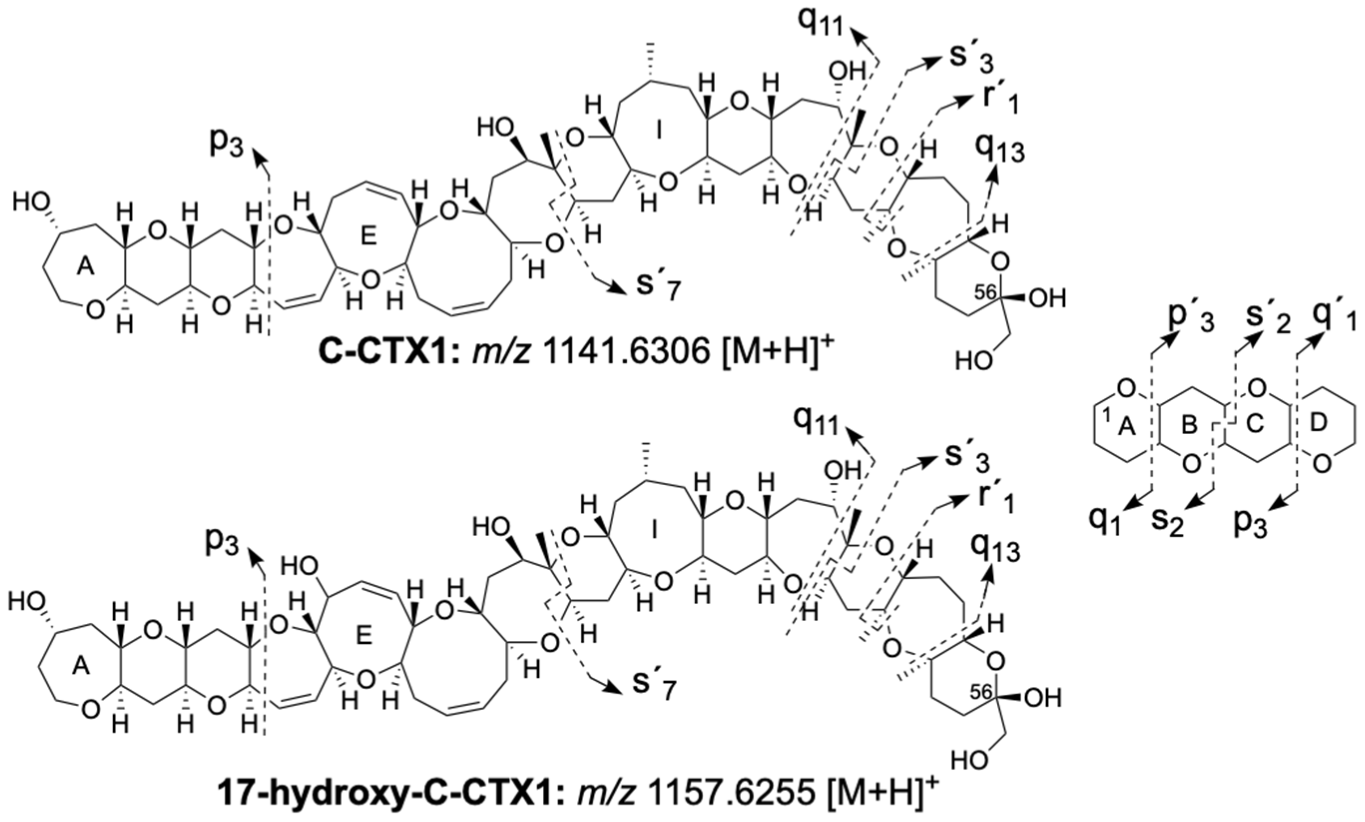
+). Additionally, the PRM mode was used to confirm the toxins based on their fragmentation. The nomenclature used for the identification of the fragment ions was proposed in [
22] and is summarized in
Figure 1. P, q and s indicate the bonds which are cleaved, the subscript number is the number of rings contained in the fragment (intact ring, or ring fragment) and the prime symbol points out fragments towards the right end of the molecule. The toxins were quantified with a calibration curve of C-CTX1 standard ranging from 0.61 to 20.00 ng/mL and each toxin was expressed in ng C-CTX1 equivalent/g fish tissue.
2.1. Ciguatoxin Profile
C-CTX1 was detected as the main toxin at a concentration of 0.46 ng C-CTX1 eq./g fish tissue. 17-hydroxy-C-CTX1 was also present at a concentration of 0.22 ng C-CTX1 eq./g fish tissue (
Table 1). Both toxins were identified with traces of their respective 56-methoxy- congeners, 0.07 ng C-CTX1/g fish tissue for 56-methoxy-C-CTX1 and 0.04 ng C-CTX1/g fish tissue for 17-hydroxy-56-methoxy-C-CTX1 (
Table 1). Additionally, traces of a new C-CTX algal analogue (C-CTX5), recently identified in [
23] in algal from the Caribbean Sea, were also detected in the sample (
Table 1). The total concentration of C-CTX1 eq. in the sample was 0.79 ng/g, which is around eight-fold above the guidance level proposed by the FDA (USA) for C-CTX1 [
24].
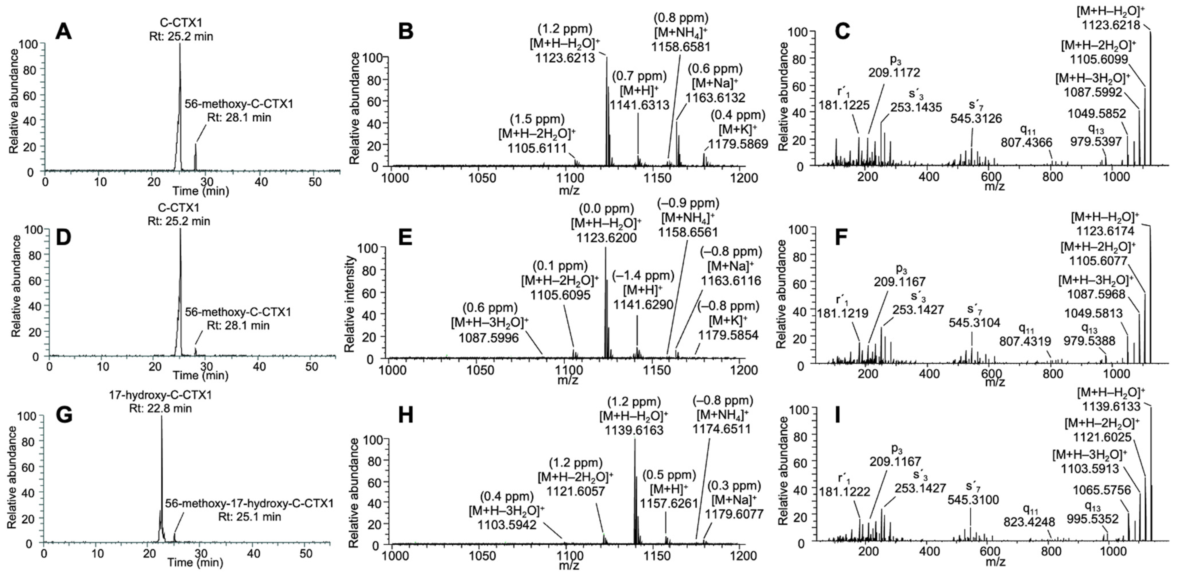
The C-CTX1 retention time (25.2 min), the ion pattern in MS1 and the MS2 fragmentation in the sample matched the C-CTX1 standard (
Figure 2A–F,
Tables S1 and S2). 17-hydroxy-C-CTX1 was detected at a retention time of 22.8 min with a prominent first water loss in MS1 at
m/
z 1139.6163 [M+H−H
2O]
+ (1.2 ppm) (
Figure 2G,H). This compound was confirmed by its fragmentation in the PRM mode matching the data previously reported in [
21] (
Figure 1 and
Figure 2I,
Table S3).
2.2. Identification and Confirmation of C-CTX5
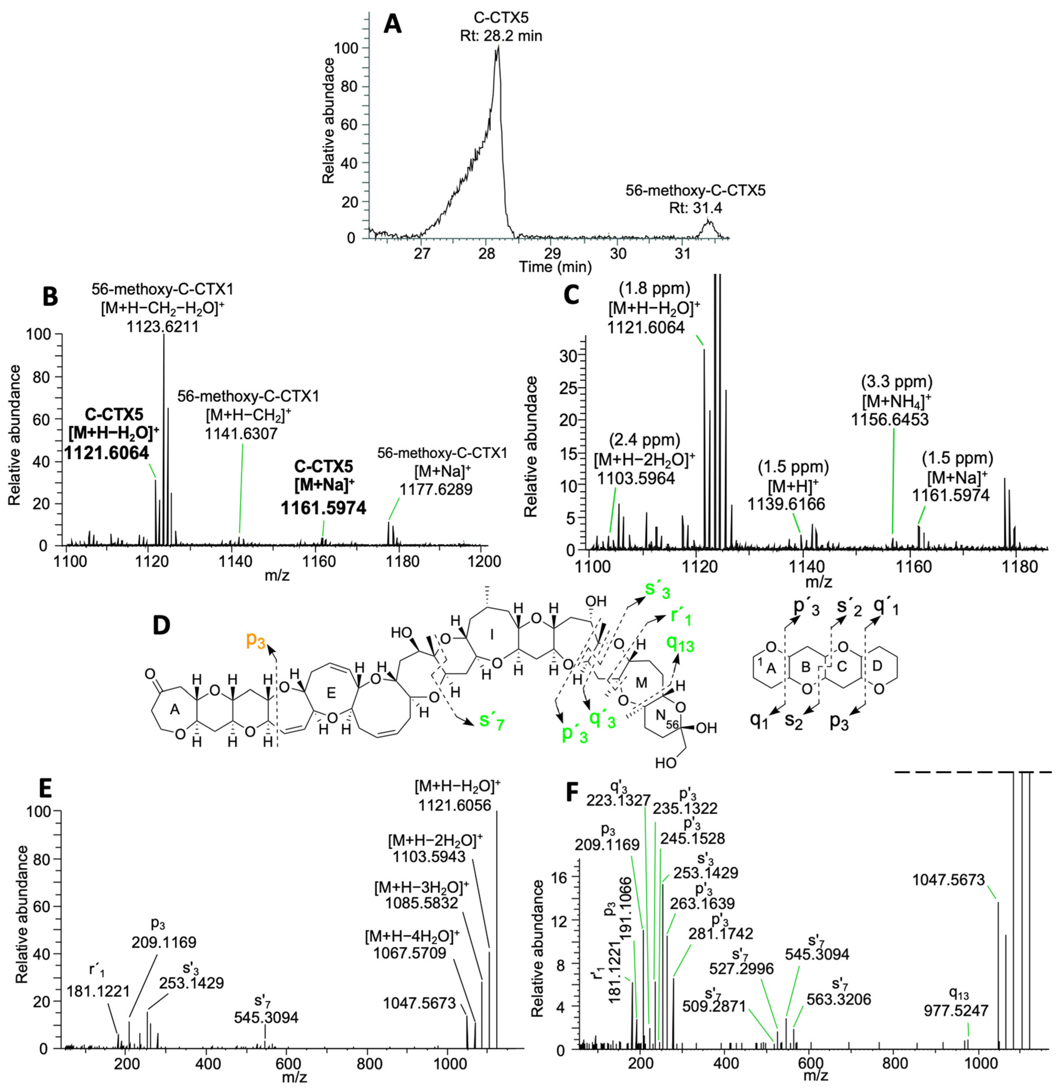
C-CTX5 was detected at 28.2 min with traces of its 56-methoxy analogue at 31.4 min (
Figure 3A). C-CTX5 was eluted as a broad chromatographic peak, as detected for C-CTX1, resulting from the rapid on-column epimerization of the ketal in C-56 due to the acidic conditions [
22]. C-CTX5 showed an ion pattern with a first water loss at
m/
z 1121.6064 [M+H−H
2O]
+ (1.8 ppm), sodium adduct at
m/
z 1161.5974 [M + Na]
+ (1.5 ppm) and additional traces of ions such as [M+H]
+, [M + NH
4]
+ and [M+H−2H
2O]
+ with Δppm < 3.5 (
Figure 3B,C). This analogue coeluted with 56-methoxy-C-CTX1 (28.1 min) (
Figure 1 and
Figure 3B,C). PRM analyses selecting C-CTX5 first water loss as precursor ion
m/
z 1121.6043 [M+H−H
2O]
+ showed a similar fragmentation pathway to that described in [
23]. Not only did the detection of successive water losses confirm C-CTX5, but it also confirmed the fragments described in [
23] from fragmentation in the G-, and H-rings (s’7) and the K-, L- and M-rings (q13, s´7, s´3 and p´3) (
Figure 3D–F,
Table S4).
Despite obtaining the same fragmentation pattern as that described in [
23], the fragment at
m/
z 209.1171 (p3) from the fragmentation in the D-ring did not match the theoretical fragment of a 3-oxo metabolite and, consequently, additional PRM analyses at different CE levels were performed to corroborate the fragmentation of C-CTX5. The PRM analyses revealed that C-CTX5 should be a 2,3 or 3,4-olefing together with a hydroxylation in the E-, F- or G-rings instead of a 3-oxo metabolite of C-CTX1. The detection of fragments from the fragmentation in the B-, C- and D-rings (p3, q2 and p2) confirmed this possibility (
Figure 4A–C,
Table S5).
3. Discussion
As mentioned earlier, characterizing fish samples involved in human intoxications associated with Ciguatera Poisoning (CP) is crucial for advancing the risk assessment of CP in emerging regions such as the Canary Islands (Spain). This is particularly important because the evaluation of this risk in fish from official controls is not always efficient, given that the concentration of ciguatoxins (CTXs) in these samples might be limited.
The use of a sensitive method such as cLC-HRMS is a valuable approach for identifying and quantifying CTX analogues with both major and minor contributions to the overall CTX toxicity. The total CTX content of the sample analyzed in this study was 0.79 ng C-CTX1 eq./g, which should be considered a reasonably high CTX concentration to produce CP symptoms.
C-CTX1 (0.46 ng/g) and 17-hydroxy-C-CTX1 (0.22 ng/g) were present in concentrations clearly above the guidance level proposed by the FDA for C-CTX1 (0.1 ng/g) [
24]. These results match the data in the literature in which, not only in the endemic areas of the Caribbean Sea, but also in the east Atlantic Ocean, C-CTX1 is always the main toxin present in the fish samples [
19,
25,
26,
27]. The presence of 56-methoxy- metabolites could be related to the artificial methoxylation of the CTXs during sample pretreatment [
28].
C-CTX5, an algal ciguatoxin recently identified in [
23] in
Gambierdisucs silvae and
G. caribeaus from the Caribbean Sea, was detected for the first time in fish from the east Atlantic Ocean. The detection of C-CTX5 in fish from this region could suggest that it might be a contributing toxin in
Gambierdiscus strains from the area. Only taking into account the MS data, the structure of C-CTX5 matches a 2,3 or 3,4-olefing together with a hydroxylation in the E-, F- or G-rings instead of a 3-oxo metabolite of C-CTX1. Mudge et al., 2023, also proposed this possibility only using the MS data. However, the structure of C-CTX5 was proposed using selected fragments from MS2 and also the results after chemical and enzymatic conversions. C-CTX5 should be isolated in higher concentrations to investigate these discrepancies and study its MS fragmentation. Also, this would allow its structure to be elucidated by NMR. Unfortunately, the concentration of C-CTX5 in the fish sample analyzed in this study was below the LOQ, which impeded the obtention of better MS1 and MS2 spectra for structural characterization purposes. The low concentration of C-CTX5 also suggests that once transferred from
Gambierdiscus, a metabolization process of C-CTX5 in fish might occur until it is converted into C-CTX1. A similar metabolization process has been reported for some Pacific CTXs (P-CTXs) [
3].
4. Conclusions
The fish sample analyzed in this study, which was consumed and linked to an outbreak of CP in the Canary Islands (Spain), was successfully characterized using a sensitive method involving Capillary Liquid Chromatography coupled to High-Resolution Mass Spectrometry (cLC-HRMS). The concentration of CTXs, expressed in C-CTX1 eq., was eight-fold above the guidance level proposed by the FDA, with C-CTX1 being the major toxin followed by 17-hydroxy-C-CTX1. Traces of putative C-CTX5 (an algal toxin) were detected for the first time in fish from the east Atlantic Ocean, suggesting that C-CTX5 might be a precursor to C-CTX1 in Gambierdiscus strains from this region. MS1 and MS2 data showed that this compound might be a 2,3 or 3,4-olefin together with a hydroxylation in the E-, F- or G-rings instead of a 3-oxo metabolite, as initially proposed. However, the low toxin amount in the sample and the absence of an authentic C-CTX5 standard make the characterization of its structure challenging. Further isolation of this compound in higher concentrations from fish or dinoflagellates would enable its complete structural characterization by NMR and confirm its identity as C-CTX5.
5. Materials and Methods
5.1. Standard and Sample
C-CTX1 standard (20 ng) was kindly provided by Dr. Robert W. Dickey (previously U.S. Food and Drug Administration) [
29].
The fish sample consisted of a raw portion of amberjack fish (Seriola sp.) tissue captured in the spring of 2023 in Fuerteventura in the Canary Islands (Spain). This fish sample was consumed and linked to an outbreak of ciguatera and was kindly provided by the Canary Islands Government Health Services through the Instituto Universitario de Sanidad Animal y Seguridad Alimentaria (IUSA) from the University of Las Palmas de Gran Canaria (ULPGC) on the course of the activities of the EuroCigua II project (GP/EFSA/KNOW/2022/03).
5.2. Sample Preparation and cLC-HRMS Analyses
Sample pretreatment and cLC-HRMS analyses were performed as described in [
21].
Supplementary Materials
The following supporting information can be downloaded at:
https://www.mdpi.com/article/10.3390/toxins16040189/s1, Table S1. Mass error of C-CTX1 fragments after PRM analyses of C-CTX1 standard (20 ng/mL) selecting
m/
z 1123.6200 [M+H–H
2O]
+ as a precursor ion at a CE of 15; Table S2. Mass error of C-CTX1 fragments after PRM analyses of C-CTX1 in amberjack sample selecting
m/
z 1123.6200 [M+H–H
2O]
+ as a precursor ion at a CE of 15; Table S3. Mass error of 17-hydroxy-C-CTX1 fragments after PRM analyses of 17-hydroxy-C-CTX1 in amberjack sample selecting
m/
z 1139.6149 [M+H–H
2O]
+ as a precursor ion at a CE of 15; Table S4. Mass error of C-CTX5 fragments after PRM analyses of C-CTX5 in amberjack sample selecting
m/
z 1121.6043 [M+H–H
2O]
+ as a precursor ion at a CE of 15; Table S5. Mass error of C-CTX5 fragments after PRM analyses of C-CTX5 in amberjack sample selecting
m/
z 1121.6043 [M+H–H
2O]
+ as a precursor ion at a CE of 40.
Author Contributions
Conceptualization, P.E. and A.G.-M.; methodology, P.E., J.O.-P., D.C. and A.P.; investigation, P.E.; resources, A.B. and A.G.-M.; data curation, P.E.; writing—original draft preparation, P.E. and A.G.-M.; writing—review and editing, P.E., J.O.-P. and A.G.-M.; supervision, J.O.-P., A.B. and A.G.-M.; funding acquisition, A.B. and A.G.-M. All authors have read and agreed to the published version of the manuscript.
Funding
Pablo Estevez (P.E.) acknowledges financial support from the Ministry for Universities (Spain) funded by the European Union (Next Generation EU) under the Margarita Salas Postdoctoral fellowship. Mass Spectrometry was provided by the Mass Spectrometry Resource at UCSF (A.L. Burlingame, Director) supported by the Dr. Miriam and Sheldon G. Adelson Medical Research Foundation (AMRF) and NIH P41GM103481 and 1S10OD016229. The authors acknowledge the financial support and the provision of the naturally contaminated sample received through the project EUROCIGUA II: “An integrated approach to assess the human health risks of ciguatoxins in fish in Europe” GP/EFSA/KNOW/2022/03, co-funded by the European Food Safety Authority (EFSA).
Institutional Review Board Statement
Not applicable.
Informed Consent Statement
Not applicable.
Data Availability Statement
Data available on request.
Acknowledgments
The authors thank Robert W. Dickey (previously U.S. Food and Drug Administration) for kindly providing the C-CTX1 pure standard used in this work. The authors acknowledge the Canary Islands Government Health Services (Spain) and the Instituto Universitario de Sanidad Animal y Seguridad Alimentaria (IUSA) from the University of Las Palmas de Gran Canaria (ULPGC) for kindly providing the contaminated amberjack used for characterization in this study on the course of the activities of the EuroCigua II project (GP/EFSA/KNOW/2022/03). The authors thank all the members of the CI8 research group at the University of Vigo.
Conflicts of Interest
The authors declare no conflicts of interest.
References
- Food and Agriculture Organization of the United Nations; World Health Organization. Report of the Expert Meeting on Ciguatera Poisoning: Rome, 19–23 November 2018; Food Safety and Quality Series; 9; World Health Organization: Geneva, Switzerland, 2020. [Google Scholar]
- Yasumoto, T.; Nakajima, I.; Bagnis, R.; Adachi, R. Finding of a Dinoflagellate as a Likely Culprit of Ciguatera. Nippon Suisan Gakkaishi 1977, 43, 1021–1026. [Google Scholar] [CrossRef]
- Ikehara, T.; Kuniyoshi, K.; Oshiro, N.; Yasumoto, T. Biooxidation of Ciguatoxins Leads to Species-Specific Toxin Profiles. Toxins 2017, 9, 205. [Google Scholar] [CrossRef] [PubMed]
- Friedman, M.A.; Fernandez, M.; Backer, L.C.; Dickey, R.W.; Bernstein, J.; Schrank, K.; Kibler, S.; Stephan, W.; Gribble, M.O.; Bienfang, P.; et al. An Updated Review of Ciguatera Fish Poisoning: Clinical, Epidemiological, Environmental, and Public Health Management. Mar. Drugs 2017, 15, 72. [Google Scholar] [CrossRef]
- Lewis, R.J. The Changing Face of Ciguatera. Toxicon 2001, 39, 97–106. [Google Scholar] [CrossRef]
- Boada, L.D.; Zumbado, M.; Luzardo, O.P.; Almeida-González, M.; Plakas, S.M.; Granade, H.R.; Abraham, A.; Jester, E.L.E.; Dickey, R.W. Ciguatera Fish Poisoning on the West Africa Coast: An Emerging Risk in the Canary Islands (Spain). Toxicon 2010, 56, 1516–1519. [Google Scholar] [CrossRef]
- Loeffler, C.R.; Spielmeyer, A.; Blaschke, V.; Bodi, D.; Kappenstein, O. Ciguatera Poisoning in Europe: A Traceback to Indian Ocean Sourced Snapper Fish (Lutjanus Bohar). Food Control 2023, 151, 109799. [Google Scholar] [CrossRef]
- Lewis, R.J.; Holmes, M.J. Origin and Transfer of Toxins Involved in Ciguatera. Comp. Biochem. Physiol. Part C Pharmacol. Toxicol. Endocrinol. 1993, 106, 615–628. [Google Scholar] [CrossRef]
- Sibat, M.; Herrenknecht, C.; Darius, H.T.; Roué, M.; Chinain, M.; Hess, P. Detection of Pacific Ciguatoxins Using Liquid Chromatography Coupled to Either Low or High Resolution Mass Spectrometry (LC-MS/MS). J. Chromatogr. A 2018, 1571, 16–28. [Google Scholar] [CrossRef] [PubMed]
- Yogi, K.; Oshiro, N.; Inafuku, Y.; Hirama, M.; Yasumoto, T. Detailed LC-MS/MS Analysis of Ciguatoxins Revealing Distinct Regional and Species Characteristics in Fish and Causative Alga from the Pacific. Anal. Chem. 2011, 83, 8886–8891. [Google Scholar] [CrossRef]
- Kato, T.; Yasumoto, T. Quantification of Representative Ciguatoxins in the Pacific Using Quantitative Nuclear Magnetic Resonance Spectroscopy. Mar. Drugs 2017, 15, 309. [Google Scholar] [CrossRef]
- Murata, M.; Ishibashi, Y.; Yasumoto, T.; Legrand, A.M. Structures of Ciguatoxin and Its Congener. J. Am. Chem. Soc. 1989, 111, 8929–8931. [Google Scholar] [CrossRef]
- Oshiro, N.; Nagasawa, H.; Watanabe, M.; Nishimura, M.; Kuniyoshi, K.; Kobayashi, N.; Sugita-Konishi, Y.; Asakura, H.; Tachihara, K.; Yasumoto, T. An Extensive Survey of Ciguatoxins on Grouper Variola Louti from the Ryukyu Islands, Japan, Using Liquid Chromatography–Tandem Mass Spectrometry (LC-MS/MS). J. Mar. Sci. Eng. 2022, 10, 423. [Google Scholar] [CrossRef]
- Nagae, M.; Igarashi, T.; Mizukoshi, K.; Kuniyoshi, K.; Oshiro, N.; Yasumoto, T. Development and Validation of an LC-MS/MS Method for the Ultra-Trace Analysis of Pacific Ciguatoxins in Fish. J. AOAC Int. 2021, 104, 1272–1281. [Google Scholar] [CrossRef] [PubMed]
- Tsumuraya, T.; Hirama, M. Rationally Designed Synthetic Haptens to Generate Anti-Ciguatoxin Monoclonal Antibodies, and Development of a Practical Sandwich ELISA to Detect Ciguatoxins. Toxins 2019, 11, 533. [Google Scholar] [CrossRef] [PubMed]
- Pérez-Arellano, J.L.; Luzardo, O.P.; Brito, A.P.; Cabrera, M.H.; Zumbado, M.; Carranza, C.; Angel-Moreno, A.; Dickey, R.W.; Boada, L.D. Ciguatera Fish Poisoning, Canary Islands. Emerg. Infect. Dis. 2005, 11, 1981–1982. [Google Scholar] [CrossRef] [PubMed]
- EFSA Panel on Contaminants in the Food Chain. Scientific Opinion on Marine Biotoxins in Shellfish—Emerging Toxins: Ciguatoxin Group. EFSA J. 2010, 8, 1627. [Google Scholar] [CrossRef]
- Otero, P.; Pérez, S.; Alfonso, A.; Vale, C.; Rodríguez, P.; Gouveia, N.N.; Gouveia, N.; Delgado, J.; Vale, P.; Hirama, M.; et al. First Toxin Profile of Ciguateric Fish in Madeira Arquipelago (Europe). Anal. Chem. 2010, 82, 6032–6039. [Google Scholar] [CrossRef] [PubMed]
- Estevez, P.; Sibat, M.; Leão-Martins, J.M.; Costa, P.R.; Gago-Martínez, A.; Hess, P. Liquid Chromatography Coupled to High-Resolution Mass Spectrometry for the Confirmation of Caribbean Ciguatoxin-1 as the Main Toxin Responsible for Ciguatera Poisoning Caused by Fish from European Atlantic Coasts. Toxins 2020, 12, 267. [Google Scholar] [CrossRef]
- Tudó, À.; Rambla-alegre, M.; Flores, C.; Sagristà, N.; Aguayo, P.; Reverté, L.; Campàs, M.; Gouveia, N.; Santos, C.; Andree, K.B.; et al. Identification of New CTX Analogues in Fish from the Madeira and Selvagens Archipelagos by Neuro-2a CBA and LC-HRMS. Mar. Drugs 2022, 20, 236. [Google Scholar] [CrossRef]
- Estevez, P.; Oses Prieto, J.; Burlingame, A.; Gago Martinez, A. Characterization of the Ciguatoxin Profile in Fish Samples from the Eastern Atlantic Ocean Using Capillary Liquid Chromatography-High Resolution Mass Spectrometry. Food Chem. 2023, 418, 135960. [Google Scholar] [CrossRef]
- Kryuchkov, F.; Robertson, A.; Miles, C.O.; Mudge, E.M.; Uhlig, S. LC–HRMS and Chemical Derivatization Strategies for the Structure Elucidation of Caribbean Ciguatoxins: Identification of C-CTX-3 and -4. Mar. Drugs 2020, 18, 182. [Google Scholar] [CrossRef]
- Mudge, E.M.; Miles, C.O.; Ivanova, L.; Uhlig, S.; James, K.S.; Erdner, D.L.; Fæste, C.K.; McCarron, P.; Robertson, A. Algal Ciguatoxin Identified as Source of Ciguatera Poisoning in the Caribbean. Chemosphere 2023, 330, 138659. [Google Scholar] [CrossRef] [PubMed]
- Dickey, R.W.; Plakas, S.M. Ciguatera: A Public Health Perspective. Toxicon 2010, 56, 123–136. [Google Scholar] [CrossRef] [PubMed]
- Pottier, I.; Vernoux, J.P.; Jones, A.; Lewis, R.J. Characterisation of Multiple Caribbean Ciguatoxins and Congeners in Individual Specimens of Horse-Eye Jack (Caranx Latus) by High-Performance Liquid Chromatography/Mass Spectrometry. Toxicon 2002, 40, 929–939. [Google Scholar] [CrossRef] [PubMed]
- Pottier, I.; Hamilton, B.; Jones, A.; Lewis, R.J.; Vernoux, J.P. Identification of Slow and Fast-Acting Toxins in a Highly Ciguatoxic Barracuda (Sphyraena Barracuda) by HPLC/MS and Radiolabelled Ligand Binding. Toxicon 2003, 42, 663–672. [Google Scholar] [CrossRef]
- Abraham, A.; Jester, E.L.E.; Granade, H.R.; Plakas, S.M.; Dickey, R.W. Caribbean Ciguatoxin Profile in Raw and Cooked Fish Implicated in Ciguatera. Food Chem. 2012, 131, 192–198. [Google Scholar] [CrossRef]
- Estevez, P.; Leao, J.M.; Yasumoto, T.; Dickey, R.W.; Gago-Martinez, A. Caribbean Ciguatoxin-1 Stability under Strongly Acidic Conditions: Characterisation of a New C-CTX1 Methoxy Congener. Food Addit. Contam. Part A 2020, 37, 519–529. [Google Scholar] [CrossRef]
- Crouch, R.C.; Martin, G.E.; Musser, S.M.; Ray Grenade, H.; Dickey, R.W. Improvements in the Sensitivity of Inverse-Detected Heteronuclear Correlation Spectra Using Micro Inverse Probes and Micro Cells: HMQC and HMBC Spectra of Caribbean Ciguatoxin—Preliminary Structural Inferences. Tetrahedron Lett. 1995, 36, 6827–6830. [Google Scholar] [CrossRef]
| Disclaimer/Publisher’s Note: The statements, opinions and data contained in all publications are solely those of the individual author(s) and contributor(s) and not of MDPI and/or the editor(s). MDPI and/or the editor(s) disclaim responsibility for any injury to people or property resulting from any ideas, methods, instructions or products referred to in the content. |
© 2024 by the authors. Licensee MDPI, Basel, Switzerland. This article is an open access article distributed under the terms and conditions of the Creative Commons Attribution (CC BY) license (https://creativecommons.org/licenses/by/4.0/).

 ,
, 


