Analytical Methods for Anatoxin-a Determination: A Review


Abstract
:1. Introduction
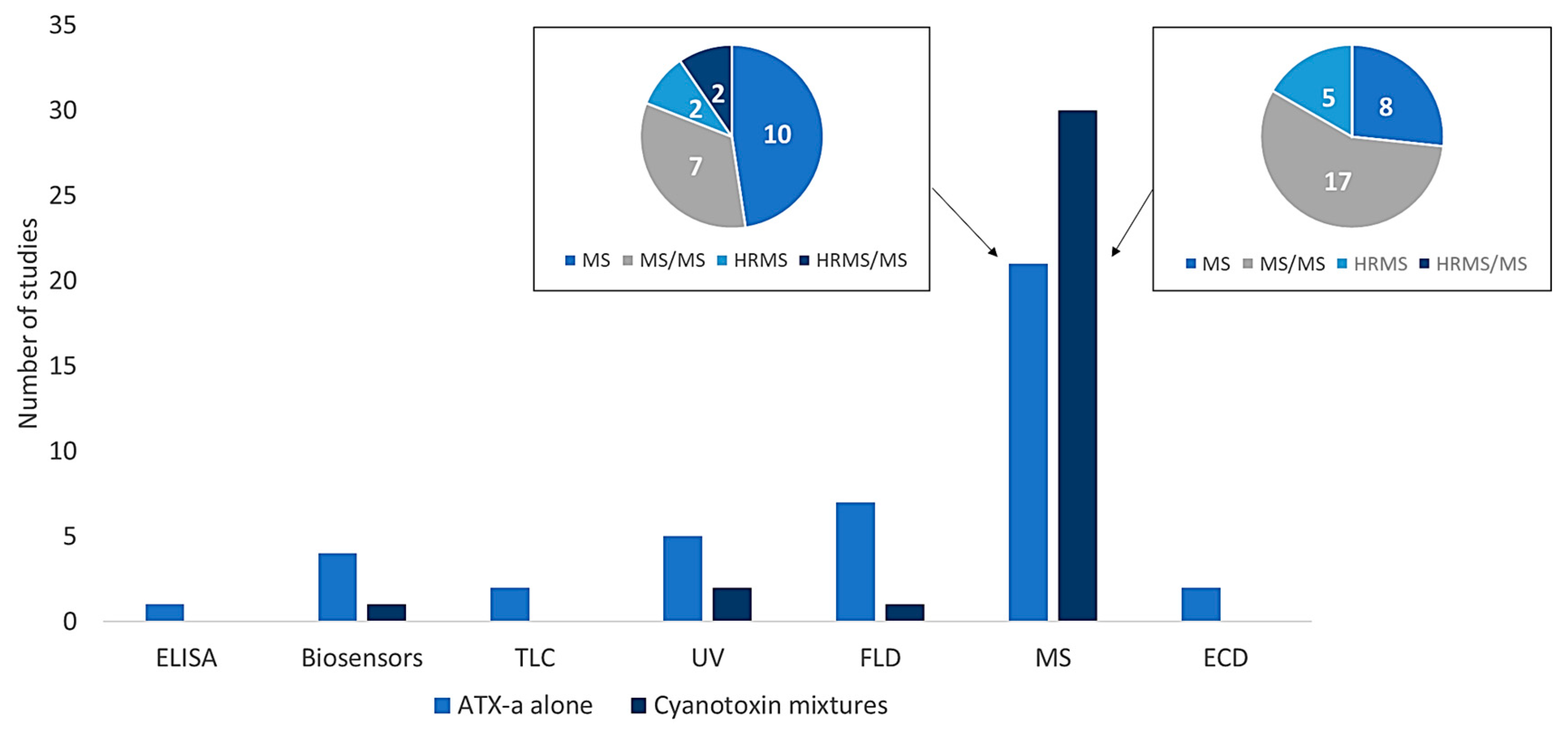
2. Analytical Methods for ATX-a Determination
2.1. Enzyme-Linked Immunosorbent Assay (ELISA)
2.2. DNA Aptamers/Biosensors
2.3. Ultraviolet and Fluorescence Detection
2.4. Mass Spectrometry Detection
Method EPA 545
2.5. Gas Chromatography–Electron Capture Detection
3. General Discussion
4. Conclusions
5. Materials and Methods
5.1. The Information Sources and Search Strategy
5.2. Inclusion and Exclusion Criteria
Supplementary Materials
Author Contributions
Funding
Institutional Review Board Statement
Informed Consent Statement
Data Availability Statement
Acknowledgments
Conflicts of Interest
References
- World Health Organization (WHO). Cyanobacterial Toxins: Anatoxin-a and Analogues; Background Document for Development of WHO Guidelines for Drinking-Water Quality and Guidelines for safe Recreational Water Environments; WHO: Geneva, Switzerland, 2020. [Google Scholar]
- Stevens, D.K.; Krieger, R.I. Stability studies on the cyanobacterial nicotinic alkaloid anatoxin-a. Toxicon 1991, 29, 167–179. [Google Scholar] [CrossRef]
- Colas, S.; Marie, B.; Lance, E.; Quiblier, C.; Tricoire-Leignel, H.; Mattei, C. Anatoxin-a: Overview on a harmful cyanobacterial neurotoxin from the environmental scale to the molecular target. Environ. Res. 2021, 193, 110590. [Google Scholar] [CrossRef]
- Bouma-Gregson, K.; Kudela, R.M.; Power, M.E. Widespread anatoxin-a detection in benthic cyanobacterial mats throughout a river network. PLoS ONE 2018, 13, e0197669. [Google Scholar] [CrossRef]
- Moreira, C.; Gomes, C.; Vasconcelos, V.; Antunes, A. Cyanotoxins Occurrence in Portugal: A New Report on Their Recent Multiplication. Toxins 2020, 12, 154. [Google Scholar] [CrossRef]
- Sabart, M.; Crenn, K.; Perrière, F.; Abila, A.; Leremboure, M.; Colombet, J.; Jousse, C.; Latour, D. Co-occurrence of microcystin and anatoxin-a in the freshwater lake Aydat (France): Analytical and molecular approaches during a three-year survey. Harmful Algae 2015, 48, 12–20. [Google Scholar] [CrossRef]
- Cerasino, L.; Salmaso, N. Co-occurrence of anatoxin-a and microcystins in Lake Garda and other deep perialpine lakes. Adv. Oceanogr. Limnol. 2020, 11, 11–21. [Google Scholar] [CrossRef]
- Blahova, L.; Sehnal, L.; Lepsova-Skacelova, O.; Szmucova, V.; Babica, P.; Hilscherova, K.; Teikari, J.; Sivonen, K.; Blaha, L. Occurrence of cylindrospermopsin, anatoxin-a and their homologs in the southern Czech Republic—Taxonomical, analytical, and molecular approaches. Harmful Algae 2021, 108, 102101. [Google Scholar] [CrossRef]
- Ríos, V.; Moreno, I.; Prieto, A.I.; Puerto, M.; Gutiérrez-Praena, D.; Soria-Díaz, M.E.; Cameán, A.M. Analysis of MC-LR and MC-RR in tissue from freshwater fish (Tinca tinca) and crayfish (Procambarus clarkii) in tench ponds (Cáceres, Spain) by liquid chromatography-mass spectrometry (LC-MS). Food Chem. Toxicol. 2013, 57, 170–178. [Google Scholar] [CrossRef]
- Guzmán-Guillén, R.; Moreno, I.; Prieto Ortega, A.I.; Soria-Díaz, M.E.; Vasconcelos, V.; Cameán, A.M. CYN determination in tissues from freshwater fish by LC-MS/MS: Validation and application in tissues from subchronically exposed tilapia (Oreochromis niloticus). Talanta 2015, 131, 452–459. [Google Scholar] [CrossRef]
- Spivak, C.E.; Witkop, B.; Albuquerque, E.X. Anatoxin-a: A novel, potent agonist at the nicotinic receptor. Mol. Pharmacol. 1980, 18, 384–394. [Google Scholar]
- Aráoz, R.; Molgó, J.; Tandeau de Marsac, N. Neurotoxic cyanobacterial toxins. Toxicon 2010, 56, 813–828. [Google Scholar] [CrossRef]
- Bruno, M.; Ploux, O.; Metcalf, J.S.; Mejean, A.; Pawlik-Skowronska, B.; Furey, A. Anatoxin-a, homoanatoxin-a, and natural analogues. In Handbook of Cyanobacterial Monitoring and Cyanotoxin analysis, 1st ed.; Meriluoto, J., Spoof, L., Codd, G.A., Eds.; John Wiley & Sons, Ltd.: New York, NY, USA, 2017; pp. 138–147. [Google Scholar]
- Plata-Calzado, C.; Prieto, A.I.; Cameán, A.M.; Jos, A. Toxic effects produced by anatoxin-a under laboratoty conditions: A review. Toxins 2022, 14, 861. [Google Scholar] [CrossRef]
- Rao Lakshmana, P.; Bhattacharya, R.; Gupta, N.; Parida, M.; Bhaskar, A.; Dubey, R. Involvement of caspase and reactive oxygen species in cyanobacterial toxin anatoxin-a-induced cytotoxicity and apoptosis in rat thymocytes and Vero cells. Arch. Toxicol. 2002, 76, 227–235. [Google Scholar] [CrossRef]
- Zhong, Y.; Shen, L.; Ye, X.; Zhou, D.; He, Y.; Li, Y.; Ding, Y.; Zhu, W.; Ding, J.; Zhang, H. Neurotoxic anatoxin-a can also exert immunotoxicity by the induction of apoptosis on Carassius auratus lymphocytes in vitro when exposed to environmentally relevant concentrations. Front. Physiol. 2020, 11, 316. [Google Scholar] [CrossRef]
- Plata-Calzado, C.; Diez-Quijada, L.; Medrano-Padial, C.; Prieto, A.I.; Cameán, A.M.; Jos, A. In vitro mutagenic and genotoxic assessment of anatoxin-a alone and in combination with cylindrospermopsin. Toxins 2023, 15, 458. [Google Scholar] [CrossRef]
- Testai, E. Anatoxin-a and analogues. In Toxic Cyanobacteria in Water. A Guide to Their Public Health Consequences, Monitoring and Management, 2nd ed.; Chorus, I., Welker, M., Eds.; CRC Press: Boca Raton, FL, USA, 2021; pp. 72–94. [Google Scholar]
- Fastner, J.; Beulker, C.; Geiser, B.; Hoffmann, A.; Kröger, R.; Teske, K.; Hoppe, J.; Mundhenk, L.; Neurath, H.; Sagebiel, D.; et al. Fatal neurotoxicosis in dogs associated with tychoplanktic, anatoxin-a producing Tychonema sp. in mesotrophic Lake Tegel, Berlin. Toxins 2018, 10, 60. [Google Scholar] [CrossRef]
- McCarron, P.; Rafuse, C.; Scott, S.; Lawrence, J.; Bruce, M.R.; Doothwright, E.; Murphy, C.; Reith, M.; Beach, D.G. Anatoxins from benthic cyanobacteria responsible for dog motalities in New Brunswick, Canada. Toxicon 2023, 227, 107086. [Google Scholar] [CrossRef]
- Biré, R.; Bertin, T.; Dom, I.; Hort, V.; Schmitt, C.; Diogène, J.; Lemée, R.; De Haro, L.; Nicolas, M. First Evidence of the presence of anatoxin-a in sea figs associated with human food poisonings in France. Mar. Drugs 2020, 18, 285. [Google Scholar] [CrossRef]
- Rellán, S.; Osswald, J.; Saker, M.; Gago-Martinez, A.; Vasconcelos, V. First detection of anatoxin-a in human and animal dietary supplements containing cyanobacteria. Food Chem. Toxicol. 2009, 47, 2189–2195. [Google Scholar] [CrossRef]
- Amzil, Z.; Derrien, A.; Terre Terrillon, A.; Savar, V.; Bertin, T.; Peyrat, M.; Duval, A.; Lhaute, K.; Arnich, N.; Hort, V.; et al. Five years monitoring the emergence of unregulated toxins in shellfish in France (EMERGTOX 2018-2022). Mar. Drugs 2023, 21, 435. [Google Scholar] [CrossRef]
- Osswald, J.; Rellan, S.; Carvalho, A.P.; Gago, A.; Vasconcelos, V. Acute effects of an anatoxin-a producing cyanobacterium on juvenile fish—Cyprinus carpio L. Toxicon 2007, 49, 693–698. [Google Scholar] [CrossRef]
- Osswald, J.; Azevedo, J.; Vasconcelos, V.; Guilhermino, L. Experimental determination of the bioconcentration factors for anatoxin-a in juvenile rainbow trout (Oncorhynchus mykiss). Proc. Int. Acad. Ecol. Environ. Sci. 2011, 1, 77–86. [Google Scholar]
- Pawlik-Skowrońska, B.; Toporowska, M.; Rechulicz, J. Simultaneous accumulation of anatoxin-a and microcystins in three fish species indigenous to lakes affected by cyanobacterial blooms. Oceanol. Hydrobiol. Stud. 2012, 41, 53–65. [Google Scholar] [CrossRef]
- Colas, S.; Duval, C.; Marie, B. Toxicity, transfer and depuration of anatoxin-a (cyanobacterial neurotoxin) in medaka fish exposed by single-dose gavage. Aquat. Toxicol. 2020, 222, 105422. [Google Scholar] [CrossRef]
- Picardo, M.; Filatova, D.; Nuñez, O.; Farré, M. Recent advances in the detection of natural toxins in freshwater environments. Trend Anal. Chem. 2019, 112, 75–86. [Google Scholar] [CrossRef]
- Sundaravadivelu, D.; Sanan, T.T.; Venkatapathy, R.; Mash, H.; Tettenhorst, D.; Danglada, L.; Frey, S.; Tatters, A.O.; Lazorchak, J. Determination of cyanotoxins and prymnesins in water, fish tissue, and other matrices: A review. Toxins 2022, 14, 213. [Google Scholar] [CrossRef]
- Harada, K.; Kimura, Y.; Ogawa, K.; Suzuki, M.; Dahlem, A.M.; Beasley, V.R.; Carmichael, W.W. A new procedure for the analysis and purification of naturally occurring anatoxin-a from the blue-green alga Anabaena flos-aquae. Toxicon 1989, 27, 1289–1296. [Google Scholar] [CrossRef]
- Ojanperä, I.; Vuori, E.; Himberg, K.; Waris, M.; Niinivaara, K. Facile detection of anatoxin-a in algal material by thin-layer chromatography with fast black K salt. Analyst 1991, 116, 265–267. [Google Scholar] [CrossRef]
- Furey, A.; Crowley, J.; Lehane, M.; James, K.J. Liquid chromatography with electrospray ion-trap mass spectrometry for the determination of anatoxins in cyanobacteria and drinking water. Rapid Commun. Mass Spectrom. 2003, 17, 583–588. [Google Scholar] [CrossRef]
- Dagnino, D.; Schripsema, J. 1H NMR quantification in very dilute toxin solutions: Application to anatoxin-a analysis. Toxicon 2005, 46, 236–240. [Google Scholar] [CrossRef]
- Ghassempour, A.; Mashkouri Najafi, N.; Mehdiania, A.; Hosseiny Davarani, S.S.; Fallahi, M.; Nakhshab, M. Analysis of anatoxin-a using polyaniline as a sorbent in solid-phase microextraction coupled to gas chromatography-mass spectrometry. J. Chromatogr. A 2005, 1078, 120–127. [Google Scholar] [CrossRef] [PubMed]
- James, K.J.; Crowley, J.; Hamilton, B.; Lehane, M.; Skulberg, O.; Furey, A. Anatoxins and degradation products, determined using hybrid quadrupole time-of-flight and quadrupole ion-trap mass spectrometry: Forensic investigations of cyanobacterial neurotoxin poisoning. Rapid Commun. Mass Spectrom. 2005, 19, 1167–1175. [Google Scholar] [CrossRef] [PubMed]
- Rawn, D.F.K.; Lau, B.; Niedzwiadek, B.; Lawrence, J.F. Improved method for the determination of anatoxin-a and two of its metabolites in blue-green algae using liquid chromatography with fluorescence detection. J. AOAC Int. 2005, 88, 1741–1747. [Google Scholar] [CrossRef] [PubMed]
- Bogialli, S.; Bruno, M.; Curini, R.; Di Corcia, A.; Laganà, A. Simple and rapid determination of anatoxin-a in lake water and fish muscle tissue by liquid-chromatography-tandem mass spectrometry. J. Chromatogr. A 2006, 1122, 180–185. [Google Scholar] [CrossRef] [PubMed]
- Rodríguez, V.; Yonamine, M.; Pinto, E. Determination of anatoxin-a in environmental water samples by solid-phase microextraction and gas chromatography-mass spectrometry. J. Sep. Sci. 2006, 29, 2085–2090. [Google Scholar] [CrossRef] [PubMed]
- Rellán, S.; Gago-Martínez, A. Improved conditions for the application of solid phase microextraction prior to HPLC-FLD analysis of anatoxin-a. J. Sep. Sci. 2007, 30, 2522–2528. [Google Scholar] [CrossRef]
- Aráoz, R.; Guérineau, V.; Rippka, R.; Palibroda, N.; Herdman, M.; Laprevote, O.; Döhren, H.; Tandeau de Marsac, N.; Erhard, M. MALDI-TOF-MS detection of the low molecular weight neurotoxins anatoxin-a and homoanatoxin-a on lyophilized and fresh filaments of axenic Oscillatoria strains. Toxicon 2008, 51, 1308–1315. [Google Scholar] [CrossRef]
- Dimitrakopoulus, I.K.; Kaloudis, T.S.; Hiskia, A.E.; Thomaidis, N.S.; Koupparis, M.A. Development of a fast and selective method for the sensitive determination of anatoxin-a in lake waters using liquid chromatography-tandem mass spectrometry and phenylalanine-d5 as internal standard. Anal. Bioanal. Chem. 2010, 397, 2245–2252. [Google Scholar] [CrossRef]
- Azevedo, J.; Osswald, J.; Guilhermino, L.; Vasconcelos, V. Development and validation of an SPE-HPLC-FL method for the determination of anatoxin-a in water and trout (Oncorhincus mykiss). Anal. Lett. 2011, 44, 1431–1441. [Google Scholar] [CrossRef]
- Lemoine, P.; Roy-Lachapelle, A.; Prévost, M.; Tremblay, P.; Solliec, M.; Sauvé, S. Ultra-fast analysis of anatoxin-a using laser diode thermal desorption-atmospheric pressure chemical ionization-tandem mass spectrometry: Validation and resolution from phenylalanine. Toxicon 2013, 61, 165–174. [Google Scholar] [CrossRef]
- Sanchez, J.A.; Otero, P.; Alfonso, A.; Ramos, V.; Vasconcelos, V.; Aráoz, R.; Molgó, J.; Vieytes, M.R.; Botana, L.M. Detection of anatoxin-a and three analogs in Anabaena spp. Cultures: New fluorescence polarization assay and toxin profile by LC-MS/MS. Toxins 2014, 6, 402–415. [Google Scholar] [CrossRef] [PubMed]
- Elshafey, R.; Siaj, M.; Zourob, M. DNA aptamers selection and characterization for development of label-free impedimetric aptasensor for neurotoxin anatoxin-a. Biosens. Bioelectron. 2015, 68, 295–302. [Google Scholar] [CrossRef]
- Roy-Lachapelle, A.; Solliec, M.; Sinotte, M.; Deblois, C.; Sauvé, S. High resolution/accurate mass (HRMS) detection of anatoxin-a in lake water using LDTD-APCI coupled to a Q-Exactive mass spectrometer. Talanta 2015, 132, 836–844. [Google Scholar] [CrossRef] [PubMed]
- Beach, D.G.; Hollingdale, C.; Quilliam, M.A. Isotope-labelling derivatization: A broadly applicable approach to quantitation of algal toxins by isotope dilution LC-MS/MS. Anal. Methods 2016, 8, 2872–2879. [Google Scholar] [CrossRef]
- Le, T.; Esteve-Turrillas, F.A.; Armenta, S.; de la Guardia, M.; Quiñones-Reyes, G.; Abad-Fuentes, A.; Abad-Somovilla, A. Dispersive magnetic immunoaffinity extraction. Anatoxin-a determination. J. Chromatogr. A 2017, 1529, 57–62. [Google Scholar] [CrossRef] [PubMed]
- Lin, Z.; Gu, J.; Li, C.; Lee, R.; Xie, L.; Chen, S.; Cao, P.; Jiang, S.; Yuan, Y.; Hong, X.; et al. A nanoparticle-decorated biomolecule-responsive polymer enables robust signaling cascade for biosensing. Adv. Mater. 2017, 29, 1702090. [Google Scholar] [CrossRef] [PubMed]
- Beach, D.G.; Rafuse, C.; Melanson, J.E.; McCarron, P. Rapid quantitative screening of cyanobacteria for production of anatoxins using direct analysis in real time high-resolution mass spectrometry. Rapid Commun. Mass Spectrom. 2021, 35, e8940. [Google Scholar] [CrossRef] [PubMed]
- Nguyen, D.; Jang, C. A simple and ultrasensitive colorimetric biosensor for anatoxin-a based on aptamer and gold nanoparticles. Micromachines 2021, 12, 1526. [Google Scholar] [CrossRef] [PubMed]
- Xia, M.; Zhou, F.; Feng, X.; Sun, J.; Wang, L.; Li, N.; Wang, X.; Wang, G. A DNAzyme-Based Dual-Stimuli responsive electrochemiluminescence resonance energy transfer platform for ultrasensitive anatoxin-a detection. Anal. Chem. 2021, 93, 11284–11290. [Google Scholar] [CrossRef] [PubMed]
- Beach, D.G.; Bruce, M.; Lawrence, J.; McCarron, P. Rapid quantification of anatoxins in benthic cyanobacterial mats using direct analysis in real-time-high resolution tandem mass spectrometry. Environ. Sci. Technol. 2022, 56, 13837–13844. [Google Scholar] [CrossRef]
- Cevallos-Cedeño, R.E.; Quiñones-Reyes, G.; Agulló, C.; Abad-Somovilla, A.; Abad-Fuentes, A.; Mercader, J.V. Rapid immunochemical methods for anatoxin-a monitoring in environmental water samples. Anal. Chem. 2022, 94, 10857–10864. [Google Scholar] [CrossRef] [PubMed]
- Beach, D.; Zamlynny, L.; MacArthur, M.; Miles, C. Liquid chromatography-high-resolution tandem mass spectrometry of anatoxins, including new conjugates and reduction products. Anal. Bioanal. Chem. 2023, 415, 5281–5296. [Google Scholar] [CrossRef]
- Dahlmann, J.; Budakowski, W.R.; Luckas, B. Liquid chromatography–electrospray ionisation-mass spectrometry based method for the simultaneous determination of algal and cyanobacterial toxins in phytoplankton from marine waters and lakes followed by tentative structural elucidation of microcystins. J. Chromatogr. A 2003, 994, 45–57. [Google Scholar] [CrossRef] [PubMed]
- Dell’Aversano, C.; Eaglesham, G.K.; Quilliam, M.A. Analysis of cyanobacterial toxins by hydrophilic interaction liquid chromatography-mass spectrometry. J. Chromatogr. A 2004, 1028, 155–164. [Google Scholar] [CrossRef] [PubMed]
- Maizels, M.; Budde, W.L. A LC/MS method for the determination of cyanobacteria toxins in water. Anal. Chem. 2004, 76, 1342–1351. [Google Scholar] [CrossRef] [PubMed]
- Vasas, G.; Gáspar, A.; Páger, C.; Surányi, G.; Mathé, C.; Hamvas, M.M.; Borbely, G. Analysis of cyanobacterial toxins (anatoxin-a, cylindrospermopsin, microcystin-LR) by capillary electrophoresis. Electrophoresis 2004, 25, 108–115. [Google Scholar] [CrossRef] [PubMed]
- Hiller, S.; Krock, B.; Cembella, A.; Luckas, B. Rapid detection of cyanobacterial toxins in precursor ion mode by liquid chromatography tandem mass spectrometry. J. Mass Spectrom. 2007, 42, 1238–1250. [Google Scholar] [CrossRef] [PubMed]
- Oehrle, S.A.; Southwell, B.; Westrick, J. Detection of various freshwater cyanobacterial toxins using ultra-performance liquid chromatography tandem mass spectrometry. Toxicon 2010, 55, 965–972. [Google Scholar] [CrossRef] [PubMed]
- Yen, H.; Lin, T.; Liao, P. Simultaneous detection of nine cyanotoxins in drinking water using dual solid-phase extraction and liquid chromatography–mass spectrometry. Toxicon 2011, 58, 209–218. [Google Scholar] [CrossRef]
- Al-Sammak, M.A.; Hoagland, K.D.; Snow, D.D.; Cassada, D. Methods for simultaneous detection of the cyanotoxins BMAA, DABA, and anatoxin-a in environmental samples. Toxicon 2013, 76, 316–325. [Google Scholar] [CrossRef]
- Rodríguez, I.; Alfonso, C.; Alfonso, A.; Otero, P.; Meyer, T.; Breitenback, U.; Botana, L.M. Toxin profile in samples collected in fresh and brackish water in Germany. Toxicon 2014, 91, 35–44. [Google Scholar] [CrossRef] [PubMed]
- Fayad, P.B.; Roy-Lachapelle, A.; Duy, S.V.; Prévost, M.; Sauvé, S. On-line solid-phase extraction coupled to liquid chromatography tandem mass spectrometry for the analysis of cyanotoxins in algal blooms. Toxicon 2015, 108, 167–175. [Google Scholar] [CrossRef] [PubMed]
- Greer, B.; McNamee, S.E.; Boots, B.; Cimarelli, L.; Guillebault, D.; Helmi, K.; Marcheggiani, S.; Panaiotov, S.; Breitenbach, U.; Akçaalan, R.; et al. A validated UPLC–MS/MS method for the surveillance of ten aquatic biotoxins in European brackish and freshwater systems. Harmful Algae 2016, 55, 31–40. [Google Scholar] [CrossRef] [PubMed]
- Pekar, H.; Westerberg, E.; Bruno, O.; Lääne, A.; Persson, K.M.; Sundström, L.F.; Thim, A. Fast, rugged and sensitive ultra-high pressure liquid chromatography tandem mass spectrometry method for analysis of cyanotoxins in raw water and drinking water—First findings of anatoxins, cylindrospermopsins and microcystin variants in Swedish source waters and infiltration ponds. J. Chromatogr. A 2016, 1429, 265–276. [Google Scholar] [CrossRef] [PubMed]
- Wang, C.C.; Petty, E.E.; Smith, C.M. Rapid and Efficient Analysis of Microcystins, Nodularin, Cylindrospermopsin, and Anatoxin-a in Drinking Water by LC Tandem MS. J. AOAC Int. 2016, 99, 1565–1571. [Google Scholar] [CrossRef] [PubMed]
- Ortiz, X.; Korenkova, E.; Jobst, K.; MacPherson, K.A.; Reiner, E.J. A high throughput targeted and non-targeted method for the analysis of microcystins and anatoxin-A using on-line solid phase extraction coupled to liquid chromatography–quadrupole time-of-flight high resolution mass spectrometry. Anal. Bioanal. Chem. 2017, 409, 4959–4969. [Google Scholar] [CrossRef]
- Roy-Lachapelle, A.; Solliec, M.; Bouchard, M.F.; Sauvé, S. Detection of cyanotoxins in algae dietary supplements. Toxins 2017, 9, 76. [Google Scholar] [CrossRef] [PubMed]
- Zervou, S.; Christoporidis, C.; Kaloudis, T.; Triantis, T.M.; Hiskia, A. New SPE-LC-MS/MS method for simultaneous determination of multi-class cyanobacterial and algal toxins. J. Hazard. Mater. 2017, 323, 56–66. [Google Scholar] [CrossRef] [PubMed]
- Haddad, S.P.; Bobbitt, J.M.; Taylor, R.B.; Lovin, L.M.; Conkle, J.L.; Chambliss, C.K.; Brooks, B.W. Determination of microcystins, nodularin, anatoxin-a, cylindrospermopsin, and saxitoxin in water and fish tissue using isotope dilution liquid chromatography tandem mass spectrometry. J. Chromatogr. A 2019, 1599, 66–74. [Google Scholar] [CrossRef]
- Li, Z.; Zhang, S.; Yu, T.; Dai, Z.; Wei, Q. Aptamer-based fluorescent sensor array for multiplexed detection of cyanotoxins on a smartphone. Anal. Chem. 2019, 91, 10448–10457. [Google Scholar] [CrossRef]
- Roy-Lachapelle, A.; Duy, S.V.; Munoz, G.; Dihn, Q.T.; Bahl, E.; Simon, D.F.; Sauvé, S. Analysis of multiclass cyanotoxins (microcystins, anabaenopeptins, cylindrospermopsin and anatoxins) in lake waters using on-line SPE liquid chromatography high-resolution Orbitrap mass spectrometry. Anal. Methods 2019, 11, 5289. [Google Scholar] [CrossRef]
- Filatova, D.; Núñez, O.; Farré, M. Ultra-Trace analysis of cyanotoxins by liquid chromatography coupled to High-Resolution Mass Spectrometry. Toxins 2020, 12, 247. [Google Scholar] [CrossRef] [PubMed]
- Tran, N.H.; Li, Y.; Reinhard, M.; Goh, K.C.; Sukarji, N.H.B.; You, L.; He, Y.; Gin, K.Y. Quantification of cylindrospermopsin, anatoxin-a and homoanatoxin-a in cyanobacterial bloom freshwater using direct injection/SPE coupled with UPLC-MS/MS. Sci. Total Environ. 2020, 731, 139014. [Google Scholar] [CrossRef] [PubMed]
- Romera-García, E.; Helmus, R.; Ballesteros-Gómez, A.; Visser, P.M. Multi-class determination of intracellular and extracellular cyanotoxins in freshwater samples by ultra-high performance liquid chromatography coupled to high resolution mass spectrometry. Chemosphere 2021, 274, 129770. [Google Scholar] [CrossRef] [PubMed]
- Skafi, M.; Duy, S.V.; Munoz, G.; Dinh, Q.T.; Simon, D.F.; Juneau, P.; Sauvé, S. Ocurrence of microcystins, anabaenopeptins and other cyanotoxins in fish from a freshwater wildlife reserve impacted by harmful cyanobacterial blooms. Toxicon 2021, 194, 44–52. [Google Scholar] [CrossRef] [PubMed]
- Aparicio-Muriana, M.M.; Carmona-Molero, R.; Lara, F.J.; García-Campaña, A.M.; Olmo-Iruela, M. Multiclass cyanotoxin analysis in reservoir waters: Tandem solid-phase extraction followed by zwitterionic hydrophilic interaction liquid chromatography-mass spectrometry. Talanta 2022, 237, 122929. [Google Scholar] [CrossRef]
- Aparicio-Muriana, M.M.; Lara, F.J.; Olmo-Iruela, M.; García-Campaña, A.M. Determination of multiclass cyanotoxins in Blue-Green Algae (BGA) dietary supplements using Hydrophilic Interaction Liquid Chromatography-Tandem Mass Spectrometry. Toxins 2023, 15, 127. [Google Scholar] [CrossRef] [PubMed]
- España Amórtegui, J.C.; Pekar, H.; Chico Retrato, M.D.; Persson, M.; Karlson, B.; Bergquist, J.; Zuberovic-Muratovic, A. LC-MS/MS analysis of cyanotoxins in bivalve mullusks-method development, validation and first evidence of occurrence of nodularin in mussels (Mytilus edulis) and Oysters (Magallana gigas) from the West Coast of Sweden. Toxins 2023, 15, 329. [Google Scholar] [CrossRef] [PubMed]
- Rios Jacinavicius, F.; Valverde Campos, T.G.; Souza Passos, L.; Pinto, E.; Geraldes, V. A rapid LC-MS/MS method for multi-class identification and quantification of cyanotoxins. Toxicon 2023, 234, 107282. [Google Scholar] [CrossRef]
- Carmona-Molero, R.; Aparicio-Muriana, M.M.; Lara, F.J.; García-Campaña, A.M.; del Olmo-Iruela, M. Capillary electrophoresis tandem mass spectrometry to determine multiclass cyanotoxins in reservoir water and spinach samples. J. Chromatogr. A 2024, 1717, 464666. [Google Scholar] [CrossRef]
- U.S. EPA. Method 546: Determination of Total Microcystins and Nodularins in Drinking Water and Ambient Water by Adda Enzyme-Linked Immunosorbent Assay. Available online: https://www.epa.gov/esam/method-546-determination-total-microcystins-and-nodularins-drinking-water-and-ambient-water (accessed on 15 October 2023).
- Quiñones-Reyes, G.; Agulló, C.; Mercader, J.V.; Abad-Somovilla, A.; Abad-Fuentes, A. Synthetic haptens and monoclonal antibodies to the cyanotoxin anatoxin-a. Angew. Chem. Int. Ed. 2019, 58, 9134–9139. [Google Scholar] [CrossRef] [PubMed]
- Song, S.; Wang, L.; Li, J.; Zhao, J.; Fan, C. Aptamer-based biosensors. Trends Anal. Chem. 2008, 27, 108–117. [Google Scholar] [CrossRef]
- Wong, S.H.; Hindin, E. Detecting an algal toxin by high-pressure liquid chromatography. J. Am. Water Works Assoc. 1982, 74, 528–529. [Google Scholar] [CrossRef]
- Zotou, A.; Jefferies, T.M.; Brough, P.A.; Gallagher, T. Determination of anatoxin-a and homoanatoxin in blue-green algal extracts by high-performance liquid chromatography and gas chromatography-mass spectrometry. Analyst 1993, 118, 753–758. [Google Scholar] [CrossRef]
- Jefferies, T.M.; Brammer, G.; Zotou, A.; Brough, P.A.; Gallagher, T. Determination of Anatoxin-a, Homoanatoxin and Propylanatoxin in Cyanobacterial Extracts by HPLC, GC-Mass Spectrometry and Capillary Electrophoresis. In Detection Methods for Cyanobacterial Toxins, 1st ed.; Codd, G.A., Jefferies, T.M., Keevil, C.W., Potter, E., Eds.; Royal Society of Chemistry: Cambridge, UK, 1994; pp. 34–39. [Google Scholar]
- James, K.; Sherlock, I.R. Determination of the cyanobacterial neurotoxin, anatoxin-a, by derivatisation using 7-fluoro-4-nitro-2,1,3-benzoxadiazole (NBD-F) and HPLC analysis with fluorometric detection. Biomed. Chromatogr. 1996, 10, 46–47. [Google Scholar] [CrossRef]
- Powell, M.W. Analysis of anatoxin-a in aqueous samples. Chromatographia 1997, 45, 25–28. [Google Scholar] [CrossRef]
- Gugger, M.; Lenoir, S.; Berger, C.; Ledreux, A.; Druart, J.; Humbert, J.; Guette, C.; Bernard, C. First report in a river in France of the benthic cyanobacterium Phormidium favosum producing anatoxin-a associated with dog neurotoxicosis. Toxicon 2005, 45, 919–928. [Google Scholar] [CrossRef] [PubMed]
- Osswald, J.; Rellán, S.; Gago, A.; Vasconcelos, V. Toxicology and detection methods of the alkaloid neurotoxin produced by cyanobacteria, anatoxin-a. Environ. Int. 2007, 33, 1070–1089. [Google Scholar] [CrossRef] [PubMed]
- James, K.; Furey, A.; Sherlock, I.R.; Stack, M.A.; Twohig, M.; Caudwell, F.B.; Skulberg, O.M. Sensitive determination of anatoxin-a, homoanatoxin-a and their degradation products by liquid chromatography with fluorometric detection. J. Chromatogr. A 1998, 798, 147–157. [Google Scholar] [CrossRef]
- Namera, A.; So, A.; Pawliszyn, J. Analysis of anatoxin-a in aqueous samples by solid-phase microextraction coupled to high-performance liquid chromatography with fluorescence detection and on-fiber derivatization. J. Chromatogr. A 2002, 963, 295–302. [Google Scholar] [CrossRef]
- James, K.; Sherlock, I.R.; Stack, M.A. Anatoxin-a in Irish freshwater and cyanobacteria, determined using a new fluorimetric liquid chromatographic method. Toxicon 1997, 35, 963–971. [Google Scholar] [CrossRef] [PubMed]
- Furey, A.; Crowley, J.; Hamilton, B.; Lehane, M.; James, K.J. Strategies to avoid the mis-identification of anatoxin-a using mass spectrometry in the forensic investigation of acute neurotoxic poisoning. J. Chromatogr. A 2005, 1082, 91–97. [Google Scholar] [CrossRef] [PubMed]
- Ross, M.M.; Kidwell, D.A.; Callahan, J.H. Mass spectrometric analysis of anatoxin-a. J. Anal. Toxicol. 1989, 13, 317–321. [Google Scholar] [CrossRef] [PubMed]
- Himberg, K. Determination of anatoxin-a, the neurotoxin of Anabaena flos-aquae cyanobacterium, in algae and water by gas chromatography-mass spectrometry. J. Chromatogr. A 1989, 481, 358–362. [Google Scholar] [CrossRef] [PubMed]
- Hormazábal, V.; Østensvik, Ø.; Underval, B.; Skulberg, O.M. Simultaneous determination of the cyanotoxins anatoxin-a, microcystin desmethyl-3, LR, RR, and YR in fish muscle using liquid chromatography-mass spectrometry. J. Liq. Chrom. Rel. Technol. 2000, 23, 185–196. [Google Scholar] [CrossRef]
- Hormazábal, V.; Østensvik, Ø.; Underval, B.; Skulberg, O.M. Simultaneous determination of the cyanotoxins anatoxin-a, microcystin desmethyl-3, LR, RR, and YR in water using liquid chromatography-mass spectrometry. J. Liq. Chrom. Rel. Technol. 2000, 23, 3155–3164. [Google Scholar] [CrossRef]
- Pietsch, J.; Fichtner, S.; Imhof, L.; Schmidt, W.; Brauch, H.J. Simultaneous determination of cyanobacterial hepato- and neurotoxins in water samples by ion-pair supported enrichment and HPLC-ESI-MS-MS. Chromatographia 2001, 54, 339–344. [Google Scholar] [CrossRef]
- U.S. EPA. Method 545: Determination of Cylindrospermopsin and Anatoxin-a in Ambient Freshwaters by Liquid Chromatography/Tandem Mass Spectrometry (LC/MS/MS), EPA 815-R-15-009. Available online: https://www.epa.gov/esam/method-545-determination-cylindrospermopsin-and-anatoxin-drinking-water-liquid-chromatography (accessed on 15 October 2023).
- Stevens, D.K.; Krieger, R.I. Analysis of anatoxin-a by GC/ECD. J. Anal. Toxicol. 1988, 12, 126–131. [Google Scholar] [CrossRef] [PubMed]
- Bumke-Vogt, C.; Mailhan, W.; Rotard, W.; Chorus, I. A highly sensitive analytical method for the neurotoxin anatoxin-a, using GC-ECD, and first application to laboratory cultures. Phycologia 1996, 35, 51–61. [Google Scholar] [CrossRef]
- Guzmán-Guillén, R.; Prieto Ortega, A.I.; Moreno, I.; González, G.; Soria-Díaz, M.E.; Vasconcelos, V.; Cameán, A.M. Development and optimization of a method for the determination of Cylindrospermopsin from strains of Aphanizomenon cultures: Intra-laboratory assessment of its accuracy by using validation standards. Talanta 2012, 100, 356–363. [Google Scholar] [CrossRef]
- Wood, S.; Rasmussen, J.P.; Holland, P.; Campbell, R.; Crowe, A.L. First report of the cyanotoxin anatoxin-a from Aphanizomenon issatschenkoi (cyanobacteria). J. Phycol. 2007, 43, 356–365. [Google Scholar] [CrossRef]
- Draisci, R.; Ferretti, E.; Palleschi, L.; Marchiafava, C. Identification of anatoxins in blue-green algae food supplements using liquid chromatography-tandem mass spectrometry. Food Addit. Contam. 2001, 18, 525–531. [Google Scholar] [CrossRef] [PubMed]
- Sánchez-Parra, E.; Boutarfa, S.; Aboal, M. Are cyanotoxins the only toxic compound potentially present in microalgae supplements? Results from a study of ecological and non-ecological products. Toxins 2020, 12, 552. [Google Scholar] [CrossRef] [PubMed]
- Buratti, F.M.; Manganelli, M.; Vichi, S.; Stefanelli, M.; Scardala, S.; Testai, E.; Funari, E. Cyanotoxins: Producing organisms, occurrence, toxicity, mechanism of action and human health toxicological risk evaluation. Arch. Toxicol. 2017, 91, 1049–1130. [Google Scholar] [CrossRef]
- Takino, M.; Daishima, S.; Yamaguchi, K. Analysis of anatoxin-a in freshwater by automated on-line derivatization-liquid chromatography-electrospray mass spectrometry. J. Chromatogr. A 1999, 862, 191–197. [Google Scholar] [CrossRef]


| Type of Sample | Analytical Method | Linear Concentration Range | Validation Parameters | More Information | References |
|---|---|---|---|---|---|
| Cyanobacteria and water samples | LC-MSn | - | LOD < 0.6 µg/L Reproducibility (RSD%): ≤7% (LC-MS/MS and LC-MS3) | LC-MS4 did not produce reproductible quantitative data. | [32] |
| Cyanobacteria cultures | GC-MS 1H NMR | 2–400 ng | LOD: 0.5 ng | Derivatization is not necessary. To improve the detection limits, SIM is necessary. | [33] |
| Water samples and cyanobacterial bloom | GC-MS | 50–10,000 ng/L | LOD: 11.2 ng/mL LOQ: 200.1 ng/mL Intra-day (RSD%): 5.15% Inter-day (RSD%): 2.7% | Three forms of PANI films and a single form of PPY film were used, showing that the leucoemeraldine form of PANI displayed a better selectivity to ATX-a. | [34] |
| Cyanobacterial samples | QqTOF-MS QIT-MS | - | - | The investigated compounds were ATX-a, HATX-a, and their degradation products. | [35] |
| Food supplements (BGA and spirulina tablets, powders, and capsules) | LC-FLD | 0.1–2.0 µg/g | LOD: 50 µg/kg (BGA) and 10 µg/kg (spirulina) Recovery: 74–108% | Derivatization reagent: NBD-F. Two transformation products of ATX-a were also analyzed: epoxyanatoxin-a (LOD: 10–55 µg/kg) and dihydroanatoxin-a (LOD: 10–65 µg/kg). | [36] |
| Water and fish muscle tissue | LC-MS/MS | - | LOD: 8 ng/L (water) and 0.2 ng/g (fish) LOQ: 13 ng/L (water) and 0.5 ng/g (fish) Recovery: 71–79% | Water samples were directly injected after filtration, whereas fish tissue required the matrix SPD technique. Provides sensitivity for analyzing ATX-a in fish at levels < 1 ng/g. | [37] |
| Water samples | GC-MS | 2.5–200 ng/mL | LOD: 2.0 ng/mL LOQ: 2.5 ng/mL Repeatability (%RSD): 6.8–10.9% | Direct derivatization of the ATX-a by adding hexylchloroformate in a sample with pH = 9. | [38] |
| Water samples (deionized water and river water) and cyanobacterial samples | LC-FLD | 6.25–1250 ng/mL | LOD: 0.18 ng/mL (deionized water) and 0.29 ng/mL (river water) Repeatability (RSD%): <15% Reproducibility (RSD%): <15% | Derivatization reagent: NBD-F SPME coupled to HPLC was used. | [39] |
| Cyanobacterial cultures | MALDI-TOF-MS | - | - | Derivatization reagent: N-methyl-N(tert-butyldimethylsilyl) trifluoroacetamide. Useful method for small sample volumes (1 µL) and has an analysis time of 1 min/sample. | [40] |
| Dietary food supplements containing cyanobacteria | LC-FLD | 0.03–0.59 µg/mL | Recovery: 84% LOD: 3 ng/g LOQ: 10 ng/g Repeatability (%RSD): 6.3% | No significant effect on response of ATX-a in the matrix sample. | [22] |
| GC-MS | 5.87–23.50 µg/mL | LOD: 24 ng/g LOQ: 70 ng/g Repeatability (%RSD): 10.9% | 13% of suppression of ATX-a response in sample matrix extract compared with solvent. | ||
| Water samples | LC-MS/MS | 0.5–2000 µg/L | Recovery: 73–97% LOD: 0.65 ng/L LOQ: 1.96 ng/L Intra-day (RSD%): 4.2–5.9% Inter-day (RSD%): 4.2–9.1% | Three different SPE cartridges were assessed—hypersep PGC SPE cartridges gave better recoveries. PHE-d5 was used as internal standard. | [41] |
| Water and lyophilized trout | LC-FLD | 0.1–23.0 µg/mL | LOD: 0.17 µg/mL (water) and 80 ng/g (d.w. trout) LOQ: 0.58 µg/mL (water) and 170 ng/g (d.w. trout) Repeatability (RSD%): 0.4–0.9% Recovery: 84–94% Intermediate precision <2% Accuracy (RSD%): 6% (water) and <0.5% (trout) | Methanol is used as the mobile phase. | [42] |
| Cyanobacterial bloom | LDTD-APCI-MS/MS | 3–250 µg/L | LOD: 1 µg/L LOQ: 3 µg/L Accuracy: 108% Inter-day (RSD%): 8% | Remove interference from PHE. | [43] |
| Cyanobacterial cultures | FP | 0.1–200 µm | LOD: 33.3 nM LOQ: 100.0 nM | No effects of the solvent (methanol) in fluorescence intensity. | [44] |
| LC-MS/MS | - | LOD: 1.5 ng/mL (5.33 nM) LOQ: 5.0 ng/mL (17.77 nM) | The sample was analyzed in positive MRM mode, searching for the transitions of the ATX-a and the most common ATX-a analogues: HATX-a, dihydroanatoxin-a, dihydrohomoanatoxin-a, epoxyanatoxin-a, and epoxyhomoanatoxin-a. | ||
| Water samples | Aptasensor (DNA aptamers) | 1–100 nM | LOD: 0.5 nM Recovery: 94.8–108.6% | The aptasensor exhibited high stability and selectivity of ATX-a against CYN and MC-LR. | [45] |
| Water samples | LDTD-APCI-HRMS | 0.5–1000 µg/L | Recovery: 96–108% LOD: 0.2 µg/L LOQ: 0.6 µg/L Accuracy and precision values (RSD%) < 15% | Resolve the misidentification of ATX-a and PHE. Two scan modes were assessed, showing that targeted-MS/MS increased the selectivity of compound detection. | [46] |
| Cyanobacteria samples | LC-MS | 0.005–1.25 µM | LOD: 0.02 mg/kg LOQ: 0.06 mg/kg Precision (RSD%): 1–12% | A novel mixed reverse-phase/weak anion exchange solid phase clean-up was employed. Also determines HATX-a (LOD: 0.04 mg/kg and LOQ: 0.12 mg/kg). | [47] |
| Water samples | IMS | 20–150 µg/L | Recovery: 91–115% LOD: 0.02 µg/L LOQ: 0.08 µg/L Repeatability (RSD%): 3–9% | Interferences with PHE avoided using the d-MagIA extraction methodology. | [48] |
| Water samples | UPSS sensor | 10−15–10−10 M | LOD: 10−14 M | Selectivity of ATX-a against CYN and brevetoxin-2. | [49] |
| Cyanobacterial samples | DART-HRMS | 0.283–206 ng/mL | LOD: 1 ng/mL Intra-day (RSD%): 10–40% Inter-day (RSD%): 33% | <2 min of analysis per sample for triplicate analysis. Also determines HATX-a and dihydroanatoxin-a. | [50] |
| Water samples | Colorimetric biosensor (DNA aptamer) | 10 pM–200 nM | LOD: 4.45 pM Repeatability (RSD%): 3.6% Recoveries: 89.72–112.43% (RSD: 4.12–10.91%) | Gold nanoparticles are used as probes. Excellent specificity toward ATX-a. | [51] |
| Water samples | ECL-RET aptasensor | 0.001–1 mg/mL | LOD: 0.00034 mg/mL | This sensor showed high assay performance for ATX-a determination. | [52] |
| Cyanobacterial samples | DART-HRMS/MS | 0.14–86 ng/mL | LOD: 4.8 ng/g cyanobacteria Recovery: 82–84% Precision (RSD%): 12–34% | Also determines HATX-a and dihydroanatoxin-a. | [53] |
| Water samples | ELISA (direct and indirect) | 0.5–500 ng/mL | Direct and indirect ELISA: LOD: 0.1 ng/mL Recovery: 82–117.4% CV < 20% Intra-day (RSD%) < 10% Inter-day (RSD%) < 20% | Detected (+)-ATX-a. Robust to pH variations. It is advisable to minimize the concentration of organic solvents, and PBS proved to be the most suitable buffer for the ELISA. | [54] |
| LFICA | - | LOD: 2 ng/mL | No influence of pH. | ||
| Cyanobacterial samples | LC-HRMS/MS | - | - | Allows for the detection of new ATX-a derivatives. | [55] |
| Type of Sample | Cyanotoxins | Analytical Method | Linear Concentration Range | Global Validation Parameters of Multitoxin Methods | More Information and Specific ATX-a Data | References |
|---|---|---|---|---|---|---|
| Phytoplankton and aqueous phytoplankton | STX, ATX-a, DA, NOD, MCs (-RR, -YR, -LR, LA, -LW, and -LF), OA, and DTX-1 | LC-MS | 1 to 50 ng on column | LOD: 0.5 to 1.0 ng RSD%: 0.6–7.1% Recoveries: 96–113% | Direct analysis after the simple preparation of the sample. The applied chromatographic conditions allow for the isolation and identification of substances suspected to be “new” microcystins. For ATX-a: LOD: 0.5 ng, RSD%: 3.2%, and recovery: 103%. | [56] |
| Cyanobacterial cultures | STX and its various analogues, ATX-a, CYN, doCYN, MCs (-LR and -RR) | LC-MS | - | LOD: 1.4–3.2 pmol injected on column in SIM mode | MS detection was carried out in the SIM and SRM modes for the positive ions, with SRM being the best performing. In samples, a simple extraction method was used, and no clean-up was performed on the crude extracts in order to demonstrate rapid analysis. For ATX-a: LOD: 1.4 pmol injected on column in SIM mode. | [57] |
| Water samples | ATX-a, MCs (-LR, -RR, and -YR), and NOD | LC-MS | 5–50 µg/mL | LOD: less than 1 µg/L RSD%: 5–19% Recoveries: 68–98% | Toxins are partitioned from water samples with extraction disks. It allows for sample analysis, including processing in 1 h. Does not provide multipoint linearity data due to the lack of sufficient quantities of analyte. For ATX-a: LOD: less than 1 µg/L, and recovery: 68%. | [58] |
| Water bloom samples and crude cyanobacterial extracts | ATX-a, MC-LR, and CYN | CZE-UV | - | LOD: 0.73–3.77 µg/mL RSD% peak area: 1.48–2.93% RSD% migration time: 0.27–0.71% | For ATX-a: LOD: 1.12 µg/mL; RSD% peak area: 2.93%; and RSD% migration time: 0.27%. | [59] |
| MEKC-UV | - | LOD: 0.89–3.65 µg/mL RSD% peak area: 1.87–2.98% RSD% migration time: 0.58–0.74% | It is recommended to use both CZE and MEKC for the analysis of the same sample in order to confirm the results. For ATX-a: LOD: 2.63 µg/mL; RSD% peak area: 2.98%; and RSD% migration time: 0.66%. | |||
| Cyanobacteria-containing samples | PSP toxins, ATXs, CYNs, MCs (-RR, -LR, -YR, -LA, -LW, and -LF), and NODs | LC-MS/MS | - | LOD: 6–700 pg LOQ: 10–1150 pg | It is necessary to clean the sample to avoid false positives. This method allows one to detect potential unknown variants of cyanobacterial toxins. For ATX-a: LOD: 700 pg and LOQ: 1150 pg | [60] |
| Water samples | ATX-a, MCs (-RR, -LR, and -LF), and CYN | LC-MS/MS | 0.5–100 ppb (µg/L) | LOD: 0.10–0.21 ppb (µg/L) Recoveries: 88–110% | This method can be used to either screen or quantify the main cyanotoxins in a few minutes. For ATX-a: LOD: 0.13 µg/L and recoveries: 88–102%. | [61] |
| Water samples | MCs (-LR, -RR, -YR, -LW, -LF, and -LA), NOD, ATX-a, and CYN | LC-MS | 8–1000 ng/L (MCs and NOD) 40–2000 ng/L (ATX-a) 40–2000 ng/L (CYN) | LOD: 2–100 ng/L (pure water) Recoveries: 83–104% | Dual-cartridge SPE extraction is necessary. Toxin recoveries were lower in reservoir water than in pure water (83–90% vs. 94–104%). Differences of less than 10% were obtained in the values of MC-LR and CYN concentrations measured by this method (SPE-LC-MS) compared to the ELISA test. For ATX-a (pure water): LOD: 46 ng/L and recovery: 96%. | [62] |
| Water, fish, and plant samples | BMAA, DAB, and ATX-a | LC-FLD | 0.01–0.70 mg/L (BMAA and ATX-a) 0.01–1 mg/L (DAB) | LOD: 5–7 µg/L | Derivatization reagent: 6-aminoquinolyl-N-hydroxysuccinimidyl carbamate. An extraction step with SPE cartridges is required. The sensitivity provided by LC-MS/MS was improved over HPLC-FLD. For ATX-a: LOD: 6 µg/L in HPLC-FLD and 3.2 µg/L in LC-MS/MS. | [63] |
| LC-MS/MS | 0.01–0.70 mg/L (BMAA and ATX-a) 0.01–1 mg/L (DAB) | LOD: 0.8–3.2 µg/L | ||||
| Water samples | CYN, MCs (-LR, -RR, and -YR), and ATX-a | LC-MS/MS | - | LOD: 1.9–3.9 ng/mL LOQ: 5–10 ng/mL | The method separates the amino acid PHE, so there is no interference with ATX-a. The most common cyanotoxins can be simultaneously identified within 12.5 min, including several ATX-a analogues. For ATX-a: LOD: 1.9 ng/mL and LOQ: 5 ng/mL. | [64] |
| Algal bloom water samples | MCs (-RR, -YR, -LR, -LY, -LW, and -LF), ATX-a, and CYN | LC-MS/MS | 0.1–10 µg/L | LOD: 0.01–0.02 µg/L LOQ: 0.03–0.06 µg/L Intra-day (RSD%): 3–9% Inter-day (RSD%): 7–13% Bias (%): 7–12% Recoveries: 72–102% | SPE extraction is required. The analysis time was 7 min per sample. The method permitted the chromatographic separation of ATX-a and PHE. For ATX-a: LOD: 0.01 µg/L, LOQ: 0.03, bias: 8–9%, intra-day (RSD%): 4–6%, inter-day (RSD%): 9–13%, and recovery: 72%. | [65] |
| Aquatic samples | MCs (-LR, -YR, -RR, -LA, -LY, and -LF), NOD, CYN, ATX-a, and DA | LC-MS/MS | 0.01–2 ng/mL | LOD: 0.3 and 5.6 ng/L LOQ: 0.8 and 18.5 ng/L Intra-day (RSD%): 1.2–9.6% Inter-day (RSD%): 1.3–12.0% Recoveries: 35.5–107.5% | SPE extraction is required. Very low recoveries are obtained for ATX-a and DA (35.5 and 65.5%, respectively). The rest of the toxins show good recoveries (93.8–107.5%). For ATX-a: LOD: 5.6 ng/L, LOQ: 18.5, ng/L: 1.3–12.0%, and recoveries: 35.5%. | [66] |
| Water samples | ATX-a, HATX-a, CYN, doCYN, NOD, and MCs (-LR, -RR, -YR, HtyR, -LA, -LF, -LW, -LY, -WR, -[Dha7]-LR, -[d-Asp3]-LR, -[d-Asp3]-RR, -HilR, -(N-methyl-l)-R, -[d-Asp3, (E)-Dhb7]-HphR, -[d-Asp3, (E)-Dhb7]-HtyR, -[d-Asp3, (E)-Dhb7]-RR, -[Dha7]-LR) | LC-MS/MS | 0.1–10 µg/L | LOQ: 0.1–0.5 µg/L RSD%: 0.9–5.4% Recoveries: 60–111% for drinking water Recoveries: 65–138% for raw water | The time of analysis, including the lysis of cell-bound toxins, is less than three hours. For ATX-a: LOQ: 0.5 µg/L and recoveries: 89–138% (raw water). | [67] |
| Drinking water samples | MCs (-RR, -YR, -LR, -WR, -LA, -LY, -LW, and -LF), NOD, ATX-a, and CYN | LC-MS | 0.1–10 µg/L (ATX-a, MC-RR, -WR, -LA, -LF, and NOD) 0.2–20 µg/L (MC-YR, and -LW) 0.3–30 µg/L (CYN, MC-LR, and -LY) | LOD < 100 ng/L (range of 30–90 ng/L) LOQ: 0.1–0.3 µg/L RSD < 20% (intra-day (RSD%): 5.1–16.6% and inter-day (RSD%): 5.8–13.7 for 1 µg/L; intra-day (RSD%): 2.5–16.0% and inter-day (RSD%) 4.6–12.3 for 200 ng/L) Accuracy: intra-day 93–107% and inter-day: 84–99% for 1 µg/L; accuracy intra-day: 89–106% and inter-day 91–105 for 200 pg/mL | Adequate separation of toxins in 12 min. The matrix effects between calibrators and samples (≤30%) were negligible. For ATX-a: LOD: 30 ng/L and RSD < 20% (intra-day (RSD%): 8.6% and inter-day (RSD%) 8.1% for 1 µg/L; intra-day (RSD%): 5.1% and inter-day (RSD%) 6.3 for 200 ng/L; accuracy: intra-day 101% and inter-day: 99% for 1 µg/L; and accuracy intra-day: 106% and inter-day 105% for 200 pg/mL | [68] |
| Cyanobacterial blooms samples | MCs (-LR, -YR, -RR, -HtyR, -HilR, -WR, -LW, -LA, -LF, -LY, -Dha7, -LR, and -Dha7-RR) and ATX-a | LC-HRMS | 0.05–4.80 µg/L | LOD: 0.004 and 0.01 μg/L Intra-day (RSD%): 1.71–4.68% Inter-day (RSD%): 1.95–3.63% Recoveries: 93.5–105.4% | Automated preconcentration of the sample (on-line SPE) was used. This method presents quantitative results in a period less than 3 h. Uncertainty of method: 4 and 14%. For ATX-a: LOD: 0.0044 µg/L, intra-day (RSD%): 3.66%, inter-day (RSD%): 3.42%, recovery: 98.9%. | [69] |
| Algal dietary supplements (Spirulina and Aphanizomenon flos-aquae) | MCs (-RR, -YR, -LR, -LA, -LY, -LW, and -LF), ATX-a, dihydroanatoxin-a, epoxyanatoxin-a, CYN, STX, and BMAA | LDTD-APCI-HRMS and LC-HRMS | 0.03–20 µg/g | LODs: 0.01–0.1 µg/gLOQs: 0.03–0.1 µg/gIntra-day (RSD%): 1–9% and 5–12% for high and low concentrations Inter-day (RSD%): 7–13% and 7–15% for high and low concentrations Recoveries: 79–97% | The ATX-a recoveries obtained using LDTD-APCI-HRMS analysis were better (92–95%) than applying UHPLC-HESI-HRMS analysis (89–90%). However, a better linearity range and better LOD and LOQ data were obtained with UHPLC-HESI-HRMS compared to LDTD-APCI-HRMS. For ATX-a: LOD: 0.04 µg/g, LOQ: 0.1 µg/g, intra-day (RSD%): 5%, inter-day (RSD%): 8–9%, and recovery: 89–90%. | [70] |
| Water samples | CYN, ATX-a, NOD, MCs ([D-Asp3]-RR, -RR, -YR, -HtyR, [D-Asp3]-LR, -LR, -HilR, -WR, -LA, -LY, -LW, and -LF), OA, and DA | LC-MS/MS | 1–250 µg/L | LODs: 1–10 ng/L RSD%: 5.5–45.8% Recoveries: 44–113% | In general, the mean recoveries and precision parameters are in agreement with guidelines for all common toxins. The worst analytical validation data (recovery and precision) were found for MC-WR, MC-LF, and MC-LW. Nevertheless, the decreased efficiency of the method, observed only for these analytes, is compensated for by the advantage of achieving the simultaneous determination of numerous toxins. For ATX-a: LODs: 1 ng/L, RSD%: 25.38%, and recovery: 62.3%. | [71] |
| Water samples and fish tissue | MCs (-LA, -LR, -LY, -RR, and -YR), NOD, ATX-a, CYN, and STX | LC-MS | 0.1–100 ng/mL 0.1–20 ng/mL (ATX-a) | LOD: 0.004–0.080 ng/mL LOQ: 0.01–0.28 ng/mL RSD% water: 0.6–7.2% RSD% fish tissue: 1.2–8.2% Recoveries water: 53–98% Recoveries fish tissue: 45–103% | SPE extraction was used for water, and liquid–liquid extraction was used for fish tissue. A zwitterionic HILIC was evaluated to separate toxins. For ATX-a: LOD: 0.004 ng/mL, LOQ: 0.01 ng/mL, RSD% water: 1.9%, RSD% fish tissue: 3.4%, recovery water: 97%, and recovery fish tissue: 103%. | [72] |
| Water samples | ATX-a, CYN, NOD, and MC-LR | Differential fluorescent sensor array (DNA aptamer) | 8–100,000 nM | LOD of fluorimeter: 0.54–1.8 nM LOD of smartphone: 1.2–2.8 nM LOQ: 8–23 nM Recoveries: 97–104.6% RSD%: 0.9–3% | The smartphone-based sensor platform showed remarkable chemical specificity against potential interfering agents in water. This assay chip runs to completion after the addition of a drop of environmental water, making it suitable for field applications. Robust to various interferences (minerals, organics, and biomacromolecules). For ATX-a: LOD of fluorimeter: 0.54 nM and LOD of smartphone: 1.2 nM. | [73] |
| Water samples | CYN, ATX-a, HATX-a, AP-A and AP-B, and MCs (-RR, [Asp3]-RR, -YR, -HtyR, -LR, [Asp3]-LR, -HilR, -WR, -LA, -LY, -LW, and -LF) | LC-HRMS | - | LOD: 8 and 53 ng/L LOQ: 36 and 176 ng/L Intra-day (RSD%): 1.2–17% Inter-day (RSD%): 3.1–19% RSD% < 20% Recoveries: 81–113% | On-line SPE extraction was used. Extraction and separation of toxins were achieved in 8 min. Low relative matrix effects (<29%). For ATX-a: LOD: 15 ng/L and LOQ: 49 ng/L. | [74] |
| Water reservoirs | CYN, ATX-a, NOD, and MCs (-LR, -RR, -YR, -LA, -LY, -LW, -LF). | LC-HRMS | 0.025–50 µg/L | LOD: 4 and 150 pg/L LOQ: 12 and 450 pg/L Intra-day (RSD%): 1.5–8.8% Inter-day (RSD%): 2–23.2% Recoveries: 9.2–84.3% at 2 ng/L Recoveries: 32.3–70.3% at 10 ng/L Recoveries: 48.7–87.8% at 20 ng/L | Two-step SPE extraction was used. The best recoveries of 70.2–87.8% were obtained for ATX-a, and the worst data recoveries (only 9.2–48.7%) were obtained with MC-LW. For ATX-a: LOD: 20 pg/L, LOQ: 60 pg/L, intra-day (RSD%): 2.1%, inter-day (RSD%): 22.6%, and recoveries: 70.2–87.8%. | [75] |
| Cyanobacterial bloom freshwater | CYN, ATX-a, and HATX-a | LC-MS/MS | 0.03–100 ng/mL | LOD by DI: 15–70 ng/L LOD by SPE: 0.6–1.3 ng/L LOQ by DI: 50–240 ng/L LOQ by SPE: 2–4 ng/L RSD%: 1.4–3.3% | ATX-a and HATX-a in freshwater samples could be performed by both DI and SPE coupled with UPLC-ESI-MS/MS; the best values were obtained by SPE. Acetaminophen-d4 (an isotopically labeled acetaminophen) is a suitable internal standard for correcting the matrix effects on the signal intensity of ATX-a and HATX-a. For ATX-a: LOD by DI: 70 ng/L, LOD by SPE: 1.3 ng/L, LOQ by DI: 240 ng/L, LOQ by SPE: 4 ng/L, and RSD%: 2.2%. | [76] |
| Water samples | ATX-a, HATX-a, CYN, and MCs (-LF, -LR, -RR, -YR, [D-Asp3]-LR, [D-Asp3]-RR) | LC-HRMS | 0.02–100 µg/L | LOD extracellular: 10–129 ng/L LOD intracellular: 3–45 ng/L LOQ extracellular: 25–129 ng/L LOQ intracellular: 8–45 ng/L Intra-day (RSD%): 2–7% Inter-day (RSD%): 2–7% Recoveries:84–119% | Matrix effects for the intracellular fraction were similar and acceptable for all analytes. For ATX-a: LOD extracellular: 10 ng/L, LOD intracellular: 3 ng/L, LOQ extracellular: 25 ng/L, LOQ intracellular: 8 ng/L, intra-day (RSD%): 2–4%, inter-day (RSD%): 4%, and recoveries: 84–100% | [77] |
| Fish muscle | ATX-a, HATX-a, CYN, 12 MCs, AP-A and AP-B, and cyanopeptolin-A | LC-MS/MS | 0.05–250 µg/kg | LOD: 0.3–10 µg/kg LOQ: 0.3–33 µg/kg Intra-day (RSD%): 1.8–11.4% Inter-day (RSD%): 7.2–25.5% Recoveries: 83.2–109.8% | For the ATX-a, the parameters were as follows: LOD: 10 µg/kg, LOQ: 33 µg/kg, intra-day (RSD%): 1.8%, inter-day (RSD%): 19%, and recovery: 97.8%. | [78] |
| Water reservoirs | MCs (-LR and -RR), NOD, CYN, ATX-a, BMAA, DAB, and AEG | LC-MS/MS | 0.004–2.00 µg/L | LOD: 0.0012–0.03 µg/L LOQ: 0.004–0.1 µg/L Intra-day (RSD%): 3.3–14.1% Inter-day (RSD%): 5.1–14% Recoveries: 70.6–101.0% | Matrix effects: 11.2–253%. Different types of stationary phases were tested. The results showed that the Oasis MCX cartridge provided satisfactory recoveries for all cyanotoxins (above 55%), with the exception of ATX-a. Moreover, different membrane filters (CA, GF, PTFE, Nylon, and PVDF) were studied, with the hydrophilic PTFE filter membrane being the most reliable filter material. Separation and detection of toxins were achieved in less than 12 min. For ATX-a: LOD: 0.03 µg/L, LOQ: 0.1 µg/L, intra-day (RSD%): 3.6–4.5%, inter-day (RSD%): 6.4–14%, recoveries: 82.3–92.8%, and matrix effect: 11.4–16.8%. | [79] |
| Algal dietary supplements (Spirulina) | MCs (-LR and -RR), NOD, ATX-a, BMAA, DAB, and AEG | LC-MS/MS | 60–2500 µg/kg | LOD: 15–90 µg/kg LOQ: 50–300 µg/kg Intra-day (RSD%): 3.7–19.5% Inter-day (RSD%): 5.6–25.1% Recoveries: 64.2–102.9% | Matrix effects: 9.1–467.8%. For ATX-a, matrix effects: 26–33%. For ATX-a: LOD: 45 µg/kg, LOQ: 150 µg/kg, intra-day (RSD%): 7.1–9.1%, inter-day (RSD%): 8.2–8.6%, and recoveries: 78.7–87.8%. | [80] |
| Shellfish | STXs, ATX-a, CYN, NOD doCYN, DTXs, DA, MCs, BMAA, DAB, AEG, azaspiracids, yessotoxins, spirolides, pectenotoxins, pinnatoxins, ovatoxins, gymnodimines, and brevetoxins | LC-MS/MS | - | LOD: 1.2–150 µg/kg LOQ: 3.5–450 µg/kg Recoveries: 48–107.5% | For ATX-a: LOD: 8 µg/kg, LOQ: 17.5 µg/kg, and recovery: 94%. | [23] |
| Bivalve mollusks and phytoplankton | ATX-a, HATX-a, CYN, MCs (-RR[D-Asp3], -RR[D-Asp3, (E)-Dhb7], -LA, -LR-[Dha7], -LR-[Asp3], -LF, -LR, -LY, -HilR, -LW, -YR, -HtyR, and -WR), and NOD | LC-MS/MS | 3.12–200 µg/kg | LOD: 2.1–4.04 µg/kg LOQ: 6.29–12.11 µg/kg Within-batch repeatability (RSD%): 1.3–7.4%; between-batch repeatability (RSD%): 5.6–18% Recoveries: 18.6–88.8% | ATX-a, HATX-a, MC-LF, MC-LW, and especially CYN presented the worst recovery data. For ATX-a: LOD: 2.51 µg/kg, LOQ: 7.52 µg/kg, and recoveries: 57.9–60.6%. | [81] |
| Cyanobacterial samples | ATX-a, HATX-a, CYN, doCYN, NOD, GNT, MCs (-RR, [C-Asp3]RR, -LA, -LR, -LY, -LW, and -YR), and STXs | LC-MS/MS | - | - | First method to detect GNT simultaneously with other cyanotoxins. | [82] |
| Water samples | MCs (-LR and -RR), NOD, CYN, ATX-a, DAB, BMAA, and AEG | CE-MS/MS | 0.03–2.5 µg/L (NOD and CYN) 0.02–0.15 µg/mL (rest of cyanotoxins) | LOD: 0.005–0.102 µg/L LOQ: 0.016–0.340 µg/L Recoveries: 53.5–105% Intra-day (RSD%): <9.8% Inter-day (RSD%): <13.7% | No significant matrix effect. The lower recoveries were obtained for DAB. For ATX-a: LOD: 0.005 µg/L and LOQ: 0.016 µg/L. | [83] |
| Spinach samples | 0.6–2.4 µg/kg (AEG, DAB, and MC-LR) 0.45–1.80 µg/kg (ATX-a, MC-RR, and BMAA) 1.28–8 µg/kg (NOD and CYN) | LOD: 0.03–0.23 µg/kg LOQ: 0.10–0.78 µg/kg Recoveries: 65.5–81.0% Precision (RSD%): 1.1–11.9% | For ATX-a: LOD: 0.03 µg/Kg and LOQ: 0.10 µg/kg. |
Disclaimer/Publisher’s Note: The statements, opinions and data contained in all publications are solely those of the individual author(s) and contributor(s) and not of MDPI and/or the editor(s). MDPI and/or the editor(s) disclaim responsibility for any injury to people or property resulting from any ideas, methods, instructions or products referred to in the content. |
© 2024 by the authors. Licensee MDPI, Basel, Switzerland. This article is an open access article distributed under the terms and conditions of the Creative Commons Attribution (CC BY) license (https://creativecommons.org/licenses/by/4.0/).
Share and Cite
Plata-Calzado, C.; Prieto, A.I.; Cameán, A.M.; Jos, A. Analytical Methods for Anatoxin-a Determination: A Review. Toxins 2024, 16, 198. https://doi.org/10.3390/toxins16040198
Plata-Calzado C, Prieto AI, Cameán AM, Jos A. Analytical Methods for Anatoxin-a Determination: A Review. Toxins. 2024; 16(4):198. https://doi.org/10.3390/toxins16040198
Chicago/Turabian StylePlata-Calzado, Cristina, Ana I. Prieto, Ana M. Cameán, and Angeles Jos. 2024. "Analytical Methods for Anatoxin-a Determination: A Review" Toxins 16, no. 4: 198. https://doi.org/10.3390/toxins16040198