2. Experimental Details
All the experiments were conducted in a cold wall type MW-CVD, as shown in
Figure 1. The 2.45 GHz microwave which radiated from an RLSA was introduced into the process chamber through a dielectric plate. High efficiency power transformation from the microwave to the plasma with low reflection power was achieved in the microwave power range of 2400–3800 W. We could control the decomposition of source gases by using two different gas injection paths. One was the gas ring that fed gases at the upper region of the chamber, and the other was the showerhead that fed gases at the lower region of the chamber, wherein a low electron temperature was maintained [
8]. To generate the plasma, Ar and H
2 gases were injected with the gas ring with flow rates of 100–250 and 100–1000 SCCM, respectively. PH
3 (15% diluted in H
2) was also injected with the gas ring to achieve a high decomposition rate and, thus, a high doping concentration with the flow rate in the range of 0–20 SCCM. SiH
4 was injected directly at the wafer surface from the showerhead gas holes with the flow rate of 0–20 SCCM. Plasma was generated at a pressure of under 500 mTorr, and we kept the pressure of 30–40 mTorr at the growth step by evacuating a turbo molecular pump (3 m
3/hr) with a base pressure of 1 × 10
−7 Torr. The growth temperature varied between 350 and 520 °C. The growth selectivity was confirmed by using 100 nm-thick thermal grown oxide wafers and bare Si wafers. Prior to Si gap-filling at the 20 nm pitch gBC pattern, the wafers were treated by the plasma NF
3 clean and loaded into the vacuum load-lock within 60 min in order to remove the native oxide and maintain the H-passivated Si surface. Grown Si structures were analyzed by X-ray diffraction (XRD) (Bruker, Billerica, MA, USA) with Cu Kα1 radiation. In-line X-ray fluorescence (XRF) (Bruker, Billerica, MA, USA) was used for measuring the P concentration in the layer, which was confirmed by secondary ion mass spectrometry (SIMS). A Secondary electron microscope (SEM) and cross-section transmission electron microscope (X-TEM) analyses were used to observe the profile, such as seam, void existence, and contact epitaxial growth (CEG) formation between the polycrystalline Si and the active Si interface. To identify the doping characteristics in the pattern, we performed two-dimensional energy dispersive spectroscopy (EDS) mapping.
3. Results and Discussion
Figure 2 shows XRD results of the Si grown on SiO
2 by thermal CVD and MW-CVD. The black line shows that no peak appeared in the range of the X-ray scan, representing amorphous Si due to the low growth temperature of 580 °C. Conversely, a high intensity Si (1 1 1) peak appeared in the X-ray scan of the Si grown by MW-CVD (red line), representing crystallized Si, although the growth temperature was low (430 °C) compared to that of the thermal CVD. In the CVD, as the energy of electrons in the plasma varied from 0 to several 10 s volts (eV), the ground-state electrons of the source gas molecules were excited into their electronic excited states. SiH
4 excited states are usually dissociating states from which dissociation occurs to SiH
3 (long life time species), SiH
2, SiH, Si (short life time species), H
2 and H [
9]. The species control of SiH
4 and H
2 dissociation is crucial to the film quality and the growth rate. SiH
3 is a dominant and mainly expected film precursor for the polycrystalline Si growth. However, the short life time species, which causes the deterioration of structural properties in the resulting films, is highly dissociated at the high electric power and the low SiH
4 flow rate conditions. In the case of H
2, the hydrogen radical enhances precursor surface diffusion by the surface hydrogen coverage, acts as an etchant of weak Si–Si bonds, incorporates into the existing crystalline site, all of which result in the promoting the growth of the crystalline phase [
10,
11,
12]. Accordingly, the applied electric power and the electron temperature of the plasma should be controlled to maximize the SiH
3 portion in SiH
4 dissociation, although a high electric power and, thus a high plasma density, are needed to enhance crystalline quality by H
2 dissociation. By inserting a showerhead and thus dividing the space into two plasma discharge spaces with different electron temperatures, source gas decomposition can be controlled. Namely, H
2 dissociation can be accelerated through injection at the high electron temperature region, and SiH
4 can be injected at the low electron temperature region to encourage the dissociation of SiH
3 and prevent the dissociation of the short life time species.
The doping concentration of the as-grown Si layer measured by XRF and SIMS is represented in
Figure 3. In-situ P incorporation during Si growth at 430 °C was linearly increased with the PH
3 flow ratio without saturation up to 2.5 × 10
21 cm
−3, thus indicating that the dopant incorporation was not saturated and further higher doping is possible. However, at 520 °C, the P-concentration was lower than that of 430 °C, and the saturation of dopant incorporation started at the PH
3 flow ratio of 0.1. PH
3 initially molecularly adsorbed on Si (1 0 0) at room temperature and gradually converted to PH
2 at 350 °C [
13]. Additionally, PH
3 decomposed to PH
2 before the surface adsorption in the MW-CVD system, because PH
3 was injected into the high electron temperature region (upper side of chamber). On the contrary, SiH
4 was injected at the low electron temperature region, and the absorption of decomposed SiH
3 was limited by the thermal reaction. The growth temperature could be used up to 520 °C, but the growth rate was so much higher than the H etch rate that it was hard to see the effect of hydrogen. At 430 °C, a hydrogen crystallinity effect and the selectivity of the epitaxy growth were obtained. Thus, we could obtain a higher P-concentration by MW-CVD compared to thermal CVD, and the higher doping was achieved by using a lower growth temperature. It has the non-pattern wafer used for thermal CVD and MW-CVD experiments under this growth condition. The P-concentration measured by XRF was confirmed by SIMS. SIMS was used for the relative comparison only as a qualitative analysis method.
Figure 3b shows the depth profile of the P-concentration in the Si layer grown by the thermal CVD (black) and the Si layer grown by MW-CVD at 520 °C. The red line shows that the P concentration measured by XRF was well matched in the range of 5 × 10
20–1.2 × 10
21 cm
−3, and the stiff change of doping concentration was achieved in MW-CVD. The peak appearing at the interface of sub Si was the MW plasma initial ignition effect, where the MW plasma was ignited higher at 700 mTorr than the process pressure.
The deposition kinetics of the Si grown by MW-CVD at 520 °C is represented in
Figure 4. The
x-intercepts, called the incubation times (
tI), at the growth on SiO
2 33–91 s (
Figure 4a) with the Ar flow rate of 100–250 SCCM indicates a possibility of selectively growth on the SiO
2 patterned Si wafer. According to Schroeder et al. [
14], reactive atomic hydrogen has a major effect on the nucleation and the growth processes, suggesting that the hydrogen atoms adsorbed on Si-like sites on SiO
2 can effectively block the nucleation of Si. However, in terms of promoting selective area growth, coincident atomic hydrogen reduces the epitaxial growth rate, essentially offsetting any increase in the
tI for growth on SiO
2. Though we fixed the H
2 flow rate at 1000 SCCM, similar results were obtained by varying the Ar flow rate at 100–250 SCCM, because the plasma density was proportional to the Ar flow rate, resulting in an increase of H
2 dissociation. This reactive atomic hydrogen radical also etched the Si atom. Granata et al. [
15] proposed that the H
2 plasma selectively etches Si toward silicon nitrides hard masks. Additionally, depending on the power density and the temperature, an amorphous-to-crystalline transition can approached. In our system, the hydrogen etch process was activated at an SiH
4 flow ratio below 0.004, as shown in
Figure 4b. Using the H
2 plasma that simultaneously promotes the deposition and the etch processes, we set up the cyclic selective growth technique by controlling the SiH
4 flow rate—specifically, at the deposition step, a high SiH
4 flow ratio (>0.004) was used within t
I at the etching step, and low SiH
4 flow ratio (<0.004) was used to refresh the dielectric surface by removing Si nuclei. We tried to fill the gBC pattern of the 20 nm pitch DRAM device using the cyclic Si selective growth technique. The pattern size of gBC was 20 × 30 nm
2, and the height was approximately 200 nm, resulting in an aspect ratio of 10.
Figure 5a shows a conventional X-TEM image of a 20 nm pitch DRAM gBC pattern after Si gap-filling and etching back (including additional thermal nitride deposition and etching-back). Though the amino-silane was used for uniform nucleation before thermal CVD growth [
1], there were large voids near the interface between the active Si and the thermal grown Si, because Si was simultaneously grown from the bottom active Si and the side wall dielectric. The TEM image also shows that the thermal grown amorphous Si at the contact area was crystallized, thus forming the CEG due to the thermal process (~800 °C) of the following nitride deposition. However, the dispersion of the contact area and the CEG height resulted in the widespread contact resistance distribution of etch cells. On the contrary, there were no voids and seam formations at the case of the Si grown by MW-CVD (X-TEM shown in
Figure 5b). The CEG is formed in all of the contact area. Additionally, although there was no additional thermal energy after the “etch-back” process, we could observe that the all the remaining Si was crystallized.
Figure 5c shows a plan view of the TEM image of the Si grown by MW-CVD. In the large area, the gBC pattern was fully filled by polycrystalline Si, thus indicating the bottom-up filled Si and the dielectric side wall were well-adhesive without slit void formations. The doping distribution of the gap-filled Si in the gBC grown by conventional thermal CVD and MW-CVD is compared in
Figure 6 by EDS. A gBC channel which filled the Si grown by conventional thermal CVD had a non-uniform P-concentration due to the undoped amino-silane seeding layer and the doping concentration degradation at the initial stage of Si deposition. The two-dimensional profile shows that P-atoms were concentrated in the center area of the gBC Si. The line profile also shows that the P-concentration of center area was five times more than that of the edge area, resulting in the reduction of the electrical contact area and, thus, the increase of contact resistance. However, the high doping concentration with a uniform doping distribution was achieved in the Si grown by MW-CVD, resulting in a large electrical contact area which was not affected by line pitch distribution.
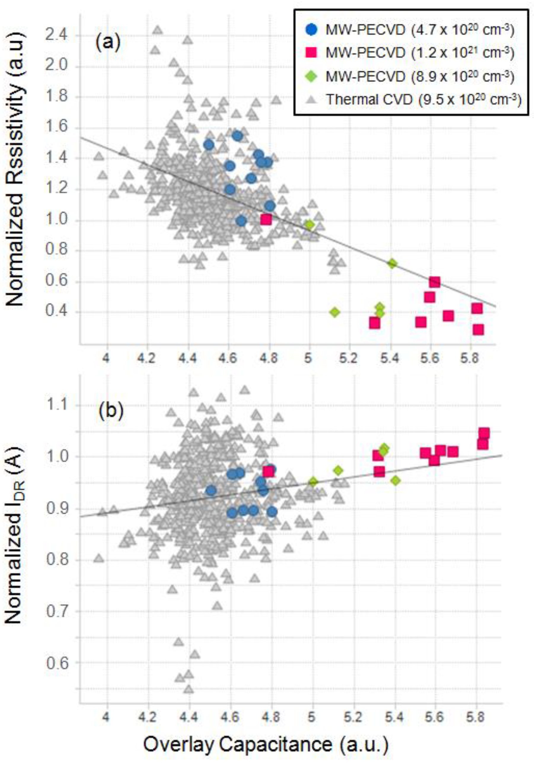
Figure 7 shows the electrical characteristics results of 20 nm pitch DRAM device with the Si gap-filled gBC grown by thermal CVD and MW-CVD. The gBC resistivity (
RC) was reduced, and the on-current (
IDR) thus increased with the doping concentration range of 4.7–8.9 × 10
20 cm
−3. Additionally, although the Si grown by MW-CVD had a low doping concentration of 4.7 × 10
20 cm
−3, similar
RC and
IDR values were obtained compared to the Si grown by the thermal CVD (9.5 × 10
20 cm
−3). The overlay capacitance determined by cell structure and the doping concentration also increased with the doping concentration of the Si grown by MW-CVD, and a similar value of 4.7 × 10
20 cm
−3 was reached with conventional device. The bottom-up Si gap-filling by MW-CVD suppressed seam and void formation and made a uniform P-concentration in the contact, resulting in an increase of electrical contact area, and, thus, a low
RC and the high
IDR.


