Article

Development of a Highly Specific Fluoroimmunoassay for the Detection of Doxycycline Residues in Water Environmental and Animal Tissue Samples

1
College of Life Sciences, Chongqing Normal University, Chongqing 401331, China
2
Fuxing Hospital, Capital Medical University, Chongqing 401331, China
3
Bioassay 3D Reconstruction Laboratory, Chongqing College of Electronic Engineering, Chongqing 401331, China
*
Author to whom correspondence should be addressed.
Micromachines 2022, 13(11), 1864; https://doi.org/10.3390/mi13111864
Submission received: 23 August 2022 / Revised: 25 October 2022 / Accepted: 28 October 2022 / Published: 30 October 2022
(This article belongs to the Special Issue Quantum Dot Frontiers)

Abstract

:
Doxycycline (DOX) and its metabolite residues in food and the environment pose a serious threat to human health and the ecological environment. In this work, a novel method, termed competitive fluoroimmunoassays (cFIA), based on monoclonal antibody (mAb) bio-conjugated CdSe/ZnS core–shell quantum dots (QDs), was developed for sensitive and rapid bioanalyses of DOX in natural water and commercial meats. After the optimization of the experimental conditions, 1 μg mL−1 of coating antigen and 0.5 μg mL−1 of QD-labeled mAb were used for the establishment of the cFIA. With this assay, the 50% inhibition concentration was found to be 0.35 ng mL−1 of DOX in phosphate-buffered saline samples, and the limit of detection was 0.039 ng mL−1 with minor cross-reactivity to other tetracycline members. The recoveries from natural water and commercial meats spiked with DOX concentrations of 10–600 ng mL−1 were 81.3–109.8%, and standard deviation were all below 12%. Levels measured with the QD-cFIA for thirty authentic samples were confirmed by high-performance liquid chromatography with good correlations. These results indicate that QD-cFIA is sultable for the rapid and quantitative detection of DOX residue in environmental and food samples.

1. Introduction

Doxycycline (DOX) is an essential member of the tetracycline family of antibiotics that is often used to treat infectious diseases in animals [1]. It is also used as a feed additive to prevent diseases and promote the growth of animals. Early research on DOX residues focused on food or biological samples [2]. Since animal manure and urine are usually used as fertilizers on farmland, more attention has been paid to the impact of residual DOXs on water and soil environments [3]. Depending on the animal species, researchers have found that up to 70% of DOX in urine or feces enters soil and surface water through the excretion of prototypes or metabolites.
According to different animal species, researchers have found that up to 70% of DOX in urine or feces enters the soil and surface water via excreta in prototypes or metabolites [4,5]. Notably, extensive use of animal fertilizers (not harmlessly treated) causes DOX to enter the soil and groundwater, thereby seriously threatening the ecological environment and the safety of agricultural products [6]. The presence of these antibiotic residues in the environment or food or biological samples can be directly toxic or can cause allergic reactions in some hypersensitive individuals, also promoting drug resistance to the microbial strain. Since these DOX residues are present in the environment or in food or biological samples, they may be directly toxic and cause allergic reactions in certain allergic individuals and can also promote resistance to microbial strains [7,8]. In addition, due to its broad-spectrum antimicrobial activity, residues in environmental media may affect the microbial abundance, community structure, and function, thus affecting the stability of the entire ecosystem [5,9].
Traditionally, the residues of DOX are measured by analytical methods. Electrochemistry [10], high-performance liquid chromatography tandem mass spectrometry [11], high-performance thin-layer chromatography [12]. and high-performance liquid chromatography (HPLC) [13,14] have been successfully used to detect DOX. However, these methods have some obvious defects, such as the need for expensive equipment and highly skilled personnel and may not be suitable for rapid screening of many samples. Additionally, immunoassays are widely used for the detection of various small molecule drugs because of their simplicity, rapidity, low cost, and high throughput. In our previous study, enzyme-linked immunosorbent assay [2,15], immunochromatographic test strip [16] and time-resolved fluroimmunoassay [17] have been developed to detect DOX in animal edible tissues (Table 1). However, these immunoassay methods based on horseradish peroxidase, colloidal gold, europium and samarium labelled antibodies to generate colored molecules as signal output have low sensitivity when detecting some trace antibiotics or biomarkers in complex biological matrices.
Recently, compared with traditional immunoassays, the use of fluoroimmunoassays (FIA) has been increasing due to their higher sensitivity, wider detection range, and lower matrix interference [18,19]. The fluorescent label may be an inorganic or organic material, such as CdSe/ZnS core–shell quantum dots (QDs), lanthanide chelates, fluorescent proteins, fluorescent microspheres, and so on [20,21]. As a class of fluorescent semiconductor nanocrystals with unique optical properties, such as a high quantum yield, high photostability, large molar extinction coefficient, wide absorption, and narrow fluorescence emission spectrum, QDs are considered to be promising fluorescent labeling materials for use in immunoassays [22]. At present, QDs have been widely used in the biomedical and food safety fields as labeling materials for immunofluorescence probes [22,23,24]. However, as yet, no FIA has been reported for DOX, and a simple and rapid QD-based FIA (QD-cFIA) is urgently needed.
In this work, the aim was to develop QD-cFIA for DOX in edible animal tissues and environmental samples using QD-labeled monoclonal antibodies (mAbs). The optimization of parameters, including the coating antigen concentration, QD-labeled mAb concentration, blocking buffer, incubation temperature, and competitive reaction time, were detailed. Finally, the proposed QD-cFIA was compared with conventional HPLC results in an analysis of environment and food samples containing DOX.

2. Materials and Methods

2.1. Materials and Instruments

Doxycycline (DOX), oxytetracycline, tetracycline, chlortetracycline, ovalbumin (OVA), and carboxylic acid-modified CdSe/ZnS core–shell QDs were purchased from Sigma–Aldrich (St. Louis, MO, USA). The coating antigens DOX-para-aminobenzoic acid–DOX (DOX-PABA-OVA) and anti-DOX mAb were obtained from our laboratory [2]. HPLC-grade acetonitrile and methanol were purchased from Fisher Chemical Company (Fair Lawn, NJ, USA). Ultraviolet absorbance was detected by using a NanoDrop-1000 spectrophotometer (Thermo Fisher Scientific Inc., Wilmington, DE, USA). The fluorescence was determined with a Varioskan LUX Multimode Microplate Reader (Thermo Fisher Scientific Inc., Wilmington, DE, USA). Deionized water was purified using the Millipore Milli-Q Ultrapure Water System (Bedford, MA, USA) and was used for all experiments. The QD-cFIA was validated with an Agilent 1260 HPLC equipped with an ultraviolet detector (Agilent, Wilmington, DE, USA).

2.2. Preparation of the Labeled Antibodies

The anti-DOX mAbs were covalently conjugated with the QDs by using carbodiimide chemistry. Briefly, 50 μL of QD was activated by adding 50 μL of 1-ethyl-3-(3-dimethylaminopropyl)carbodiimide hydrochloride (4 mg mL−1) and 50 μL of N-Hydroxysuccinimide (0.15 mg mL−1), and the mixture was incubated for 30 min at room temperature with gentle shaking. Then, the solution’s pH was adjusted to 7.4, and 300 μL of mAb (6 mg mL−1) was added to the ester-activated QD and then incubated for 2 h under dark conditions at room temperature. The reaction was separated by centrifugation at 18,000 rpm for 30 min at 4 °C, and the precipitates were dispersed in 0.05 M borate buffer (pH 8.0) until further use.

2.3. QD-cFIA Procedure Used for DOX Detection

As shown in Figure 1, the QD-cFIA procedure was carried out as follows: (1) The coating antigen (DOX-PABA-OVA) was diluted to a certain concentration with 0.05 M carbonate-buffered saline buffer (pH 9.6, 100 μL per well) and then added to a 96-well microplate. (2) After incubation overnight at 37 °C, the microplates were washed five times with 0.05 M sodium borate buffer (pH 9.0) containing 0.05% Tween-20 and then blocked with 1% OVA (200 μL per well) for 0.5 h at 37 °C. (3) After washing three times, 50 μL of the sample or standard solution and 50 μL of QD-labeled mAb were added to each well and incubated for 1 h at 37 °C. (4) The fluorescence intensity (F) of each well was measured with a Microplate Reader after washing and padding dry.

2.4. Optimization of Experimental Conditions

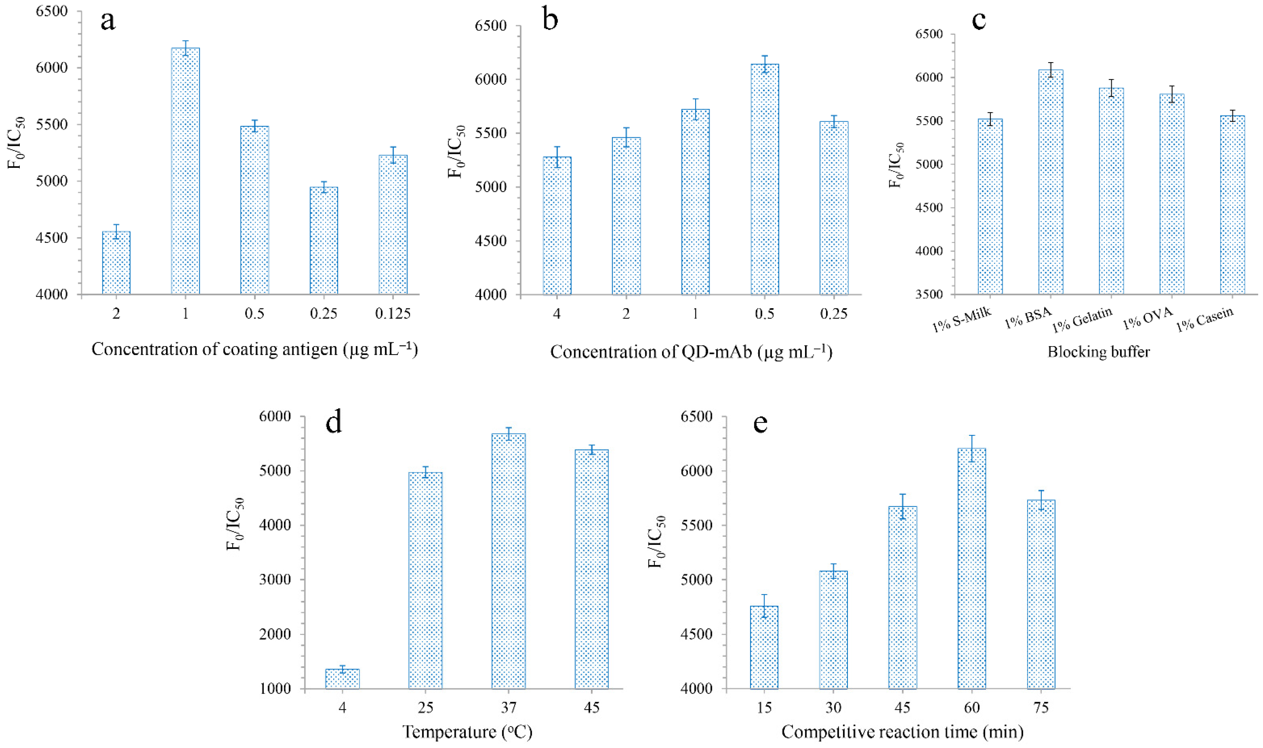
The experimental parameters were optimized in order to improve the sensitivity of the QD-cFIA. Coating antigens were diluted to concentrations ranging from 2 to 0.125 μg mL−1 by serial dilution with 0.05 M carbonate-buffered saline buffer (pH 9.6). The QD-labeled mAb (0.5 mg mL−1) was diluted with sodium borate buffer to concentrations of 4, 2, 1, 0.5, and 0.25 μg mL−1, respectively. The blocking buffers were selected, including 1% S-Milk, 1% BSA, 1% Gelatin, 1% OVA, and 1% Casein. The incubation temperatures used for the competitive binding assays were 4 °C, 25 °C, 37 °C, and 45 °C, respectively. The competitive reaction times were 15, 30, 45, 60, and 75 min, respectively. The concentration of DOX was diluted from 1000 to 0.01 ng mL−1 in sequence. F0/IC50 was used as the main index to evaluate the performance of QD-cFIA (F0: the fluorescence signal absence of analytes; IC50 represents the half-maximal inhibition concentration), where the highest F0/IC50 ratio was the most desirable [25].

2.5. Standard Curve and Specificity of QD-cFIA

To establish a standard curve, a series of DOX standard solutions were prepared by diluting DOX standards in PBS at a concentration of 0.01–100 ng mL−1. The standard curves for DOX were obtained by plotting the mean F/F0 against the logarithm of the analyte concentration under optimized parameters (F0 and F represent the fluorescence intensities without and with the analyte, respectively). Origin Pro 7.0 software was used to analyze the IC50 and limit of detection (LOD, IC10) using a four-parameter logistic equation of the Sigmoidal curve [26]. The specificity of QD-cFIA was evaluated by cross reactivity (CR). The CR values were calculated as follows: CR% = (IC50 of analyte/IC50 of analog) × 100 [2].

2.6. Accuracy and Precision Studies

The accuracy and precision of QD-cFIA were evaluated by the recovery rate and relative standard deviation (RSD). Animal tissue and water samples that had been shown to be DOX-free were used for matrix effect, accuracy, and precision studies. According to the Chinese MRLs standard, different concentrations of DOX were spiked to edible animal tissues, which were 0.5, 1 and 2 times the MRLs, respectively. Liver samples were spiked with DOX at final concentrations of 150, 300, and 600 μg kg−1 in sample buffer, and muscle and fish samples had final DOX concentrations of 50, 100, and 200 μg kg−1. All water samples were spiked with DOX at concentrations of 10, 50, and 100 μg L−1. DOX residues were extracted and purified from the samples using the modified QuEChERS (quick, easy, cheap, effective, rugged and safe) extraction method [27]. Each analysis was performed in quintuplicate.

2.7. The Correlation of QD-cFIA with HPLC

Thirty authentic samples of chicken liver, chicken muscle, fish muscle, tap water, river water, and contaminated water were collected from farms in Chongqing and Guizhou, China, where DOX had been used. All the samples were prepared using the procedure described above. Then, each sample was divided into two parts: one was analyzed using QD-cFIA and the other was analyzed using HPLC. For HPLC, the samples were analyzed in accordance with the method provided in the national standards of the Ministry of Agriculture of the People’s Republic of China (No. 958–2–2007). HPLC was performed on an Agilent ZORBAX SB–C18 (150 mm × 4.6 mm I.D., 5 μm, New Jersey, USA) using a mixture of methanol/acetonitrile/oxalic acid (7:8:85, v/v/v) as the mobile phase at a flow rate of 1.0 mL/min. The operating temperature of the column was set to 35 °C while the wavelength of the UV detector was set to 355 nm, and the injection volume was 50 μL.

3. Results and Discussion

3.1. Characterization of the QD-Labeled Antibodies

The fluorescence spectra of the QD-labeled mAbs had a maximum emission wavelength of 520 nm (λex = 400 nm), the same as that of the QDs, indicating that the excellent optical properties of the QDs conjugated with the mAbs were still preserved. The fluorescence intensity of QD-labeled mAbs was slightly weaker than that of the QDs due to the loss of QDs and fluorescence quenching during the reaction. In addition, the following QD-cFIA results indicated that the QDs were successfully linked to the mAbs against DOX.

3.2. Optimization of QD-cFIA

The F0/IC50 ratio was used to estimate the optimal parameter, with the highest ratio corresponding to the optimal parameter for QD-cFIA. QD-cFIA showed the highest F0/IC50 ratios when the coating antigen and QD-labeled mAbs were concentrated at 1 μg mL−1 (Figure 2a) and 0.5 μg mL−1 (Figure 2b), respectively. In this work, the effect of blocking buffers on the QD-cFIA performance was also studied. As shown in Figure 2c, 1% BSA was used as the blocking buffer to obtain the best sensitivity for the assay. An optimal incubation temperature of 37 °C was chosen (Figure 2d) and 60 min was selected as the optimal incubation time for the competitive binding assay (Figure 2e). Therefore, 1 μg mL−1 of coating antigen, 0.5 μg mL−1 of QD-labeled mAb, 1% BSA, 37 °C, and 60 min were selected as the optimal parameters for QD-cFIA in the experiment.

3.3. Standard Curve and Specificity of QD-cFIA

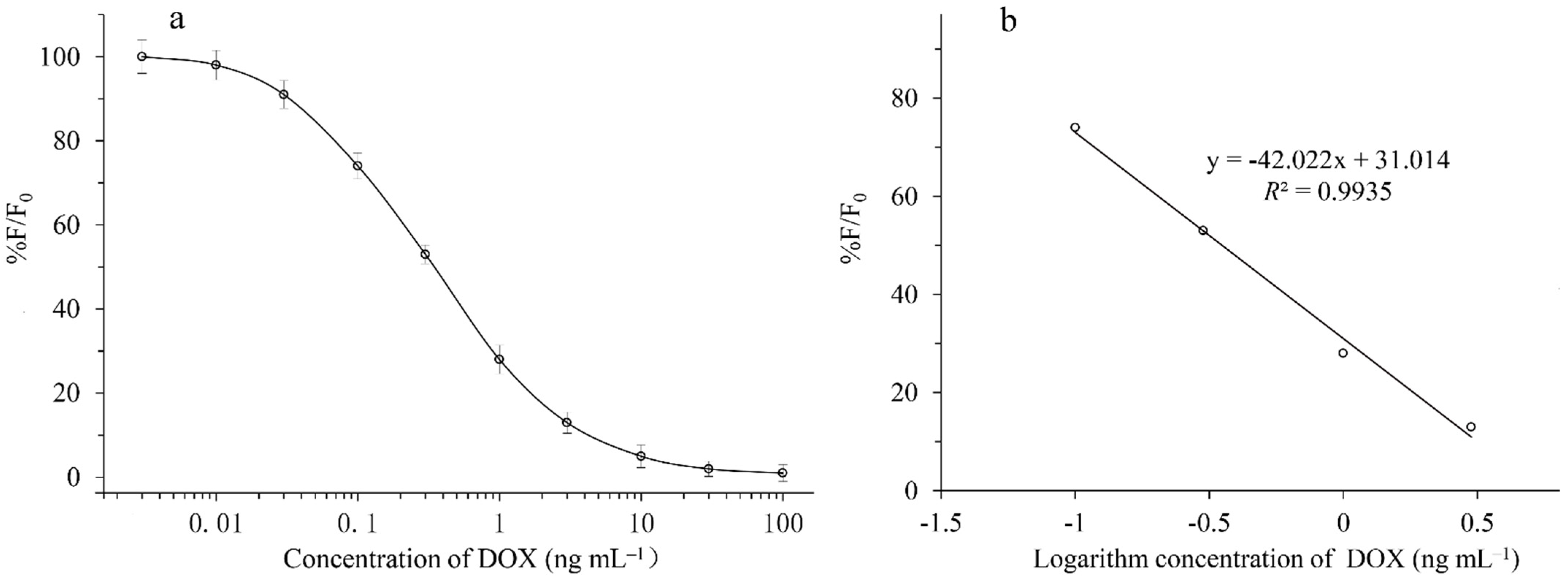
Competitive curves with final DOX concentrations of 0.003, 0.01, 0.03, 0.1, 0.3, 1, 3, 10, 30, and 100 ng mL−1 were run in PBS under the optimized parameters (Figure 3a). A standard curve with good linearity (Figure 3b) was prepared by using DOX concentrations of 0.03, 0.1, 1, and 3 ng mL−1 in PBS. The standard curves that were based on the DOX solvent calibration showed good linearity with R2 values equal to 0.9935. The IC50, LOD, and linear range of the method were 0.35, 0.039, and 0.03–3 ng mL−1, respectively. As shown in Table 2, the QD-cFIA showed negligible CR with the analogues (CR < 0.01%) except for 4-epi-doxycycline, which showed a CR of 51.5%. These results are similar to those reported in previous studies using ic-ELISA [2].

3.4. Accuracy and Precision

The accuracy and precision values of QD-cFIA were determined by analyzing the recovery and RSD. As illustrated in Table 3, the recovery rate of spiked samples was in the range of 81.3–109.8%, and the RSD was 4.2–11.9%. The results indicate an excellent performance by the QD-cFIA during the detection of DOX residues in edible animal tissue and environmental samples.

3.5. Correction of Immunoassays and HPLC

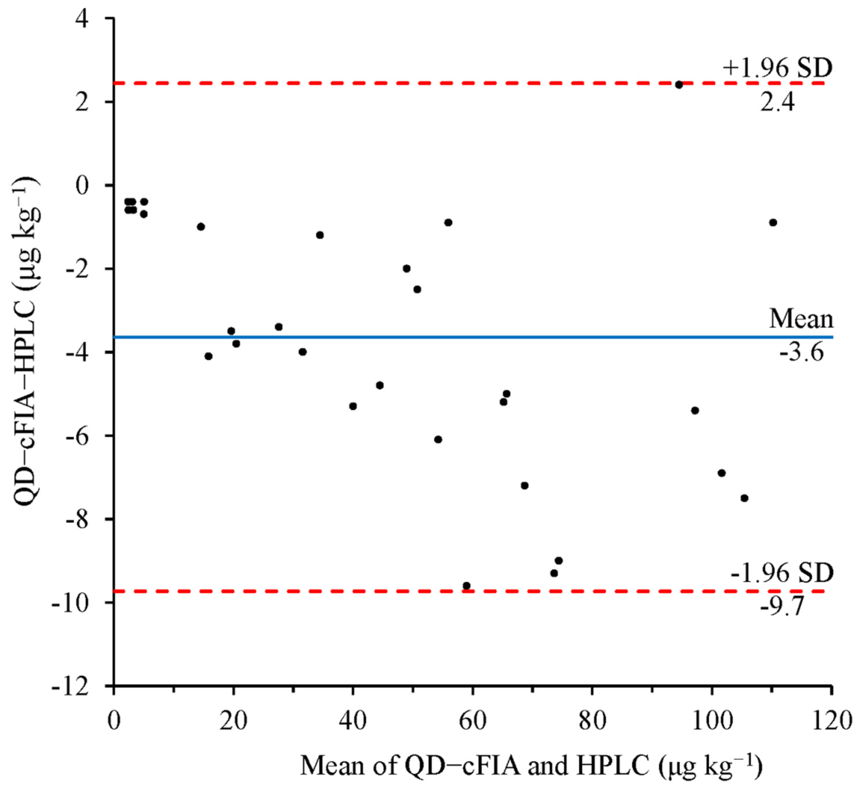
The developed QD-CFIA method and the HPLC reference method were used to compare and analyze the natural DOX-contaminated food and environmental samples to evaluate the reliability of the QD-cFIA method. Using QD-cFIA, we found that the samples were contaminated with various levels of DOX, ranging from 2.2 to 109.8 μg kg−1. The HPLC results were basically consistent with those of QD-cFIA, and the positive results ranged from 2.6 to 110.7 μg kg−1. The Bland–Altman plot for DOX measured by QD-cFIA and HPLC is shown in Figure 4. The results show that the mean bias was −3.6 μg kg−1, and the 95% limits of the agreement defined as the mean ± 1.96 SD were between –9.7 and 2.4 μg kg−1. It is evident that the results from the QD-cFIA and HPLC were not significantly different (significant level α = 0.05).

4. Conclusions

In this study, the proposed method was the first application of the QD-cFIA for the determination of DOX in edible animal tissue and environmental samples. The QD-cFIA showed high specificity for DOX and a qualitative LOD of 0.03 ng mL−1. Six samples were spiked with DOX and detected using the proposed QD-cFIA method, and an excellent recovery and satisfactory coefficient of variation were obtained. Additionally, the reliability of the developed QD-cFIA was confirmed by HPLC for parallel analysis of the natural DOX-contaminated food and environmental samples, further demonstrating that the established assay method can act as a sensitive and reliable tool for the detection of DOX residue in environmental and food samples.

Author Contributions

Y.X. and L.X.; Formal analysis, Y.X. and L.X.; Funding acquisition, Y.X. and T.L.; Investigation, L.Y.; Methodology, Y.X., L.X. and L.Y.; Software, L.X.; Validation, Y.X. and T.L.; Writing—original draft, T.L.; Writing—review and editing, Y.X. and T.L. All authors have read and agreed to the published version of the manuscript.

Funding

The authors would like to thank the Chongqing of University Innovation Research Group Project (No.CXQT20031) and the Chongqing Natural Science Foundation project (cstc2021jcyj-msmX0314).

Institutional Review Board Statement

This article does not contain any studies with human subjects. All animal experiments that were described in the present study were performed in adherence to Chongqing Normal University Animal Experiment Center Guidelines and approved by the Animal Ethics Committee.

Informed Consent Statement

Informed consent was obtained from all subjects involved in the study.

Data Availability Statement

The data presented in this study are available on request from the corresponding author.

Conflicts of Interest

The authors declare no conflict of interest.

References

  1. Jiang, C.Q.; Zhang, N. Enzyme-amplified lanthanide luminescence based on complexation reaction--a new technique for the determination of doxycycline. J. Pharmaceut. Biomed. 2004, 35, 1301–1306. [Google Scholar] [CrossRef]
  2. Le, T.; Zhao, Z.; Wei, W.; Bi, D. Development of a highly sensitive and specific monoclonal antibody-based enzyme-linked immunosorbent assay for determination of doxycycline in chicken muscle, liver and egg. Food Chem. 2012, 134, 2442–2446. [Google Scholar] [CrossRef] [PubMed]
  3. Zhang, W.L.; Zhang, L.F.; Zhang, K.Y.; Wang, X.Y.; Xue, F.Q. Determination of tetracyclines and their epimers in agricultural soil fertilized with swine manure by ultra-high-performance liquid chromatography tandem mass spectrometry. J. Integr. Agric. 2012, 11, 1189–1198. [Google Scholar] [CrossRef]
  4. Lyu, N.; Yin, F.H.; Chen, Y.; Gao, Z.J.; Liu, Y.; Shi, L. Effects of elevated atmospheric CO2 and nitrogen application on cotton biomass, nitrogen utilization and soil urease activity. J. Appl. Ecol. 2015, 26, 3337–3344. [Google Scholar]
  5. Yan, Q.; Li, X.; Ma, B.; Zou, Y.; Wang, Y.; Liao, X.; Liang, J.; Mi, J.; Wu, Y. Different Concentrations of Doxycycline in Swine Manure Affect the Microbiome and Degradation of Doxycycline Residue in Soil. Front. Microbiol. 2018, 9, 3129. [Google Scholar] [CrossRef]
  6. Aga, D.S.; O’Connor, S.; Ensley, S.; Payero, J.O.; Snow, D.; Tarkalson, D. Determination of the persistence of tetracycline antibiotics and their degradates in manure-amended soil using enzyme-linked immunosorbent assay and liquid chromatography-mass spectrometry. J. Agric. Food Chem. 2005, 53, 7165–7171. [Google Scholar] [CrossRef]
  7. Girma, K.; Tilahun, Z.; Haimanot, D.; Tadele, T. Review on detection of antimicrobial residues in raw bulk milk in dairy farms. J. Basic. Appl. Sci. 2014, 6, 87–97. [Google Scholar]
  8. Cinquina, A.L.; Longo, F.; Anastasi, G.; Giannetti, L.; Cozzani, R. Validation of a high-performance liquid chromatography method for the determination of oxytetracycline, tetracycline, chlortetracycline and doxycycline in bovine milk and muscle. J. Chromatogr. A 2003, 987, 227–233. [Google Scholar] [CrossRef]
  9. Xu, N.; Dong, J.; Zhou, W.; Liu, Y.T.; Ai, X.H. Determination of Doxycycline, 4-epidoxycycline, and 6-epidoxycycline in Aquatic Animal Muscle Tissue by an Optimized Extraction Protocol and Ultra-performance Performance Liquid Chromatography with Ultraviolet Detection. Anal. Lett. 2019, 52, 452–464. [Google Scholar] [CrossRef]
  10. Gajda, A.; Posyniak, A.; Zmudzki, J.; Tomczyk, G. Determination of doxycycline in chicken fat by liquid chromatography with UV detection and liquid chromatography-tandem mass spectrometry. J. Chromatogr. B 2013, 928, 113–120. [Google Scholar] [CrossRef]
  11. Spielmeyer, A.; Ahlborn, J.; Hamscher, G. Simultaneous determination of 14 sulfonamides and tetracyclines in biogas plants by liquid-liquid-extraction and liquid chromatography tandem mass spectrometry. Anal. Bioanal. Chem. 2014, 406, 2513–2524. [Google Scholar] [CrossRef] [PubMed]
  12. da Silveira, V.G.; Oliveira, M.; de Almeida, C.A.A.; Hoff, R.; Mallmann, C.A. Liquid Chromatography-Tandem Mass Spectrometry Determination and Depletion Profile of Chlortetracycline, Doxycycline, and Oxytetracycline in Broiler Chicken Muscle After Oral Administration. Food Anal. Methods 2018, 11, 2181–2194. [Google Scholar] [CrossRef]
  13. Liu, Y.; Yang, H.L.; Yang, S.; Hu, Q.W.; Cheng, H.B.; Liu, H.Y.; Qiu, Y.S. High-performance liquid chromatography using pressurized liquid extraction for the determination of seven tetracyclines in egg, fish and shrimp. J. Chromatogr. B 2013, 15, 11–17. [Google Scholar] [CrossRef] [PubMed]
  14. Yu, H.; Tao, Y.; Chen, D.; Wang, Y.; Yuan, Z. Development of an HPLC–UV method for the simultaneous determination of tetracyclines in muscle and liver of porcine, chicken and bovine with accelerated solvent extraction. Food Chem. 2011, 124, 1131–1138. [Google Scholar] [CrossRef]
  15. Le, T.; Yu, H.; Guo, Y.; Ngom, B.; Shen, Y.; Bi, D. Development of an indirect competitive ELISA for the detection of doxycycline residue in animal edible tissues. Food Agric. Immunol. 2009, 20, 111–124. [Google Scholar] [CrossRef]
  16. Le, T.; Yu, H.; Wang, X.; Ngom, B.; Guo, Y.; Bi, D. Development and validation of an immunochromatographic test strip for rapid detection of doxycycline residues in swine muscle and liver. Food Agric. Immunol. 2011, 22, 235–246. [Google Scholar] [CrossRef] [Green Version]
  17. Le, T.; Yi, S.; Wei, S.; Liu, J. A competitive dual-label time-resolved fluoroimmunoassay for the simultaneous detection of chlortetracycline and doxycycline in animal edible tissues. Food Agric. Immunol. 2015, 26, 804–812. [Google Scholar] [CrossRef]
  18. Zhu, Y.; Chao, J.; Zhu, F.; Zhu, N.; Zhang, Q.; Gyimah, E.; Yakubu, S.; Zou, Y.; Zhang, Z. Ratiometric fluorescence immunoassay based on FAM-DNA-functionalized CdSe/ZnS QDs for the sensitive detection of tetrabromobisphenol A in foodstuff and the environment. Anal. Bioanal. Chem. 2020, 412, 3605–3613. [Google Scholar] [CrossRef]
  19. Zhang, F.; Liu, B.; Zhang, Y.; Wang, J.; Lu, Y.; Deng, J.; Wang, S. Application of CdTe/CdS/ZnS quantum dot in immunoassay for aflatoxin B1 and molecular modeling of antibody recognition. Anal. Chim. Acta 2019, 1047, 139–149. [Google Scholar] [CrossRef]
  20. Esteve-Turrillas, F.A.; Abad-Somovilla, A.; Quinones-Reyes, G.; Agullo, C.; Mercader, J.V.; Abad-Fuentes, A. Monoclonal antibody-based immunoassays for cyprodinil residue analysis in QuEChERS-based fruit extracts. Food Chem. 2015, 187, 530–536. [Google Scholar] [CrossRef] [Green Version]
  21. Le, T.; Zhu, L.; Yu, H. Dual-label quantum dot-based immunoassay for simultaneous determination of Carbadox and Olaquindox metabolites in animal tissues. Food Chem. 2016, 199, 70–74. [Google Scholar] [CrossRef] [PubMed]
  22. Xie, Y.; Zhang, L.; Sun, Q.; Zhang, Z.; Le, T. Immunochromatographic Assay for Simultaneous and Quantitative Detection of 3-Methyl-Quinoxaline-2-Carboxylic Acid and Quinoxaline-2-Carboxylic Acid Residues in Animal Tissues Based on Highly Luminescent Quantum Dot Beads. Food Anal. Methods 2018, 11, 334–342. [Google Scholar] [CrossRef]
  23. Le, T.; Xie, Y.; Zhu, L.Q.; Zhang, L. Rapid and sensitive detection of 3-amino-2-oxazolidinone using a quantum dot-based immunochromatographic fluorescent biosensor. J. Agric. Food Chem. 2016, 64, 8678–8683. [Google Scholar] [CrossRef] [PubMed]
  24. Xie, Y.; Zhang, L.; Yang, X.; Le, T. Development of a quantum dot-based immunochromatography test strip for rapid screening of oxytetracycline and 4-epioxytetracycline in edible animal tissues. Food Addit. Contam. A 2017, 34, 371–378. [Google Scholar] [CrossRef] [PubMed]
  25. Sun, Y.; Li, Y.; Meng, X.; Qiao, B.; Hu, P.; Meng, X.; Lu, S.; Ren, H.; Liu, Z.; Zhou, Y. Fluorescence-linked immunosorbent assay for detection of phenanthrene and its homolog. Anal. Biochem. 2018, 547, 45–51. [Google Scholar] [CrossRef] [PubMed]
  26. Hua, X.; Ding, Y.; Yang, J.; Ma, M.; Shi, H.; Wang, M. Direct competitive fluoroimmunoassays for detection of imidaclothiz in environmental and agricultural samples using quantum dots and europium as labels. Sci. Total Environ. 2017, 583, 222–227. [Google Scholar] [CrossRef] [PubMed]
  27. Grande-Martínez, Á.; Moreno-González, D.; Arrebola-Liébanas, F.J.; Garrido-Frenich, A.; García-Campaña, A.M. Optimization of a modified QuEChERS method for the determination of tetracyclines in fish muscle by UHPLC-MS/MS. J. Pharmaceut. Biomed. 2018, 155, 27–32. [Google Scholar] [CrossRef]
Figure 1. Schematic diagram of QD-cFIA.
Micromachines 13 01864 g001
Figure 2. (a) Effect of DOX-PABA-OVA concentrations (2, 1, 0.5, 0.25 and 0.125 μg mL−1) on F0/IC50. (b) The ratio of F0/IC50 with different blocking buffer (1% S-Milk, 1% BSA, 1% Gelatin, 1% OVA and 1% Casein). (c) The ratio of F0/IC50 under different concentrations of QDs-labeled mAb (4, 2, 1, 0.5 and 0.25 μg mL−1). (d) The ratio of F0/IC50 under different incubation temperatures: 4 °C, 25 °C, 37 °C and 45 °C, respectively. (e) The ratio of F0/IC50 under different competitive reaction time: 15, 30, 45, 60 and 75 min, respectively. The error bars represent the standard deviation of three measurments.
Micromachines 13 01864 g002
Figure 3. (a) Standard curves of our developed QD-cFIA with increasing DOX concentrations, from top to bottom: 0.003, 0.01, 0.03, 0.1, 0.3, 1, 3, 10, 30, and 100 ng mL−1, respectively. (b) Good linearity of the calibration curves was achieved for DOX in the range of 0.1–3 ng mL−1. Error bars represented the standard deviation.
Micromachines 13 01864 g003
Figure 4. Bland–Altman plots to show comparisons between detection results obtained by the QD-cFIA and HPLC in fortified real samples.
Micromachines 13 01864 g004
Table 1. A review of immunoassay methods for the determination of DOX in recent years.
MethodAntibody TypeIC50 (ng mL−1)LOD (ng mL−1)SampleReferences
Enzyme-linked immunosorbent assayMonoclonal antibody1.32 0.14liver, muscle and egg[2]
Enzyme-linked immunosorbent assayPolyclonal antibody8.741.96Liver and muscle[15]
Immunochromatographic test stripPolyclonal antibody22.07.0Liver and muscle[16]
Time-resolved fluroimmunoassayPolyclonal antibody1.060.04liver, muscle and egg[17]
QD-cFIAMonoclonal antibody0.350.039Chicken (liver, muscle), fish muscle, tap water, river, water and contaminated waterThis work
Table 2. Cross-reactivity of analogues related to DOX by QD-cFIA.
CompoundIC50 (ng m L−1)CR (%)
DOX0.35100
4-epi-doxycycline0.6851.5
Oxytetracycline>10,000<0.01
4-epi- oxytetracycline>10,000<0.01
Tetracycline>10,000<0.01
4-epi-tetracycline>10,000<0.01
Chlortetracycline>10,000<0.01
4-epi-chlortetracycline chlortetracycline>10,000<0.01
Demeclocycline>10,000<0.01
Table 3. Accuracy and precision of DOX in spiked samples by QD-cFIA (n = 5).
SampleSpiked (ng mL−1, ng g−1)Mean Recovery ±SD (%)RSD (%)
Chicken liver150105.4 ± 10.49.9
30098.7 ± 10.810.9
60088.5 ± 9.711.0
Chicken muscle5098.8 ± 6.66.7
100109.8 ± 9.48.6
200108.4 ± 7.56.9
Fish muscle10081.3 ± 9.711.9
20091.8 ± 7.27.8
400109.3 ± 11.210.3
Tap water10102.3 ± 6.46.3
50103.1 ± 7.67.4
10098.8 ± 4.64.7
River water10103.3 ± 4.34.2
50101.5 ± 9.59.4
10099.6 ± 11.211.2
Contaminated water10106.3 ± 9.69.0
50100.8 ± 7.97.8
10097.3 ± 9.59.8
Publisher’s Note: MDPI stays neutral with regard to jurisdictional claims in published maps and institutional affiliations.