Article

Simple and Label-Free Detection of Carboxylesterase and Its Inhibitors Using a Liquid Crystal Droplet Sensing Platform

Department of Chemistry, Gachon University, Seongnam-daero 1342, Sujeong-gu, Seongnam-si 13120, Gyeonggi-do, Korea
*
Author to whom correspondence should be addressed.
Micromachines 2022, 13(3), 490; https://doi.org/10.3390/mi13030490
Submission received: 4 March 2022 / Revised: 18 March 2022 / Accepted: 20 March 2022 / Published: 21 March 2022
(This article belongs to the Special Issue Microfluidics in Analytical Chemistry)

Abstract

:
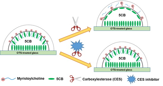
In this study, we developed a liquid crystal (LC) droplet-based sensing platform for the detection of carboxylesterase (CES) and its inhibitors. The LC droplet patterns in contact with myristoylcholine chloride (Myr) exhibited dark cross appearances, corresponding to homeotropic anchoring of the LCs at the aqueous/LC interface. However, in the presence of CES, Myr was hydrolyzed; therefore, the optical images of the LC patterns changed to bright fan-shaped textures, corresponding to a planar orientation of LCs at the interface. In contrast, the presence of CES inhibitors, such as benzil, inhibits the hydrolysis of Myr; as a result, the LC patterns exhibit dark cross textures. This principle led to the development of an LC droplet-based sensing method with a detection limit of 2.8 U/L and 10 μM, for CES detection and its inhibitor, respectively. The developed biosensor not only enables simple and label-free detection of CES but also shows high promise for the detection of CES inhibitors.

Graphical Abstract

1. Introduction

Liquid crystals (LCs) are promising materials for biosensing owing to their unique physical properties such as long-range orientational order, optical anisotropy, and molecular sensitivity [1,2]. Thus, LCs have been widely used to develop novel sensing systems for monitoring biochemical events [3,4]. LC-based sensing systems can transform chemical/biological events into optical responses that can be observed under a polarized light microscope (POM) [4,5]. Moreover, these sensing systems can operate in ambient environments without the need for molecular labels or electrical power [5,6,7]. Recently, LC-based sensors for monitoring various biochemical events have been developed and reported [5,7,8,9,10]. Among these, LC droplet-based sensors have been widely used to investigate enzymatic activity because the large surface-area-to-volume ratio of the LC droplets can increase the sensitivity of the detection [3,11]. The LC droplets were formed by simply dropping the solution of LC dissolved in organic solvents onto the surfaces of glass slides. The LC patterns exhibited bright fan-shaped appearances when in contact with non-surfactant solutions (e.g., deionized water), whereas dark cross textures were observed after introducing surfactant solutions [11,12].
Carboxylesterases (CESs) are an important class of enzymes that catalyze the hydrolysis of carboxylic esters and play a crucial role in the detoxification and metabolism of many drugs [13,14,15]. Moreover, the level of CES can serve as an indicator of hepatocellular carcinoma [16]. Thus, the accurate measurement of CESs is very helpful for better understanding their biological functions as well as for the early diagnosis of hepatocellular carcinoma.
To date, several detection methods, including fluorescence [14,15,17], chromatography [18], and luminescence [19,20], have been developed for CES detection. However, these methods have limitations including the need for complex instrumentation, labeling of samples, and long operating times. Recently, a simple and sensitive sensing system for CES detection based on a surfactant-decorated LC/aqueous interface was reported [21]. However, some drawbacks need to be addressed. The copper grids used to prepare LC/aqueous interfaces are easily oxidized and deformed, which limits their reusability [11]. Moreover, when preparing LC thin films, excess LCs are removed from copper grid waste materials.
In this study, we developed a simple and sensitive LC droplet-based biosensor for CES detection. The structure diagram and principle of the developed sensor are illustrated in Figure 1. The LC droplet patterns were formed by spreading 1.5 µL of a heptane solution containing 2% (v/v) 5CB onto the OTS-modified glass slides after the evaporation of heptane. OTS with a hydrophobic tail was used to align the 5CB molecules at the glass/LC interface in a homeotropic orientation. Myristoylcholine (Myr, the substrate of the CES enzyme) was used as a cationic surfactant to induce homeotropic anchoring of LCs at the LC/aqueous interfaces, corresponding to the dark cross appearances of the LC droplets observed under POM (Figure 1B(a)). However, in the presence of CES, the optical images of the LC droplet patterns changed to bright fan-shaped textures owing to the enzymatic hydrolysis of Myr by CES (Figure 1B(b)). Moreover, the effect of the inhibitor on CES activity was investigated. This biosensor enables simple and fast detection of CES and its inhibitors with high selectivity.

2. Materials and Methods

2.1. Materials and Apparatus

The materials, reagents, and apparatus used in this study are listed in the Supplementary Materials section.

2.2. Preparation of Octyltrichlorosilane (OTS)-Modified Glass Slides

OTS-modified glass slides were prepared as previously described [22,23]. The details are provided in the Supplementary Materials section.

2.3. Preparation of LC Droplet Patterns and Fabrication of LC Cells

LC droplet patterns were prepared by dropping 1.5 µL of 5CB (2% (v/v) in heptane) onto the OTS-modified substrate. The solvent was evaporated at room temperature (~25 °C) and LC droplet patterns were formed. Subsequently, approximately 100 µL of aqueous solution was transferred onto the LC droplets at 25 °C. The LC cells were fabricated by pairing the covering glass slides and OTS-modified glass slides face to face (Figure 1A). Two small pieces of glass (26 × 0.5 mm, thickness = 1 mm) were used to produce a gap between two glass slides.

2.4. Preparation of the Aqueous Solution

All aqueous solutions were prepared in phosphate-buffered saline (PBS, 10 mM, pH = 7.4). During the CES detection assay, varying concentrations of CES were mixed with 10 µM Myr and incubated at 37 °C for 30 min. For the enzyme inhibition assay, a mixture of CES (0.1 µg/mL) and benzil at various concentrations was pre-incubated at 37 °C for 2 h. After the addition of 10 µM Myr, the mixtures were incubated at 37 °C for 30 min. Subsequently, 100 µL of the incubated solution was added to the LC droplet at 25 °C.

3. Results and Discussion

3.1. Optimization of the Myr Concentration

In our system, the LC droplet patterns were prepared by spreading 1.5 µL of an anhydrous heptane solution containing 2% (v/v) 5CB onto the OTS-modified glass slides. Here, Myr was used as a cationic surfactant to induce homeotropic anchoring of LCs at the LC/aqueous interfaces, corresponding to the dark cross appearances of the LC droplets observed under POM. Previous studies have demonstrated that the LC droplet patterns exhibited bright fan-shaped appearances when in contact with non-surfactant solutions (e.g., PBS solution), whereas dark cross textures were observed after introducing surfactant solutions [11,12]. Therefore, we first investigated the orientational behavior of 5CB at the aqueous/LC interface by comparing the optical responses of the LC droplets in contact with PBS and Myr solution. When the LC droplet pattern was immersed in PBS solution (pH = 7.4), we observed bright fan-shaped images coupled to the planar orientation of LCs at the interface (Figure 2a). However, the LCs adopted dark cross textures after the addition of 50 µM Myr solution (Figure 2b), corresponding to the homeotropic orientation of LCs at the aqueous/LC interface. This indicated that the addition of Myr altered the ordering of LCs at the interface from a planar to homeotropic orientation.
Next, we determined the minimum Myr concentration (CMyr) to induce homeotropic orientation of the LCs by examining the optical response of LCs after reducing CMyr. As shown in Figure 2, dark cross appearances were observed in LCs when CMyr ≥ 10 µM (Figure 2b–d), and bright fan-shaped images were observed when CMyr < 10 µM (Figure 2e,f). Therefore, we determined that the critical CMyr for inducing homeotropic orientation of LCs at the interface was approximately 10 µM.

3.2. Feasibility and Detection Limit of the LC Droplet-Based Biosensor for CES Detection

According to previous reports, CES can catalyze the hydrolysis of carboxylic acid esters into their corresponding acids and alcohols [21,24]. Thus, to verify the feasibility of the developed sensor, we studied the orientational transition of LCs at the interface after introducing a mixture of Myr and CES. As shown in Figure 3a, bright fan-shaped images were observed after adding the mixture of Myr (10 µM) and 100 mg/L CES to the LC droplet patterns, suggesting a planar orientation of LCs at the interface. This indicated that Myr was completely hydrolyzed by CES and demonstrated that CES activity could be monitored using the developed LC droplet biosensor.
Next, the limit of detection (LOD) for CES was determined by lowering the concentration of CES. The LC patterns showed bright fan-shaped images after incubating with pre-incubated mixtures containing 10 µM Myr and 10 mg/L, 1 mg/L, or 0.1 mg/L CES (Figure 3b–d). However, dark cross appearances were observed when the CES concentration was reduced to 0.01 mg/L or lower (Figure 3e,f), which was attributed to the incomplete hydrolysis of Myr by AChE. Therefore, we concluded that the LOD for CES using this LC droplet sensing system was 0.1 mg/L (2.8 U/L).

3.3. Specificity of the LC Droplet-Based Biosensor in CES Detection

The specificity of the developed sensor was also investigated. Various interfering substances, including urease, lipase, α-chymotrypsin, lysozyme, urea, glucose, and CaCl2, were used to test the specificity of the sensor. As shown in Figure 4, a bright fan-shaped texture was observed in the case of CES (Figure 4a), whereas dark cross appearances were obtained for other interfering substances (Figure 4b–h). These results demonstrate the high specificity of the proposed biosensor for CES detection.
It has been reported that benzil is a very potent CES inhibitor and exhibits selective inhibitory effects toward CES [25,26,27]. Therefore, to further confirm the specificity of the CES sensor, we investigated the optical images of LCs coupled with enzymatic hydrolysis of Myr with CES, which was pre-incubated with various benzil concentrations (Cbenzil). Optical images of the LC droplets are presented in Figure 5. Dark cross appearances were obtained when Cbenzil ≥ 10 µM (Figure 5a–c), indicating that CES activity was prevented by benzil. However, a bright fan-shaped texture was observed when the concentration of benzil was reduced to 1 µM (Figure 5d), suggesting that the concentration of benzil was too low to completely inhibit the activity of CES. These results further confirmed the high specificity of the LC droplet-based biosensor for CES detection. In addition, they also demonstrate the potential application of the LC droplet-based sensing system for the detection or screening of CES inhibitors.

3.4. Detection of CES in Human Urine Samples

To evaluate the practicability of the developed biosensor, its ability to detect CES in human urine was examined. The human urine was collected from a healthy volunteer. The filtered human urine sample was diluted 10 times with PBS and then spiked with standard CES solution at a concentration of 5 mg/L. The spiked urine samples were analyzed using the developed biosensor. Human urine without the addition of CES was used as a control. The LC pattern displayed a dark cross in the control sample (Figure 6a), confirming there was no CES in the human urine. However, in the urine sample spiked with 5 mg/L CES, bright fan-shaped images were observed (Figure 6b), suggesting that the CES changed the orientation of LCs at the interface. These results prove the potential application of the developed sensing system for CES monitoring in real samples.
Before concluding, we compared the analytical characteristics of the developed LC droplet-based biosensor with other reported methods for CES detection such as spectroscopic, fluorescence, and bioluminescence. These detection methods are limited by several drawbacks such as the need for complex instrumentation, labeling of samples, and long operating times. In contrast, the proposed strategy offers a simple, rapid, label-free, and cost-effective method for the detection of CES with high selectivity. Using this method, a low detection limit of 0.1 mg/L (2.8 U/L) for CES was obtained, which is comparable to or lower than the LODs of other reported methods (Table 1).

4. Conclusions

In summary, an LC droplet-based sensing platform was developed to monitor the enzymatic activity of CES, based on the optical responses of LC droplet patterns when in contact with surfactant or non-surfactant solutions. A dark cross appearance was obtained after the introduction of a cationic surfactant solution, Myr, onto the LC droplet patterns, whereas a bright fan-shaped optical image was observed after the addition of a pre-incubated mixture of Myr and CES, owing to the enzymatic hydrolysis of Myr by CES. However, the presence of CES inhibitors, such as benzil, inhibited CES activity resulting in a dark cross appearance. The proposed LC droplet-based biosensor exhibited high specificity toward CES with a low detection limit of 0.1 mg/L (2.8 U/L). In addition, the detection limit of the CES inhibitor was as low as 10 μM. Moreover, the developed biosensor was successfully applied to detect CES in a real human urine sample. This LC droplet-based biosensor not only enables simple, rapid, and label-free detection of CES but also shows high potential for application in the detection or screening of CES inhibitors.

Supplementary Materials

Supporting information can be downloaded at: https://www.mdpi.com/article/10.3390/mi13030490/s1.

Author Contributions

Conceptualization, D.-K.N. and C.-H.J.; methodology, D.-K.N. and C.-H.J.; investigation, D.-K.N.; data curation, D.-K.N.; writing—original draft preparation, D.-K.N.; writing—review and editing, D.-K.N.; visualization, D.-K.N.; supervision, C.-H.J.; funding acquisition, C.-H.J. All authors have read and agreed to the published version of the manuscript.

Funding

This research was funded by Ministry of Education, grant number NRF-2019R1A2C1003862. The APC was funded by Duy-Khiem Nguyen.

Acknowledgments

This work was supported by the Basic Science Research Program of the National Research Foundation of Korea (NRF) funded by the Ministry of Education (NRF-2019R1A2C1003862).

Conflicts of Interest

The authors declare no conflict of interest.

References

  1. Hussain, A.; Pina, A.S.; Roque, A.C.A. Bio-recognition and detection using liquid crystals. Biosens. Bioelectron. 2009, 25, 1–8. [Google Scholar] [CrossRef] [PubMed]
  2. Hartono, D.; Xue, C.Y.; Yang, K.L.; Yung, L.Y.L. Decorating liquid crystal surfaces with proteins for real-time detection of specific protein–protein binding. Adv. Funct. Mater. 2009, 19, 3574–3579. [Google Scholar] [CrossRef]
  3. Wang, Y.; Hu, Q.; Tian, T.; Yu, L. Simple and sensitive detection of pesticides using the liquid crystal droplet patterns platform. Sens. Actuators B Chem. 2017, 238, 676–682. [Google Scholar] [CrossRef]
  4. Liao, S.; Qiao, Y.; Han, W.; Xie, Z.; Wu, Z.; Shen, G.; Yu, R. Acetylcholinesterase liquid crystal biosensor based on modulated growth of gold nanoparticles for amplified detection of acetylcholine and inhibitor. Anal. Chem. 2012, 84, 45–49. [Google Scholar] [CrossRef] [PubMed]
  5. Yang, X.; Li, H.; Zhao, X.; Liao, W.; Zhang, C.X.; Yang, Z. A novel, label-free liquid crystal biosensor for Parkinson’s disease related alpha-synuclein. Chem. Commun. 2020, 56, 5441. [Google Scholar] [CrossRef]
  6. Rouhbakhsh, Z.; Verdian, A.; Rajabzadeh, G. Design of a liquid crystal-based aptasensing platform for ultrasensitive detection of tetracycline. Talanta 2020, 206, 120246. [Google Scholar] [CrossRef] [PubMed]
  7. Wang, Y.; Hu, Q.; Guo, Y.; Yu, L. A cationic surfactant-decorated liquid crystal sensing platform for simple and sensitive detection of acetylcholinesterase and its inhibitor. Biosens. Bioelectron. 2015, 72, 25–30. [Google Scholar] [CrossRef]
  8. Wu, W.; Wang, W.; Qi, L.; Wang, Q.; Yu, L.; Lin, J.M.; Hu, Q. Screening of Xanthine Oxidase Inhibitors by Liquid Crystal-Based Assay Assisted with Enzyme Catalysis-Induced Aptamer Release. Anal. Chem. 2021, 93, 6151–6157. [Google Scholar] [CrossRef] [PubMed]
  9. McUmber, A.C.; Noonan, P.S.; Schwartz, D.K. Surfactant–DNA interactions at the liquid crystal–aqueous interface. Soft Matter 2012, 8, 4335–4342. [Google Scholar] [CrossRef]
  10. Munir, S.; Park, S.Y. Liquid crystal-based DNA biosensor for myricetin detection. Sens. Actuators B Chem. 2016, 233, 559–565. [Google Scholar] [CrossRef]
  11. Liu, D.; Jang, C.H. A new strategy for imaging urease activity using liquid crystal droplet patterns formed on solid surfaces. Sens. Actuators B Chem. 2014, 193, 770–773. [Google Scholar] [CrossRef]
  12. Wei, Y.; Jang, C.H. Optical imaging of cholylglycine by using liquid crystal droplet patterns on solid surfaces. J. Mater. Sci. 2016, 51, 2033–2040. [Google Scholar] [CrossRef]
  13. Di, L. The impact of carboxylesterases in drug metabolism and pharmacokinetics. Curr. Drug Metab. 2019, 20, 91–102. [Google Scholar] [CrossRef] [PubMed]
  14. Zhang, Y.; Chen, W.; Feng, D.; Shi, W.; Li, X.; Ma, H. A spectroscopic off-on probe for simple and sensitive detection of carboxylesterase activity and its application to cell imaging. Analyst 2012, 137, 716. [Google Scholar] [CrossRef]
  15. Li, W.; Zhai, C.; Wang, S.; Huang, W.; Liu, Y.; Li, Z. Detection of carboxylesterase by a novel hydrosoluble near-infrared fluorescence probe. RSC Adv. 2019, 9, 40689. [Google Scholar] [CrossRef] [Green Version]
  16. Na, K.; Lee, E.Y.; Lee, H.J.; Kim, K.Y.; Lee, H.; Jeong, S.K.; Jeong, A.S.; Cho, S.Y.; Kim, S.A.; Song, S.Y.; et al. Human plasma carboxylesterase 1, a novel serologic biomarker candidate for hepatocellular carcinoma. Proteomics 2009, 9, 3989–3999. [Google Scholar] [CrossRef]
  17. Dai, J.; Zhao, Y.; Hou, Y.; Zhong, G.; Gao, R.; Wu, J.; Shen, B.; Zhang, X. Detection of carboxylesterase 1 and carbamates with a novel fluorescent protein chromophore based probe. Dyes Pigment. 2021, 192, 109444. [Google Scholar] [CrossRef]
  18. Koitka, M.; Hochel, J.; Obst, D.; Rottmann, A.; Gieschen, H.; Borchert, H.H. Determination of rat serum esterase activities by an HPLC method using S-acetylthiocholine iodide and p-nitrophenyl acetate. Anal. Biochem. 2008, 381, 113–122. [Google Scholar] [CrossRef] [PubMed]
  19. Steinkamp, T.; Schweppe, F.; Krebs, B.; Karst, U. A tripod ligand as new sensitiser for the enzyme amplified lanthanide luminescence determination of esterase. Analyst 2003, 128, 29. [Google Scholar] [CrossRef]
  20. Wang, D.D.; Jin, Q.; Zou, L.W.; Hou, J.; Lv, X.; Lei, W.; Cheng, H.L.; Ge, G.B.; Yang, L. A bioluminescent sensor for highly selective and sensitive detection of human carboxylesterase 1 in complex biological samples. Chem. Commun. 2016, 52, 3183–3186. [Google Scholar] [CrossRef]
  21. Zhou, L.; Kang, Q.; Fang, M.; Yu, L. Label-free, rapid, and sensitive detection of carboxylesterase using surfactant-doped liquid crystal sensor. J. Mol. Liq. 2019, 296, 111921. [Google Scholar] [CrossRef]
  22. Brake, J.M.; Abbott, N.L. An Experimental System for Imaging the Reversible Adsorption of Amphiphiles at Aqueous-Liquid Crystal Interfaces. Langmuir 2002, 18, 6101–6109. [Google Scholar] [CrossRef]
  23. Duong, T.D.S.; Jang, C.H. A label-free liquid crystal droplet-based sensor used to detect lead ions using single-stranded DNAzyme. Colloid Surface A 2020, 604, 125304. [Google Scholar] [CrossRef]
  24. Jin, Q.; Feng, L.; Wang, D.D.; Wu, J.J.; Hou, J.; Dai, Z.R.; Sun, S.G.; Wang, J.Y.; Ge, G.B.; Cui, J.N.; et al. A highly selective near-infrared fluorescent probe for carboxylesterase 2 and its bioimaging applications in living cells and animals. Biosens. Bioelectron. 2016, 83, 193–199. [Google Scholar] [CrossRef] [Green Version]
  25. Zou, L.W.; Jin, Q.; Wang, D.D.; Qian, Q.K.; Hao, D.C.; Ge, G.B.; Yang, L. Carboxylesterase inhibitors: An update. Curr. Med. Chem. 2018, 25, 1627–1649. [Google Scholar] [CrossRef]
  26. Wadkins, R.M.; Hyatt, J.L.; Wei, X.; Yoon, K.J.P.; Wierdl, M.; Edwards, C.C.; Morton, C.L.; Obenauer, J.C.; Damodaran, K.; Beroza, P.; et al. Identification and characterization of novel benzil (diphenylethane-1,2-dione) analogues as inhibitors of mammalian carboxylesterases. J. Med. Chem. 2005, 48, 2906–2915. [Google Scholar] [CrossRef] [PubMed]
  27. Fleming, C.D.; Bencharit, S.; Edwards, C.C.; Hyatt, J.L.; Tsurkan, L.; Bai, F.; Fraga, C.; Morton, C.L.; Howard-Williams, E.L.; Potter, P.M.; et al. Structural Insights into Drug Processing by Human Carboxylesterase 1: Tamoxifen, Mevastatin, and Inhibition by Benzil. J. Mol. Biol. 2005, 352, 165–177. [Google Scholar] [CrossRef]
Figure 1. (A) Structure diagram of a liquid crystal (LC) cell; (B) Schematic illustration of the LC orientation transition at the aqueous/LC interface and the corresponding optical images: (a) after the addition of myristoylcholine chloride (Myr) solution; and (b) after introducing a mixture of Myr and carboxylesterase (CES).
Micromachines 13 00490 g001
Figure 2. Optical images of liquid crystal (LC) droplets in contact with (a) PBS solution, and (b) 50 µM, (c) 25 µM, (d) 10 µM, (e) 7.5 µM, and (f) 5 µM myristoylcholine chloride (Myr). Scale bar: 100 μm.
Micromachines 13 00490 g002
Figure 3. Optical images of liquid crystal (LC) droplets in contact with a pre-incubated mixture of 10 μM myristoylcholine chloride (Myr) and varying concentrations of carboxylesterase: (a) 100 mg/L, (b) 10 mg/L, (c) 1 mg/L, (d) 0.1 mg/L, (e) 0.01 mg/L, and (f) 0.005 mg/L. Scale bar: 100 μm.
Micromachines 13 00490 g003
Figure 4. Optical images of liquid crystal (LC) droplets in contact with a pre-incubated mixture of 10 μM myristoylcholine chloride (Myr) and (a) 1 μg/mL carboxylesterase (CES), (b) 100 μg/mL urease, (c) 10 μg/mL lipase, (d) 100 μg/mL α-chymotrypsin, (e) 1 μg/mL lysozyme, (f) 100 μg/mL urea, (g) 100 μg/mL glucose, and (h) 100 μg/mL CaCl2. Scale bar: 100 μm.
Micromachines 13 00490 g004
Figure 5. Optical images of liquid crystal (LC) droplets in contact with a mixture of 10 μM myristoylcholine chloride (Myr) and 0.1 mg/L carboxylesterase (CES) pre-incubated with different concentrations of benzil: (a) 1 mM, (b) 100 μM, (c) 10 μM, and (d) 1 μM. Scale bar: 100 μm.
Micromachines 13 00490 g005
Figure 6. Optical images of liquid crystal (LC) droplets in contact with a mixture of 10 μM myristoylcholine chloride (Myr) and (a) human urine (control), (b) human urine spiked with 5 mg/L carboxylesterase (CES). Scale bar: 100 μm.
Micromachines 13 00490 g006
Table 1. Comparison of the developed LC droplet-based strategy with other reported methods for carboxylesterase detection.
MethodsLinear RangeDetection LimitReference
Spectroscopic probe sensor40–300 U/L0.086 U/L[14]
Near-infrared fluorescence probe10–300 U/L3.4 U/L[15]
Bioluminescence sensor0.01–6 mg/L0.01 mg/L[20]
Surfactant-doped LC-based sensor a18–180 U/L1 mg/L (18 U/L)[21]
LC droplet-based biosensor-0.1 mg/L (2.8 U/L)This study
a LC, liquid crystal.
Publisher’s Note: MDPI stays neutral with regard to jurisdictional claims in published maps and institutional affiliations.