31 pages, 934 KiB  
Review
CD123 as a Therapeutic Target in the Treatment of Hematological Malignancies
by Ugo Testa, Elvira Pelosi and Germana Castelli
Cancers 2019, 11(9), 1358; https://doi.org/10.3390/cancers11091358 - 12 Sep 2019
Cited by 103 | Viewed by 9253
Abstract
The interleukin-3 receptor alpha chain (IL-3R), more commonly referred to as CD123, is widely overexpressed in various hematological malignancies, including acute myeloid leukemia (AML), B-cell acute lymphoblastic leukemia, hairy cell leukemia, Hodgkin lymphoma and particularly, blastic plasmacytoid dendritic neoplasm (BPDCN). Importantly, CD123 is [...] Read more.
The interleukin-3 receptor alpha chain (IL-3R), more commonly referred to as CD123, is widely overexpressed in various hematological malignancies, including acute myeloid leukemia (AML), B-cell acute lymphoblastic leukemia, hairy cell leukemia, Hodgkin lymphoma and particularly, blastic plasmacytoid dendritic neoplasm (BPDCN). Importantly, CD123 is expressed at both the level of leukemic stem cells (LSCs) and more differentiated leukemic blasts, which makes CD123 an attractive therapeutic target. Various agents have been developed as drugs able to target CD123 on malignant leukemic cells and on the normal counterpart. Tagraxofusp (SL401, Stemline Therapeutics), a recombinant protein composed of a truncated diphtheria toxin payload fused to IL-3, was approved for use in patients with BPDCN in December of 2018 and showed some clinical activity in AML. Different monoclonal antibodies directed against CD123 are under evaluation as antileukemic drugs, showing promising results either for the treatment of AML minimal residual disease or of relapsing/refractory AML or BPDCN. Finally, recent studies are exploring T cell expressing CD123 chimeric antigen receptor-modified T-cells (CAR T) as a new immunotherapy for the treatment of refractory/relapsing AML and BPDCN. In December of 2018, MB-102 CD123 CAR T developed by Mustang Bio Inc. received the Orphan Drug Designation for the treatment of BPDCN. In conclusion, these recent studies strongly support CD123 as an important therapeutic target for the treatment of BPDCN, while a possible in the treatment of AML and other hematological malignancies will have to be evaluated by in the ongoing clinical studies. Full article
17 pages, 3248 KiB  
Review
Kinase Inhibitors and Ovarian Cancer
by Periklis Katopodis, Dimple Chudasama, Gurleen Wander, Louise Sales, Juhi Kumar, Manreen Pandhal, Vladimir Anikin, Jayanta Chatterjee, Marcia Hall and Emmanouil Karteris
Cancers 2019, 11(9), 1357; https://doi.org/10.3390/cancers11091357 - 12 Sep 2019
Cited by 21 | Viewed by 12298
Abstract
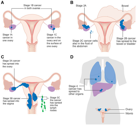
Ovarian cancer is fifth in the rankings of cancer deaths among women, and accounts for more deaths than any other gynecological malignancy. Despite some improvement in overall-(OS) and progression-free survival (PFS) following surgery and first-line chemotherapy, there is a need for development of [...] Read more.
Ovarian cancer is fifth in the rankings of cancer deaths among women, and accounts for more deaths than any other gynecological malignancy. Despite some improvement in overall-(OS) and progression-free survival (PFS) following surgery and first-line chemotherapy, there is a need for development of novel and more effective therapeutic strategies. In this mini review, we provide a summary of the current landscape of the clinical use of tyrosine kinase inhibitors (TKIs) and mechanistic target of rapamycin (mTOR) inhibitors in ovarian cancer. Emerging data from phase I and II trials reveals that a combinatorial treatment that includes TKIs and chemotherapy agents seems promising in terms of PFS despite some adverse effects recorded; whereas the use of mTOR inhibitors seems less effective. There is a need for further research into the inhibition of multiple signaling pathways in ovarian cancer and progression to phase III trials for drugs that seem most promising. Full article
(This article belongs to the Collection Targeting Solid Tumors)
Show Figures

Figure 1

11 pages, 1957 KiB  
Article
Low Numbers of Vascular Vessels Correlate to Progression in Hormone-Naïve Prostate Carcinomas Undergoing Radical Prostatectomy
by Julia Smentoch, Jolanta Szade, Anna J. Żaczek, Elke Eltze, Axel Semjonow, Burkhard Brandt and Natalia Bednarz-Knoll
Cancers 2019, 11(9), 1356; https://doi.org/10.3390/cancers11091356 - 12 Sep 2019
Cited by 7 | Viewed by 2848
Abstract
Vascularization influences tumor development by supporting the nutrition and dissemination of tumor cells. On the other hand, a low number of vascular vessels (VVlow) may induce hypoxia, accounting for selection of resistant clone(s) of tumor cells. This study aimed to evaluate [...] Read more.
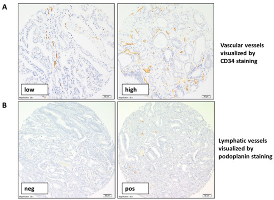
Vascularization influences tumor development by supporting the nutrition and dissemination of tumor cells. On the other hand, a low number of vascular vessels (VVlow) may induce hypoxia, accounting for selection of resistant clone(s) of tumor cells. This study aimed to evaluate the prognostic significance of vascular (VV) and lymphatic vessels (LV) in prostate cancer (PCa). Tumor samples from 400 PCa patients undergoing radical prostatectomy (RP) were prepared in duplex as tissue microarrays. Numbers of VV and LV were evaluated using immunohistochemistry detecting CD34 and podoplanin, respectively, and correlated to clinical data, biochemical recurrence (BR), and proteins analyzed in tumor cells. VVlow and LV were found in 32% and 43% of patients with informative PCa samples, respectively. VVlow correlated with a shorter time to BR 3, 5, and 10 years after RP in hormone-naïve patients (p = 0.028, p = 0.027 and p = 0.056, respectively). It was also shown to be an independent prognostic factor 5 years after surgery (multivariate analysis, p = 0.046). Tumors characterized by VVlow expressed the epithelial cell adhesion molecule, EpCAM, less frequently (p = 0.016) and revealed a borderline correlation to increased levels of tumor cell invasion marker Loxl-2 (p = 0.059). No correlations were found for LV. In summary, VVlow in hormone-naïve patients undergoing RP has prognostic potential and seems to be related to an aggressive phenotype of tumor cells. Full article
(This article belongs to the Special Issue New Biomarkers in Cancers)
Show Figures

Figure 1

20 pages, 363 KiB  
Review
Current Treatment Options for Metastatic Hormone-Sensitive Prostate Cancer
by Carlo Cattrini, Elena Castro, Rebeca Lozano, Elisa Zanardi, Alessandra Rubagotti, Francesco Boccardo and David Olmos
Cancers 2019, 11(9), 1355; https://doi.org/10.3390/cancers11091355 - 12 Sep 2019
Cited by 55 | Viewed by 9503
Abstract
The possible treatments options for metastatic hormone-sensitive prostate cancer (mHSPC) have dramatically increased during the last years. The old backbone, which androgen-deprivation therapy (ADT) is the exclusive approach for hormone-naïve patients, has been disrupted. Despite the fact that several high-quality, randomized, controlled phase [...] Read more.
The possible treatments options for metastatic hormone-sensitive prostate cancer (mHSPC) have dramatically increased during the last years. The old backbone, which androgen-deprivation therapy (ADT) is the exclusive approach for hormone-naïve patients, has been disrupted. Despite the fact that several high-quality, randomized, controlled phase 3 trials have been conducted in this setting, no direct comparison is currently available among the different strategies. Inadequate power, absence of preplanning and small sample size frequently affect the subgroup analyses according to disease volume or patient’s risk. The choice between ADT alone and ADT combined with docetaxel, abiraterone acetate, enzalutamide, apalutamide or radiotherapy to the primary tumor remains challenging. Factors that are related to the tumor, patient or drug side effects, currently guide these clinical decisions. This comprehensive review aims to indirectly compare the phase 3 trials in the mHSPC setting, in order to extrapolate data useful for treatment selection, providing also perspectives on future biomarkers. Full article
(This article belongs to the Special Issue Prostate Cancer: Past, Present, and Future)
27 pages, 3665 KiB  
Review
Purine-Metabolising Enzymes and Apoptosis in Cancer
by Marcella Camici, Mercedes Garcia-Gil, Rossana Pesi, Simone Allegrini and Maria Grazia Tozzi
Cancers 2019, 11(9), 1354; https://doi.org/10.3390/cancers11091354 - 12 Sep 2019
Cited by 55 | Viewed by 7474
Abstract
The enzymes of both de novo and salvage pathways for purine nucleotide synthesis are regulated to meet the demand of nucleic acid precursors during proliferation. Among them, the salvage pathway enzymes seem to play the key role in replenishing the purine pool in [...] Read more.
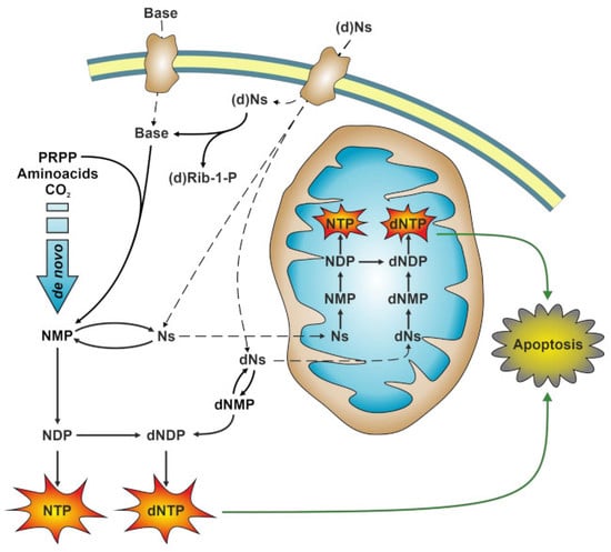
The enzymes of both de novo and salvage pathways for purine nucleotide synthesis are regulated to meet the demand of nucleic acid precursors during proliferation. Among them, the salvage pathway enzymes seem to play the key role in replenishing the purine pool in dividing and tumour cells that require a greater amount of nucleotides. An imbalance in the purine pools is fundamental not only for preventing cell proliferation, but also, in many cases, to promote apoptosis. It is known that tumour cells harbour several mutations that might lead to defective apoptosis-inducing pathways, and this is probably at the basis of the initial expansion of the population of neoplastic cells. Therefore, knowledge of the molecular mechanisms that lead to apoptosis of tumoural cells is key to predicting the possible success of a drug treatment and planning more effective and focused therapies. In this review, we describe how the modulation of enzymes involved in purine metabolism in tumour cells may affect the apoptotic programme. The enzymes discussed are: ectosolic and cytosolic 5′-nucleotidases, purine nucleoside phosphorylase, adenosine deaminase, hypoxanthine-guanine phosphoribosyltransferase, and inosine-5′-monophosphate dehydrogenase, as well as recently described enzymes particularly expressed in tumour cells, such as deoxynucleoside triphosphate triphosphohydrolase and 7,8-dihydro-8-oxoguanine triphosphatase. Full article
(This article belongs to the Special Issue Apoptosis in Cancer)
Show Figures

Graphical abstract

16 pages, 748 KiB  
Article
CYP2D6 Genotype Predicts Plasma Concentrations of Tamoxifen Metabolites in Ethiopian Breast Cancer Patients
by Jemal Hussien Ahmed, Eyasu Makonnen, Alan Fotoohi, Abraham Aseffa, Rawleigh Howe and Eleni Aklillu
Cancers 2019, 11(9), 1353; https://doi.org/10.3390/cancers11091353 - 12 Sep 2019
Cited by 13 | Viewed by 3873
Abstract
Tamoxifen displays wide inter-individual variability (IIV) in its pharmacokinetics and treatment outcome. Data on tamoxifen pharmacokinetics and pharmacogenetics from black African breast cancer patient populations is lacking. We investigated the pharmacokinetic and pharmacogenetic profile of tamoxifen and its major active metabolite, endoxifen, in [...] Read more.
Tamoxifen displays wide inter-individual variability (IIV) in its pharmacokinetics and treatment outcome. Data on tamoxifen pharmacokinetics and pharmacogenetics from black African breast cancer patient populations is lacking. We investigated the pharmacokinetic and pharmacogenetic profile of tamoxifen and its major active metabolite, endoxifen, in Ethiopian breast cancer patients. A total of 81 female breast cancer patients on adjuvant tamoxifen therapy were enrolled. Tamoxifen (Tam) and its major metabolites, N-desmethyltamoxifen (NDM), 4-hydroxy-tamoxifen (4-HT), and (Z)-endoxifen (E) were quantified using LC-MS/MS. Genotyping for CYP2D6, CYP2C9, CYP2C19, CYP3A5, POR, and ABCB1 and UGT2B15 and copy number variation for CYP2D6 were done. The proportion of patients with low endoxifen level (<5.9 ng/mL) was 35.8% (median concentration 7.94 ng/mL). The allele frequency of CYP2D6 gene deletion (*5) and duplication (*1×N or *2×N) was 4.3% and 14.8%, respectively. Twenty-six percent of the patients carried duplicated or multiplicated CYP2D6 gene. An increase in CYP2D6 activity score was associated with increased endoxifen concentration and MRE/NDM (p < 0.001). The IIV in endoxifen concentration and MRE/NDM was 74.6% and 59%, respectively. CYP2D6 diplotype explained 28.2% and 44% of the variability in absolute endoxifen concentration and MRE/NDM, respectively. The explanatory power of CYP2D6 diplotype was improved among ABCB1c.4036G carriers (43% and 65.2%, respectively for endoxifen concentration and MRE/NDM) compared to A/A genotype. CYP2C9, CYP2C19, and CYP3A5 genotypes had no significant influence on endoxifen concentration or MRE/NDM. In conclusion, we report a high rate of low endoxifen level as well as large IIV in tamoxifen and its metabolite concentrations. CYP2D6 is significant predictor of plasma endoxifen level in a gene-dose dependent manner. Full article
Show Figures

Figure 1

21 pages, 3345 KiB  
Article
Growth Hormone Upregulates Melanocyte-Inducing Transcription Factor Expression and Activity via JAK2-STAT5 and SRC Signaling in GH Receptor-Positive Human Melanoma
by Reetobrata Basu, Prateek Kulkarni, Yanrong Qian, Christopher Walsh, Pranay Arora, Emily Davis, Silvana Duran-Ortiz, Kevin Funk, Diego Ibarra, Colin Kruse, Samuel Mathes, Todd McHugh, Alison Brittain, Darlene E. Berryman, Edward O. List, Shigeru Okada and John J. Kopchick
Cancers 2019, 11(9), 1352; https://doi.org/10.3390/cancers11091352 - 12 Sep 2019
Cited by 21 | Viewed by 6529
Abstract
Growth hormone (GH) facilitates therapy resistance in the cancers of breast, colon, endometrium, and melanoma. The GH-stimulated pathways responsible for this resistance were identified as suppression of apoptosis, induction of epithelial-to-mesenchymal transition (EMT), and upregulated drug efflux by increased expression of ATP-binding cassette [...] Read more.
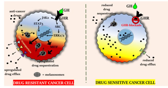
Growth hormone (GH) facilitates therapy resistance in the cancers of breast, colon, endometrium, and melanoma. The GH-stimulated pathways responsible for this resistance were identified as suppression of apoptosis, induction of epithelial-to-mesenchymal transition (EMT), and upregulated drug efflux by increased expression of ATP-binding cassette containing multidrug efflux pumps (ABC-transporters). In extremely drug-resistant melanoma, ABC-transporters have also been reported to mediate drug sequestration in intracellular melanosomes, thereby reducing drug efficacy. Melanocyte-inducing transcription factor (MITF) is the master regulator of melanocyte and melanoma cell fate as well as the melanosomal machinery. MITF targets such as the oncogene MET, as well as MITF-mediated processes such as resistance to radiation therapy, are both known to be upregulated by GH. Therefore, we chose to query the direct effects of GH on MITF expression and activity towards conferring chemoresistance in melanoma. Here, we demonstrate that GH significantly upregulates MITF as well as the MITF target genes following treatment with multiple anticancer drug treatments such as chemotherapy, BRAF-inhibitors, as well as tyrosine-kinase inhibitors. GH action also upregulated MITF-regulated processes such as melanogenesis and tyrosinase activity. Significant elevation in MITF and MITF target gene expression was also observed in mouse B16F10 melanoma cells and xenografts in bovine GH transgenic (bGH) mice compared to wild-type littermates. Through pathway inhibitor analysis we identified that both the JAK2-STAT5 and SRC activities were critical for the observed effects. Additionally, a retrospective analysis of gene expression data from GTEx, NCI60, CCLE, and TCGA databases corroborated our observed correlation of MITF function and GH action. Therefore, we present in vitro, in vivo, and in silico evidence which strongly implicates the GH–GHR axis in inducing chemoresistance in human melanoma by driving MITF-regulated and ABC-transporter-mediated drug clearance pathways. Full article
(This article belongs to the Special Issue JAK-STAT Signalling Pathway in Cancer)
Show Figures

Graphical abstract

18 pages, 1714 KiB  
Review
Early Steps of Mammary Stem Cell Transformation by Exogenous Signals; Effects of Bisphenol Endocrine Disrupting Chemicals and Bone Morphogenetic Proteins
by Nora Jung, Veronique Maguer-Satta and Boris Guyot
Cancers 2019, 11(9), 1351; https://doi.org/10.3390/cancers11091351 - 12 Sep 2019
Cited by 8 | Viewed by 3779
Abstract
Estrogens are major regulators of the mammary gland development, notably during puberty, via estrogen receptor (ER) activation, leading to the proliferation and differentiation of mammary cells. In addition to estrogens, the bone morphogenetic proteins (BMPs) family is involved in breast stem cell/progenitor commitment. [...] Read more.
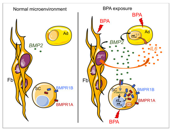
Estrogens are major regulators of the mammary gland development, notably during puberty, via estrogen receptor (ER) activation, leading to the proliferation and differentiation of mammary cells. In addition to estrogens, the bone morphogenetic proteins (BMPs) family is involved in breast stem cell/progenitor commitment. However, these two pathways that synergistically contribute to the biology of the normal mammary gland have also been described to initiate and/or promote breast cancer development. In addition to intrinsic events, lifestyle habits and exposure to environmental cues are key risk factors for cancer in general, and especially for breast cancer. In the latter case, bisphenol A (BPA), an estrogen-mimetic compound, is a critical pollutant both in terms of the quantities released in our environment and of its known and speculated effects on mammary gland biology. In this review, we summarize the current knowledge on the actions of BMPs and estrogens in both normal mammary gland development and breast cancer initiation, dissemination, and resistance to treatment, focusing on the dysregulations of these processes by BPA but also by other bisphenols, including BPS and BPF, initially considered as safer alternatives to BPA. Full article
Show Figures

Figure 1

15 pages, 2172 KiB  
Article
Human Ovarian Cancer Tissue Exhibits Increase of Mitochondrial Biogenesis and Cristae Remodeling
by Anna Signorile, Domenico De Rasmo, Antonella Cormio, Clara Musicco, Roberta Rossi, Francesco Fortarezza, Luigi Leonardo Palese, Vera Loizzi, Leonardo Resta, Giovanni Scillitani, Ettore Cicinelli, Francesca Simonetti, Anna Ferretta, Silvia Russo, Antonio Tufaro and Gennaro Cormio
Cancers 2019, 11(9), 1350; https://doi.org/10.3390/cancers11091350 - 12 Sep 2019
Cited by 43 | Viewed by 4099
Abstract
Ovarian cancer (OC) is the most lethal gynecologic cancer characterized by an elevated apoptosis resistance that, potentially, leads to chemo-resistance in the recurrent disease. Mitochondrial oxidative phosphorylation was found altered in OC, and mitochondria were proposed as a target for therapy. Molecular evidence [...] Read more.
Ovarian cancer (OC) is the most lethal gynecologic cancer characterized by an elevated apoptosis resistance that, potentially, leads to chemo-resistance in the recurrent disease. Mitochondrial oxidative phosphorylation was found altered in OC, and mitochondria were proposed as a target for therapy. Molecular evidence suggests that the deregulation of mitochondrial biogenesis, morphology, dynamics, and apoptosis is involved in carcinogenesis. However, these mitochondrial processes remain to be investigated in OC. Eighteen controls and 16 OC tissues (serous and mucinous) were collected. Enzymatic activities were performed spectrophotometrically, mitochondrial DNA (mtDNA) content was measured by real-time-PCR, protein levels were determined by Western blotting, and mitochondrial number and structure were measured by electron microscopy. Statistical analysis was performed using Student’s t-test, Mann-Whitney U test, and principal component analysis (PCA). We found, in OC, that increased mitochondrial number associated with increased peroxisome proliferator-activated receptor gamma coactivator 1-alpha (PGC1α) and mitochondrial transcription factor A (TFAM) protein levels, as well as mtDNA content. The OC mitochondria presented an increased maximum length, as well as reduced cristae width and junction diameter, associated with increased optic atrophy 1 protein (OPA1) and prohibitin 2 (PHB2) protein levels. In addition, in OC tissues, augmented cAMP and sirtuin 3 (SIRT3) protein levels were observed. PCA of the 25 analyzed biochemical parameters classified OC patients in a distinct group from controls. We highlight a “mitochondrial signature” in OC that could result from cooperation of the cAMP pathway with the SIRT3, OPA1, and PHB2 proteins. Full article
Show Figures

Figure 1

20 pages, 3169 KiB  
Article
Estrogens Counteract Platinum-Chemosensitivity by Modifying the Subcellular Localization of MDM4
by Rossella Lucà, Giorgia di Blasio, Daniela Gallo, Valentina Monteleone, Isabella Manni, Laura Fici, Marianna Buttarelli, Germana Ciolli, Marsha Pellegrino, Emanuela Teveroni, Silvia Maiullari, Alessandra Ciucci, Alessandro Apollo, Francesca Mancini, Maria Pia Gentileschi, Gian Franco Zannoni, Alfredo Pontecorvi, Giovanni Scambia and Fabiola Moretti
Cancers 2019, 11(9), 1349; https://doi.org/10.3390/cancers11091349 - 12 Sep 2019
Cited by 5 | Viewed by 2670
Abstract
Estrogen activity towards cancer-related pathways can impact therapeutic intervention. Recent omics data suggest possible crosstalk between estrogens/gender and MDM4, a key regulator of p53. Since MDM4 can either promote cell transformation or enhance DNA damage-sensitivity, we analysed in vivo impact of estrogens on [...] Read more.
Estrogen activity towards cancer-related pathways can impact therapeutic intervention. Recent omics data suggest possible crosstalk between estrogens/gender and MDM4, a key regulator of p53. Since MDM4 can either promote cell transformation or enhance DNA damage-sensitivity, we analysed in vivo impact of estrogens on both MDM4 activities. In Mdm4 transgenic mouse, Mdm4 accelerates the formation of fibrosarcoma and increases tumor sensitivity to cisplatin as well, thus confirming in vivo Mdm4 dual mode of action. Noteworthy, Mdm4 enhances chemo- and radio-sensitivity in male but not in female animals, whereas its tumor-promoting activity is not affected by mouse gender. Combination therapy of transgenic females with cisplatin and fulvestrant, a selective estrogen receptor degrader, was able to recover tumor cisplatin-sensitivity, demonstrating the relevance of estrogens in the observed sexual dimorphism. Molecularly, estrogen receptor-α alters intracellular localization of MDM4 by increasing its nuclear fraction correlated to decreased cell death, in a p53-independent manner. Importantly, MDM4 nuclear localization and intra-tumor estrogen availability correlate with decreased platinum-sensitivity and apoptosis and predicts poor disease-free survival in high-grade serous ovarian carcinoma. These data demonstrate estrogen ability to modulate chemo-sensitivity of MDM4-expressing tumors and to impinge on intracellular trafficking. They support potential usefulness of combination therapy involving anti-estrogenic drugs. Full article
Show Figures

Figure 1

17 pages, 853 KiB  
Review
18F-Facbc in Prostate Cancer: A Systematic Review and Meta-Analysis
by Riccardo Laudicella, Domenico Albano, Pierpaolo Alongi, Giovanni Argiroffi, Matteo Bauckneht, Sergio Baldari, Francesco Bertagna, Michele Boero, Giuseppe De Vincentis, Angelo Del Sole, Giuseppe Rubini, Lorenzo Fantechi, Viviana Frantellizzi, Gloria Ganduscio, Priscilla Guglielmo, Anna Giulia Nappi, Laura Evangelista and on the behalf of Young AIMN Working Group
Cancers 2019, 11(9), 1348; https://doi.org/10.3390/cancers11091348 - 11 Sep 2019
Cited by 34 | Viewed by 4564
Abstract
Trans-1-amino-3-18F-fluorocyclobutanecarboxylic-acid (anti-[18F]-FACBC) has been approved for the detection of prostate cancer (PCa) in patients with elevated prostate-specific-antigen following prior treatment. This review and meta-analysis aimed to investigate the diagnostic performance of 18F-FACBC positron emission tomography/computed-tomography (PET/CT) in the [...] Read more.
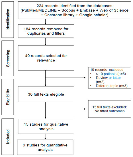
Trans-1-amino-3-18F-fluorocyclobutanecarboxylic-acid (anti-[18F]-FACBC) has been approved for the detection of prostate cancer (PCa) in patients with elevated prostate-specific-antigen following prior treatment. This review and meta-analysis aimed to investigate the diagnostic performance of 18F-FACBC positron emission tomography/computed-tomography (PET/CT) in the detection of primary/recurrent PCa. A bibliographic search was performed including several databases, using the following terms: “FACBC”/“fluciclovine” AND “prostate cancer”/“prostate” AND “PET”/“Positron Emission Tomography”. Fifteen and 9 studies were included in the systematic reviews and meta-analysis, respectively. At patient-based analysis, the pooled sensitivity and specificity of 18F-FACBC-PET/CT for the assessment of PCa were 86.3% and 75.9%, respectively. The pooled diagnostic odds-ratio value was 16.453, with heterogeneity of 30%. At the regional-based-analysis, the pooled sensitivity of 18F-FACBC-PET/CT for the evaluation of primary/recurrent disease in the prostatic bed was higher than in the extra-prostatic regions (90.4% vs. 76.5%, respectively); conversely, the pooled specificity was higher for the evaluation of extra-prostatic region than the prostatic bed (89% vs. 45%, respectively). 18F-FACBC-PET/CT seems to be promising in recurrent PCa, particularly for the evaluation of the prostatic bed. Additional studies to evaluate its utility in clinical routine are mandatory. Full article
(This article belongs to the Special Issue Role of Medical Imaging in Cancers)
Show Figures

Figure 1

22 pages, 2408 KiB  
Article
The miR-205-5p/BRCA1/RAD17 Axis Promotes Genomic Instability in Head and Neck Squamous Cell Carcinomas
by Fabio Valenti, Andrea Sacconi, Federica Ganci, Giuseppe Grasso, Sabrina Strano, Giovanni Blandino and Silvia Di Agostino
Cancers 2019, 11(9), 1347; https://doi.org/10.3390/cancers11091347 - 11 Sep 2019
Cited by 32 | Viewed by 3627
Abstract
Defective DNA damage response (DDR) is frequently associated with tumorigenesis. Abrogation of DDR leads to genomic instability, which is one of the most common characteristics of human cancers. TP53 mutations with gain-of-function activity are associated with tumors under high replicative stress, high genomic [...] Read more.
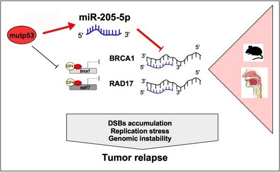
Defective DNA damage response (DDR) is frequently associated with tumorigenesis. Abrogation of DDR leads to genomic instability, which is one of the most common characteristics of human cancers. TP53 mutations with gain-of-function activity are associated with tumors under high replicative stress, high genomic instability, and reduced patient survival. The BRCA1 and RAD17 genes encode two pivotal DNA repair proteins required for proper cell-cycle regulation and maintenance of genomic stability. We initially evaluated whether miR-205-5p, a microRNA (miRNA) highly expressed in head and neck squamous cell carcinoma (HNSCC), targeted BRCA1 and RAD17 expression. We found that, in vitro and in vivo, BRCA1 and RAD17 are targets of miR-205-5p in HNSCC, leading to inefficient DNA repair and increased chromosomal instability. Conversely, miR-205-5p downregulation increased BRCA1 and RAD17 messenger RNA (mRNA) levels, leading to a reduction in in vivo tumor growth. Interestingly, miR-205-5p expression was significantly anti-correlated with BRCA1 and RAD17 targets. Furthermore, we documented that miR-205-5p expression was higher in tumoral and peritumoral HNSCC tissues than non-tumoral tissues in patients exhibiting reduced local recurrence-free survival. Collectively, these findings unveil miR-205-5p’s notable role in determining genomic instability in HNSCC through its selective targeting of BRCA1 and RAD17 gene expression. High miR-205-5p levels in the peritumoral tissues might be relevant for the early detection of minimal residual disease and pre-cancer molecular alterations involved in tumor development. Full article
Show Figures

Graphical abstract

30 pages, 5601 KiB  
Review
Anti-Tumor Potential of IMP Dehydrogenase Inhibitors: A Century-Long Story
by Rand Naffouje, Punita Grover, Hongyang Yu, Arun Sendilnathan, Kara Wolfe, Nazanin Majd, Eric P. Smith, Koh Takeuchi, Toshiya Senda, Satoshi Kofuji and Atsuo T. Sasaki
Cancers 2019, 11(9), 1346; https://doi.org/10.3390/cancers11091346 - 11 Sep 2019
Cited by 70 | Viewed by 11025
Abstract
The purine nucleotides ATP and GTP are essential precursors to DNA and RNA synthesis and fundamental for energy metabolism. Although de novo purine nucleotide biosynthesis is increased in highly proliferating cells, such as malignant tumors, it is not clear if this is merely [...] Read more.
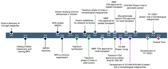
The purine nucleotides ATP and GTP are essential precursors to DNA and RNA synthesis and fundamental for energy metabolism. Although de novo purine nucleotide biosynthesis is increased in highly proliferating cells, such as malignant tumors, it is not clear if this is merely a secondary manifestation of increased cell proliferation. Suggestive of a direct causative effect includes evidence that, in some cancer types, the rate-limiting enzyme in de novo GTP biosynthesis, inosine monophosphate dehydrogenase (IMPDH), is upregulated and that the IMPDH inhibitor, mycophenolic acid (MPA), possesses anti-tumor activity. However, historically, enthusiasm for employing IMPDH inhibitors in cancer treatment has been mitigated by their adverse effects at high treatment doses and variable response. Recent advances in our understanding of the mechanistic role of IMPDH in tumorigenesis and cancer progression, as well as the development of IMPDH inhibitors with selective actions on GTP synthesis, have prompted a reappraisal of targeting this enzyme for anti-cancer treatment. In this review, we summarize the history of IMPDH inhibitors, the development of new inhibitors as anti-cancer drugs, and future directions and strategies to overcome existing challenges. Full article
(This article belongs to the Special Issue Metabolic Reprogramming and Vulnerabilities in Cancer)
Show Figures

Figure 1

14 pages, 1706 KiB  
Article
Targeting Autophagy by MPT0L145, a Highly Potent PIK3C3 Inhibitor, Provides Synergistic Interaction to Targeted or Chemotherapeutic Agents in Cancer Cells
by Chun-Han Chen, Tsung-Han Hsieh, Yu-Chen Lin, Yun-Ru Liu, Jing-Ping Liou and Yun Yen
Cancers 2019, 11(9), 1345; https://doi.org/10.3390/cancers11091345 - 11 Sep 2019
Cited by 14 | Viewed by 3400
Abstract
Anticancer therapies reportedly promote pro-survival autophagy in cancer cells that confers drug resistance, rationalizing the concept to combine autophagy inhibitors to increase their therapeutic potential. We previously identified that MPT0L145 is a PIK3C3/FGFR inhibitor that not only increases autophagosome formation due to fibroblast [...] Read more.
Anticancer therapies reportedly promote pro-survival autophagy in cancer cells that confers drug resistance, rationalizing the concept to combine autophagy inhibitors to increase their therapeutic potential. We previously identified that MPT0L145 is a PIK3C3/FGFR inhibitor that not only increases autophagosome formation due to fibroblast growth factor receptor (FGFR) inhibition but also perturbs autophagic flux via PIK3C3 inhibition in bladder cancer cells harboring FGFR activation. In this study, we hypothesized that combined-use of MPT0L145 with agents that induce pro-survival autophagy may provide synthetic lethality in cancer cells without FGFR activation. The results showed that MPT0L145 synergistically sensitizes anticancer effects of gefitinib and gemcitabine in non-small cell lung cancer A549 cells and pancreatic cancer PANC-1 cells, respectively. Mechanistically, drug combination increased incomplete autophagy due to impaired PIK3C3 function by MPT0L145 as evidenced by p62 accumulation and no additional apoptotic cell death was observed. Meanwhile, drug combination perturbed survival pathways and increased vacuolization and ROS production in cancer cells. In conclusion, the data suggest that halting pro-survival autophagy by targeting PIK3C3 with MPT0L145 significantly sensitizes cancer cells to targeted or chemotherapeutic agents, fostering rational combination strategies for cancer therapy in the future. Full article
(This article belongs to the Special Issue The Role of Autophagy in Cancer Progression and Drug Resistance)
Show Figures

Figure 1

14 pages, 11105 KiB  
Article
The Potential Use of Electrochemotherapy in the Treatment of Uveal Melanoma: In Vitro Results in 3D Tumor Cultures and In Vivo Results in a Chick Embryo Model
by Miltiadis Fiorentzis, Arne Viestenz, Udo Siebolts, Berthold Seitz, Sarah E. Coupland and Joana Heinzelmann
Cancers 2019, 11(9), 1344; https://doi.org/10.3390/cancers11091344 - 11 Sep 2019
Cited by 25 | Viewed by 4017
Abstract
Uveal melanoma (UM) is the most common primary intraocular tumor that arises from neoplastic melanocytes in the choroid, iris, and ciliary body. Electrochemotherapy (ECT) has been successfully established for the treatment of skin and soft tissue metastatic lesions, deep-seated tumors of the liver, [...] Read more.
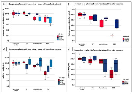
Uveal melanoma (UM) is the most common primary intraocular tumor that arises from neoplastic melanocytes in the choroid, iris, and ciliary body. Electrochemotherapy (ECT) has been successfully established for the treatment of skin and soft tissue metastatic lesions, deep-seated tumors of the liver, bone metastases, and unresectable pancreas lesions. The aim of this study was to evaluate the effect of ECT in vitro in 3D spheroid culture systems in primary and metastatic UM cell lines. We also investigated the chick embryo chorioallantoic membrane (CAM) as an in vivo model system for the growth and treatment of UM tumors using ECT. The cytotoxic effect of ECT in 3D spheroids was analyzed seven days following treatment by assessment of the size and MTT [(3-(4,5-dimethylthiazol-2-yl)-2,5-diphenyltetrazolium bromide) tetrazolium reduction] assay. The cytotoxicity of ECT after intratumoral or intraarterial administration was evaluated histologically. In vitro and in vivo ECT caused a significant reduction in tumor size and viability compared to electroporation or chemotherapy in both sections of our study. The current results underline the effectiveness of ECT in the treatment of UM and prepare the way for further investigation of its potential application in UM. Full article
(This article belongs to the Special Issue Uveal Melanoma)
Show Figures

Figure 1