Supramolecular Hydrogen-Bond Motifs in Chiral and Racemic Molecular Salts: A Comparison of (S)-2-Methyl Piperizinium Hydrogen Phosphite Monohydrate, C5H14N2·HPO3·H2O and (R,S)-2-Methyl Piperizinium Hydrogen Phosphite 2.23 Hydrate, C5H14N2·HPO3·2.23H2O

Abstract
: The crystal structures of C5H14N2·HPO3·H2O (1) and C5H14N2·HPO3·2.23H2O (2) are described and compared. Compound 1 contains homochiral (S)-2-methyl piperizinium cations, hydrogen phosphite ions and water molecules. The components are linked by N–H⋯O and O–H⋯O hydrogen bonds into a three-dimensional network. In compound 2, racemic (R,S)-2-methyl piperizinium cations combine with the same anions and water molecules to generate a far more complex, high symmetry “supramolecular” structure, which features distinctive R66(12) loops and helical C(2) chain hydrogen-bonding motifs involving the water molecules. Crystal data: 1 (C5H17N2O4P), Mr = 200.18, orthorhombic, P212121 (No. 19), Z = 4, a = 8.564 (5) Å, b = 9.593 (6) Å, c = 11.607 (6) Å, V = 953.6 (9) Å3, R(F) = 0.066, wR(F2) = 0.081. 2 (C5H19.47N2O5.24P), Mr = 222.49, trigonal, R3̄ (No. 148), Z = 18, a = 31.075 (2) Å, c = 6.1875 (4) Å, V = 5174.5 (6) Å3, R(F) = 0.044, wR(F2) = 0.107.1. Introduction
Supramolecular chemistry—the “chemistry of molecular assemblies and intermolecular bonds” [1] is an area of contemporary chemical research to which crystallography makes a key contribution. The (non-covalent) intermolecular interaction par excellence is arguably the hydrogen bond and much work has been carried out in this area, for which a recent two-volume review edited by Mingos et al. [2] may serve as a starting point for further reading.
As a small contribution to this area, we now describe the syntheses and crystal structures of two related molecular salts, viz., C5H14N2·HPO3·H2O (1) and C5H14N2·HPO3·2.23H2O (2), which contain homochiral (S)-2-methyl piperizinium and racemic (R,S)-2-methyl piperizinium cations, respectively, combined with the same simple inorganic anion and water molecules of crystallization. This also follows on from our previous studies of hydrogen-bonding patterns in molecular salts containing oxo-anions, for example piperizinium hydrogen phosphite monohydrate, C4H12N2·HPO3·H2O [3], in which supramolecular anion–water chains occur.
2. Results and Discussion
2.1. Structure of C5H14N2·HPO3·H2O (1)
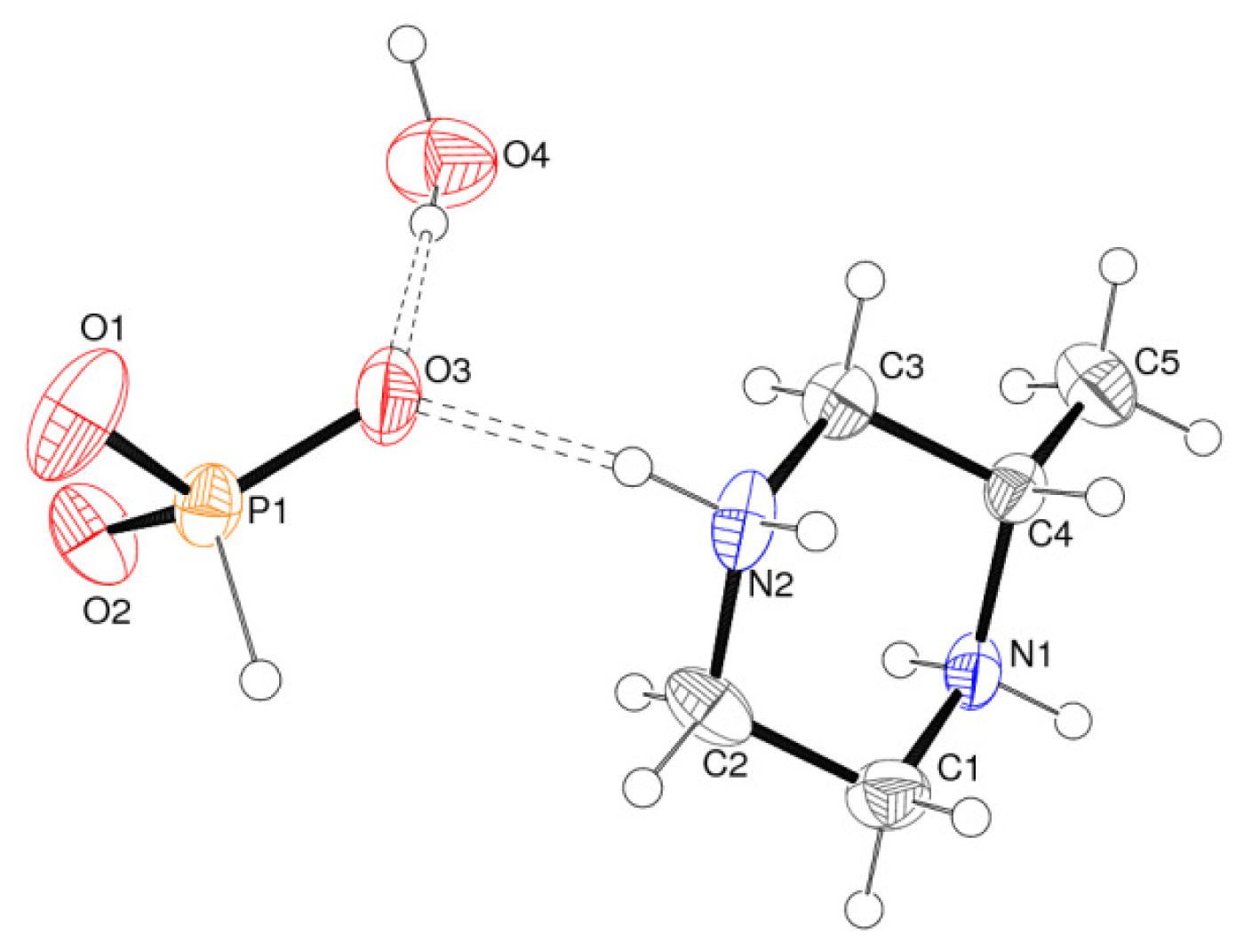
Compound 1 is a hydrated molecular salt in which double proton transfer from phosphorus acid to (S)-2-methyl piperizine has occurred: the asymmetric unit contains one [C5H14N2]2+ cation, one [HPO3]2− anion and one water molecule of crystallisation (Figure 1). Its space group of P212121 (see experimental section) is of course consistent with the presence of the homochiral organic molecule. The geometrical parameters for the component species in 1 are unexceptional: the cation [mean C–C = 1.515 (7) Å; mean C–N = 1.486 (7) Å] adopts a typical chair conformation with the pendant methyl group in an equatorial position, as is usually seen for this species in the solid state [4]. The N2–C3–C4–C5 and C1–N1–C4–C5 torsion angles are 175.5 (5) and 179.7 (6)°, respectively. The hydrogen phosphite dianion possesses typical geometrical parameters [mean P–O = 1.506 (4) Å; mean O–P–O = 112.0 (3)°] and the P atom is displaced from the plane of its three attached O atoms by 0.434 (3) Å. As expected [5], the P–H vertex is not involved in bonding (or hydrogen bonding) to other species.
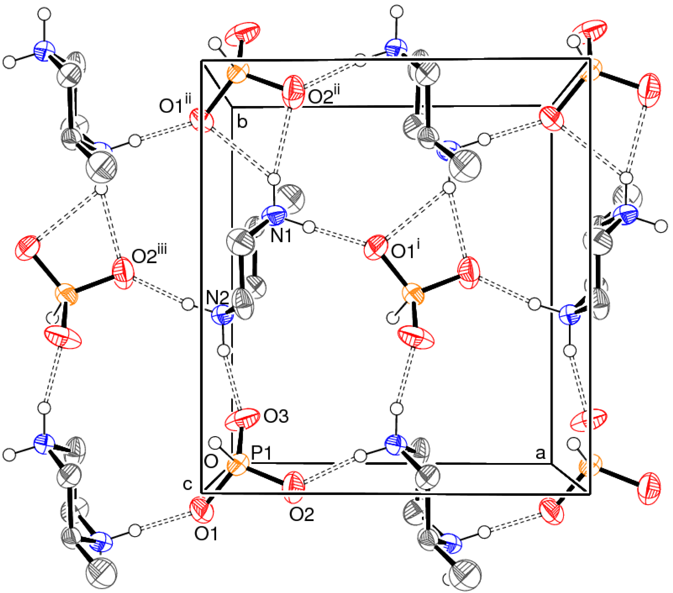
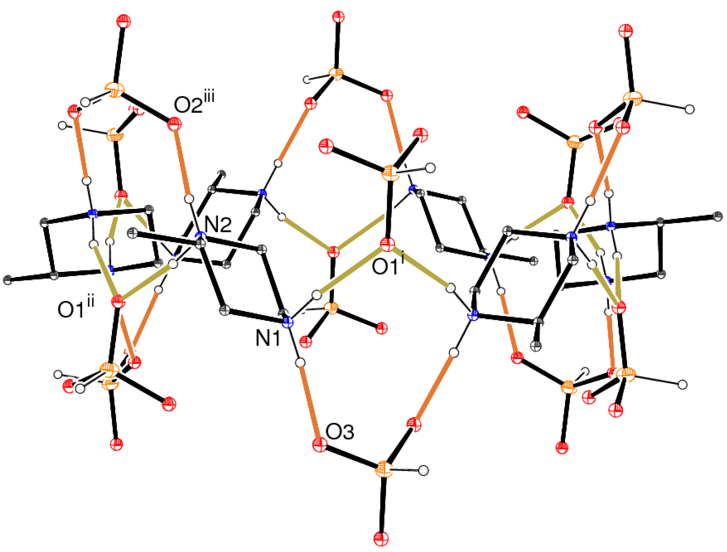
The packing for 1 features N–H⋯O and O–H⋯O hydrogen bonds (Table 1). All four N–H vertices of the cation make N–H⋯O links to different hydrogen phosphite O atoms. The bond involving H2b is a bifurcated N–H⋯(O,O) link to two oxygen atoms in the same dianion (bond angle sum for H2b = 359.9°). Taken together, these bonds link the cations and anions into (001) sheets (Figure 2). Within the sheets, R24(11) and R34(15) loops, using the graph-set notation of Bernstein et al. [6], are apparent.
Finally, the water molecule of crystallisation in 1 makes two O–H⋯O hydrogen bonds to link the (001) cation/anion sheets into a three-dimensional array. Overall, the phosphite oxygen atoms O1, O2 and O3 accept two, three and two hydrogen bonds each, respectively.
2.2. Structure of C5H14N2·HPO3·2.23H2O (2)
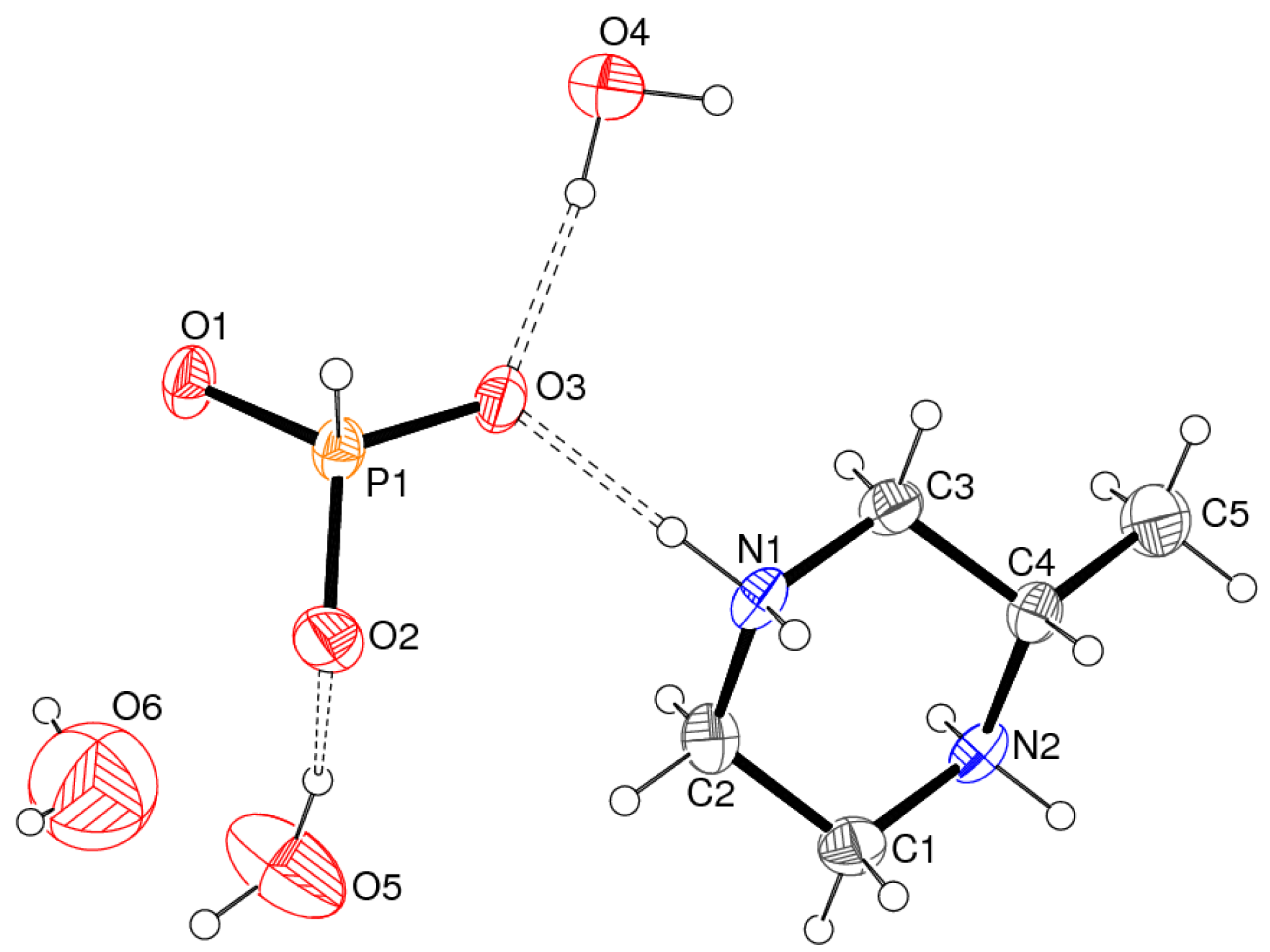
The asymmetric unit of compound 2 contains one cation, one anion and three water molecules of crystallisation, one of which is partially occupied (Figure 3). The pendant methyl group of the cation has an equatorial orientation and the N1–C3–C4–C5 and C1–N2–C4–C5 torsion angles are 176.6 (2) and −177.5 (2)°, respectively. Otherwise, the key geometrical parameters in 2 barely differ from those for the equivalent species 1: mean C–C = 1.513 (4) Å; mean C–N = 1.482 (4) Å; mean P–O = 1.512 (2) Å; mean O–P–O = 112.5 (2)°; displacement of the P atom from its three attached O atoms = 0.4240 (13) Å. The centrosymmetric crystal symmetry (space group R3̄ generates a statistical mixture of the two enantiomers of the organic cation, which is consistent with the racemic starting material.
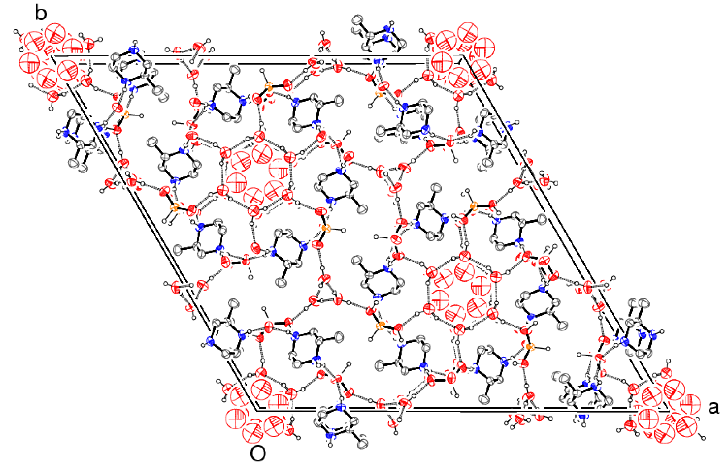
The complex unit-cell packing in 2 (Figure 4) can be broken down as follows. Alternating six-rings of the O5- and O6-water molecules stack about the 3̄ symmetry axis located at (x = 0, y = 0) and symmetry-equivalent positions. The O5 species are well ordered and almost in the same plane as the 3̄-axis inversion centre at (0, 0, 1/2). They combine to form a distinctive “daisy-chain” motif of a six-ring of O5–H19⋯O5 hydrogen bonds, resulting in an R66(12) loop [6]. So far as we can ascertain from the present refinement, within any one loop, the hydrogen bonds are well ordered and all aligned in the same sense (either clockwise or anticlockwise). The O5–H18 vertex makes a hydrogen bond to an O atom of a nearby hydrogen phosphite group.
Due to two short, symmetry generated, oxygen⋯oxygen contacts [O⋯O = 1.610 (18) Å], the O6 water molecule must be partially occupied with a maximum site occupancy of ⅓ [the refined site occupancy of 0.230 (11) is slightly lower than this value]. The H atoms associated with this disordered species were geometrically placed in reasonable positions to form hydrogen bonds, and their locations should be regarded as less certain than those of the other atoms.
The 3̄ axis generates six hydrogen phosphite groups to encapsulate each O5 water-molecule ring and a [C5H14N2]2+ cation wedges between each adjacent phosphite pair and interacts with them by means of N–H⋯O bonds. These hydrogen bonds also link to further rings of anions both above and below the plane of the cations, to result in infinite [001] tubes: the cations in any six-ring circuit have alternating R and S chiralities at their C4 atoms. The cation/anion tubes (Figure 5) thus serve as “supramolecular wrappers” to encapsulate the O5/O6 water molecules, or alternately, we may envisage that the O5/O6 water molecule columns serve to “template” the ionic components of the structure.
Finally, the hydrogen phosphite O3 atom accepts a hydrogen bond from the O4 water molecule, by way of an O4–H16⋯O3 interaction and an O4–H17⋯O4 (water-to-water) hydrogen bond completes the structure. These infinite [001] chains of water-molecule hydrogen bonds entwine about either a 31 or a 32 symmetry axis [at (x = 1/3, y = 1/3) and symmetry-equivalent locations], hence generating C(2) helices of hydrogen bonds. Around any O5-centred column, the O4–H17⋯O4 helices propagate in alternating clockwise and anticlockwise senses.
3. Experimental Section
Compound 1 was prepared by dissolving (S)-2-methyl piperizine (Aldrich, 99%, 0.10 g; 1.00 mmol) and H3PO3 (0.082 g; 1.00 mmol) in 2 mL distilled water at room temperature. A few tiny, colorless rods of 1 were observed after a few days as the water evaporated from a watch glass. Repeated attempts to grow larger, better quality crystals were unsuccessful and glassy/amorphous residues were usually the result.
To prepare compound 2, (R,S)-2-methyl piperizine (Aldrich, 98%, 1.00 g; 10.0 mmol) and H3PO3 (0.82 g; 10.0 mmol) were dissolved in 10 mL distilled water at room temperature. Colorless needle-shaped crystals of 2 grew over the course of a few days as the solvent evaporated.
The single-crystal data for 1 and 2 were collected using a Bruker SMART1000 CCD diffractometer (graphite monochromated Mo Kα radiation, λ = 0.71073 Å) at room temperature. Suitable crystals were selected and mounted on thin glass fibers with cyanoacrylate adhesive and intensity data were collected with the aid of the SMART program. Data reduction with SAINT then proceeded in each case and the structures were solved by direct methods with SHELXS. The resulting atomic models were refined against |F|2 with SHELXL [7] and the “observed data” threshold for calculating the R(F) residuals was set as I > 2σ(I).
The small crystal of 1 was a feeble scatterer, which may correlate with the high RInt value. Disappointingly, refinement of the Flack absolute structure parameter [8] for 1 was inconclusive [refined value = −0.2 (3)] and therefore the S configuration for atom C5 was assigned based on the stated absolute structure of the starting material. The C-, N- and P -bound H atoms were placed in idealised locations (C–H = 0.96-0.97 Å, N–H = 0.90 Å, P–H = 1.32 Å) and refined as riding atoms. The water H atoms were located in a difference map and refined as riding atoms in their as-found relative locations. The constraint Uiso(H) = 1.2Ueq(carrier) was applied in all cases.
For 2, the C-, N- and P-bound H atoms were treated in the same way as the equivalent atoms in 1. The H atoms attached to O4 and O5 were located in a difference map and refined as riding atoms in their as-found relative locations. The H atoms attached to O6 were placed in chemically reasonable positions to achieve O–H⋯O hydrogen bonds and refined as riding atoms. The constraint Uiso(H) = 1.2Ueq(carrier) was applied in all cases. Atom O6 and its attached H atoms have refined site occupancies of 0.230 (11). A PLATON [9] void-space analysis indicated a small amount of free space in the vicinity of (0, 0, 1/2), in the centre of the O5 daisy chain. An O atom placed there refined to zero site occupancy in a few cycles. Full refinement details are given in the deposited cifs.
Crystal data for 1: colorless rod, 0.04 × 0.01 × 0.01 mm, C5H17N2O4P, Mr = 200.18, orthorhombic, P212121 (No. 19), a = 8.564 (5) Å, b = 9.593 (6) Å, c = 11.607 (6) Å, V = 953.6 (9) Å3, Z = 4, F(000) = 432, T = 293 K, ρcalc = 1.394 g cm−3, μ = 0.272 mm−1, 5970 reflections measured (−10 ≤ h ≤ 10, −11 ≤ k ≤ 11, −8 ≤ l ≤ 14; 5.50° ≤ 2θ ≤ 52.00°), RInt = 0.247, 1104 merged reflections, 507 with I > 2σ(I), 110 parameters, R(F) = 0.066, wR(F2) = 0.081, w = 1/[σ2(Fo2) + 0.0035P2], where P = (Fo2 + 2 Fc2)/3, min./max. Δρ = −0.21, +0.22 e Å−3. Cambridge Database deposition number: CSD-840886.
Crystal data for 2: colorless needle, 0.45 × 0.04 × 0.03 mm, C5H19.46N2O5.23P, Mr = 222.49, trigonal, R3̄ (No. 148), a = 31.075 (2) Å, c = 6.1875 (4) Å, V = 5174.5 (6) Å3, Z = 18, F(000) = 2167, T = 296 K, ρcalc = 1.285 g cm−3, μ = 0.240 mm−1, 11694 reflections measured (−37 ≤ h ≤ 38, −38 ≤ k ≤ 27, −7 ≤ l ≤ 7; 4.54° ≤ 2θ ≤ 52.00°), RInt = 0.067, 2270 merged reflections, 1419 with I > 2σ(I), 120 parameters, R(F) = 0.044, wR(F2) = 0.107, w = 1/[σ2(Fo2) + 0.0541P2], where P = (Fo2 + 2 Fc2)/3, min./max. Δρ = −0.15, +0.24 e Å−3. Cambridge Database deposition number: CSD-840887.
4. Conclusions
The crystal structures of the two title hydrated molecular salts have been described. Although they contain the same simple building units, differing only in the homochiral/racemic nature of the cations, their crystal structures are very different. The structure of 1 might be regarded as “typical” of a hydrated molecular salt [10,11] and similar networks of hydrogen bonds have been seen in many related structures [12,13]: it seems that the chiral nature of the cation in 1 imparts no notable properties whatsoever to the extended structure. Indeed, the supramolecular connectivity in 1 closely resembles that in the centrosymmetric C4H12N2·HPO3·H2O [3].
The complex, high symmetry, centrosymmetric structure of 2 is quite different, and the differences seem to extend beyond the replacement of the chiral organic cation by its racemic congener. It is perhaps most notable that hydrogen bonding involving the water molecules in 2 appear to play such a critical role in establishing the supramolecular structure in terms of the “daisy chains” and helices described above, although we most certainly do not claim that these motifs are “novel” or previously unseen in other structures [14].


| Bond | D–H | H⋯A | D⋯A | D–H⋯A |
|---|---|---|---|---|
| N1–H1A⋯O1i | 0.90 | 1.72 | 2.617 (6) | 179 |
| N1–H1B⋯O2ii | 0.90 | 2.19 | 3.073 (6) | 167 |
| N1–H1B⋯O1ii | 0.90 | 2.33 | 2.958 (6) | 127 |
| N2–H2B⋯O3 | 0.90 | 1.79 | 2.662 (6) | 164 |
| N2–H2A⋯O2iii | 0.90 | 1.79 | 2.688 (6) | 172 |
| O4–H2⋯O3 | 0.84 | 1.91 | 2.741 (7) | 172 |
| O4–H3⋯O2iv | 0.93 | 1.91 | 2.838 (7) | 179 |
Symmetry codes:(i)½+x, ½−y, 1−z;(ii)x, 1+y, z;(iii)−½+x, ½−y, 1−z;(iv)½−x, −y, −1/2+z.
| Bond | D–H | H⋯A | D⋯A | D–H⋯A |
|---|---|---|---|---|
| N1–H1C⋯O3 | 0.90 | 1.75 | 2.648 (3) | 176 |
| N1–H1D⋯O1i | 0.90 | 1.83 | 2.701 (3) | 164 |
| N2–H2C⋯O1ii | 0.90 | 1.80 | 2.680 (3) | 166 |
| N2–H2D⋯O2iii | 0.90 | 1.77 | 2.665 (3) | 173 |
| O4–H41⋯O3 | 0.93 | 1.82 | 2.746 (3) | 172 |
| O4–H42⋯O4iv | 0.92 | 1.81 | 2.724 (2) | 171 |
| O5–H51⋯O2 | 0.98 | 1.89 | 2.830 (3) | 161 |
| O5–H52⋯O5v | 0.89 | 2.05 | 2.829 (3) | 147 |
| O6–H61⋯O5vi | 0.90 | 2.01 | 2.912 (7) | 180 |
| O6–H62⋯O6vii | 0.90 | 2.04 | 2.942 (5) | 180 |
Symmetry codes:(i)x, y, 1+z;(ii)y, y−x, 1−z;(iii)y, y−x, 2−z;(iv)2/3−y, 1/3+x−y, 1/3+z;(v)x−y, x, 1−z;(vi)y, y−x, −z;(vii)–y, x−y, z.
References and Notes
- Steed, J.W., Atwood, J.L., Eds.; Supramolecular Chemistry; Wiley–Blackwell: New York, NY, USA, 2009.
- Mingos, D.M.P., Ed.; Supramolecular Assembly via Hydrogen Bonds; Springer: Berlin, Germany, 2004.
- Harrison, W.T.A. Piperizinium hydrogenphosphite monohydrate. Acta Cryst. 2004, E60, o1577–o1579. [Google Scholar]
- Katagiri, H.; Morimoto, M.; Sakai, K. A pair of diastereomeric 1:2 salts of (R)- and (S)-2-methylpiperazine with (2S,3S)-tartaric acid. Acta Cryst. 2010, C66, o20–o24. [Google Scholar]
- Powell, D.R.; Smith, S.K.; Farrar, T.C.; Ross, F.K. Neutron and X-ray diffraction study of magnesium phosphite hexahydrate, [Mg(H2O)6]2+[HPO3]2−. Acta Cryst. 1994, C50, 342–346. [Google Scholar]
- Bernstein, J.; Davis, R.E.; Shimoni, L.; Chang, N.-L. Patterns in hydrogen bonding: functionality and graph-set analysis in crystals. Angew. Chem. Int. Ed. 1995, 34, 1555–1573. [Google Scholar]
- Sheldrick, G.M. A short history of SHELX. Acta Cryst. 2008, A64, 112–122. [Google Scholar]
- Flack, H.D. On enantiomorph–polarity estimation. Acta Cryst. 1983, A39, 876–881. [Google Scholar]
- Spek, A.L. Structure validation in chemical crystallography. Acta Cryst. 2009, D65, 148–155. [Google Scholar]
- Choudhury, A.; Natarajan, S.; Rao, C.N.R. Simple linear-chain cobalt phosphates. J. Chem. Soc. Dalton Trans. 2000, 2595–2598. [Google Scholar]
- Wilkinson, H.S.; Harrison, W.T.A. 2-Methylpiperizinium bis(dihydrogenarsenate). Acta Cryst. 2007, E63, m900–m901. [Google Scholar]
- Harrison, W.T.A. (S)-2-Methylpiperizinium dichloride 0.42 hydrate. Acta Cryst. 2008, E64, o878. [Google Scholar]
- Baisch, U.; Rubini, K.; Braga, D. Remarkable structural similarities between organic co-crystals and a metal-organic coordination network—insights into hydrogen bonded aliphatic ammonium chlorides. CrystEngComm 2008, 10, 1939–1947. [Google Scholar]
- Mascal, M.; Infantes, L.; Chisholm, J. Water oligomers in crystal hydrates—what's news and what isn't? Angew. Chem. Int. Ed. 2006, 45, 32–36. [Google Scholar]
© 2011 by the authors; licensee MDPI, Basel, Switzerland. This article is an open access article distributed under the terms and conditions of the Creative Commons Attribution license (http://creativecommons.org/licenses/by/3.0/).
Share and Cite
Harrison, W.T.A. Supramolecular Hydrogen-Bond Motifs in Chiral and Racemic Molecular Salts: A Comparison of (S)-2-Methyl Piperizinium Hydrogen Phosphite Monohydrate, C5H14N2·HPO3·H2O and (R,S)-2-Methyl Piperizinium Hydrogen Phosphite 2.23 Hydrate, C5H14N2·HPO3·2.23H2O. Crystals 2011, 1, 236-243. https://doi.org/10.3390/cryst1040236
Harrison WTA. Supramolecular Hydrogen-Bond Motifs in Chiral and Racemic Molecular Salts: A Comparison of (S)-2-Methyl Piperizinium Hydrogen Phosphite Monohydrate, C5H14N2·HPO3·H2O and (R,S)-2-Methyl Piperizinium Hydrogen Phosphite 2.23 Hydrate, C5H14N2·HPO3·2.23H2O. Crystals. 2011; 1(4):236-243. https://doi.org/10.3390/cryst1040236
Chicago/Turabian StyleHarrison, William T. A. 2011. "Supramolecular Hydrogen-Bond Motifs in Chiral and Racemic Molecular Salts: A Comparison of (S)-2-Methyl Piperizinium Hydrogen Phosphite Monohydrate, C5H14N2·HPO3·H2O and (R,S)-2-Methyl Piperizinium Hydrogen Phosphite 2.23 Hydrate, C5H14N2·HPO3·2.23H2O" Crystals 1, no. 4: 236-243. https://doi.org/10.3390/cryst1040236
APA StyleHarrison, W. T. A. (2011). Supramolecular Hydrogen-Bond Motifs in Chiral and Racemic Molecular Salts: A Comparison of (S)-2-Methyl Piperizinium Hydrogen Phosphite Monohydrate, C5H14N2·HPO3·H2O and (R,S)-2-Methyl Piperizinium Hydrogen Phosphite 2.23 Hydrate, C5H14N2·HPO3·2.23H2O. Crystals, 1(4), 236-243. https://doi.org/10.3390/cryst1040236