Article

Comparative Analysis of Hg2Br2 and Hg2BrxCl2-x Crystals Grown via PVT

1
Department of Advanced Material Engineering, Chungbuk National University, Chungbuk 28644, Korea
2
Optical Material Group, Green Optics, Chungbuk 28126, Korea
*
Author to whom correspondence should be addressed.
Crystals 2020, 10(12), 1096; https://doi.org/10.3390/cryst10121096
Submission received: 10 November 2020 / Revised: 24 November 2020 / Accepted: 25 November 2020 / Published: 30 November 2020
(This article belongs to the Section Inorganic Crystalline Materials)

Abstract

:
In this study, we comparatively analyzed the physical properties of Hg2Br2 and Hg2BrxCl2−x crystals synthesized via physical vapor transport (PVT). Prior to crystal growth, the elemental mapping images obtained through scanning electron microscopy/energy-dispersive spectroscopy clearly showed the presence of Cl atoms (2.62%) in Hg2BrxCl2−x powder; however, Cl atoms were not detected in Hg2Br2 powder. After crystal growth by PVT, each single crystal was characterized using various analysis techniques, such as X-ray diffraction, X-ray photoelectron spectroscopy, and Raman spectroscopy. It was observed that the introduction of Cl elements into Hg2Br2 crystals strongly affected the crystallinity and Raman vibration modes. Hence, this in-depth material characterization study proposed a feasible method to screen impurities in mercury halide crystals.

Graphical Abstract

1. Introduction

Acousto-optic tunable filters (AOTFs) are used to rapidly and dynamically select a specific wavelength from a broadband laser source. An AOTF module comprises a piezoelectric transducer and an acousto-optic modulator (AOM) single crystal. The vibration frequency of the piezoelectric transducer determines the diffraction properties of the incident light on the AOM crystal, thereby enabling the diffraction of specific wavelengths. AOTFs are used in a variety of applications, such as medical diagnostics, mineral exploration, air quality monitoring, and the detection of harmful biological agents [1,2,3,4]. In particular, AOTFs exhibit excellent performance in selectively identifying toxic aerosols in air. This application of AOTFs must be further studied to effectively detect other toxic substances. The emission and absorption spectra related to the toxic chemical agents are primarily found in the infrared region of 8–12 μm. Therefore, AOM crystal materials covering this range should be actively explored [5,6,7].
Recently, there has been a tremendous increase in the application of acousto-optic devices, which in turn has led to an increase in the number of studies on improved AOM materials, such as Te, thallium arsenic selenide (Tl3AsSe3 or TAS), and mercurous halides (Hg2Cl2 and Hg2Br2). Even if Te has the highest performance index, it is readily degraded by heat because of its small band gap and weak mechanical strength. In addition, TAS is a highly toxic material and requires high facility construction costs [8]. Conversely, mercurous bromide (Hg2Br2) single crystals have several advantages, such as a broad optical transmission band (0.4–30 μm), a wide birefringence, a high refractive index, and a relatively high figure of merit. It also has a boiling point (~390 °C) lower than its melting point (~405 °C), and this characteristic facilitates the physical vapor transport (PVT) process. Moreover, this process can form a high-quality single-crystalline Hg2Br2 [9]. Although numerous studies have intensively examined Hg2Cl2 and Hg2Br2 crystals individually, few studies have explored a comparison between Hg2Br2 and Hg2BrxCl2−x crystals in terms of their physical properties. Since the inclusion of Cl atoms occasionally causes a serious lattice distortion in the Hg2Br2 single crystal, it is also necessary to carefully investigate the influence of Cl atoms on Hg2Br2 growth.
Herein, using diverse material analysis tools, we compared Hg2Br2 and Hg2BrxCl2−x crystals synthesized through PVT. Before crystal growth, scanning electron microscopy/energy-dispersive spectroscopy (SEM/EDS) analysis of mercury halide powders showed that source materials with ideal composition ratios could be prepared even after the powder purification process. The as-grown single crystals were analyzed by X-ray photoelectron spectroscopy (XPS), which confirmed Hg-Cl binding energy in the Hg2BrxCl2−x crystal. Additionally, the introduction of Cl in Hg2Br2 strongly affected the crystallinity and Raman vibration modes. We believe that this in-depth comparative study will contribute significantly to the study of reproducible crystal growth of high-quality AOM materials that can be used in AOTFs.

2. Materials and Methods

2.1. Purification and Crystal Growth Process of Mercury Halide Powders

First, the Hg2Br2 raw powders were purified. The quartz ampoule was washed with high-purity acetone, isopropyl alcohol, and ultrapure water to remove the diverse impurities adsorbed on its inner surface. The ampoule was loaded into a furnace with four heating zones and then dried at 150 °C for 1 h at 10−3 Torr. Subsequently, the Hg2Br2 powder (purity = 99.9%) was inserted into the pre-cleaned ampoule and dried at 100 °C for 1 h at 10−5 Torr to remove moisture. The raw powder was purified by PVT at 300 °C for 10 h under vacuum, and then the chamber was cooled at a slow rate of 1 °C/min to prevent the nucleation of Hg and its related secondary phases. The Hg2Br2 powder was purified to an extremely high purity of 99.999%, as mentioned in a previous study [10].
The purified Hg2Br2 powder was then charged into a quartz ampoule to synthesize single crystals under high vacuum conditions (10−6 Torr). To stabilize the purified Hg2Br2 raw materials, the lower and upper heaters were heated to 335 and 360 °C, respectively. As a result, the sublimated Hg2Br2 powder condensed in the top region of the quartz ampoule. In the Hg2Br2 crystal growth process, seed crystals formed and grew. The seeds could be stably formed by optimizing the temperature of the lower heater, and the crystals grew at a rate of 0.2 to 5 mm/day. The cooling process was slowly carried out to prevent cracks caused by thermal shock. The obtained single crystals had a diameter of 35 mm and were 70–80 mm in length.

2.2. Material Characterization of Mercury Halide

The purified Hg2Br2 powder was characterized by SEM (Ultra Plus, Zeiss, Oberkochen, Germany). The grown crystals were characterized by XPS (PHI Quantera-II by Ulvac-PHI, Osaka, Japan) and X-ray diffraction (XRD) (JP/SmartLab, Rigaku, Tokyo, Japan) to identify the stoichiometry of Hg2Br2. Raman spectroscopy was used to study the atomic vibration modes using a 532-nm green laser as the excitation source.

3. Result and Discussion

To examine the stoichiometry of Hg2Br2 and Hg2BrxCl2−x purified powders, we analyzed the elemental distribution of the powders using SEM/EDS elemental mapping and spectrum analysis (Figure 1). The elemental distributions of Hg, Br, and Cl were confirmed through the 500× magnified surface of each crystal. As shown in Figure 1a, both Hg and Br atoms were present uniformly throughout the surface, and no Cl atoms were detected. Cl atoms were detected in Hg2BrxCl2−x powder (Figure 1b). In the point spectrum, Br, Hg, and Cl peaks were approximately at 1.5, 2.2, and 2.6 keV, respectively. The atomic percentages of Hg and Br in the Hg2Br2 powder were 50.11 and 49.89 at.%, respectively, which meant that the powder’s composition was Hg2Br1.99. The atomic percentages of Hg, Br, and Cl in the Hg2BrxCl2−x powder were 49.72, 47.66, and 2.62 at.%, respectively. Hence, the stoichiometry of the purified powder was Hg2Br1.99Cl0.1. Even if there was some error rate on the atomic percentage on the compounds, the Cl atoms could be clearly observed. We experimentally verified that the meticulous purification process retained the composition ratio of the original compound.
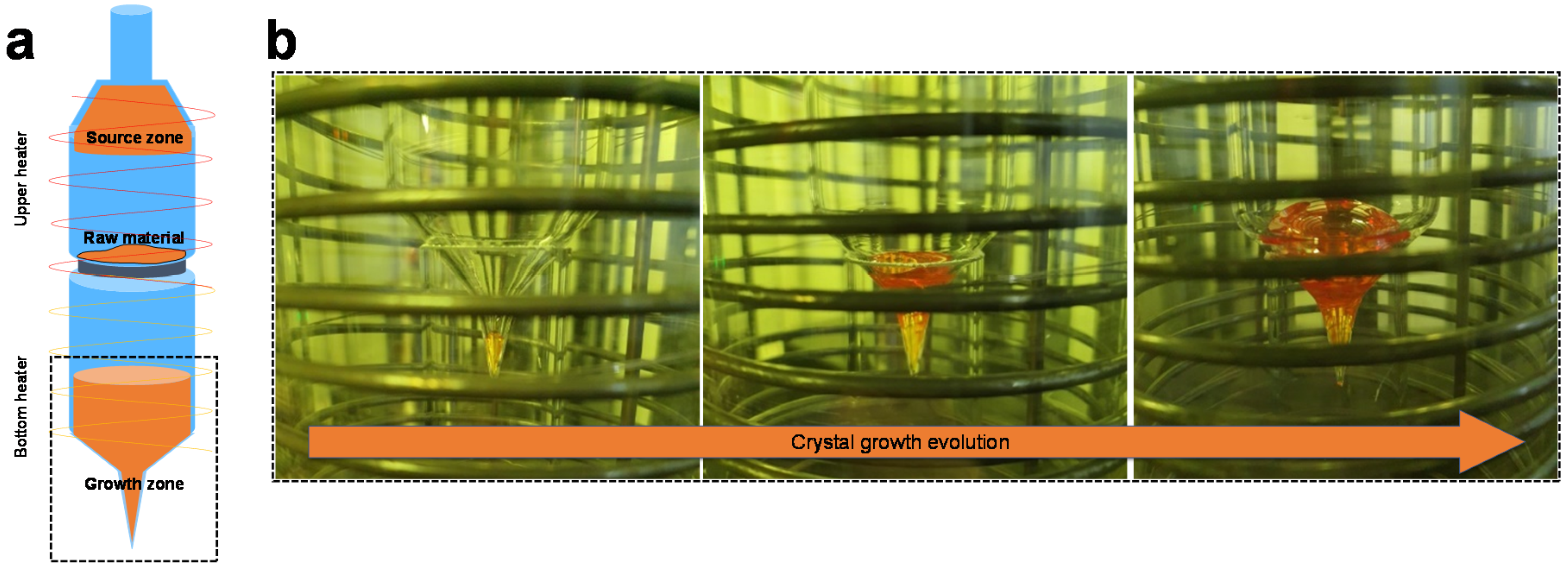
The PVT process was selected for the synthesis of Hg2Br2 single crystals because Hg2Br2 can be easily sublimated at a low temperature. The PVT equipment designed for this experiment consisted of three parts: a unit to control the process temperature or time, a driving unit to move the ampoule, and a chamber–heater unit around the ampoule. The ampoule was divided into three zones: a collected source zone, a raw material zone, and a crystal growth zone (Figure 2a). A conical tip was located on the lower section of the growth zone, while the upper stabilization section was cylindrical. To synthesize Hg2Br2 single crystals, the prepared Hg2Br2 powder was first loaded onto a 150-μm pore size ceramic membrane filter to avoid movement of the bulk powder to the bottom growing zone of the ampoule. The Hg2Br2 powder that sublimated from the raw material zone was collected at the top of the ampoule (source zone). Seeds were formed at the end of the ampoule tip and crystal growth evolved as depicted in Figure 2b. The crystals grew at a rate of 0.2 to 5 mm/day. The cooling process was also performed slowly to prevent serious thermal cracking. The resulting crystals were ~35 mm in diameter and were 70–80 mm in length. Kim et al. previously reported that Hg2Br2 nucleation is governed by the temperature gradient between the upper and lower regions of the ampoule [11]. Accordingly, a supersaturation area that critically affected the seed formation was identified by optimizing the temperature profile.
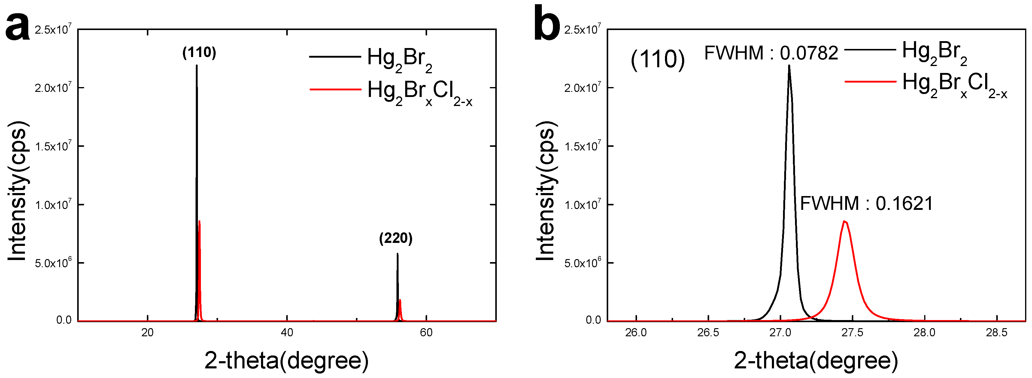
As shown in Figure 3a, the as-grown Hg2Br2 and Hg2BrxCl2−x crystals exhibited a primary peak in the (110) plane and another short peak in its family (220) plane [12]. The 2-theta positions of the major planes in Hg2Br2 and Hg2BrxCl2−x were 27.25° and 27.45°, respectively (Figure 3b). The full width at half maximum (FWHM) values of the (110) planes in the Hg2Br2 and Hg2BrxCl2−x crystals were 0.0782 and 0.1621, respectively. FWHM values reflect the amount of lattice distortion, and it is highly likely that a crystal with nonuniformity in lattice strain has a comparatively high FWHM value. Consequently, because the FWHM value of the Hg2Br2 crystal was lower than that of Hg2BrxCl2−x, it could be assumed that the former was a crystal without lattice distortions. Furthermore, the Hg2Br2 crystal showed higher peak intensities across all planes, which implied better crystallinity compared to the Hg2BrxCl2−x crystal. Therefore, the crystallinity of the Hg2Br2 crystal is better than that of the Hg2BrxCl2−x crystal. Lattice mismatching in the Hg2BrxCl2−x crystal was unavoidable owing to the large discrepancy between the Hg-Cl (2.27 Å) and Hg–Br (2.64 Å) lattice distances [13,14].
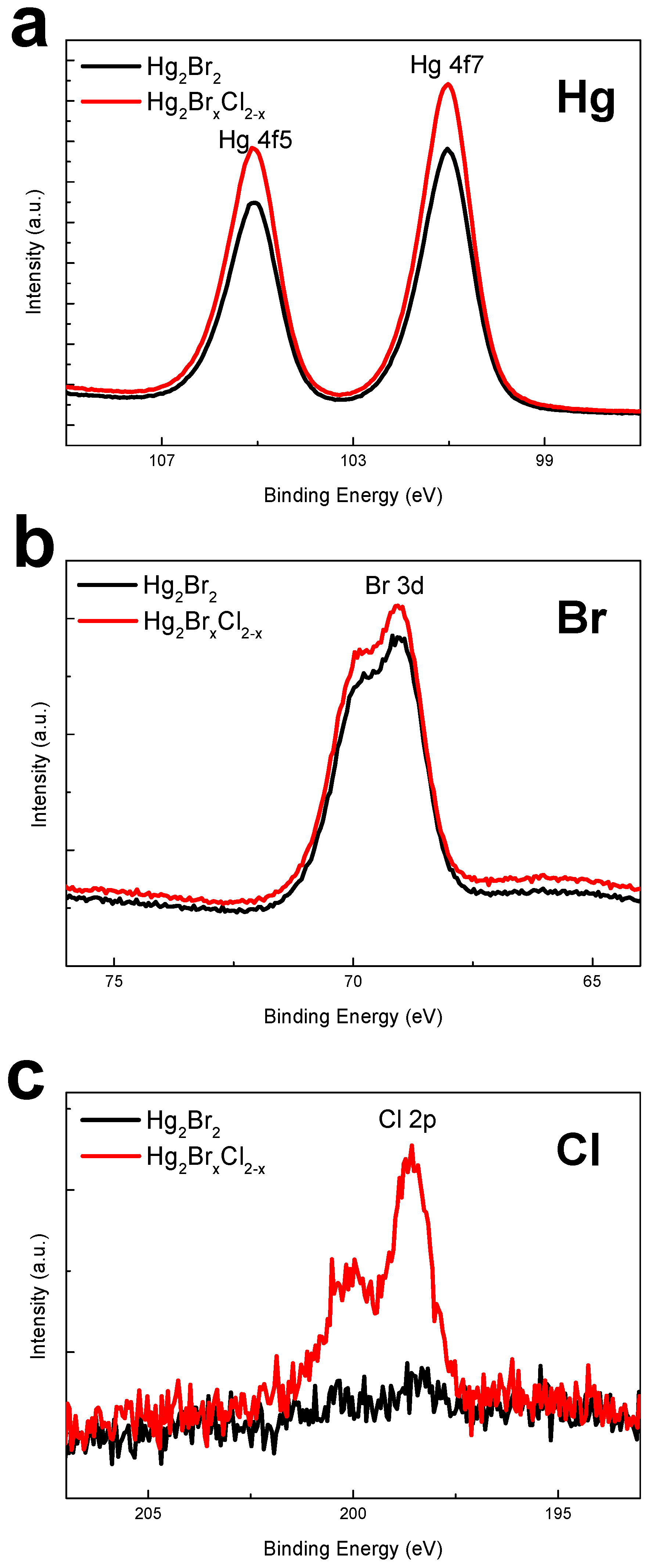
After etching the surface to prevent possible carbon contamination, XPS analysis was conducted to check the state of the chemical compounds. Figure 4a,b demonstrated that although there was a small disparity between the intensities of the two crystals, the positions of the peaks in the Hg 4f5 and Hg 4f7 spectra were almost the same. The binding energies of the Hg 4f7 electrons in Hg2Br2 and Hg2BrxCl2−x crystals were 101.03 and 101.02 eV, respectively. Similarly, the binding energies of the Hg 4f5 electrons in the two crystals were 105.08 and 105.12 eV, respectively. The Br 3d peaks of the two crystals also had approximately the same positions (69.08 and 69.12 eV, respectively). Moreover, these results were consistent with the XPS results obtained in a previous study for purified Hg2Br2 powder [15,16]. Nevertheless, the peaks in the Cl 2p spectra of the two crystals were different (Figure 4c). No clear peaks were observed for the Hg2Br2 crystal, whereas for the Hg2BrxCl2−x crystal, an initial peak was observed at a binding energy of 198.57 eV. Studies have reported that when a Hg2Br2 crystal contained Cl atoms, the Cl peak split into two peaks, Cl 2p3/2 and Cl 2p1/2 [17]. Using stoichiometry calculations, we concluded that Hg, Br, and Cl atoms in the Hg2Br2 and Hg2BrxCl2−x crystals were distributed in ratios of 2:2.19:0 and 2:1.88:0.28, respectively.
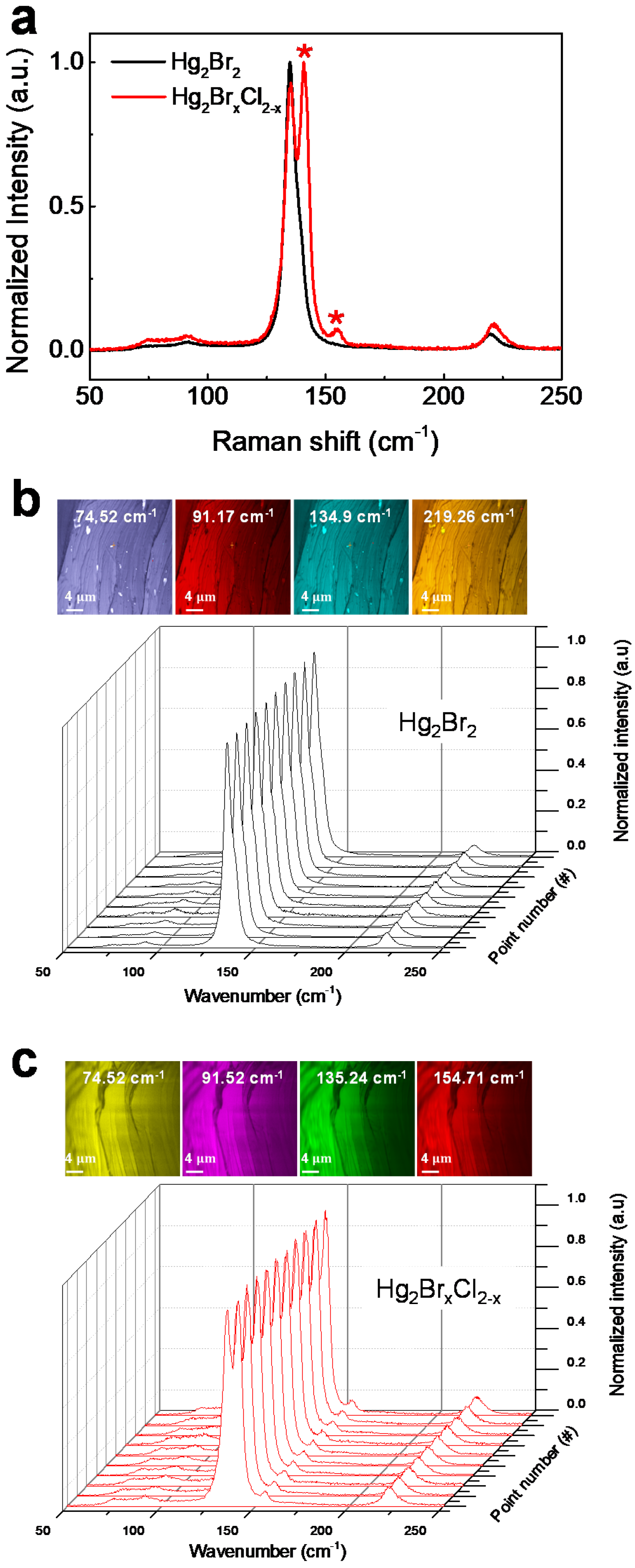
Raman spectroscopy was used to evaluate and compare the atomic vibration spectra of the two crystals (Figure 5). Group theory was applied to the selection rules for the vibrational spectra of Hg2Br2, and the analysis showed that four modes (35.5, 91, 136, and 221 cm−1) must be active in the Raman spectrum: two doubly degenerate peaks (Eg of 35.5 and 91 cm−1) and two fully symmetric peaks (A1g of 136 and 221 cm−1) [18,19,20]. The Eg symmetry was related to the rocking of the linear molecule and the zigzag bending vibrations. The A1g symmetry corresponded to the Hg–Hg and Br–Hg stretching vibrations. As illustrated in Figure 5a, both crystals clearly exhibited peaks related to the vibration mode of Hg2Br2. Nonetheless, the first vibration peak at 35.5 cm−1 was not detectable because of the measurement limit of the Raman spectroscopy equipment. The most noticeable difference between the vibration modes of the two crystals was that the Hg-Cl-related additional peaks (140.7 and 154.7 cm−1) were detected only in the Hg2BrxCl2−x crystal. This was almost the same as the position of the vibration peak of Hg2Cl2 reported in a previous study [21]. Figure 5b,c display the Raman mapping images and the 10-point data from the (110) plane of Hg2Br2 and Hg2BrxCl2-x crystals. The uniformity of the vibration peaks was validated by mapping the crystal.

4. Conclusions

In conclusion, we comparatively investigated crystals grown from different mercury halide powders (Hg2Br2 and Hg2BrxCl2−x). After purification of the raw powders, SEM/EDS analysis clearly confirmed the presence of Cl atoms (2.62%) in the Hg2BrxCl2−x powder, whereas no Cl atoms were found in the Hg2Br2 powder. The XRD results revealed that there was a considerable decrease in crystallinity of Hg2Br2 crystals by the introduction of Cl atom, which was due to the disparity between the Hg–Br and Hg-Cl lattice distances. The binding energy related to Cl atoms was measured through XPS. The presence of the Hg-Cl bonds triggered additional vibrational peaks in the crystal spectra of Hg2Br2. Therefore, this crystal characterization study can contribute to the development of a feasible technique for detecting unwanted elements in mercury halide crystals.

Author Contributions

K.K. and S.-G.W. designed and carried out the experiment for crystal growth. O.K., G.-E.J. and B.C. characterized the crystals. O.K. and B.C. wrote the paper. All authors have read and agreed to the published version of the manuscript.

Funding

This study was supported by the Civil-Military Technology Development Program of the Institute of Civil-Military Technology Cooperation funded by the Ministry of Trade, Industry and Energy (MOTIE) and the Defense Acquisition Program Administration (17-CM-DP-02). This research was also financially supported by MOTIE and the Korea Institute for Advancement of Technology (KIAT) through the National Innovation Cluster R & D program (P0006704–Development of energy-saving advanced parts).

Acknowledgments

The authors would like to thank Tae Hyeon Kim, Hee Tae Lee, and In Hoi Kwon.

Conflicts of Interest

The authors declare that there are no known conflict of interest in relation to the submitted work.

References

  1. Gupta, N. Acousto-optic tunable filters for infrared imaging. In Proceedings of the the Acousto-Optics and Photoacoustics, Warsaw, Poland, 30 August–2 September 2005; Volume 5953. [Google Scholar]
  2. Amarasinghe, P.M.; Kim, J.S.; Chen, H.; Trivedi, S.; Qadri, S.B.; Soos, J.; Diestler, M.; Zhang, D.; Gupta, N.; Jensen, J.L.; et al. Growth of high quality mercurous halide single crystals by physical vapor transport method for AOM and radiation detection applications. J. Cryst. Growth 2016, 450, 96. [Google Scholar] [CrossRef]
  3. Gupta, N.; Dahmani, R.; Choy, S. Acousto-optic tunable filter based visible- to near-infrared spectropolarimetric imager. Opt. Eng. 2002, 41, 1033–1038. [Google Scholar] [CrossRef]
  4. Kim, J.; Trivedi, S.B.; Soos, J.; Gupta, N.; Palosz, W. Development of mercurous halide crystals for acousto-optic devices. In Proceedings of the Imaging Spectrometry XII, San Diego, CA, USA, 28–29 August 2007; Volume 6661. [Google Scholar]
  5. Harris, S.E.; Wallace, R.W. Acousto-optic tunable filter. J. Opt. Soc. Am. 1969, 59, 744. [Google Scholar] [CrossRef]
  6. Chang, I.C. Tunable acousto-optic filters: An overview. In Proceedings of the Acousto-Optics, San Diego, CA, USA, 26–27 August 1976; Volume 90, pp. 12–22. [Google Scholar]
  7. Gupta, N.; Voloshinov, V. Hyperspectral imaging performance of a TeO2 acousto-optic tunable filter in the ultraviolet region. Opt. Soc. Am. 2005, 30, 985. [Google Scholar]
  8. Xu, J.; Stroud, R. Acousto-Optic Devices: Princioles, Design and Applications, 1st ed.; Wiley-Interscience: New York, NY, USA, 1992; Volume 12. [Google Scholar]
  9. Kim, J.; Trivedi, S.B.; Soos, J.; Gupta, N.; Palosz, W. Growth of Hg2Cl2 and Hg2Br2 single crystals by physical vapor transport. J. Cryst. Growth 2008, 310, 2457–2463. [Google Scholar] [CrossRef]
  10. Kim, T.H.; Lee, H.T.; Kwon, I.H.; Kang, Y.; Woo, S.; Jang, G.; Cho, B. High Purification of Hg2Br2 Powder for Acousto-Optic Tunable Filters Utilizing a PVT Process. Korean J. Mater. Res. 2018, 28, 732–737. [Google Scholar] [CrossRef]
  11. Kim, T.H.; Lee, H.T.; Kang, Y.M.; Jang, G.E.; Kwon, I.H.; Cho, B. In-depth Investigation of Hg2Br2 Crystal Growth and Evolution. Materials 2019, 12, 4224. [Google Scholar] [CrossRef] [Green Version]
  12. Thirsk, H.R. The structure and orientation of calomel formed on liquid mercury by anodic polarization. Proc. Phys. Soc. Sect. B 1953, 66, 129. [Google Scholar] [CrossRef]
  13. Maxwell, L.R.; Mosley, V.M. Internuclear Distances in Se2, Te2, and HgCl by Electron Diffraction. Phys. Rev. 1940, 57, 21. [Google Scholar] [CrossRef]
  14. Chung, C. Crystal structure of mercury(II) bromide dithiocarbamate, [HgBr2(S2CNEt2)Hg(S2CNEt2)]n, an inorganic polymer. Can. J. Chem. 1978, 56, 564–566. [Google Scholar]
  15. Ding, T.; Zhang, J.; Hong, J.; Zhu, J.; Chen, H. Sonochemical synthesis of taper shaped HgSe nanorods in polyol solvent. J. Cryst. Growth 2004, 260, 527. [Google Scholar] [CrossRef]
  16. Rahane, S.B.; Hensarling, R.M.; Sparks, B.J.; Stafford, M.; Patton, D.L. Synthesis of multifunctional polymer brush surfaces via sequential and orthogonal thiol-click reactions. J. Mater. Chem. 2012, 22, 932. [Google Scholar] [CrossRef]
  17. Li, B.; Zhou, L.; Wu, D.; Peng, H.; Yan, K.; Zhou, Y.; Liu, Z. Photochemical Chlorination of Graphene. ACS Nano 2011, 5, 5957–5961. [Google Scholar] [CrossRef] [PubMed]
  18. Roginskii, E.M.; Krylov, A.S.; Markov, Y.F. Lattice dynamics and baric behavior of phonons in the Hg2Br2 model ferroelastics. Phys. Solid State 2019, 61, 325. [Google Scholar] [CrossRef]
  19. Barta, C.; Kaplyanskii, A.A.; Markov, Y.F. The Raman scattering spectra of the Hg2Cl2 and Hg2Br2 single crystals. Sov. Phys. Solid State 1973, 15, 1896. [Google Scholar]
  20. Barta, C.; Kaplyanskii, A.A.; Kulakov, V.V.; Markov, Y.F. Raman-spectra of I and II orders of Hg2Cl2, Hg2Br2, Hg2J2 single-crystals. Opt. Spectrosk. 1974, 37, 95. [Google Scholar]
  21. Papatheodorou, G.N.; Boviatsis, I.V.; Voyiatzis, G.A. In situ Raman spectra of electrode products during electrolysis of HgCl2 in molten LiCl-KCl eutectic. J. Appl. Electrochem. 1992, 22, 517–521. [Google Scholar] [CrossRef]
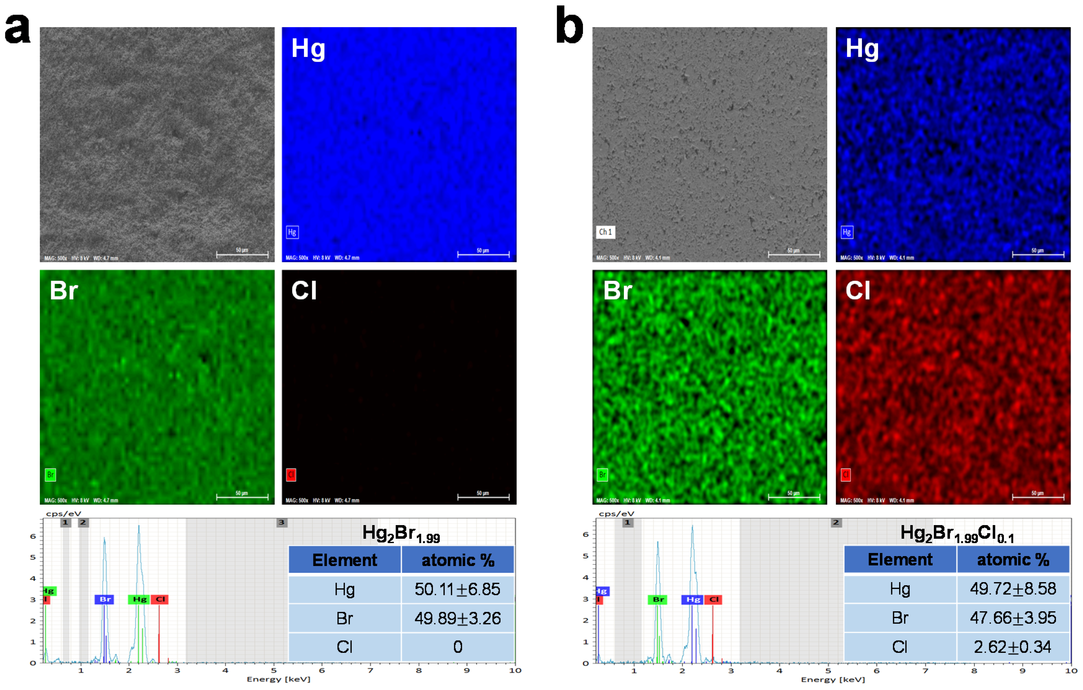
Figure 1. The scanning electron microscopy/energy-dispersive spectroscopy elemental maps and spectrum analysis of (a) Hg2Br2 and (b) Hg2BrxCl2−x.
Crystals 10 01096 g001
Figure 2. (a) Schematic of the physical vapor transport system designed for Hg2Br2 crystal growth. (b) Crystal growth evolution process in quartz ampoule.
Crystals 10 01096 g002
Figure 3. (a) Comparison of the X-ray diffraction (XRD) spectra of Hg2Br2 and Hg2BrxCl2−x crystals. (b) Full width at half maximum (FWHM) of Hg2Br2 and Hg2BrxCl2−x crystals at (110) peak.
Crystals 10 01096 g003
Figure 4. Narrow-scan X-ray photoelectron spectra of (a) Hg 4f, (b) Br 3d, and (c) Cl 2p for Hg2Br2 and Hg2BrxCl2−x crystals.
Crystals 10 01096 g004
Figure 5. (a) Comparison of single point Raman spectra of Hg2Br2 and Hg2BrxCl2−x crystals. The major peaks distinguished from Hg2Br2 peaks are marked with asterisks. Raman mapping images and data of (b) Hg2Br2 and (c) Hg2BrxCl2−x with (110) crystal surface plane.
Crystals 10 01096 g005
Publisher’s Note: MDPI stays neutral with regard to jurisdictional claims in published maps and institutional affiliations.