Article

Multifunctional Electrochemical Properties of Synthesized Non-Precious Iron Oxide Nanostructures

1
Nanochemistry Laboratory, Department of Chemistry, Jamia Millia Islamia, New Delhi 110025, India
2
King Abdullah Institute for Nanotechnology, King Saud University, Riyadh 11451, Saudi Arabia
3
Enzyme Technology Laboratory, Department of Biosciences, Jamia Millia Islamia, New Delhi 110025, India
4
Department of Chemistry, College of Science, King Saud University, Riyadh 11451, Saudi Arabia
5
School of Science and Technology, Glocal University, Saharanapur UP-247001, India
*
Authors to whom correspondence should be addressed.
Crystals 2020, 10(9), 751; https://doi.org/10.3390/cryst10090751
Submission received: 17 July 2020 / Revised: 13 August 2020 / Accepted: 25 August 2020 / Published: 26 August 2020
(This article belongs to the Special Issue Nanomaterials for Energy and Recycling)

Abstract

:
Magnetic Fe3O4 nanostructures for electrochemical water splitting and supercapacitor applications were synthesized by low temperature simple wet-chemical route. The crystal structure and morphology of as-acquired nanostructures were examined by powder X-ray diffraction and transmission electron microscopy. Magnetic measurements indicate that the as-synthesized Fe3O4 nanostructures are ferromagnetic at room temperature. The synthesized nanostructures have a high-specific surface area of 268 m2/g, which affects the electrocatalytic activity of the electrode materials. The purity of the as-synthesized nanostructures was affirmed by Raman and X-ray Photoelectron studies. The electrochemical activity of the magnetic iron oxide nanoparticles (MIONPs) for the hydrogen evolution reaction (HER) and supercapacitors were investigated in alkaline medium (0.5 M KOH) versus Ag/AgCl at room temperature. The electrocatalysts show low onset potential (~0.18 V) and Tafel slope (~440 mV/dec) for HER. Additionally, the specific capacitance of MIONPs was investigated, which is to be ~135 ± 5 F/g at 5 mV/s in 1 M KOH.

1. Introduction

Water splitting or electrolysis of water is receiving much attention from researchers due to the diminution of fossil fuels and the increased environmental hazards on account of generating various type of by-products such as dust, smoke, soot, CO2, etc. [1]. The generation of CO2 affects the world in the form of global warming, which strikes our lives through rising sea levels due to polar ice cap melt, desertification, and reduced agricultural yields [2,3]. It is necessary to lookup for ample, everlasting, eco-friendly, CO2-free, and sustainable alternative energy sources [4,5]. Several alternative sources of fuels have already been proposed such as biodiesel, methanol, ethanol, hydrogen, boron, natural gas, liquefied petroleum gas (LPG), electricity, solar fuels, etc. One of these alternative fuels, hydrogen has been believed as a perfect alternative source of fuel, since it has high specific energy, is carbon-neutral, and is the most abundant element in the universe [6,7]. Today 90% of all the H2 gas has been produced by hydrocarbon reforming, in which hydrocarbons are catalytically oxidized on metal surfaces (Pt, Pd, etc.) at elevated temperatures and results into a mixture of carbon monoxide, carbon dioxide, and hydrogen gas [8,9].
In the present study, we have described electrochemical water splitting to produce H2 using nanoparticles. Magnetite nanoparticles (Fe3O4) attract much attention as they can be used in magnetic storage [10], targeted drug delivery [11], hyperthermia treatments [12], Magnetic resonance imaging (MRI) contrast agents [13], and energy storage systems [14] etc. Supercapacitors are the most promising candidates for electrical storage systems since they have a long cycle life (>100,000 cycles), high power density, simple principle, and fast charge/discharge rate. There are two categories of supercapacitors: electric double-layer capacitors (EDLCs) viz. graphene [15], activated carbon [16], carbon nanotubes [17], etc., as electrode materials and pseudocapacitors such as conducting polymers and metal oxides [18,19,20]. The electrostatic charge accumulation at the electrode-electrolyte interface is responsible for charge storage in EDLCs, whereas the redox reaction in the electrode is accountable for charge storage in pseudocapacitors. The Fe3O4 is considered as electrode material for pseudocapacitors, on account of its natural abundance, low toxicity, low cost, high theoretical specific capacity (2299 F/g) [21], large potential window (−1.2 to 0.25 V), high conductivity (σ = 2 × 10⁴ Sm−1), etc. Since iron oxide nanoparticles could be easily separated from the reaction media by a simple magnetic separation; therefore, it can be used as a promising material for catalytic reactions.
Transition metal oxides have garnered attention for water splitting due to their fascinating electrocatalytic activity, low-cost, low toxicity, and earth abundance. They show excellent oxygen evolution reaction (OER) potential, but due to low hydrogen desorption ability, their HER reactivity is poor [22,23]. Transition metal oxides have good oxygen evolution reaction (OER) and oxygen reduction reaction (ORR) performance within single material in a synergistic effect owing to the ability to mix. Currently, scientists are working to develop efficient metal oxide-based catalysts for HER with high stability, low-toxicity, and fast reaction kinetics with desirable efficiencies. The efficiency of the catalytic materials could be tuned by combining them with other materials such as graphene, carbon nanotubes, organometallic frameworks, etc. In 1977, Nakamura [24] used iron oxide for thermochemical water splitting. Afterward, scientists have studied several metal oxide materials for water splitting [25,26,27,28,29]. At present, there is no report regarding the electrochemical water splitting for HER using MIONPs.
Numerous chemical methods have been reported in the literature for the synthesis of various metal-oxide nanoparticles such as reverse micelle [30,31], hydrothermal [32], solvothermal [33,34], sol-gel [35], co-precipitation [36], and polymeric precursor [37,38,39] methods. Luo et al. synthesized core-shell nano-heterostructures of Fe3O4@NiFexOy at low temperature, which showed utmost electrocatalytic performance for OER in carbonate electrolyte [40]. Zhang et al. constructed the phosphorus-doped inverse spinel Fe3O4 electrocatalyst on iron foam (P-Fe3O4/IF) for alkaline H2 production. The synthesized electrocatalyst showed noteworthy performance at a very low overpotential of 1.38 V at 100 mA/cm2 [41]. Further, phosphorus-doped Fe3O4 nanoflowers were developed on a 3D graphene (P-Fe3O4@3DG) substrate using hydrothermal and low-temperature phosphating reaction. The P-Fe3O4@3DG composite possessed exceptional performance for HER at a low overpotential of 1.23 V at 10 mA/cm2, prominent stability up to 50 h and Tafel slope value of 65 mV/dec. The synthesized electrocatalyst exhibited excellent performance in a wide range of pH [42]. Herein, we report the wet-chemical synthesis of magnetic Fe3O4 nanoparticles for clean energy. The three-electrode electrochemical work station has been used to investigate the catalytic performance of MIONPs for HER. Additionally, the supercapacitive behavior of MIONPs has also been studied. The as-synthesized nanoparticles were successfully investigated by powder X-ray diffraction (PXRD), high-resolution transmission electron microscopy (HRTEM), BET surface area analysis, magnetization study, Raman, and X-ray photoelectron spectroscopy.

2. Materials and Methods

2.1. Synthesis of Magnetic Iron Oxide Nanoparticles

Iron (II) sulfate heptahydrate was purchased from Fisher Scientific, (98% purity), sodium hydroxide pellets, and sodium nitrate were bought from Merck Ltd. (99% purity). All the chemicals were used as received without further purification.
Magnetic iron oxide nanoparticles (MIONPs) were synthesized by a simple wet-chemical route as per the literature [43]. In a typical procedure, 3 mmol of Fe2+ salt was dissolved in 120 mL distilled water, and the solution was stirred till the temperature of 80 °C was reached. Now, 30 mmol of NaOH and 3 mmol of NaNO3 were dissolved in 30 mL of distilled water and added dropwise to the above solution with constant stirring. The mixture was refluxed for 2 h at 80 °C forming black precipitates. The precipitates were collected by a permanent magnet and washed several times with double distilled water till the pH~7. The black precipitates were dried at 100 °C in an open-air oven for 12 h to obtain the pure Fe3O4 phase of MIONPs.

2.2. Physical Characterization

Rigaku Ultima IV X-ray diffractometer with Ni-filtered Cu-Kα radiation (λ = 1.5416 Å) was used to determine the crystalline structure of MIONPs. The diffraction pattern was collected at a step size of 0.05° and a step time of 1 s and 2θ range from 20° to 70°. Transmission electron microscopic (TEM) analysis was carried out on an FEI Technai G2 20 HRTEM with an accelerating voltage of 200 kV. TEM samples were prepared by putting a drop of dispersed sample on a carbon-coated copper grid and dried in air for 1 h. The surface area and the pore size of as-synthesized iron oxide nanoparticles were determined using BET surface area analyzer (Model: Nova 2000e, Quantachrome Instruments Limited, USA) at liquid nitrogen temperature (77 K). Before analysis, the samples were degassed at 150 °C for 3 h in a vacuum degassing mode to remove the surface contaminants such as water vapor and adsorbed gases. The specific surface area was calculated using the multipoint BET equation. The pore size distributions were determined from the N2 desorption isotherms at 77 K using the Dubinin–Astakhov (DA) method. The Raman spectrum was recorded on a Renishaw Raman spectrometer with an Ar-laser source (488 nm) at ambient conditions. The X-ray photoelectron spectroscopy (XPS) scans were carried out to confirm the oxidation state of the MIONPs using Kratos-Axis-Ultra DLD (Chestnut Ridge, NY, USA) spectrometer with Al Kα X-ray source. The magnetization study has been performed using a magnetic property measurement system Vibrating Sample Magnetometer (VSM 7300, Lakeshore, Carson, CA, USA) under an external magnetic field of ± 120 kOe at 300 K.

2.3. Electrochemical Measurements

The cyclic voltammetry (CV), linear sweep voltammetry (LSV), chrono-amperometry (CA), and Tafel studies were carried out with a three electrode electrochemical work station (potentiostat/galvanostat, CHI 660E, China) at room temperature in alkaline medium (0.5 M KOH) to investigate the redox behavior of the as-synthesized nanoparticles via electrolysis of water. Pt wire was used as a counter electrode, Ag/AgCl as the reference electrode, and glassy carbon as the working electrode. The working electrodes were prepared by putting a drop of slurry (containing 2.5 mg of catalysts to 0.5 mL of isopropanol with 0.1 mL of Nafion solution followed by the sonication for 10 min) onto the glassy carbon and then dried at 60 °C in a vacuum oven [44]. The loaded amount of the nanoparticles was of ~0.24 mg/cm2 on the GC electrode, and the area of the working electrode was 0.07 cm2. Freshly prepared electrodes were used for the electrochemical measurements. The CV of the iron oxide nanoparticles for HER was investigated in a peak window from 0 to −1.4 V versus Ag/AgCl at the various scan rates (i.e., 25, 50, 100 mV/s). The LSV and Tafel measurements for electrolysis of water were also carried out at 25 mV/s and 10 mV/s, respectively, in 0.5 M KOH vs. Ag/AgCl. The stability and activity of the iron oxide electrodes were also tested by CA measurements for HER at the fixed potential (i.e., −1.4 V vs. Ag/AgCl) for 20 s in 0.5 M KOH. The supercapacitive performance was also investigated in the potential window from −0.3 to +0.5 V at the scan rate of 5–100 mV/s in 1 M KOH. The electrode for specific capacitance study was prepared by pasting the slurry (MIONPs: Carbon black: PVDF of 75:15:10) onto the surface of nickel foam (1 cm2). The loaded amount of the sample was 1 mg. Charge-discharge (CD) studies of MIONPs were carried out in the potential window of 550 mV for 50 segments.

3. Results and Discussion

3.1. X-ray Diffraction Studies

Powder X-ray diffraction pattern of as-synthesized nanoparticles confided the presences of the magnetite phase (Figure 1). The diffraction peaks indexed as planes (220), (311), (222), (400), (331), (422), (333), and (440) correspond to a cubic unit cell with lattice parameter a = 8.394, the most potent reflection at the (311) plane is also the characteristic of cubic phase which confirms that the structure obtained for magnetite nanoparticles is an inverse spinel-type oxide (Space group: Fd3m) [45]. All the peaks could be indexed in pure cubic phase Fe3O4 and are in good agreement with standard magnetite, JCPDS No. 079-0417.

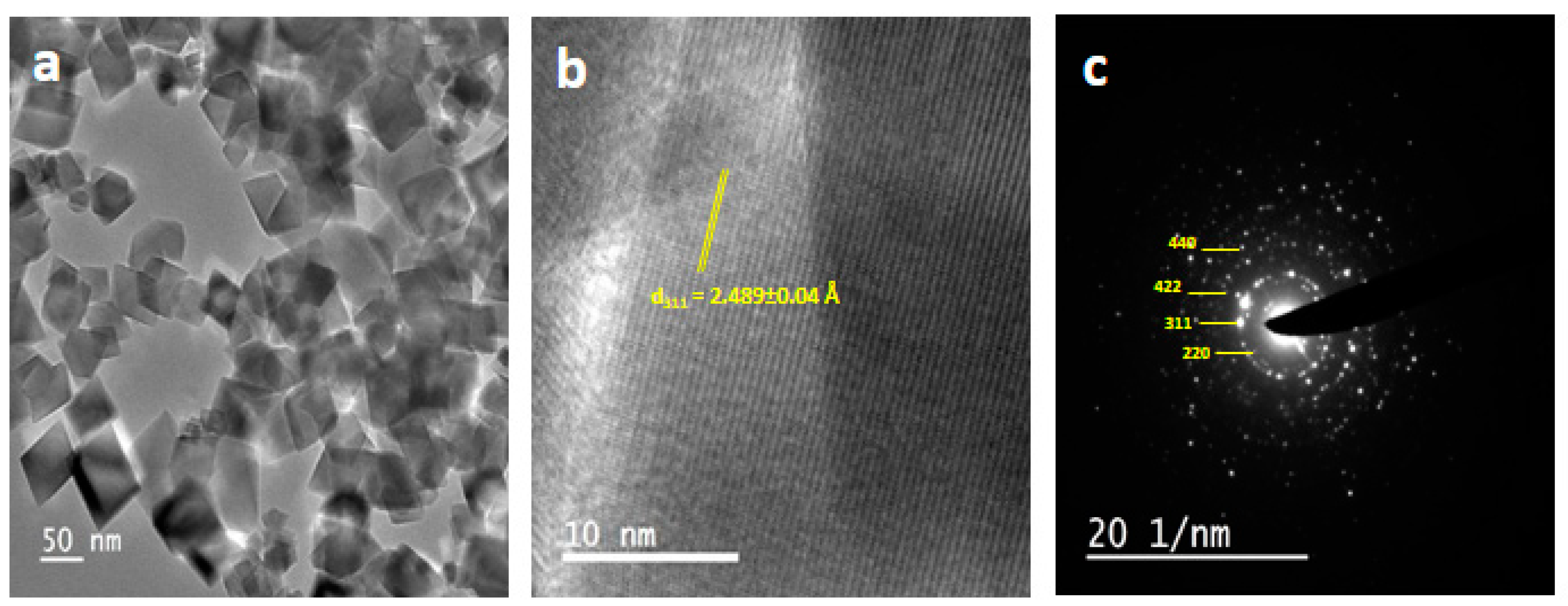
3.2. Electron Microscopic Studies

The morphological study was carried out by transmission electron microscopy (TEM) to recognize the size and morphology of MIONPs. The TEM image of MIONPs reveals that the nanoparticles are monodispersed and cube-shaped, as also shown in Figure 2a. The average particle size was calculated using ImageJ software and found to be 42 nm. Furthermore, high-resolution transmission electron microscopy (HRTEM) analysis was done to interpret the crystallinity, phase, and growth direction of as-synthesized MIONPs. Figure 2b shows a typical HRTEM image of MIONPs which visualizes well-resolved lattice fringes with an average lattice distance of 2.489 ± 0.04 Å, which corresponds to the most intense peak (311) of MIONPs. The selected area electron diffraction (SAED) pattern of MIONPs (Figure 2c) shows sharp diffraction spots, indicating the well-developed nanocrystalline structure. The different lattice planes of MIONPs (440), (422), (311), and (220) were assigned to the diffraction rings in the SAED pattern, which are in accordance with the XRD pattern.

3.3. Magnetic Properties

The vibrating sample magnetometer (VSM) was used to investigate the magnetic behavior of Fe3O4 nanoparticles at room temperature between −10 to 10 kOe. Figure 3 clearly showed the ferromagnetic nature of the as-synthesized nanoparticles. The saturation magnetization (Ms), remanent magnetization (Mr), and coercivity (Hc) of MIONPs were 68 emu/g, 13.90 emu/g, and 100 Oe, respectively. The saturation magnetization (Ms) value for MIONPs was lower than the Ms value of the corresponding bulk Fe3O4 (92 emu/g) [46]. The difference might be attributed to a smaller size, larger surface area/volume ratio, and spin canting effects at the surface [47].

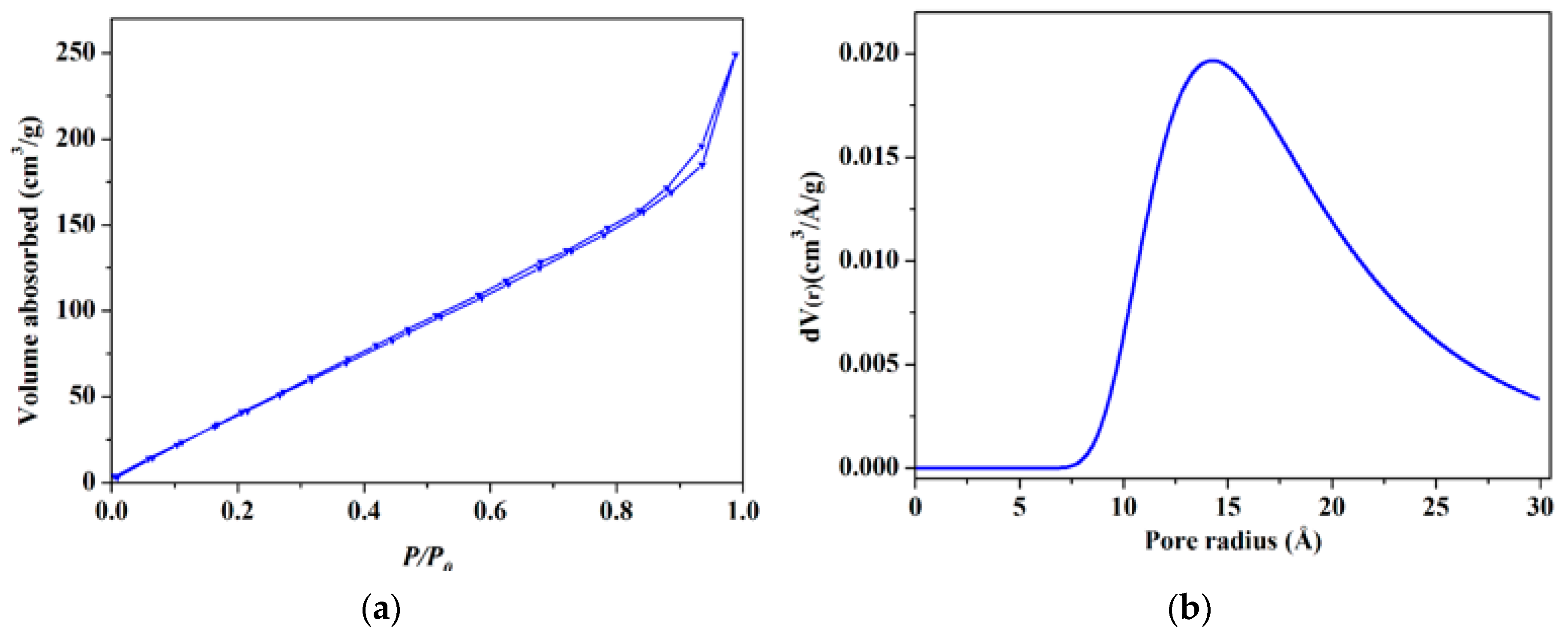
3.4. BET Surface Area Studies

The multipoint BET method was used to determine the specific surface area of as-prepared MIONPs. Figure 4a represents the nitrogen adsorption-desorption isotherm for MIONPs. The isotherm indicates that the as-synthesized nanoparticles have a porosity of type IV, which is associated with capillary condensation in mesoporous, and the isotherm shows a distinct hysteresis loop in the range of 0.7–1.0 P/P0 [48]. The specific surface area of as-synthesized nanoparticles was found to be 268 m2/g, which is comparatively higher than earlier reports [49,50]. Figure 4b shows the DA plot, which gives a pore radius of 14.8 Å for MIONPs.

3.5. Raman Studies

Figure 5 represents the Raman spectrum of MIONPs recorded in the wavelength range of 200–1000 cm−1. The spectrum shows a sharp peak at 670 cm−1, corresponding to A1g mode, depicting a symmetric stretch of oxygen atoms along with Fe–O bonds [51]. The peaks at ~515 and 535 cm−1 could be attributed to Eg and T2g modes, respectively. The T2g mode ascribes the asymmetric stretch of Fe and O atoms [52,53]. The Raman spectra confirm the absence of any other form of iron oxide, i.e., hematite or maghemite, indicating the purity of the as-synthesized nanoparticles [54].

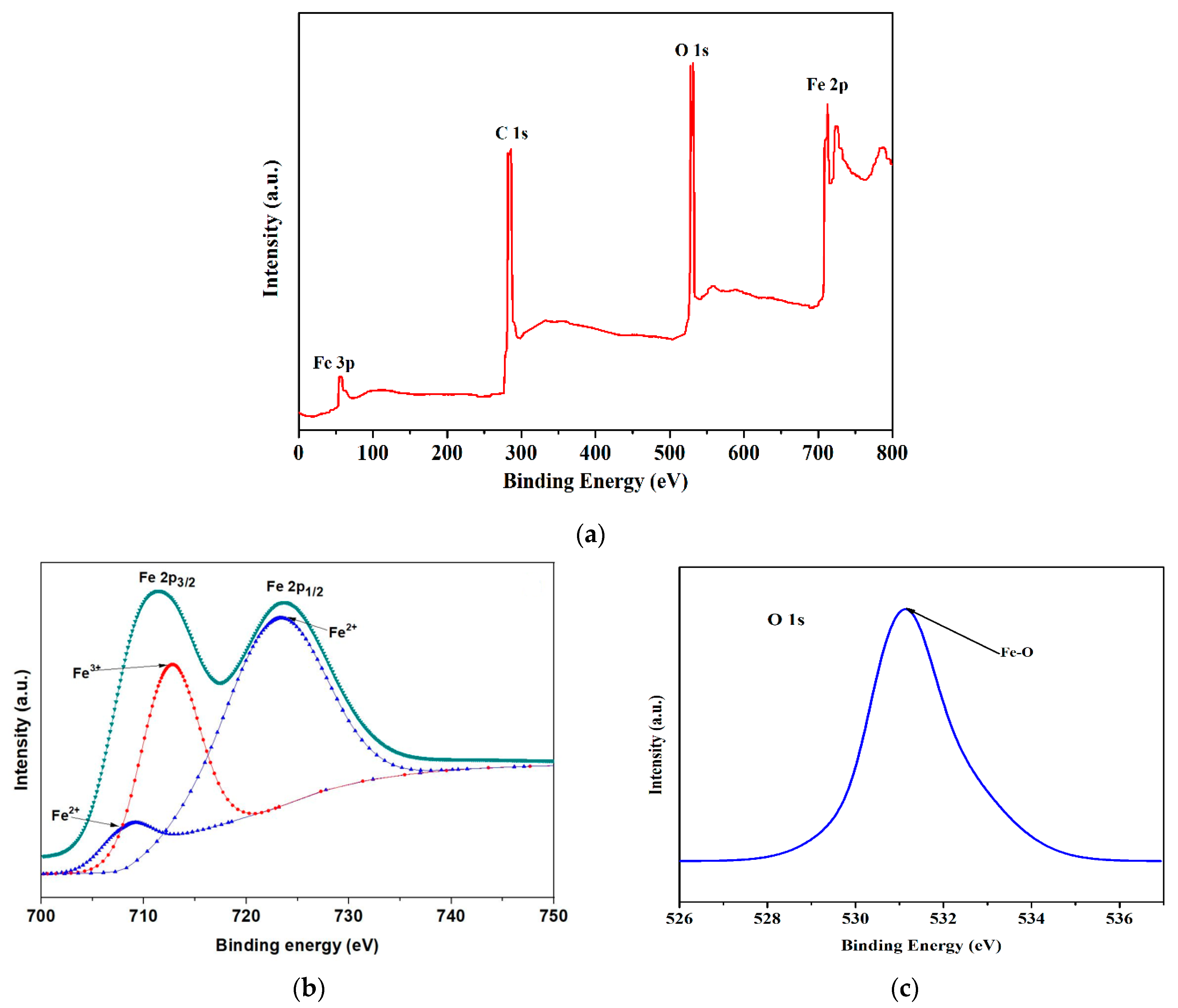
3.6. X-ray Photoelectron Spectroscopic Studies

The X-ray photoelectron spectroscopic study was carried out to determine the electronic structure and chemical composition of MIONPs. The full XPS spectrum shown in Figure 6a reveals the presence of oxygen and iron of MIONPs. The high-resolution Fe 2p spectrum (Figure 6b) shows two spin-orbit peaks at 711.3, and 723.5 eV corresponding to binding energies of Fe 2p3/2 and Fe 2p1/2 peaks of Fe3O4, respectively [53,54]. The peaks are broad due to the presence of both Fe2+ and Fe3+ ions, as Fe3O4 is the blend of FeO and Fe2O3. Further, the non-occurrence of satellite peaks at around 718 eV, which is the unique feature of the electronic structure of Fe2O3 [55], affirms the presence of pure Fe3O4. The other peaks around 708.3 and 723 eV (Fe2+) and 712.8 eV (Fe3+) could be assigned to the Fe–O bond [55]. Figure 6c shows the high-resolution O 1s spectrum with a strong peak at 531 eV, representing the Fe–O bonds. The Fe 2p and O 1s spectra are in good agreement with the standard Fe3O4 spectrum, hence confirming that the as-synthesized nanoparticles were pure Fe3O4.

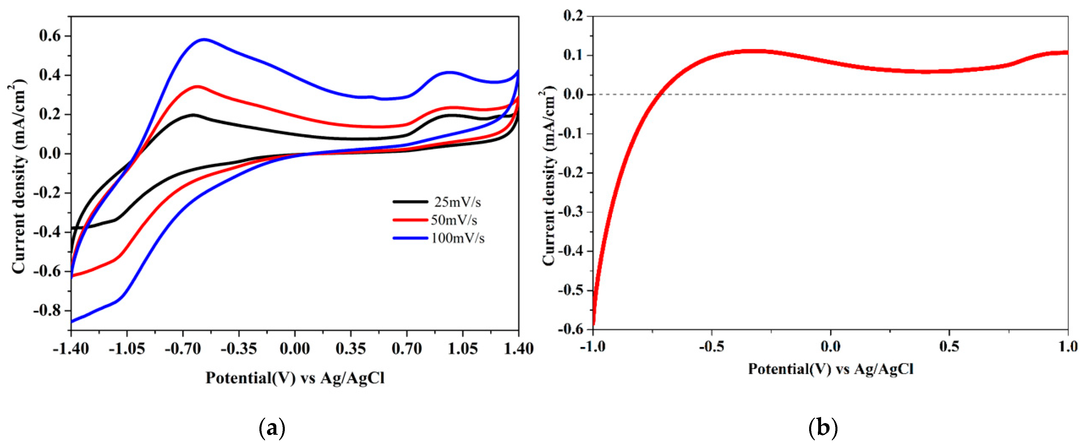
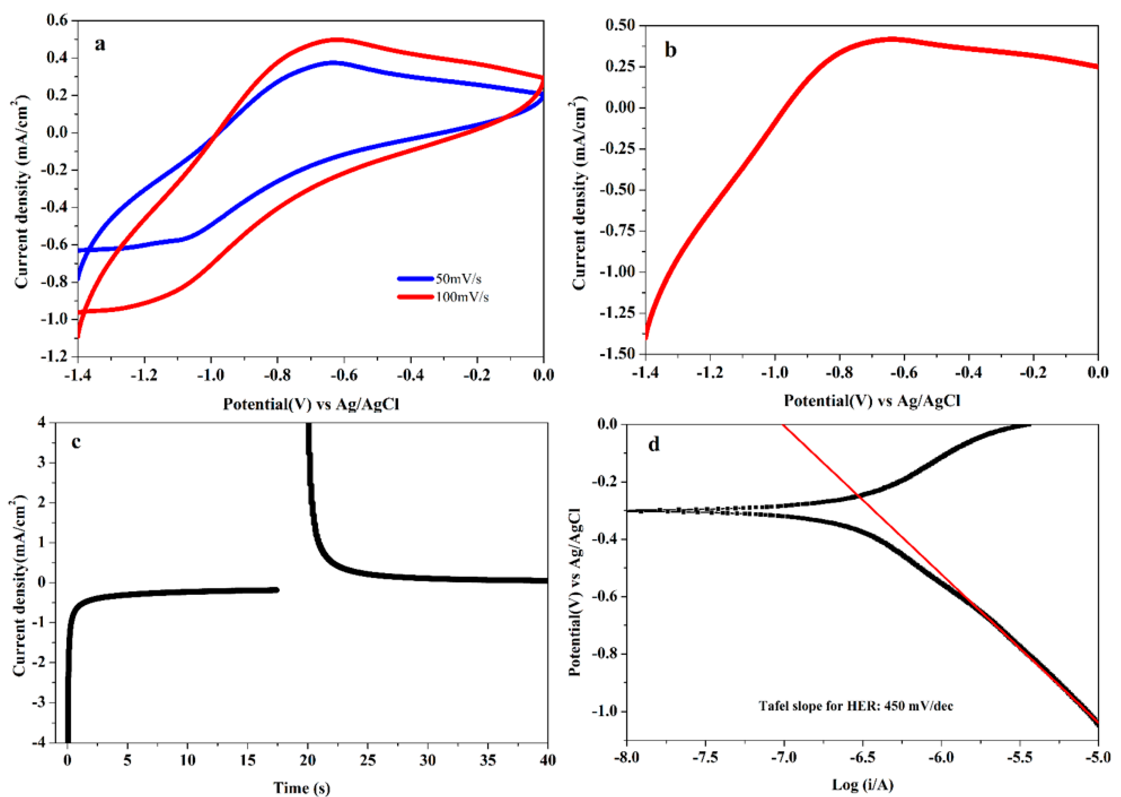
3.7. Electrocatalytic Studies

The electrocatalytic activity of as-synthesized MIONPs for HER and super-capacitive performances were conducted in 0.5 M KOH electrolyte solution. The cyclic voltammograms of MIONPs at the various scan rates (i.e., 25, 50, 100 mV/s) are depicted in Figure 7a. The redox behavior of MIONPs was observed in the applied potential window, with a redox peak at −0.65 V vs. Ag/AgCl (~1.25 V vs. RHE) ascribed to the redox reaction of iron (i.e., Fe2+/Fe3+) as reported elsewhere [56,57]. The LSV measurement was carried out to measure the current for the water-splitting reaction, with an onset potential of −0.62 V vs. Ag/AgCl (0.35 V vs. RHE) for the HER as shown in Figure 7b. The electrochemical analysis affirms that the observed cathodic faradaic steady current was efficient during the cathodic sweep (i.e., HER), while the low anodic faradaic steady current was observed toward the anodic sweep (i.e., OER). This is notable that the resulting current density (current/area of the electrode) of the electrode is directly related to the amount of H2 and O2 evolution from the water electrolysis. The electrochemical reactions for the water splitting reaction at the cathodic/anodic potentials are shown as given:
H2O(l) → 2H+ + 2e + ½ O2(g) (at anode) (OER)
2H+ + 2e → H2 (at cathode) (HER)
The catalytic activity of MIONPs towards HER was analyzed in 0.5 M KOH in the potential range from 0 to −1.4 V vs. Ag/AgCl. Figure 8a,b shows the CV and LSV profile of MIONPs for H2 generation at a scan rate of 100 mV/s. The linear sweep polarization curve reveals the generation of hydrogen at an overpotential value of ~0.75 V vs. Ag/AgCl (~0.18 V vs. RHE) is expected to achieve a low current density of 0.45 mA/cm2. The current density is directly correlated with the amount of H2 evolved. The current density of MIONPs was calculated by the current/geometric electroactive surface area of electrode, and the geometric electroactive surface area of the working electrode could be estimated from the Randles–Sevik equation [58]. The resulting current density of MIONPs depends on the size, surface area, morphology, and orientation of electrocatalysts [59,60]. The MIONPs exhibits high surface area with strong dipole interaction within the Fe3O4 nanoparticles, which provides enough space to the dissemination of the electrolyte to the active sites of the electrode, enhancing the electrochemical activity. The stability and activity of MIONPs were tested by CA studies at the steady potential of −1.4 V vs. Ag/AgCl for 40 s, as shown in Figure 8c. Chronoamperometric measurements (i.e., the epotentiostatic quantitative measurements) were done to check the stability and electrocatalytic activity of the electrode materials at a fixed potential with time. Note that the electrochemical water-splitting reaction stops and the current drop drastically to zero due to the switched off the potential (Figure 8c). The mechanism of HER activity was demonstrated by the Tafel plot (Figure 8d), which is derived by the polarization curve using the Tafel equation:
η = b × log(J/Jo)
where η, b, J, and Jo corresponds to the over potential, Tafel slope, current density, and exchange current density of the electrocatalytic reaction, respectively. The Tafel slope value of the MIONPs was found to be ~440 mV/dec from the linear fitted Tafel polarization curve of MIONPs for HER. Note that the experimental error could be ± 5. The effective electro-active catalysts for water splitting could lower the Tafel slope values to sustain the high activity and stability to enhance the efficiency due to the loss of low energy during the electrochemical reactions.
Further, we also investigated the supercapacitive behavior of MIONPs at 5 mV/s scan rate in 1 M KOH with a three-electrode system. The CV curves of the MIONPs (Figure 9a) were recorded at a scan rate from 5 to 100 mV/s in the potential window from −0.3 to 0.5 V vs. Ag/AgCl. The area under the CV curves suggests efficient supercapacitive behavior of the electrode material. The change in the current response could be due to the large surface area of nanoparticles [61]. A hysteresis under the broad potential range reveals that the as-prepared material could be useful in energy storage applications. The specific capacitance of MIONPs at 5 mV/s was calculated using the following equations:
Cs = (∫I(V)dv)/(vmΔV)
The term symbols of the above equation could be identified as Cs, ∫I(V)dv, v, m, and ΔV of the specific capacitance, an integrated area under the CV curve, scan rate, mass of the catalysts, and voltage window, respectively. Figure 9b shows the specific capacitance of MIONPs from CV tests with different scan rates, and the high specific capacitance is found to be ~135 ± 5 F/g at the scan rate of 5 mV/s. The stability/durability of the MIONPs was checked by galvanostatic charge-discharge (GCD) measurements at 1 × 10⁻⁵ A in a potential window of 550 mV for 50 segments, and it was observed that the electrode material shows excellent cycling stability with consistent specific capacitance, i.e., ~95% of the specific capacitance is retained (Figure 9c). The supercapacitive performance of MIONPs could be due to good electrical conductivity and the high surface area of the material. A comparison of the specific capacitance of the material under consideration (MIONPs) with the reported literature has been tabulated in Table 1.

4. Conclusions

In summary, we have thrived, easily recoverable magnetic iron oxide nanostructures at low temperatures via a simple co-precipitation method. These magnetic nanoparticles have an inverse spinel structure, which was confirmed by PXRD. The used precursors can distinctly evolve the cubic shaped nanoparticles having a size ~42 nm. The magnetic studies confirmed the ferromagnetic nature of nanoparticles. The current report successfully demonstrated the ability of magnetic iron oxide nanoparticles for electrochemical water splitting and supercapacitor applications. The large surface area of nanoparticles, as confirmed by BET, provides the surface for water-splitting reactions. This contribution can be important for developing a versatile synthetic pathway for easily recoverable magnetic nanoparticles as well as clean energy and storage.

Author Contributions

Conceptualization, T.A.; Data curation, J.A.; Formal analysis, R.P. and M.S.; Funding acquisition, M.A.M.K.; Investigation, T.A.; Methodology, R.P.; Resources, M.A.M.K., J.A. and T.A.; Writing—original draft, R.P. and T.A.; Writing—review & editing, J.A. and T.A. All authors have read and agreed to the published version of the manuscript.

Funding

This research was funded by the MHRD-SPARC scheme of Govt. of India, grant number: SPARC/2018-2019/P843/SL and King Saud University, Riyadh, Saudi Arabia, grant number: RSP-2020/130.

Acknowledgments

T.A. thanks for Govt. of India for financial support. R.P. specially thanks to UGC, New Delhi, for the Senior Research Fellowship. The authors also acknowledge the measurement support provided through the DST PURSE program at CIF, Jamia Millia Islamia, and thank JNCASR, Bengaluru, for the electron microscopic and VSM studies. The authors extend their sincere appreciation to King Saud University, Riyadh, Saudi Arabia, for funding this research.

Conflicts of Interest

The authors declare no conflict of interest.

References

  1. Habas, S.E.; Platt, H.A.S.; van Hest, M.F.A.M.; Ginley, D.S. Low-Cost Inorganic Solar Cells: From Ink To Printed Device. Chem. Rev. 2010, 110, 6571–6594. [Google Scholar] [CrossRef]
  2. Lewis, N.S.; Nocera, D.G. Powering the planet: Chemical challenges in solar energy utilization. Proc. Natl. Acad. Sci. USA 2006, 103, 15729–15735. [Google Scholar] [CrossRef] [Green Version]
  3. Solomon, S.; Plattner, G.-K.; Knutti, R.; Friedlingstein, P. Irreversible climate change due to carbon dioxide emissions. Proc. Natl. Acad. Sci. USA 2009, 106, 1704–1709. [Google Scholar] [CrossRef] [PubMed] [Green Version]
  4. Bard, A.J.; Fox, M.A. Artificial Photosynthesis: Solar Splitting of Water to Hydrogen and Oxygen. Acc. Chem. Res. 1995, 28, 141–145. [Google Scholar] [CrossRef]
  5. Chow, J.; Kopp, R.J.; Portney, P.R. Energy Resources and Global Development. Science 2003, 302, 1528–1531. [Google Scholar] [CrossRef] [PubMed] [Green Version]
  6. Balat, M. Potential importance of hydrogen as a future solution to environmental and transportation problems. Int. J. Hydrog. Energy 2008, 33, 4013–4029. [Google Scholar] [CrossRef]
  7. Campen, A.; Mondal, K.; Wiltowski, T. Separation of hydrogen from syngas using a regenerative system. Int. J. Hydrog. Energy 2008, 33, 332–339. [Google Scholar] [CrossRef]
  8. Muradov, N.Z.; Veziroǧlu, T.N. From hydrocarbon to hydrogen–carbon to hydrogen economy. Int. J. Hydrog. Energy 2005, 30, 225–237. [Google Scholar] [CrossRef]
  9. Haryanto, A.; Fernando, S.; Murali, N.; Adhikari, S. Current Status of Hydrogen Production Techniques by Steam Reforming of Ethanol: A Review. Energy Fuels 2005, 19, 2098–2106. [Google Scholar] [CrossRef]
  10. Fried, T.; Shemer, G.; Markovich, G. Ordered Two-Dimensional Arrays of Ferrite Nanoparticles. Adv. Mater. 2001, 13, 1158–1161. [Google Scholar] [CrossRef]
  11. Kim, J.; Lee, J.E.; Lee, S.H.; Yu, J.H.; Lee, J.H.; Park, T.G.; Hyeon, T. Designed Fabrication of a Multifunctional Polymer Nanomedical Platform for Simultaneous Cancer-Targeted Imaging and Magnetically Guided Drug Delivery. Adv. Mater. 2008, 20, 478–483. [Google Scholar] [CrossRef]
  12. Lee, H.; Yu, M.K.; Park, S.; Moon, S.; Min, J.J.; Jeong, Y.Y.; Kang, H.-W.; Jon, S. Thermally Cross-Linked Superparamagnetic Iron Oxide Nanoparticles: Synthesis and Application as a Dual Imaging Probe for Cancer in Vivo. J. Am. Chem. Soc. 2007, 129, 12739–12745. [Google Scholar] [CrossRef] [PubMed]
  13. Ahmad, T.; Phul, R. Magnetic Iron Oxide Nanoparticles as Contrast Agents: Hydrothermal Synthesis, Characterization and Properties. Solid State Phenom. 2015, 232, 111–145. [Google Scholar] [CrossRef]
  14. Nithya, V.D.; Sabari Arul, N. Progress and development of Fe3O4 electrodes for supercapacitors. J. Mater. Chem. A 2016, 4, 10767–10778. [Google Scholar] [CrossRef]
  15. Zhang, X.; Zhang, H.; Li, C.; Wang, K.; Sun, X.; Ma, Y. Recent advances in porous graphene materials for supercapacitor applications. RSC Adv. 2014, 4, 45862–45884. [Google Scholar] [CrossRef]
  16. Divyashree, A.; Hegde, G. Activated carbon nanospheres derived from bio-waste materials for supercapacitor applications—A review. RSC Adv. 2015, 5, 88339–88352. [Google Scholar] [CrossRef]
  17. Pan, H.; Li, J.; Feng, Y. Carbon Nanotubes for Supercapacitor. Nanoscale Res. Lett. 2010, 5, 654. [Google Scholar] [CrossRef] [Green Version]
  18. Wang, C.; Xu, J.; Yuen, M.-F.; Zhang, J.; Li, Y.; Chen, X.; Zhang, W. Hierarchical Composite Electrodes of Nickel Oxide Nanoflake 3D Graphene for High-Performance Pseudocapacitors. Adv. Funct. Mater. 2014, 24, 6372–6380. [Google Scholar] [CrossRef]
  19. Mitchell, E.; Gupta, R.K.; Mensah-Darkwa, K.; Kumar, D.; Ramasamy, K.; Gupta, B.K.; Kahol, P. Facile synthesis and morphogenesis of superparamagnetic iron oxide nanoparticles for high-performance supercapacitor applications. New J. Chem. 2014, 38, 4344–4350. [Google Scholar] [CrossRef]
  20. Chaudhari, N.K.; Chaudhari, S.; Yu, J.-S. Cube-like α-Fe2O3 Supported on Ordered Multimodal Porous Carbon as High Performance Electrode Material for Supercapacitors. ChemSusChem 2014, 7, 3102–3111. [Google Scholar] [CrossRef] [PubMed]
  21. Chen, X.; Chen, K.; Wang, H.; Xue, D. Composition Design upon Iron Element toward Supercapacitor Electrode Materials. Mater. Focus 2015, 4, 78–80. [Google Scholar] [CrossRef]
  22. Zhang, T.; Wu, M.-Y.; Yan, D.-Y.; Mao, J.; Liu, H.; Hu, W.-B.; Du, X.-W.; Ling, T.; Qiao, S.-Z. Engineering oxygen vacancy on NiO nanorod arrays for alkaline hydrogen evolution. Nano Energy 2018, 43, 103–109. [Google Scholar] [CrossRef]
  23. Yang, L.; Zhou, H.; Qin, X.; Guo, X.; Cui, G.; Asiri, A.M.; Sun, X. Cathodic electrochemical activation of Co3O4 nanoarrays: A smart strategy to significantly boost the hydrogen evolution activity. Chem. Commun. 2018, 54, 2150–2153. [Google Scholar] [CrossRef] [PubMed]
  24. Nakamura, T. Hydrogen production from water utilizing solar heat at high temperatures. Sol. Energy 1977, 19, 467–475. [Google Scholar] [CrossRef]
  25. Scheffe, J.R.; Li, J.; Weimer, A.W. A spinel ferrite/hercynite water-splitting redox cycle. Int. J. Hydrog. Energy 2010, 35, 3333–3340. [Google Scholar] [CrossRef]
  26. Gokon, N.; Kondo, K.; Hatamachi, T.; Sato, M.; Kodama, T. Oxygen-releasing step of nickel ferrite based on Rietveld analysis for thermochemical two-step water-splitting. Int. J. Hydrog. Energy 2013, 38, 4935–4944. [Google Scholar] [CrossRef]
  27. Bader, R.; Venstrom, L.J.; Davidson, J.H.; Lipiński, W. Thermodynamic Analysis of Isothermal Redox Cycling of Ceria for Solar Fuel Production. Energy Fuels 2013, 27, 5533–5544. [Google Scholar] [CrossRef]
  28. Loutzenhiser, P.G.; Steinfeld, A. Solar syngas production from CO2 and H2O in a two-step thermochemical cycle via Zn/ZnO redox reactions: Thermodynamic cycle analysis. Int. J. Hydrog. Energy 2011, 36, 12141–12147. [Google Scholar] [CrossRef]
  29. Ahmed, J.; Poltavets, V.V.; Prakash, J.; Alshehri, S.M.; Ahamad, T. Sol-Gel synthesis, structural characterization and bifunctional catalytic activity of nanocrystalline delafossite CuGaO2 particles. J. Alloy. Compd. 2016, 688, 1157–1161. [Google Scholar] [CrossRef]
  30. Ganguli, A.K.; Ahmad, T. Nanorods of Iron Oxalate Synthesized Using Reverse Micelles: Facile Route for α-Fe2O3 and Fe3O4 Nanoparticles. J. Nanosci. Nanotechnol. 2007, 7, 2029–2035. [Google Scholar] [CrossRef]
  31. Ahmad, T.; Ganguli, A.K. Structural and Dielectric Characterization of Nanocrystalline (Ba,Pb)ZrO3 Developed by Reverse Micellar Synthesis. J. Am. Ceram. Soc. 2006, 89, 3140–3146. [Google Scholar] [CrossRef]
  32. Ganguly, A.; Ahmad, T.; Ganguli, A.K. Silica Mesostructures: Control of Pore Size and Surface Area Using a Surfactant-Templated Hydrothermal Process. Langmuir 2010, 26, 14901–14908. [Google Scholar] [CrossRef] [PubMed]
  33. Ahmad, T.; Khatoon, S.; Coolahan, K.; Lofland, S.E. Solvothermal synthesis, optical and magnetic properties of nanocrystalline Cd1−xMnxO (0.04 < x = 0.10) solid solutions. J. Alloy. Compd. 2013, 558, 117–124. [Google Scholar] [CrossRef]
  34. Khatoon, S.; Coolahan, K.; Lofland, S.E.; Ahmad, T. Optical and magnetic properties of solid solutions of In2−xMnxO3 (0.05, 0.10 and 0.15) nanoparticles. J. Alloy. Compd. 2012, 545, 162–167. [Google Scholar] [CrossRef]
  35. Sui, R.; Charpentier, P. Synthesis of Metal Oxide Nanostructures by Direct Sol–Gel Chemistry in Supercritical Fluids. Chem. Rev. 2012, 112, 3057–3082. [Google Scholar] [CrossRef] [PubMed]
  36. Ahmad, T.; Phul, R.; Khatoon, N.; Sardar, M. Antibacterial efficacy of Ocimum sanctum leaf extract-treated iron oxide nanoparticles. New J. Chem. 2017, 41, 2055–2061. [Google Scholar] [CrossRef]
  37. Ahmad, T.; Lone, I.H.; Ansari, S.G.; Ahmed, J.; Ahamad, T.; Alshehri, S.M. Multifunctional properties and applications of yttrium ferrite nanoparticles prepared by citrate precursor route. Mater. Des. 2017, 126, 331–338. [Google Scholar] [CrossRef]
  38. Ahmad, T.; Phul, R.; Alam, P.; Lone, I.H.; Shahazad, M.; Ahmed, J.; Ahamad, T.; Alshehri, S.M. Dielectric, optical and enhanced photocatalytic properties of CuCrO2 nanoparticles. RSC Adv. 2017, 7, 27549–27557. [Google Scholar] [CrossRef] [Green Version]
  39. Alduhaish, O.; Ubaidullah, M.; Al-Enizi, A.M.; Alhokbany, N.; Alshehri, S.M.; Ahmed, J. Facile Synthesis of Mesoporous α-Fe2O3@g-C3N4-NCs for Efficient Bifunctional Electrocatalytic Activity (OER/ORR). Sci. Rep. 2019, 9, 14139. [Google Scholar] [CrossRef]
  40. Luo, Z.; Martí-Sànchez, S.; Nafria, R.; Joshua, G.; de la Mata, M.; Guardia, P.; Flox, C.; Martínez-Boubeta, C.; Simeonidis, K.; Llorca, J.; et al. Fe3O4@NiFexOy Nanoparticles with Enhanced Electrocatalytic Properties for Oxygen Evolution in Carbonate Electrolyte. ACS Appl. Mater. Interfaces 2016, 8, 29461–29469. [Google Scholar] [CrossRef] [Green Version]
  41. Zhang, J.; Shang, X.; Ren, H.; Chi, J.; Fu, H.; Dong, B.; Liu, C.; Chai, Y. Modulation of Inverse Spinel Fe3O4 by Phosphorus Doping as an Industrially Promising Electrocatalyst for Hydrogen Evolution. Adv. Mater. 2019, 31, 1905107. [Google Scholar] [CrossRef] [PubMed]
  42. Li, S.; Jian, X.; Liu, J.; Guo, S.; Zhou, C.; Zhang, P.; Yang, Y.; Chen, L. Phosphorus-doped Fe3O4 nanoflowers grown on 3D porous graphene for robust pH-Universal hydrogen evolution reaction. Int. J. Hydrog. Energy 2020, 45, 4435–4443. [Google Scholar] [CrossRef]
  43. Tancredi, P.; Botasini, S.; Moscoso-Londoño, O.; Méndez, E.; Socolovsky, L. Polymer-assisted size control of water-dispersible iron oxide nanoparticles in range between 15 and 100 nm. Colloids Surf. A Physicochem. Eng. Asp. 2015, 464, 46–51. [Google Scholar] [CrossRef]
  44. Ahmed, J.; Mao, Y. Delafossite CuAlO2 Nanoparticles with Electrocatalytic Activity toward Oxygen and Hydrogen Evolution Reactions. In Nanomaterials for Sustainable Energy; American Chemical Society: Washington, DC, USA, 2015; Volume 1213, pp. 57–72. [Google Scholar]
  45. Bernal, J.D.; Dasgupta, D.R.; Mackay, A.L. The oxides and hydroxides of iron and their structural inter-relationships. Clay Miner. Bull. 2018, 4, 15–30. [Google Scholar] [CrossRef]
  46. Zou, G.; Xiong, K.; Jiang, C.; Li, H.; Li, T.; Du, J.; Qian, Y. Fe3O4 Nanocrystals with Novel Fractal. J. Phys. Chem. B 2005, 109, 18356–18360. [Google Scholar] [CrossRef] [PubMed]
  47. Linderoth, S.; Hendriksen, P.V.; Bo/dker, F.; Wells, S.; Davies, K.; Charles, S.W.; Mo/rup, S. On spin-canting in maghemite particles. J. Appl. Phys. 1994, 75, 6583–6585. [Google Scholar] [CrossRef] [Green Version]
  48. Brunauer, S.; Deming, L.S.; Deming, W.E.; Teller, E. On a Theory of the van der Waals Adsorption of Gases. J. Am. Chem. Soc. 1940, 62, 1723–1732. [Google Scholar] [CrossRef]
  49. Wang, L.; Ji, H.; Wang, S.; Kong, L.; Jiang, X.; Yang, G. Preparation of Fe3O4 with high specific surface area and improved capacitance as a supercapacitor. Nanoscale 2013, 5, 3793–3799. [Google Scholar] [CrossRef]
  50. Wang, N.; Zhu, L.; Wang, D.; Wang, M.; Lin, Z.; Tang, H. Sono-assisted preparation of highly-efficient peroxidase-like Fe3O4 magnetic nanoparticles for catalytic removal of organic pollutants with H2O2. Ultrason. Sonochemistry 2010, 17, 526–533. [Google Scholar] [CrossRef]
  51. Zhang, X.; Niu, Y.; Meng, X.; Li, Y.; Zhao, J. Structural evolution and characteristics of the phase transformations between α-Fe2O3, Fe3O4 and γ-Fe2O3 nanoparticles under reducing and oxidizing atmospheres. CrystEngComm 2013, 15, 8166–8172. [Google Scholar] [CrossRef]
  52. Wang, Y.; Zhang, L.; Gao, X.; Mao, L.; Hu, Y.; Lou, X.W. One-Pot Magnetic Field Induced Formation of Fe3O4/C Composite Microrods with Enhanced Lithium Storage Capability. Small 2014, 10, 2815–2819. [Google Scholar] [CrossRef]
  53. Li, G.; Li, R.; Zhou, W. A Wire-Shaped Supercapacitor in Micrometer Size Based on Fe3O4 Nanosheet Arrays on Fe Wire. Nano Micro Lett. 2017, 9, 46. [Google Scholar] [CrossRef] [Green Version]
  54. Alajmi, M.F.; Ahmed, J.; Hussain, A.; Ahamad, T.; Alhokbany, N.; Amir, S.; Ahmad, T.; Alshehri, S.M. Green synthesis of Fe3O4 nanoparticles using aqueous extracts of Pandanus odoratissimus leaves for efficient bifunctional electrocatalytic activity. Appl. Nanosci. 2018, 8, 1427–1435. [Google Scholar] [CrossRef]
  55. Fujii, T.; de Groot, F.M.F.; Sawatzky, G.A.; Voogt, F.C.; Hibma, T.; Okada, K. In situ XPS analysis of various iron oxide films grown by NO2-assisted molecular-beam epitaxy. Phys. Rev. B 1999, 59, 3195–3202. [Google Scholar] [CrossRef] [Green Version]
  56. Ferraz, L.C.C.; Carvalho, W.M.; Criado, D.; Souza, F.L. Vertically Oriented Iron Oxide Films Produced by Hydrothermal Process: Effect of Thermal Treatment on the Physical Chemical Properties. ACS Appl. Mater. Interfaces 2012, 4, 5515–5523. [Google Scholar] [CrossRef] [PubMed]
  57. Roberts, J.J.P.; Westgard, J.A.; Cooper, L.M.; Murray, R.W. Solution Voltammetry of 4 nm Magnetite Iron Oxide Nanoparticles. J. Am. Chem. Soc. 2014, 136, 10783–10789. [Google Scholar] [CrossRef] [PubMed]
  58. Aljabali, A.A.A.; Barclay, J.E.; Butt, J.N.; Lomonossoff, G.P.; Evans, D.J. Redox-active ferrocene-modified Cowpea mosaic virus nanoparticles. Dalton Trans. 2010, 39, 7569–7574. [Google Scholar] [CrossRef] [PubMed] [Green Version]
  59. Stoerzinger, K.A.; Qiao, L.; Biegalski, M.D.; Shao-Horn, Y. Orientation-Dependent Oxygen Evolution Activities of Rutile IrO2 and RuO2. J. Phys. Chem. Lett. 2014, 5, 1636–1641. [Google Scholar] [CrossRef]
  60. Ahmed, J.; Trinh, P.; Mugweru, A.M.; Ganguli, A.K. Self-assembly of copper nanoparticles (cubes, rods and spherical nanostructures): Significant role of morphology on hydrogen and oxygen evolution efficiencies. Solid State Sci. 2011, 13, 855–861. [Google Scholar] [CrossRef]
  61. Wang, J.; Tang, H.; Ren, H.; Yu, R.; Qi, J.; Mao, D.; Zhao, H.; Wang, D. pH-Regulated Synthesis of Multi-Shelled Manganese Oxide Hollow Microspheres as Supercapacitor Electrodes Using Carbonaceous Microspheres as Templates. Adv. Sci. 2014, 1, 1400011. [Google Scholar] [CrossRef] [Green Version]
  62. Du, X.; Wang, C.; Chen, M.; Jiao, Y.; Wang, J. Electrochemical Performances of Nanoparticle Fe3O4/Activated Carbon Supercapacitor Using KOH Electrolyte Solution. J. Phys. Chem. C 2009, 113, 2643–2646. [Google Scholar] [CrossRef]
  63. Guan, D.; Gao, Z.; Yang, W.; Wang, J.; Yuan, Y.; Wang, B.; Zhang, M.; Liu, L. Hydrothermal synthesis of carbon nanotube/cubic Fe3O4 nanocomposite for enhanced performance supercapacitor electrode material. Mater. Sci. Eng. B 2013, 178, 736–743. [Google Scholar] [CrossRef]
  64. Zhao, X.; Johnston, C.; Crossley, A.; Grant, P.S. Printable magnetite and pyrrole treated magnetite based electrodes for supercapacitors. J. Mater. Chem. 2010, 20, 7637–7644. [Google Scholar] [CrossRef]
  65. Alshehri, S.M.; Ahmed, J.; Alhabarah, A.N.; Ahamad, T.; Ahmad, T. Nitrogen-Doped Cobalt Ferrite/Carbon Nanocomposites for Supercapacitor Applications. ChemElectroChem 2017, 4, 2952–2958. [Google Scholar] [CrossRef]
  66. Meng, W.; Chen, W.; Zhao, L.; Huang, Y.; Zhu, M.; Huang, Y.; Fu, Y.; Geng, F.; Yu, J.; Chen, X.; et al. Porous Fe3O4/carbon composite electrode material prepared from metal-organic framework template and effect of temperature on its capacitance. Nano Energy 2014, 8, 133–140. [Google Scholar] [CrossRef]
Figure 1. Powder X-Ray diffraction pattern of MIONPs.
Crystals 10 00751 g001
Figure 2. (a) TEM micrograph, (b) HR-TEM image and (c) SAED pattern of MIONPs.
Crystals 10 00751 g002
Figure 3. The magnetization curve (M-H) measured at room temperature for MIONPs; the inset shows a magnified view of the hysteresis loop.
Crystals 10 00751 g003
Figure 4. (a) Nitrogen adsorption/desorption isotherm and (b) DA pore size distribution plot of MIONPs.
Crystals 10 00751 g004
Figure 5. The Raman spectrum recorded at room temperature for MIONPs.
Crystals 10 00751 g005
Figure 6. (a) Full XPS spectrum, (b) High-resolution XPS spectrum of Fe 2p and (c) High-resolution XPS spectrum of O 1s of MIONPs.
Crystals 10 00751 g006
Figure 7. (a) Cyclic voltammograms (CV) of the MIONPs and (b) Linear sweep voltammetry (LSV) obtained for MIONPs in 0.5 M KOH.
Crystals 10 00751 g007
Figure 8. (a) Cyclic voltammograms of MIONPs in 0.5 M KOH by applying the potential range from 0 to −1.4 V vs. Ag/AgCl with a scan rate of 50 mV/s and 100 mV/s; (b) Linear sweep voltammetry for MIONPs at 100 mV/s; (c) Chronoamperometric (CA) studies of MIONPs on glassy carbon electrode at −1.4 V; and (d) Tafel plot at 10 mV/s for H2 generation.
Crystals 10 00751 g008
Figure 9. (a) Cyclic voltammograms at 5 to 100 mV/s scan rates, (b) Specific capacitances of the MIONPs electrode as a function of scan rate and (c) charge/discharge profile of MIONPs.
Crystals 10 00751 g009
Table 1. Comparison of specific capacitances with electrochemical parameters of as-acquired MIONPs with the literature.
CatalystElectrolyteMethodScan Rate (mV/s)Cs (F/g)Ref.
Fe3O43 M KOHSol-Gel 20185[19]
(AC)-Fe3O46 M KOHMicrowave1037.9[62]
CNT/Fe3O46 M KOHHydrothermal10117.2[63]
Pyrrole treated Fe3O4 film0.1 M Na2SO3Hydrothermal/spray deposition10190[64]
CoFe2O45.0 M KOHPolymeric Route594[65]
Porous Fe3O4/carbon composite1 M KOHCalcination50139[66]
Fe3O40.5 M KOHCo-precipitation5135Present

