Article

Crystal Structures, Thermal and Luminescent Properties of Gadolinium(III) Trans-1,4-cyclohexanedicarboxylate Metal-Organic Frameworks

by
Pavel A. Demakov
1,*,
Alena A. Vasileva
1,2,
Vladimir A. Lazarenko
3,
Alexey A. Ryadun
1 and
Vladimir P. Fedin
1
1
Nikolaev Institute of Inorganic Chemistry, Siberian Branch of the Russian Academy of Sciences, 630090 Novosibirsk, Russia
2
Department of Natural Sciences, Novosibirsk State University, 630090 Novosibirsk, Russia
3
National Research Centre “Kurchatov Institute”, 123182 Moscow, Russia
*
Author to whom correspondence should be addressed.
Crystals 2021, 11(11), 1375; https://doi.org/10.3390/cryst11111375
Submission received: 27 October 2021 / Revised: 8 November 2021 / Accepted: 10 November 2021 / Published: 11 November 2021

Abstract

:
Four new gadolinium(III) metal-organic frameworks containing 2,2′-bipyridyl (bpy) or 1,10-phenanthroline (phen) chelate ligands and trans-1,4-cyclohexanedicarboxylate (chdc2−) were synthesized. Their crystal structures were determined by single-crystal X-ray diffraction analysis. All four coordination frameworks are based on the binuclear carboxylate building units. In the compounds [Gd2(bpy)2(chdc)3]·H2O (1) and [Gd2(phen)2(chdc)3]·0.5DMF (2), the six-connected {Ln2(L)2(OOCR)6} blocks form a 3D network with the primitive cubic (pcu) topology. In the compounds [Gd2(NO3)2(phen)2(chdc)2]·2DMF (3) and [Gd2Cl2(phen)2(chdc)2]·0.3DMF·2.2dioxane (4), the four-connected {Ln2(L)2(X)2OOCR)4} units (where X = NO3 for 3 or Cl for 4) form a 2D square-grid (sql) network. The solid-state luminescent properties were investigated for the synthesized frameworks. Bpy-containing compound 1 shows no luminescence, possibly due to the paramagnetic quenching by Gd3+ cation. In contrast, the phenathroline-containing MOFs 24 possess yellow emission under visible excitation (λex = 460 nm) with the tuning of the characteristic wavelength by the coordination environment of the metal center.

Graphical Abstract

1. Introduction

Metal-organic frameworks (MOFs) are an important class of coordination compounds extensively studied in recent years. Their porosity as well as a wide variability of metal centers and organic ligands unveil a route to design materials with highly tunable adsorption, catalytic, optical and other physico-chemical properties. In particular, lanthanide(III) MOFs deserve a great interest due to the unique fn electron configuration of the metal center and subsequent applications in magnetic and luminescent materials [1,2,3,4,5,6,7,8,9].
Gadolinium(III), having a half-filled f7 sublevel, takes a special place in the lanthanide row. This is a most relevant paramagnetic center in the development of contrast agents for magnetic-resonance tomography and visualization [10,11,12,13,14]. The most intensive electron transitions in Gd3+ occur in the ultraviolet region of 290–318 nm with the narrow-banded emission and have been applied in common lasers. Under a soft UV and visible excitation, Gd(III) is a non-emissive center and the luminescence of its coordination compounds is ligand-centered [15,16,17,18,19,20,21,22,23].
Trans-1,4-cyclohexanedicarboxylate is quite rare, but is still a commercially available example of an aliphatic ligand, which saturated backbone is known to have extremely low UV/vis absorbance and no luminescent activity. Several examples of the distribution of such unusual optical properties to the coordination frameworks using aliphatic ligands have been reported in the literature to date [24,25,26,27]. In this work, four new gadolinium(III) trans-1,4-cyclohexanedicarboxyalte metal-organic frameworks based on the binuclear carboxylate blocks with the additionally coordinated N-donor chelate ligands were synthesized and characterized. The determined crystallographic formulas of the compounds are [Gd2(bpy)2(chdc)3]·H2O (1, bpy = 2,2′-bipyridyl), [Gd2(phen)2(chdc)3]·0.5DMF (2, phen = 1,10-phenanthroline), [Gd2(NO3)2(phen)2(chdc)2]·2DMF (3) and [Gd2Cl2(phen)2(chdc)2]·0.3DMF·2.2dioxane (4). A new molecular complex [Gd(DMF)2(phen)]Cl3 (5) not containing trans-1,4-cyclohexanedicarboxylate bridge (see Figure A1 in Appendix A) was crystallized during this work. The successful synthesis of such a series with a non-photoactive bridging ligand and a non-emissive paramagnetic metal center makes it possible to investigate the impact of the metal ion coordination environment on the N-donor ligand-centered luminescence in the corresponding coordination networks.

2. Materials and Methods

2.1. Materials

Trans-1,4-cyclohexanedicarboxylic acid (H2chdc, >97.0%), 2,2′-bipyridyl (bpy, >99.0%) and 1,10-phenanthroline monohydrate (phen·H2O, >98.0%) were received from TCI. Gd(NO3)3·6H2O (99.9% REO) was received from Dalchem. GdCl3·6H2O (high-purity grade) was received from Krystall. N,N-dimethylformamide (DMF, reagent grade) and dioxane were received from Vekton.

2.2. Instruments

IR spectra in KBr pellets were recorded in the range 4000−400 cm−1 on a Bruker Scimitar FTS 2000 spectrometer. Elemental analysis was conducted with a VarioMICROcube analyzer. Powder X-ray diffraction (PXRD) analysis was performed at room temperature on a Shimadzu XRD-7000 diffractometer (Cu-Kα radiation, λ = 1.54178 Å, or Co-Kα radiation, λ = 1.78897 Å). Thermogravimetric analysis was carried out using a Netzsch TG 209 F1 Iris instrument under Ar flow (30 cm3·min−1) at a 10 K·min−1 heating rate. Photoluminescence spectra were recorded with a spectrofluorometer Horiba Jobin Yvon Fluorolog 3 equipped with ozone-free Xe-lamp 450W power, cooled photon detector R928/1860 PFR technologies with refrigerated chamber PC177CE-010 and double grating monochromators. The spectra were corrected for source intensity and detector spectral response by standard correction curves. The absolute quantum yield was measured using a G8 (GMP SA, Switzerland) spectralon-coated integrating sphere, which was connected to a Fluorolog 3 spectrofluorometer. Diffraction data for single crystals of 1 were obtained on the ‘Belok’ beamline [28,29] (λ = 0.74539 Å) of the National Research Center ‘Kurchatov Institute’ (Moscow, Russian Federation) using a Rayonix SX165 CCD detector. The data were indexed, integrated and scaled, and absorption correction was applied using the XDS program package [30]. Diffraction data for single crystals of 25 were collected on an automated Agilent Xcalibur diffractometer equipped with an area AtlasS2 detector (graphite monochromator, λ(MoKα) = 0.71073 Å). Integration, absorption correction and determination of unit cell parameters were performed using the CrysAlisPro program package [31]. The structures were solved by the dual-space algorithm (SHELXT [32]) and refined by the full-matrix least squares technique (SHELXL [33]) in the anisotropic approximation (except hydrogen atoms). Positions of hydrogen atoms of organic ligands were calculated geometrically and refined in the riding model. The crystallographic data and details of the structure refinements are summarized in Appendix Table A1.

2.3. Synthetic Procedures

Synthesis of [Gd2(bpy)2(chdc)3]·H2O (1): 90.4 mg (0.20 mmol) of Gd(NO3)3·6H2O, 62.4 mg (0.40 mmol) of bpy, 68.8 mg (0.40 mmol) of H2chdc and 44.8 mg (0.80 mmol) of KOH were mixed in a 30 mL Teflon-lined stainless-steel autoclave and dispersed in 20 mL of H2O. The obtained suspension was heated at 180 °C for 12 h. After cooling to room temperature, the obtained white precipitate was filtered off, washed with H2O and dried in air. Yield: 79.2 mg (84%). IR spectrum main bands (KBr, cm−1; see Figure S5): 3452 (s., br., νO−H); 3115 and 3088 (w., νCsp2−H); 2927 and 2858 (m., νCsp3−H); 1541 (s., νCOOas); 1417 (s., νCOOs). Elemental analysis data, calculated for [Gd2(bpy)2(chdc)3]·(%): C, 46.5; H, 4.1; N, 4.9. Found (%): C, 46.4; H, 4.0; N, 4.8. PXRD data: Figure S1.
Synthesis of [Gd2(phen)2(chdc)3]·0.5DMF (2): 148.8 mg (0.40 mmol) of GdCl3·6H2O, 72.0 mg (0.40 mmol) of phen·H2O and 137 mg (0.80 mmol) of H2chdc were mixed in a 20 mL glass vial and dissolved in 10 mL of DMF. The obtained solution was heated at 110 °C for 72 h. After cooling to room temperature, the obtained white precipitate was filtered off, washed with DMF and dried in air. Yield: 90.5 mg (37%). IR spectrum main bands (KBr, cm−1): 3446 (w., br., νO−H); 3078 and 3062 (m., νCsp2−H); 3010 and 2997 (w., νCsp2−H); 2927 and 2856 (m., νCsp3−H); 1685 (m., νCOamide); 1598 and 1587 (s., νCOOas); 1409 (s.,νCOOs). Elemental analysis data, calculated for [Gd2(phen)2(chdc)3]·0.5DMF·H2O (%): C, 47.9; H, 4.1; N, 5.1. Found (%): C, 47.9; H, 4.1; N, 5.4. TG data: 4% weight loss before 300 °C; calculated for 0.5DMF + H2O: 4%. PXRD data: Figure S2.
Synthesis of [Gd2(NO3)2(phen)2(chdc)2]·2DMF (3): 45.1 mg (0.10 mmol) of Gd(NO3)·6H2O, 18.0 mg (0.10 mmol) of phen·H2O and 17.2 mg (0.10 mmol) of H2chdc were mixed in a 5 mL glass vial and dissolved in 3.50 mL of DMF. Then, a 10 μL drop of HNO3 (65%) was added. The obtained solution was heated at 110 °C for 36 h. After cooling to room temperature, the obtained white precipitate was filtered off, washed with DMF and dried in air. Yield: 43.7 mg (96%). IR spectrum main bands (KBr, cm−1): 3420 (w., br., νO−H); 3078 and 3062 (m., νCsp2−H); 2936 and 2858 (m., νCsp3−H); 1681 (m., νCOamide); 1587 (s., νCOOas); 1426 (s., νCOOs); 1292 (m., νNO). Elemental analysis data, calculated for [Gd2(NO3)2(phen)2(chdc)2]·2DMF·2H2O (%): C, 41.8; H, 4.1; N, 8.5. Found (%): C, 41.9; H, 4.0; N, 7.6. TG data: 13% weight loss at 120 °C; calculated for 2DMF + 2H2O: 14%. PXRD data: Figure S3.
Synthesis of [Gd2Cl2(phen)2(chdc)2]·0.3DMF·2.2dioxane (4): 74.3 mg (0.20 mmol) of GdCl3·6H2O, 36.0 mg (0.20 mmol) of phen·H2O and 34.4 mg (0.20 mmol) of H2chdc were mixed in a 3 mL glass vial and dissolved in the mixture of 1 mL of DMF and 1.50 mL of 1,4-dioxane. Then, 25 μL of HCl (36%) was added. The obtained solution was heated at 100 °C for 36 h. After cooling to room temperature, the obtained white precipitate was filtered off, washed with DMF, then with dioxane, and dried in air. Yield: 54.0 mg (43%). IR spectrum main bands (KBr, cm−1): 3392 (m., br., νO−H); 3080 and 3049 (w., νCsp2−H); 2935 and 2856 (m., νCsp3−H); 1654 (w., νCOamide); 1598 and 1587 (s., νCOOas); 1425 (s., νCOOs). Elemental analysis data, calculated for [Gd2Cl2(phen)2(chdc)2]·0.3DMF·3H2O (%): C, 42.3; H, 3.8; N, 5.2. Found (%): C, 41.8; H, 3.5; N, 5.4. TG data: 6% weight loss at 130 °C; calculated for 0.3DMF + 3H2O: 6%. PXRD data: Figure S4.

3. Results and Discussion

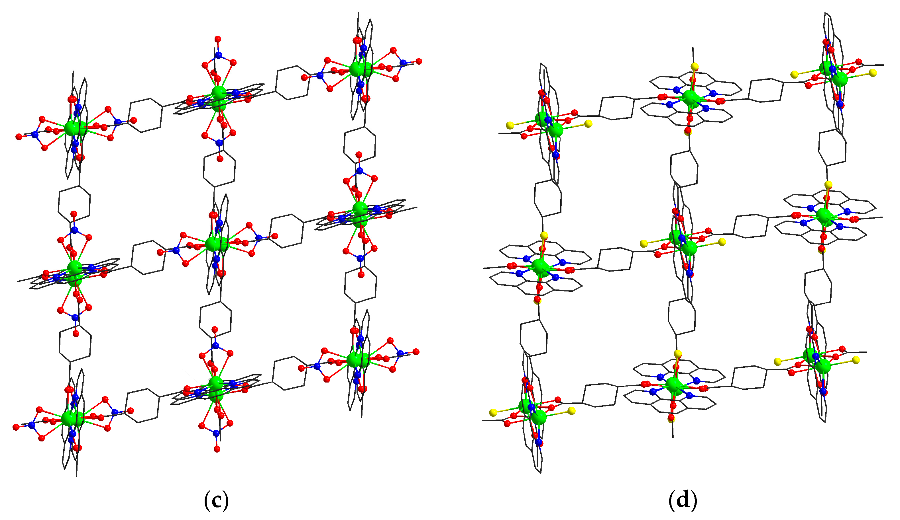
3.1. Synthesis and Crystal Structure Description

Compounds [Gd2(bpy)2(chdc)3]·H2O (1), [Gd2(phen)2(chdc)3]·0.5DMF (2) and [Gd2(NO3)2(phen)2(chdc)2]·2DMF (3) crystallize in the monoclinic crystal system with P21/n space group and are isostructural to the series previously reported by our group: [Ln2(bpy)2(chdc)3]·xH2O (x = 0…1), [Ln2(phen)2(chdc)3]·0.5DMF [34] and [Ln2(NO3)2(phen)2(chdc)2]·2DMF [35] (Ln3+ = Y3+, Eu3+ or Tb3+), respectively. Highly effective luminescence with quantum yields up to 63% was revealed for these structures, including [Y2(bpy)2(chdc)3] based on the non-emissive and diamagnetic Y3+. Unlike the yttrium(III), Gd3+ is paramagnetic but still non-emissive in the soft UV and visible region; therefore, the investigation of isostructural Gd(III)-based compounds 13 and the cognate new metal-organic framework 4 could provide significantly new information concerning the luminescent properties of such type series of coordination networks.
Compounds 2 and 3 were obtained in quite similar solvothermal conditions at 110 °C, using N,N-dimethylformamide (DMF) as a solvent. The main difference between their synthetic methods is the starting Gd(III) salt (chloride for 2 or nitrate for 3) to be used. Thus, it was shown that the anion plays a main structure-forming role in this system, as the presence of NO3 leads to the crystallization of nitrate-containing layered MOF 3. On the contrary, poorly coordinated Cl cannot compete with the carboxylate in such conditions and affords a three-dimensional framework 2, which contains the only RCOO fragments additionally coordinated to the {Gd(phen)}3+ moiety. Interestingly, the replacement of pure DMF by the mixture of DMF and dioxane (2:3) leads to the formation of the chloride-containing structure [Gd2Cl2(phen)2(chdc)2]·0.3DMF·2.2dioxane (4) instead of the carboxylate 2. A significant dilution of DMF by dioxane apparently weakens the solvation of Cl and, therefore, strengthens its coordination ability compared to the carboxylate. Thus, the crystallization of two-dimensional framework 4 seems to be anion/solvent-controlled again and suggests a simple route for the synthesis of lanthanide(III) networks with coordinated halogenides, since only one example of the compound containing {Ln2(Cl)2(L)2(OOCR)4} building units (L = any 2,2′-bipyridyl derivative) has been reported to date [36].
In all the structures 13, Gd(III) adopts a similar capped square-antiprismatic environment consisting of two N atoms of diimine chelate ligand and seven O atoms, which belong to the carboxylic groups in 1 and 2 or to carboxylic groups and terminal nitrate in 3 (Figure 1a–c). The selected coordination bond lengths are listed in Table 1. Two symmetry-equivalent Gd(III) ions form binuclear carboxylate blocks {Gd2(bpy)2(RCOO-κ2)2(μ-RCOO-κ11)2(μ-RCOO-κ12)2} (1), {Gd2(phen)2(RCOO-κ2)2(μ-RCOO-κ11)2(μ-RCOO-κ12)2} (2) and {Gd2(phen)2(ONO22)2(μ-RCOO-κ11)2(μ-RCOO-κ12)2} (3). The coordination frameworks in 1 and 2, consisting of six-connected building units and trans-1,4-cyclohexanedicarboxylate bridges, adopt a very distorted three-dimensional primitive cubic topology (pcu) and contain small voids (Figure 2a,b) interconnected by very narrow (2 × 2 Å2) windows with 5% calculated total void volume. In the coordination framework in 3, four-connected binuclear blocks are interconnected by cyclohexane moieties into a two-dimensional square-grid network (sql) (Figure 2c). The layers in 3 are packed in a one-layer (AA) manner to form channels with 26% general void volume. These channels are occupied by the localized DMF solvent molecules.
Compound [Gd2Cl2(phen)2(chdc)2]·0.3DMF·2.2dioxane (4) crystallizes in the monoclinic crystal system with the P21/n space group. The coordination environment of Gd(III) consists of two N atoms of diimine chelate ligand, five O atoms of the carboxylic groups and one Cl atom. The Gd−N and Gd−O bond lengths are close to those in 13 (see Table 1) and the Gd−Cl distance is 2.649(2) Å (Figure 1 and Figure 2). The structure of a binuclear carboxylate block {Gd2(phen)2(Cl)2(μ-RCOO-κ11)2(μ-RCOO-κ12)2} in 4 is analogous to the nitrate-containing unit in 3, except for the reduction of the coordination number to 8 due to the substitution of the bidentate nitrate anion with the larger chloride (Figure 1d), which acts as a monodentate ligand. Four-connected binuclear blocks in 4 are interconnected by cyclohexane moieties (Figure 2d) in a similar AA manner to 3, with the channels of 32% general void volume. These channels are occupied by solvent molecules. Only one dioxane molecule per formula unit was localized directly, while the non-ordered residual electron density was analyzed by the PLATON/SQUEEZE [37] procedure (69 e in 265 Å per formula unit) and assigned to 0.3DMF + 1.2dioxane (69.6 e and ca. 209 Å volume estimated from the liquid densities).

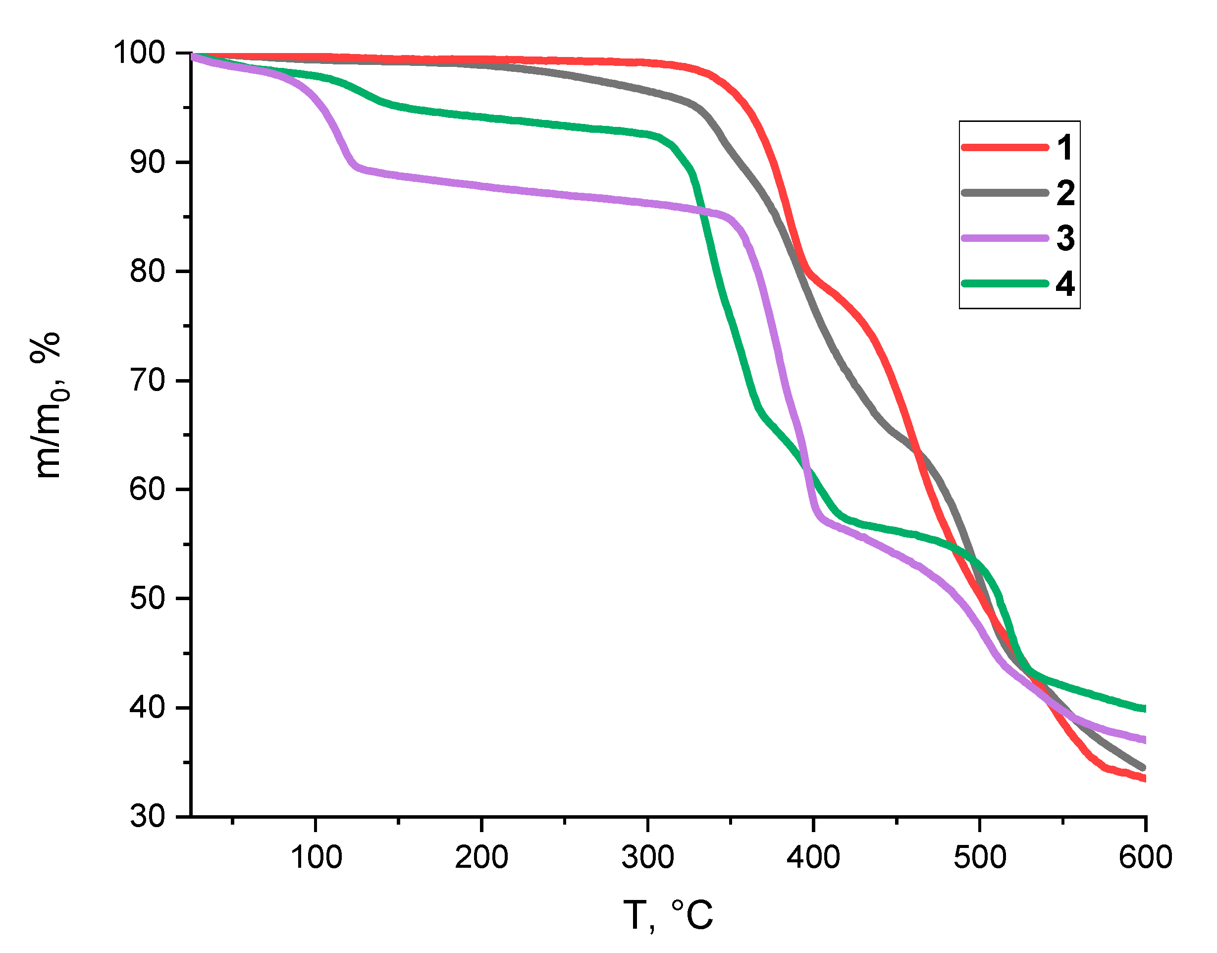
3.2. Thermal Properties

Thermogravimetric analyses for the compounds 14 were performed (Figure 3). For 1, the stepwise decomposition starts at ca. 350 °C. Only 1% weight loss before 300 °C corresponds well to the low content of guest solvent molecules (calculated as 1.5%) determined by X-ray crystallography, and 33% residual weight at 600 °C matches well to the gadolinium(III) oxide (calculated as 32%).
2 slowly losses solvent molecules at the temperature up to 300 °C, much higher than the boiling points of both DMF and water. Such feature is apparently attributed to the low size of the windows (~2 Å) in the coordination framework of 2 and the resulting kinetic hindrance of the guest diffusion. The first step of lattice decomposition occurs in the range 340–440 °C and corresponds well to the loss of phen molecules (66% residue at 440 °C; calculated for Gd2(chdc)3: 66.5%). Further weight loss starting at ca. 460 °C corresponds to the decomposition of the bridging ligand and leads to the Gd2O3 (34% residue at 600 °C, calculated: 29%) being apparently contaminated by carbon admixture due to the incomplete evaporation of the organic moieties.
Compound 3 loses solvents at ca. 120 °C. The first step of coordination lattice decomposition occurs in the range 350–400 °C and corresponds well to the loss of phen molecules (58% residue at 440 °C; calculated for Gd2(NO3)2(chdc)2: 59%). Further weight loss, corresponding to the decomposition of the nitrate and chdc2− ligands, starts at ca. 430 °C. The TG profile of 4 is close to 3 and includes solvent loss at ca. 130 °C and two steps of the lattice decomposition occurring in the range 320–410 °C and above 490 °C. In summary, thermal stability characteristics of the coordination framework in 14 are high [38,39,40,41] and quite similar to each other. However, their stability is limited by the evaporation of the neutral N-donor chelate, occurring below 400 °C.

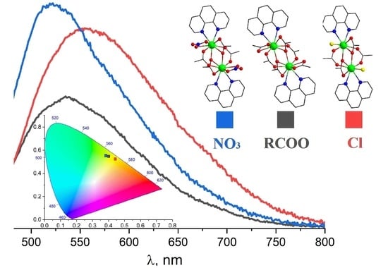
3.3. Luminescence Spectrocopy

Solid-state luminescence measurements were performed for the synthesized compounds. The bpy-containing 1 appeared to possess no luminescent activity, possibly due to the paramagnetic quenching of the emission by the Gd(III) cation. In contrast, the phen-containing compounds 24 demonstrate yellow wide-banded emission under a visible light excitation at λex = 460 nm. The corresponding emission spectra are shown in Figure 4a. The maxima of the spectra appear at λ = 537 nm for three-dimensional 2 based on the six-connected carboxylate units, λ = 522 nm for the nitrate-containing 3 and λ = 556 nm for the chloride-containing 4. The observed red-shift of both the maxima and the characteristic wavelengths in the row nitrate < carboxylate < chloride apparently correlates to the electron donor properties of the corresponding ligands. The calculated (x,y) coordinates on the CIE 1931 chromaticity diagram and characteristic wavelength values are shown in Figure 4b and visualize the integral yellow color of the wide-banded emission of 24.

4. Conclusions

To summarize, four new gadolinium(III) metal-organic frameworks were synthesized and characterized. Compounds 1 and 2 containing six-connected binuclear metal-carboxylate blocks adopt a distorted primitive cubic topology with narrow pores. A partial substitution of carboxylate by nitrate or chloride in the Gd(III) coordination sphere leads to two-dimensional square-layered networks 3 and 4. Thermal and luminescent properties of the synthesized compounds were investigated. Phenanthroline-based structures 24 emit in the yellow region under a visible blue excitation at 460 nm. The observed red-shift in the row of coordinated ligands, nitrate < carboxylate < chloride was attributed to the donor ability of the corresponding ligands.

Supplementary Materials

The following are available online at https://www.mdpi.com/article/10.3390/cryst11111375/s1: Figure S1: Experimental PXRD pattern for 1 compared to the theoretical one; Figure S2: Experimental PXRD pattern for 2 compared to the theoretical one; Figure S3: Experimental PXRD pattern for 3 compared to the theoretical one; Figure S4: Experimental PXRD pattern for 4 compared to the theoretical one; Figure S5: IR spectra for 15.

Author Contributions

P.A.D., original draft preparation, single-crystal XRD, graphing; A.A.V., synthesis, characterization, graphing; V.A.L., synchrotron single-crystal XRD; A.A.R., solid-state luminescence measurements; V.P.F., manuscript review and editing, project administration and funding acquisition. All authors have read and agreed to the published version of the manuscript.

Funding

The research was supported by the Ministry of Science and Higher Education of the Russian Federation, No. 121031700321-3 and No. 121031700321-8.

Institutional Review Board Statement

Not applicable.

Informed Consent Statement

Not applicable.

Data Availability Statement

CCDC 2118161–2118165 contain the supplementary crystallographic data for this paper. These data can be obtained free of charge from The Cambridge Crystallographic Data Center at https://www.ccdc.cam.ac.uk/structures/.

Conflicts of Interest

The authors declare no conflict of interest.

Appendix A. Crystal Structure of a New Complex [Gd(DMF)2(phen)]Cl3 (5)

Single crystals of 5 were obtained during the screening syntheses: 37.2 mg (0.10 mmol) of GdCl3·6H2O, 18.0 mg (0.10 mmol) of phen·H2O and 34.4 mg (0.20 mmol) of H2chdc were mixed in a 3 mL glass vial and dissolved in the mixture of 1 mL of DMF and 1.50 mL of acetone. The obtained solution was heated at 45 °C for 72 h. After cooling to room temperature, the obtained white precipitate was filtered off, washed with DMF and dried in air. IR spectrum main bands (KBr, cm−1): 3375 (s., br., νO−H); 2937 and 2858 (w., νCsp3−H); 1654 (s., νCOamide).
A molecular complex [Gd(DMF)2(phen)]Cl3 (5) crystallizes in the triclinic crystal system with the P-1 space group with one independent Gd atom and Z = 2. The coordination environment of Gd(III) consists of two N atoms of phenanthroline, two O atoms of the coordinated solvent and three Cl atoms. The Gd−N bond length is 2.584(2) Å, the Gd−O bond lengths are 2.334(2) Å and 2.374(2) Å and the Gd−Cl bond lengths are 2.6408(8) Å, 2.6694(8) Å and 2.6921(7) Å, which are close to the distance in the structure of 4: 2.649(2) Å.
Figure A1. A molecular complex [Gd(DMF)2(phen)]Cl3. Gd atoms are green, N atoms are blue, O atoms are red, Cl atoms are yellow. H atoms are not shown.
Crystals 11 01375 g0a1

Appendix B. The Crystallographic Data for 1–5

Table A1. Crystallographic data and refinement details for the structures 15.
12345
Chemical formulaC44H48Gd2N4O13C49.5H49.5Gd2N4.50O12.5C46H50Gd2N8O16C47.7H51.7Cl2Gd2N4.3O11.7C18H22Cl3GdN4O2
Mr, g/mol1155.361221.931285.441257.83589.99
Crystal systemMonoclinicMonoclinicMonoclinicMonoclinicTriclinic
Space groupP21/nP21/nP21/nP21/nP¯1
Temperature, K100140140140140
a, Å10.422(2)10.4479(4)12.3986(6)13.2219(5)9.2772(4)
b, Å17.551(4)17.9554(7)16.8287(6)13.3512(7)9.6641(4)
c, Å12.294(3)12.8866(7)12.7182(5)15.5831(7)12.2954(5)
α, °90909090102.460(4)
b, °103.90(3)99.104(4)112.863(5)109.100(5)94.032(3)
γ, °9090909092.587(3)
V, Å32183.0(8)2387.02(19)2445.20(19)2599.4(2)1071.67(8)
Z22222
F(000)1144121212761247578
D(calc.), g/cm31.7581.7001.7461.6071.828
m, mm−13.4622.822.772.693.49
Crystal size, mm0.20 × 0.04 × 0.040.30 × 0.14 × 0.100.30 × 0.21 × 0.110.25 × 0.17 × 0.140.65 × 0.25 × 0.20
θ range for data collection, °2.42 ≤ θ ≤ 26.682.3 ≤ θ ≤ 25.42.0 ≤ θ ≤ 25.42.1 ≤ θ ≤ 25.42.2 ≤ θ ≤ 25.3
No. of reflections: measured/independent/observed [I > 2σ(I)]14,396/3987/361510,535/4383/352011,222/4487/392111,529/4755/38279676/3919/3637
Rint0.03560.02490.02510.02310.0368
Index ranges−12 ≤ h ≤ 12
−21 ≤ k ≤ 20
−14 ≤ l ≤ 14
−12 ≤ h ≤ 12
−21 ≤ k ≤ 19
−15 ≤ l ≤ 15
−14 ≤ h ≤ 12
−15 ≤ k ≤ 20
−12 ≤ l ≤ 15
−11 ≤ h ≤ 15
−16 ≤ k ≤ 14
−18 ≤ l ≤ 18
−11 ≤ h ≤ 9
−11 ≤ k ≤ 11
−14 ≤ l ≤ 14
Final R indices [I > 2σ(I)]R1 = 0.0205
wR2 = 0.0482
R1 = 0.0286
wR2 = 0.0630
R1 = 0.0227
wR2 = 0.0480
R1 = 0.0475
wR2 = 0.1365
R1 = 0.0231
wR2 = 0.0459
R indices (all data)R1 = 0.0240
wR2 = 0.0496
R1 = 0.0437
wR2 = 0.0673
R1 = 0.0296
wR2 = 0.0502
R1 = 0.0572
wR2 = 0.1443
R1 = 0.0261
wR2 = 0.0470
Goodness-of-fit on F21.0261.0431.0611.0701.049
Largest diff. peak/hole, e30.525, −0.6010.73, −0.800.66, −0.782.36, −0.860.63, −0.68

References

  1. Nonat, A.M.; Charbonnière, L.J. Upconversion of light with molecular and supramolecular lanthanide complexes. Coord. Chem. Rev. 2020, 409, 213192. [Google Scholar] [CrossRef]
  2. Belousov, Y.A.; Drozdov, A.A.; Taydakov, I.V.; Marchetti, F.; Pettinari, R.; Pettinari, C. Lanthanide azolecarboxylate com-pounds: Structure, luminescent properties and applications. Coord. Chem. Rev. 2021, 445, 214084. [Google Scholar] [CrossRef]
  3. Xu, H.; Cao, C.-S.; Kang, X.-M.; Zhao, B. Lanthanide-based metal–organic frameworks as luminescent probes. Dalton Trans. 2016, 45, 18003–18017. [Google Scholar] [CrossRef] [PubMed]
  4. Zhao, S.-N.; Wang, G.; Poelman, D.; Van Der Voort, P. Luminescent Lanthanide MOFs: A Unique Platform for Chemical Sensing. Materials 2018, 11, 572. [Google Scholar] [CrossRef] [PubMed] [Green Version]
  5. Hao, Y.; Chen, S.; Zhou, Y.; Zhang, Y.; Xu, M. Recent Progress in Metal–Organic Framework (MOF) Based Luminescent Che-modosimeters. Nanomaterials 2019, 9, 974. [Google Scholar] [CrossRef] [PubMed] [Green Version]
  6. Zhan, Z.; Jia, Y.; Li, D.; Zhang, X.; Hu, M. A water-stable terbium-MOF sensor for the selective, sensitive, and recyclable detection of Al3+ and CO32− ions. Dalton Trans. 2019, 48, 15255–15262. [Google Scholar] [CrossRef]
  7. Saraci, F.; Quezada-Novoa, V.; Donnarumma, P.R.; Howarth, A.J. Rare-earth metal–organic frameworks: From structure to applications. Chem. Soc. Rev. 2020, 49, 7949–7977. [Google Scholar] [CrossRef] [PubMed]
  8. Litvinova, Y.M.; Gayfulin, Y.M.; Samsonenko, D.G.; Dorovatovskiy, P.V.; Lazarenko, V.A.; Brylev, K.A.; Mironov, Y.V. Coordination polymers based on rhenium octahedral chalcocyanide cluster [Re6Se8(CN)6]4– and lanthanide ions solvated with dimethylformamide. Inorg. Chim. Acta 2021, 528, 120597. [Google Scholar] [CrossRef]
  9. Yao, C.-X.; Zhao, N.; Liu, J.-C.; Chen, L.-J.; Liu, J.-M.; Fang, G.-Z.; Wang, S. Recent Progress on Luminescent Metal-Organic Framework-Involved Hybrid Materials for Rapid Determination of Contaminants in Environment and Food. Polymers 2020, 12, 691. [Google Scholar] [CrossRef] [PubMed] [Green Version]
  10. Caravan, P. Protein-Targeted Gadolinium-Based Magnetic Resonance Imaging (MRI) Contrast Agents: Design and Mechanism of Action. Accounts Chem. Res. 2009, 42, 851–862. [Google Scholar] [CrossRef] [PubMed]
  11. Narmani, A.; Farhood, B.; Haghi-Aminjan, H.; Mortezazadeh, T.; Aliasgharzadeh, A.; Mohseni, M.; Najafi, M.; Abbasi, H. Gadolinium nanoparticles as diagnostic and therapeutic agents: Their delivery systems in magnetic resonance imaging and neutron capture therapy. J. Drug Deliv. Sci. Technol. 2018, 44, 457–466. [Google Scholar] [CrossRef]
  12. Clough, T.J.; Jiang, L.; Wong, K.-L.; Long, N.J. Ligand design strategies to increase stability of gadolinium-based magnetic resonance imaging contrast agents. Nat. Commun. 2019, 10, 1420. [Google Scholar] [CrossRef] [PubMed] [Green Version]
  13. Zairov, R.; Pizzanelli, S.; Dovzhenko, A.P.; Nizameev, I.; Orekhov, A.S.; Arkharova, N.; Podyachev, S.N.; Sudakova, S.; Mustafina, A.R.; Calucci, L. Paramagnetic Relaxation Enhancement in Hydrophilic Colloids Based on Gd(III) Complexes with Tetrathia- and Calix[4]arenes. J. Phys. Chem. C 2020, 124, 4320–4329. [Google Scholar] [CrossRef]
  14. Parker, D.; Suturina, E.; Kuprov, I.; Chilton, N.F. How the Ligand Field in Lanthanide Coordination Complexes Determines Magnetic Susceptibility Anisotropy, Paramagnetic NMR Shift, and Relaxation Behavior. Accounts Chem. Res. 2020, 53, 1520–1534. [Google Scholar] [CrossRef]
  15. Tan, Q.-H.; Wang, Y.-Q.; Guo, X.-Y.; Liu, H.-T.; Liu, Z.-L. A gadolinium MOF acting as a multi-responsive and highly selective luminescent sensor for detecting o-, m-, and p-nitrophenol and Fe3+ ions in the aqueous phase. RSC Adv. 2016, 6, 61725–61731. [Google Scholar] [CrossRef]
  16. Casanovas, B.; Speed, S.; Maury, O.; El Fallah, M.S.; Font-Bardí, M.; Vicente, R. Dinuclear LnIII Complexes with 9-Anthracenecarboxylate Showing Field-Induced SMM and Visible/NIR Luminescence. Eur. J. Inorg. Chem. 2018, 34, 3859–3867. [Google Scholar] [CrossRef]
  17. Casanovas, B.; Speed, S.; Maury, O.; Font-Bardia, M.; Vicente, R. Homodinuclear lanthanide 9-anthracenecarboxylate complexes: Field induced SMM and NIR-luminescence. Polyhedron 2019, 169, 187–194. [Google Scholar] [CrossRef]
  18. Casanovas, B.; Speed, S.; Vicente, R.; Font-Bardia, M. Sensitization of visible and NIR emitting lanthanide(III) ions in a series of dinuclear complexes of formula [Ln2(μ-2-FBz)2(2-FBz)4(terpy)2]·2(2-HFBz)·2(H2O). Polyhedron 2019, 173, 114113. [Google Scholar] [CrossRef]
  19. Taydakov, I.V.; Belousov, Y.A.; Lyssenko, K.A.; Varaksina, E.; Drozdov, A.A.; Marchetti, F.; Pettinari, R.; Pettinari, C. Synthesis, phosphorescence and luminescence properties of novel europium and gadolinium tris-acylpyrazolonate complexes. Inorg. Chim. Acta 2019, 502, 119279. [Google Scholar] [CrossRef]
  20. Bryleva, Y.A.; Artem’Ev, A.V.; Glinskaya, L.A.; Komarov, V.Y.; Bogomyakov, A.S.; Rakhmanova, M.I.; Larionov, S.V. A series of bis(2-phenethyl)dithiophosphinate-based Ln(III) complexes: Synthesis, magnetic and photoluminescent properties. Inorg. Chim. Acta 2021, 516, 120097. [Google Scholar] [CrossRef]
  21. Kim, J.H.; Lepnev, L.S.; Utochnikova, V.V. Dual vis-NIR emissive bimetallic naphthoates of Eu–Yb–Gd: A new approach toward Yb luminescence intensity increase through Eu → Yb energy transfer. Phys. Chem. Chem. Phys. 2021, 23, 7213–7219. [Google Scholar] [CrossRef]
  22. Gontcharenko, V.; Kiskin, M.; Dolzhenko, V.; Korshunov, V.; Taydakov, I.; Belousov, Y. Mono- and Mixed Metal Complexes of Eu3+, Gd3+, and Tb3+ with a Diketone, Bearing Pyrazole Moiety and CHF2-Group: Structure, Color Tuning, and Kinetics of Energy Transfer between Lanthanide Ions. Molecules 2021, 26, 2655. [Google Scholar] [CrossRef] [PubMed]
  23. Utochnikova, V.V.; Aslandukov, A.N.; Vashchenko, A.A.; Goloveshkin, A.S.; Alexandrov, A.A.; Grzibovskis, R.; Bunzli, J.-C.G. Identifying lifetime as one of the key parameters responsible for the low brightness of lanthanide-based OLEDs. Dalton Trans. 2021, 50, 12806–12813. [Google Scholar] [CrossRef] [PubMed]
  24. Llabres-Campaner, P.J.; Pitarch-Jarque, J.; Ballesteros-Garrido, R.; Abarca, B.; Ballesteros, R.; García-España, E. Bicy-clo[2.2.2]octane-1,4-dicarboxylic acid: Towards transparent metal-organic frameworks. Dalton Trans. 2017, 46, 7397–7402. [Google Scholar] [CrossRef]
  25. Demakov, P.A.; Sapchenko, S.A.; Samsonenko, D.G.; Dybtsev, D.N.; Fedin, V.P. Coordination polymers based on zinc(ii) and manganese(ii) with 1,4-cyclohexanedicarboxylic acid. Russ. Chem. Bull. 2018, 67, 490–496. [Google Scholar] [CrossRef]
  26. Yin, J.; Yang, H.; Fei, H. Robust, Cationic Lead Halide Layered Materials with Efficient Broadband White-Light Emission. Chem. Mater. 2019, 31, 3909–3916. [Google Scholar] [CrossRef]
  27. Demakov, P.A.; Poryvaev, A.S.; Kovalenko, K.A.; Samsonenko, D.G.; Fedin, M.V.; Fedin, V.P.; Dybtsev, D.N. Structural Dy-namics and Adsorption Properties of the Breathing Microporous Aliphatic Metal–Organic Framework. Inorg. Chem. 2020, 59, 15724–15732. [Google Scholar] [CrossRef]
  28. Svetogorov, R.; Dorovatovskii, P.V.; Lazarenko, V.A. Belok/XSA Diffraction Beamline for Studying Crystalline Samples at Kurchatov Synchrotron Radiation Source. Cryst. Res. Technol. 2020, 55, 1900184. [Google Scholar] [CrossRef]
  29. Lazarenko, V.A.; Dorovatovskii, P.V.; Zubavichus, Y.V.; Burlov, A.S.; Koshchienko, Y.V.; Vlasenko, V.G.; Khrustalev, V.N. High-Throughput Small-Molecule Crystallography at the ‘Belok’ Beamline of the Kurchatov Synchrotron Radiation Source: Transition Metal Complexes with Azomethine Ligands as a Case Study. Crystals. 2017, 7, 325. [Google Scholar] [CrossRef] [Green Version]
  30. Kabsch, W. XDS. Acta Crystallogr. 2010, D66, 125–132. [Google Scholar] [CrossRef] [Green Version]
  31. CrysAlisPro, Version: 1.171.38.46. Rigaku Oxford Diffraction; Rigaku Americas Holding Company, Inc. : The Woodlands, TX, USA, 2015.
  32. Sheldrick, G.M. SHELXT—Integrated space-group and crystal-structure determination. Acta Crystallogr. Sect. A Found. Adv. 2015, 71, 3–8. [Google Scholar] [CrossRef] [Green Version]
  33. Sheldrick, G.M. Crystal structure refinement with SHELXL. Acta Crystallogr. Sect. C Struct. Chem. 2015, 71, 3–8. [Google Scholar] [CrossRef] [PubMed]
  34. Demakov, P.A.; Ryadun, A.A.; Dorovatovskii, P.V.; Lazarenko, V.A.; Samsonenko, D.G.; Brylev, K.A.; Fedin, V.P.; Dybtsev, D.N. Intense multi-colored luminescence in a series of rare-earth metal–organic frameworks with aliphatic linkers. Dalton Trans. 2021, 50, 11899–11908. [Google Scholar] [CrossRef] [PubMed]
  35. Demakov, P.A.; Vasileva, A.A.; Volynkin, S.S.; Ryadun, A.A.; Samsonenko, D.G.; Fedin, V.P.; Dybtsev, D.N. Cinnamal Sensing and Luminescence Color Tuning in a Series of Rare-Earth Metal−Organic Frameworks with Trans-1,4-cyclohexanedicarboxylate. Molecules 2021, 26, 5145. [Google Scholar] [CrossRef]
  36. Lu, Y.-B.; Jiang, X.-M.; Zhu, S.-D.; Du, Z.-Y.; Liu, C.-M.; Xie, Y.-R.; Liu, L.-X. Anion Effects on Lanthanide(III) Tetrazole-1-acetate Dinuclear Complexes Showing Slow Magnetic Relaxation and Photofluorescent Emission. Inorg. Chem. 2016, 55, 3738–3749. [Google Scholar] [CrossRef] [PubMed]
  37. Spek, A.L. Single-crystal structure validation with the program PLATON. J. Appl. Crystallogr. 2003, 36, 7–13. [Google Scholar] [CrossRef] [Green Version]
  38. Barsukova, M.; Samsonenko, D.G.; Fedin, V.P. Crystal structure of metal-organic frameworks based on terbium and 1,4-naphthalenedicarboxylic acid. J. Struct. Chem. 2020, 61, 1090–1096. [Google Scholar] [CrossRef]
  39. Demakov, P.A.; Ryadun, A.A.; Samsonenko, D.G.; Dybtsev, D.N.; Fedin, V.P. Structure and luminescent properties of europium(III) coordination polymers with thiophene ligands. J. Struct. Chem. 2020, 61, 1965–1974. [Google Scholar] [CrossRef]
  40. Barsukova, M.O.; Cherezova, S.V.; Sapianik, A.A.; Lundovskaya, O.V.; Samsonenko, D.G.; Fedin, V.P. Lanthanide contraction effect and white-emitting luminescence in a series of metal–organic frameworks based on 2,5-pyrazinedicarboxylic acid. RSC Adv. 2020, 10, 38252–38259. [Google Scholar] [CrossRef]
  41. Cherezova, S.V.; Barsukova, M.O.; Samsonenko, D.G.; Fedin, V.P. Crystal structure of dense metal-organic frameworks based on sc(III) and two types of ligands. J. Struct. Chem. 2021, 62, 897–904. [Google Scholar] [CrossRef]
Figure 1. Secondary building units in 1 (a), 2 (b), 3 (c) and 4 (d). Gd atoms are green, N atoms are blue, O atoms are red, Cl atoms are yellow. H atoms are not shown.
Crystals 11 01375 g001
Figure 2. Fragments of three-dimensional coordination frameworks in 1 (a) and 2 (b). Coordination layers in 3 (c) and 4 (d), Atom colors are similar to those in Figure 1.
Crystals 11 01375 g002aCrystals 11 01375 g002b
Figure 3. TG plots for compounds 14.
Crystals 11 01375 g003
Figure 4. Emission spectra for 24 at λex = 460 nm (a), CIE 1931 chromaticity diagram for 24 (b).
Crystals 11 01375 g004
Table 1. Selected bond lengths in the structures 14.
Bond1234
Gd−N, Å2.566(2),
2.600(3)
2.499(19)−2.59(3)2.553(2), 2.591(3)2.564(7), 2.579(5)
Gd−O(COOchelate), Å2.3482(19)–2.514(2)2.334(3)–2.539(3)2.330(2)–2.573(2)2.294(5)–2.513(5)
Gd–O(COOnon-chelate), Å
or
Gd–O(NO3,non-chelate), Å
2.429(2)–2.4800(19)2.463(3)–2.465(3)--
--2.458(2)–2.529(2)-
Publisher’s Note: MDPI stays neutral with regard to jurisdictional claims in published maps and institutional affiliations.

