Article

Biosynthesis of Silver Nanoparticles by Conyza canadensis and Their Antifungal Activity against Bipolaris maydis

1
College of Agriculture, Anhui Science and Technology University, Chuzhou 233100, China
2
College of Mechanical Engineering, Anhui Science and Technology University, Chuzhou 233100, China
*
Authors to whom correspondence should be addressed.
These authors contribute equally.
Crystals 2021, 11(12), 1443; https://doi.org/10.3390/cryst11121443
Submission received: 22 October 2021 / Revised: 19 November 2021 / Accepted: 22 November 2021 / Published: 23 November 2021
(This article belongs to the Topic Iron Concentrate Particles)

Abstract

:
Silver nanoparticles were biosynthesized from Conyza canadensis leaf extract with the help of a microwave oven. The UV-vis spectrum showed the maximum absorption at 441 nm, corresponding to the surface plasmon resonance of silver nanoparticles. Transmission electron microscope and scanning electron microscope images showed that the synthesized silver nanoparticles were spherical or near-spherical with an average diameter of 43.9 nm. X-ray diffraction demonstrated nanoparticles with a single-phase cubic structure. As-synthesized silver nanoparticles displayed prominent antifungal activity against Bipolaris maydis. The colony inhibition rate reached 88.6% when the concentration of nanosilver colloid was 100 μL·mL−1 (v/v). At such a concentration, no colony formation was observed on the solid plate. The diameter of the inhibition zone was 13.20 ± 1.12 mm. These results lay the foundation for the comprehensive control of plant pathogens using an environmentally friendly approach.

Graphical Abstract

1. Introduction

The use of nanotechnology, which appeared later than bio-engineering, has expanded in the last few decades [1,2,3]. It makes traditional substances into nanoscale materials, which display excellent performance, including optical, magnetic, physical, and chemical characteristics, compared with their bulk counterparts [4,5,6]. Among these materials, silver nanoparticles have proved to be one of the most important types. Consequently, significant research has been conducted on the synthesis, characterization, and application of silver nanoparticles [7,8,9,10,11,12,13,14].
Silver was used to disinfect and dress wounds in ancient times; its antimicrobial property has drawn much attention since then. Silver nanoparticles improve the antimicrobial activity of silver and have proven to be efficient. They inhibit bacteria, fungi, and viruses. Silver nanoparticles synthesized using the chemical approach exhibited an excellent antibacterial effect against Staphylococcus aureus (ATCC 6538p) at 50 µg.mL−1; the bacteria cells, enzymatic activity, and proteins changed [15]. The minimum bactericidal concentration of silver nanoparticles synthesized by Descurainia sophia against Agrobacterium tumefaciens (strain GV3850) and A. rhizogenes (strain 15843) was 4 and 8 µg.mL−1, respectively [16]. The silver nanoparticles synthesized by Phoenix dactylifera showed a significant inhibitory effect on Rhizoctonia solani (AG2_2) cultures; the concentration of 25 µg/mL prevented approximately 83% of the mycelium growth of the fungus [17]. Silver nanoparticles synthesized using turnip leaf extract interacted with several wood-degrading fungal pathogens, such as Gloeophyllum abietinum, Gloeophyllum trabeum, Chaetomium globosum, and Phanerochaete sordida. The result showed cellulose and lignocellulose degradation for all of the tested fungi [18]. The in vitro anti-HBV (Hepatitis B Virus) activities of silver nanoparticles (about 10 and 50 nm) were determined using the HepAD38 cell line. The results showed that silver nanoparticles could inhibit the in vitro production of HBV RNA and extracellular virions [19]. The anti-HIV (Human Immunodeficiency Virus) activity of silver nanoparticles was proven in the early stage of viral replication and post-entry stages of its life cycle; the results made it possible to inhibit a wide variety of circulating HIV-1 strains [20].
In this study, the silver nanoparticles biosynthesized from Conyza canadensis extract were applied to determine their antifungal activity against Bipolaris maydis. Fungicides have made an undeniable contribution to disease control. However, in the current scenario, several drawbacks became apparent, such as pesticide residue, environmental pollution, pathogens resistance, and so forth. It is necessary to explore neotype and stable fungistats to replace or assist fungicides. As non-antibiotic agents, silver nanoparticles could overcome such problems to the greatest degree.

2. Materials and Methods

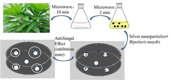
2.1. Fungus Isolation and Plant Tissue

The single-spore isolate of B. maydis was preserved in the Laboratory of Plant Protection at Anhui Science and Technology University. C. Canadensis was gathered from the plantation of the same campus.

2.2. Biosynthesis of Silver Nanoparticles

Air-dried C. Canadensis leaves were cut into tiny pieces (1 cm × 1 cm) after washing them thoroughly with distilled water. About 10 g of leaves was added to 100 mL of deionized water and heated in a microwave oven at 800 W for 10 min. AgNO3 solution (100 mmol·L−1) was freshly prepared as stock solution. The leaf extract was filtered with Whatman No.1 paper. After that, 1 mL leaf filtrate and 1 mL AgNO3 stock solution were added to 98 mL deionized water, making the final concentration of AgNO3 1 mmol·L−1. Finally, the mixture was put into a microwave oven and heated at 800 W for 2 min. The color and UV-vis spectrum of the solution were measured.

2.3. Characterization of Silver Nanoparticles

Several analysis tools, such as a UV-vis absorption spectrometer, transmission electron microscope (TEM), scanning electron microscope (SEM), and X-ray powder diffractometer (XRD), and energy-dispersive X-ray spectroscopy (EDX) were applied to determine the maximum absorption peak, morphology, crystal structure, particle size, and size distribution so as to obtain the detailed information of synthesized silver nanoparticles.

2.4. Antifungal Activity of Silver Nanoparticles against B. maydis

2.4.1. Inhibition of Colony Growth

Different volumes (1, 2, 3, 4, and 5 mL) of nanosilver colloid and varied volumes (4, 3, 2, 1, and 0 mL) of sterile water were added to 45 mL melted PDA (Potato dextrose agar) separately, and the concentration of nanosilver colloid was set as 20, 40, 60, 80, and 100 μL·mL−1 (v/v), respectively. The control contained 45 mL melted PDA medium and 5 mL sterile water. Fungus blocks (φ = 5 mm) were drilled from cultivated B. maydis, and one specimen was placed in the center of each Petri dish, then inoculated, followed by incubation at 28 °C for 3–5 days. Each control and treatment was performed in three replicates. The inhibition rate of silver nanoparticles against B. maydis was calculated using the following equation.
Inhibition rate = [(Diameter of control colony − Diameter of treatment colony)/(Diameter of control colony − Diameter of fungus block)] × 100%

2.4.2. Influence on Colony Formation

The PDA plates contained different concentrations (20, 40, 60, 80, and 100 μL·mL−1, v/v) of nanosilver colloids, respectively. About 45 mL melted PDA medium with 5 mL sterile water was set as control. The spores’ suspension of the B. maydis isolate was adjusted to 106 mL−1 with a counting chamber. Then, 100 μL of the spore suspension was spread evenly on cooled PDA plates using a sterile spreading rod. Each control and treatment was performed in three replicates. Finally, the plates were incubated at 28 °C for 2–3 days after standing for 3–5 min.

2.4.3. Measurement of the Inhibition Zone

The inhibition zone created near the agar well that was drilled into the PDA plate displayed the antifungal activity of silver nanoparticles. About 100 μL of the spore suspension (106 mL−1) was evenly smeared on a solid PDA plate. Five wells (φ = 8 mm) were drilled by a hole puncher, and different volumes (20, 30, 40, and 50 μL) of nanosilver colloid and 50 μL sterile water were dripped into corresponding agar well. After incubation for several days, an inhibition zone appeared near the agar well if the filling liquid had antifungal activity. The diameter of the inhibition zone varied as well. However, no inhibition zone was created near the well that contained sterile water. Each control and treatment was performed in three replicates. The plates were incubated at 28 °C for 2–3 days after standing for 3–5 min.

3. Results and Discussion

3.1. Biosynthesis of Silver Nanoparticles

Silver (Ag0) was formed from AgNO3 (Ag+) and plant extract. Figure 1 shows that the solution color changed from faint yellow to reddish-brown after adding 1 mmol·L−1 AgNO3, indicating the formation of silver nanoparticles [21,22]. The maximum absorption peak appeared at 441 nm, according to the surface plasmon resonance of silver nanoparticles [18]. Silver nanoparticles could be synthesized by physical, chemical, and biological methods. Among these methods, the last one is preferred by researchers owing to its unique virtues, such as the abundance of synthesis materials, easy operation, eco-friendly nature, and so forth [23,24,25,26,27]. Hence, the biosynthesis of silver nanoparticles by the microwave-assisted method has been the least time-consuming approach until now.

3.2. TEM Analysis of Silver Nanoparticles

Figure 2a–c shows that silver nanoparticles synthesized from the C. canadensis leaf extract were primarily near-spherical, with rare aggregations. For definite particle size and size distribution, 200 particles were randomly selected for measurement using Image J software. The results showed that the biosynthesized silver nanoparticles were between 18.1 and 75.0 nm, with an average size of 43.9 nm (Figure 2d).

3.3. SEM Analysis of Silver Nanoparticles

The whole morphology of silver nanoparticles was scanned using SEM. The images are shown in Figure 3a–c. The EDX graph in Figure 3d demonstrates the existence of silver, while the other signals, such as K, C, and O, may be attributed to the C. canadensis leaf extract on the substrate.

3.4. XRD Analysis of Silver Nanoparticles

The XRD pattern of biosynthesized silver nanoparticles is shown in Figure 4. It demonstrates the existence of Ag with a monoclinic crystalline system. The reflections with 2θ values of 22.26°, 26.52°, 28.18°, 30.88°, 32.62°, 36.80°, 43.10°, 44.80°, and 63.20° observed on the spectrum should be indexed to silver faces of (111), (200), and (220) [28]; the 2θ value of 76.10° might be related to (420) plane [29].

3.5. Inhibition of Colony Growth

Figure 5 shows that various concentrations of silver nanoparticles exhibited prominent antifungal activity against the colonies of B. maydis. For the control, the colony diameter was 7.5 cm, and it decreased dramatically as the concentration of silver nanoparticles increased. When the concentration of silver nanoparticles was 100 μL·mL−1, the diameter reached its minimum value of 1.3 cm, and the inhibition rate reached 88.6%.

3.6. Influence on Colony Formation

Figure 6 shows that that colony of B. maydis was dramatically inhibited by silver nanoparticles. A mass of colonies appeared all over the control plate. When the concentration of silver nanoparticles increased from 20 to 100 μL·mL−1 (v/v), no colony formation occurred for all the treatments. The excellent antimicrobial activity of silver nanoparticles, including antibacterial, antifungal, and antiviral activities, has been widely demonstrated [19,20,30,31,32,33,34]. In fact, not only do the same silver nanoparticles show a diverse inhibitory effect against various pathogens [35,36,37], but silver nanoparticles synthesized by different methods exhibited different inhibitory activity against the same pathogen [16,17]. Although several studies reported the antifungal effect of silver nanoparticles against phytopathogens, the effect against B. maydis has rarely been reported. The inhibition effect of silver nanoparticles could be influenced by several parameters, such as the concentration of AgNO3, pH, quantities of synthesis materials, stabilizers, modified substances, and so forth.

3.7. Inhibition Zone Diameter

The diameter of the inhibition zone also reflected the antimicrobial activity against fungus or bacteria. As shown in Table 1, the colony and mycelia of B. maydis covered the agar well without silver nanoparticles, with no inhibition zone. An obvious inhibition zone was created near the agar wells that contained different volumes of silver nanoparticles; it reached 13.20 ± 1.12 mm when the well was filled with 50 μL of silver nanoparticles. Besides the agar well diffusion approach used to measure the diameter of the inhibition zone, the paper disk diffusion method was also applied; the antimicrobial activity varied based on different nanomaterials, pathogens, and so forth [38,39].

4. Conclusions

In this study, silver nanoparticles were biosynthesized from the C. Canadensis leaf extract using the microwave approach. Several analysis tools, such as UV-vis, TEM, SEM, XRD, and EDX, were applied to characterize synthesized silver nanoparticles systematically. The antifungal activity of silver nanoparticles against B. maydis was tested through colony growth, colony formation, and inhibition zone. All the results demonstrated that the synthesized silver nanoparticles exhibited prominent antifungal activity against B. maydis. As is known to us, C. Canadensis plays two main roles: one is as a farmland weed that does great harm to agriculture, and the other is as Chinese herbal medicine that has an anti-inflammatory effect. This study developed the value of C. Canadensis, turning waste into treasure. The results also provide a novel approach and fungistat to control plant pathogens, and it contributes to reducing the dosage of chemical pesticides to alleviate agriculture problems, such as environmental pollution and drug residue.

Author Contributions

Conceptualization, W.H. and H.Y.; methodology, Y.Y. and X.C.; formal analysis, C.W. and K.Y.; investigation, Y.Y. and C.W.; writing—original draft preparation, Y.Y. and C.W.; writing—review and editing, W.H. and H.Y.; project administration, X.C. and K.Y. All authors have read and agreed to the published version of the manuscript.

Funding

This work was funded by the Key Research and Development Program of Anhui Province (202004a06020004), Natural Science Fund of Education Department of Anhui Province (KJ2018A0543, KJ2019A0814, KJ2019ZD57), Talent Introduction Project in Anhui Science and Technology University (NXYJ201602).

Institutional Review Board Statement

Not applicable.

Informed Consent Statement

Not applicable.

Data Availability Statement

Available on request.

Conflicts of Interest

The authors declare no conflict of interest.

References

  1. Sun, S.W.; Xu, L.; Zou, Q.; Wang, G.H. BP4RNAseq: A babysitter package for retrospective and newly generated RNA-seq data analyses using both alignment-based and alignment-free quantification method. Bioinformatics 2020, 37, 1319–1321. [Google Scholar] [CrossRef] [PubMed]
  2. Hou, C.; Yin, M.; Lan, P.; Wang, H.R.; Nie, H.; Ji, X.L. Recent progress in the research of Angelica sinensis (Oliv.) Diels polysaccharides: Extraction, purification, structure and bioactivities. Chem. Biol. Technol. Agric. 2021, 8, 13–26. [Google Scholar] [CrossRef]
  3. Ji, X.L.; Cheng, Y.Q.; Tian, J.Y.; Zhang, S.Q.; Jing, Y.S.; Shi, M.M. Structural characterization of polysaccharide from jujube (Ziziphus jujuba Mill.) fruit. Chem. Biol. Technol. Agric. 2021, 8, 54–60. [Google Scholar] [CrossRef]
  4. Osuwa, J.C.; Anusionwu, P.C. Some advances and prospects in nanotechnology: A review. Asian J. Inf. Technol. 2011, 10, 96–100. [Google Scholar] [CrossRef]
  5. Khademhosseini, A.; Parak, W.J.; Weiss, P.S. Nanoscience and Nanotechnology around the World. ACS Nano 2016, 10, 4883–4884. [Google Scholar] [CrossRef] [Green Version]
  6. Li, H.; Wang, F. Core-shell chitosan microsphere with antimicrobial and vascularized functions for promoting skin wound healing. Mater. Design 2021, 204, 109683. [Google Scholar] [CrossRef]
  7. Zhao, L.; Wang, H.; Huo, K.; Cui, L.; Zhang, W.; Ni, H. Antibacterial nano-structured titania coating incorporated with silver nanoparticles. Biomaterials 2011, 32, 5706–5716. [Google Scholar] [CrossRef] [PubMed]
  8. Ren, Y.Y.; Yang, H.; Wang, T.; Wang, C. Green synthesis and antimicrobial activity of monodisperse silver nanoparticles synthesized using Ginkgo biloba leaf extract. Phys. Lett. A 2016, 380, 3773–3777. [Google Scholar] [CrossRef]
  9. Bakshi, M.; Singh, H.B.; Abhilash, P.C. The unseen impact of nanoparticles: More or less? Curr. Sci. 2014, 106, 350–352. [Google Scholar]
  10. Kumari, A.; Guliani, A.; Singla, R.; Yadav, R.; Yadav, S.K. Silver nanoparticles synthesized using plant extracts show strong antibacterial activity. IET Nanobiotechnol. 2015, 9, 142–152. [Google Scholar] [CrossRef]
  11. Huang, W.D.; Wang, C.J.; Duan, H.M.; Bi, Y.L.; Wu, D.G.; Du, J.L.; Yu, H.B. Synergistic Antifungal Effect of Biosynthesized Silver Nanoparticles Combined with Fungicides. Int. J. Agric. Biol. 2018, 20, 1225–1229. [Google Scholar]
  12. Wang, C.; Kim, Y.J.; Singh, P.; Mathiyalagan, R.; Jin, Y.; Yang, D.C. Green synthesis of silver nanoparticles by Bacillus methylotrophicus, and their antimicrobial activity. Artif. Cell Nanomed. B 2016, 44, 1127–1132. [Google Scholar]
  13. Huang, W.D.; Bao, Y.; Duan, H.M.; Bi, Y.L.; Yu, H.B. Antifungal effect of green synthesized silver nanoparticles against Setosphaeriaturcica. IET Nanobiotechnol. 2017, 11, 803–808. [Google Scholar] [CrossRef]
  14. Ho, C.H.; Odermatt, E.K.; Berndt, I.; Tiller, J.C. Long-term active antimicrobial coatings for surgical sutures based on silver nanoparticles and hyper branched polylysine. J. Biomat. Sci.-Polym. E 2013, 24, 1589–1600. [Google Scholar] [CrossRef]
  15. Li, W.R.; Xie, X.B.; Shi, Q.S.; Duan, S.S.; Ouyang, Y.S.; Chen, Y.B. Antibacterial effect of silver nanoparticles on Staphylococcus aureus. Biometals 2011, 24, 135–141. [Google Scholar] [CrossRef] [PubMed]
  16. Khatami, M.; Mehnipor, R.; Poor, M.H.S.; Jouzani, G.S. Facile Biosynthesis of Silver Nanoparticles Using Descurainia sophia and Evaluation of Their Antibacterial and Antifungal Properties. J. Clust. Sci. 2016, 27, 1601–1612. [Google Scholar] [CrossRef]
  17. Khatami, M.; Pourseyedi, S. Phoenix dactylifera (date palm) pit aqueous extract mediated novel route for synthesis high stable silver nanoparticles with high antifungal and antibacterial activity. IET Nanobiotechnol. 2015, 9, 184–190. [Google Scholar] [CrossRef]
  18. Narayanan, K.B.; Park, H.H. Antifungal activity of silver nanoparticles synthesized using turnip leaf extract (Brassica rapa L.) against wood rotting pathogens. Eur. J. Plant. Pathol. 2014, 140, 185–192. [Google Scholar] [CrossRef]
  19. Lu, L.; Sun, R.W.Y.; Chen, R.; Hui, C.K.; Ho, C.M.; Luk, J.M.; Lau, J.K.; Che, C.M. Silver nanoparticles inhibit hepatitis B virus replication. Antivir Ther. 2008, 13, 253–262. [Google Scholar]
  20. Lara, H.H.; Ayala-Nunez, N.V.; Ixtepan-Turrent, L.; Rodriguez-Padilla, C. Mode of antiviral action of silver nanoparticles against HIV-1. J. Nanobiotechnol. 2010, 8, 1–6. [Google Scholar] [CrossRef]
  21. Khalil, N.M.; El-Ghany, M.N.A.; Rodríguez-Couto, S. Antifungal and anti-mycotoxin effificacy of biogenic silver nanoparticles produced by Fusarium chlamydosporum and Penicillium chrysogenum at non-cytotoxic doses. Chemosphere 2019, 218, 477–486. [Google Scholar] [CrossRef]
  22. Huang, W.D.; Yan, M.H.; Duan, H.M.; Bi, Y.L.; Cheng, X.X.; Yu, H.B. Synergistic Antifungal Activity of Green Synthesized Silver Nanoparticles and Epoxiconazole against Setosphaeria turcica. J. Nanomater. 2020, 3, 1–7. [Google Scholar] [CrossRef] [Green Version]
  23. Hussain, I.; Singh, N.B.; Singh, A.; Singh, H.; Singh, S.C. Green synthesis of nanoparticles and its potential application. Biotechnol. Lett. 2016, 38, 545–560. [Google Scholar] [CrossRef]
  24. Albrecht, M.A.; Evan, C.W.; Raston, C.L. Green chemistry and the health implications of nanoparticles. Green Chem. 2006, 8, 417–432. [Google Scholar] [CrossRef]
  25. Roy, S.; Das, T.K. Plant mediated green synthesis of silver nanoparticles-a review. Int. J. Plant Biol. Res. 2015, 3, 1044–1054. [Google Scholar]
  26. Mittal, A.K.; Chisti, Y.; Banerjee, U.C. Synthesis of metallic nanoparticles using plant extracts. Biotechnol. Adv. 2013, 31, 346–356. [Google Scholar] [CrossRef]
  27. Song, J.Y.; Kim, B.S. Rapid biological synthesis of silver nanoparticles using plant leaf extracts. Bioproc. Biosyst. Eng. 2009, 32, 79–84. [Google Scholar] [CrossRef]
  28. Sathiya, C.K.; Akilandeswari, S. Fabrication and characterization of silver nanoparticles using Delonixelata leaf broth. Spectrochim. Acta A 2014, 128, 337–341. [Google Scholar] [CrossRef]
  29. Philip, D. Green synthesis of gold and silver nanoparticles using Hibiscus rosasinensis. Physica E Low Dimens. Syst. Nanostruct. 2010, 42, 1417–1424. [Google Scholar] [CrossRef]
  30. Elechiguerra, J.L.; Burt, J.L.; Morones, J.R. Interaction of silver nanoparticles with HIV-1. J. Nanobiotechnol. 2005, 3, 1–6. [Google Scholar] [CrossRef] [Green Version]
  31. Rai, M.; Yadav, A.; Gade, A. Silver nanoparticles as a new generation of antimicrobials. Biotechnol. Adv. 2009, 27, 76–83. [Google Scholar] [CrossRef]
  32. Nagajyothi, P.C.; Lee, K.D. Synthesis of plant-mediated silver nanoparticles using Dioscorea batatas rhizome extract and evaluation of their antimicrobial activities. J. Nanomater. 2011, 2011, 1–7. [Google Scholar] [CrossRef] [Green Version]
  33. Kaviyaa, S.; Santhanalakshmia, J.; Viswanathanb, B.; Muthumaryc, J.; Srinivasanc, K. Biosynthesis of silver nanoparticles using Citrus sinensis peel extract and its antibacterial activity. Spectrochim. Acta A 2011, 79, 594–598. [Google Scholar] [CrossRef]
  34. Khalil, M.M.H.; Ismail, E.H.; Baghdady, K.Z.E.; Mohamed, D. Green synthesis of silver nanoparticles using olive leaf extract and its antibacterial activity. Arab. J. Chem. 2014, 7, 1131–1139. [Google Scholar] [CrossRef] [Green Version]
  35. Kathiraven, T.; Sundaramanickam, A.; Shanmugam, N.; Balasubramanian, T. Green synthesis of silver nanoparticles using marine algae Caulerpa racemosa and their antibacterial activity against some human pathogens. Appl. Nanosci. 2014, 5, 499–504. [Google Scholar] [CrossRef] [Green Version]
  36. Waghmare, S.R.; Mulla, M.N.; Marathe, S.R.; Sonawane, K.D. Ecofriendly production of silver nanoparticles using Candida utilis and its mechanistic action against pathogenic microorganisms. Biotechnology 2015, 5, 33–38. [Google Scholar] [CrossRef] [Green Version]
  37. Sarsar, V.; Selwal, M.K.; Selwal, K.K. Biogenic synthesis, optimisation and antibacterial efficacy of extracellular silver nanoparticles using novel fungal isolate Aspergillus fumigatus MA. IET Nanobiotechnol. 2016, 10, 215–221. [Google Scholar] [CrossRef]
  38. Abboud, Y.; Saffaj, T.; Chagraoui, A.; El Bouari, A.; Brouzi, K.; Tanane, O.; Ihssane, B. Biosynthesis, characterization and antimicrobial activity of copper oxide nanoparticles (CONPs) produced using brown alga extract (Bifurcaria bifurcata). Appl. Nanosci. 2014, 4, 571–576. [Google Scholar] [CrossRef] [Green Version]
  39. Khatami, M.; Heli, H.; Jahani, P.M. Copper/copper oxide nanoparticles synthesis using Stachys lavandulifolia and its antibacterial activity. IET Nanobiotechnol. 2017, 11, 709–713. [Google Scholar] [CrossRef]
Figure 1. Biosynthesis of silver nanoparticles mediated by C. Canadensis. (a) Image of leaf extract without and with AgNO3, (b) UV-vis absorption spectrum of silver nanoparticles.
Crystals 11 01443 g001
Figure 2. TEM images (ac) and size distribution (d) of silver nanoparticles.
Crystals 11 01443 g002
Figure 3. SEM images (ac) and EDX spectrum (d) of silver nanoparticles.
Crystals 11 01443 g003
Figure 4. XRD pattern of silver nanoparticles.
Crystals 11 01443 g004
Figure 5. Inhibition of the colony growth of silver nanoparticles against B. maydis. (a) control isolate, (bf) various concentration (20, 40, 60, 80, and 100 μL·mL−1, v/v) of silver nanoparticles.
Crystals 11 01443 g005
Figure 6. Colony formation of B. maydis on PDA plates. (af) different concentration (0, 20, 40, 60, 80, and 100 μL·mL−1, v/v) of silver nanoparticles.
Crystals 11 01443 g006
Table 1. Diameter of inhibition zone of silver nanoparticles against B. maydis.
Volume of Silver Nanoparticles (μL)Inhibition Zone Diameter (mm)
00.00 ± 0.00
207.00 ± 0.87
308.60 ± 2.05
4011.20 ± 1.86
5013.20 ± 1.12
Publisher’s Note: MDPI stays neutral with regard to jurisdictional claims in published maps and institutional affiliations.

