Article

Correlation between Ion Composition of Oligomineral Water and Calcium Oxalate Crystal Formation

by
Manuela Rossi
1,2,*,†,
Biagio Barone
3,†,
Dante Di Domenico
3,
Rodolfo Esposito
4,
Antonio Fabozzi
4,
Gerardino D’Errico
4,5,* and
Domenico Prezioso
3
1
Department of Earth, Environment and Resources Sciences, University of Naples Federico II, Complesso Universitario di Monte Sant’Angelo, Via Cintia 4, 80126 Naples, Italy
2
Institute of Cristallography-CNR, Via G. Amendola, 122/O, 70126 Bari, Italy
3
Department of Neuroscience, Reproductive Sciences and Dentistry, University of Naples Federico II, Via Pansini 5, 80131 Naples, Italy
4
Department of Chemical Sciences, University of Naples Federico II, Complesso Universitario di Monte Sant’Angelo, Via Cintia 4, 80126 Naples, Italy
5
CSGI, Consorzio Interuniversitario per lo Sviluppo dei Sistemi a Grande Interfase, Via della Lastruccia 3, 50019 Sesto Fiorentino, Italy
*
Authors to whom correspondence should be addressed.
These authors contributed equally to this work.
Crystals 2021, 11(12), 1507; https://doi.org/10.3390/cryst11121507
Submission received: 11 November 2021 / Revised: 28 November 2021 / Accepted: 30 November 2021 / Published: 3 December 2021
(This article belongs to the Section Inorganic Crystalline Materials)

Abstract

:
The ion content of drinking water might be associated with urinary stone formation, representing a keystone of conservative nephrolithiasis management. However, the effects of specific ions on calcium oxalate crystal formation and their mechanism of action are still highly controversial. We report an investigation of the effects of oligomineral waters with similar total salt amount but different ion composition on calcium oxalate (CaOx) precipitation in vitro, combining gravimetric and microscopic assays. The results suggest that the “collective” physicochemical properties of the aqueous medium, deriving from the ion combination rather than from a single ionic species, are of importance. Particularly, the ability of ions to strengthen/weaken the aqueous medium structure determines an increase/decrease in the interfacial energy, modulating the formation and growth of CaOx crystals.

1. Introduction

Nephrolithiasis, i.e., the formation of kidney stones, is one of the most common and painful urologic disorders, affecting up to 10–12% of men and 5–6% of women worldwide [1]. In Europe, the average probability of kidney stone formation is 5–9% per year, with a particularly high incidence in Mediterranean and Scandinavian countries [2]. The etiology of this condition resides in the deposition and aggregation of crystalline concretions, which are derived from altered metabolic processes, in renal tissue and pelvis [3]. The large majority of urinary stones (about 75% of the total) are composed of calcium oxalate (CaC2O4, referred to as CaOx below) with a small percentage of calcium phosphate [4].
Several different risk factors have been identified and associated with nephrolithiasis, e.g., age, gender, obesity, family history, diet, environment, comorbidities (as hypertension, diabetes, metabolic syndrome) as well as medications [5,6,7]. Among the others, the quantity and quality of water intake have been proposed to be associated with urinary stone formation, representing a possible means of conservative nephrolithiasis management. Concerning the quantity indeed, an increased consumption of water and/or other water-based beverages (and conversely, an increased urinary volume) up to about two liters per day is considered one of the most important preventive measures for stone incidence and recurrence [8,9]. Concerning the quality, different studies have reported and suggested a possible association between stone formation and several physicochemical parameters of drinking water [10,11]. Water hardness, in particular, has been proposed to play a pivotal role [12]. The presence of specific ions, rather than their overall quantity, seems to be of importance: Ca2+ concentration has been directly correlated with kidney stone formation, while an inverse correlation has been reported for Mg2+ [13]. The effect of other ions and organic molecules has also been investigated [14,15].
Due to the increased people’s awareness about drinking habits and the obligation to report the values of physicochemical parameters on labels of bottled mineral waters, claims concerning their correlation with healthy effects, as a reduced occurrence of nephrolithiasis or hydric retention, are likely to significantly drive the consumer choice [16,17]. In this context, physicochemical investigation on the effect of ion composition on CaOx crystallization is strategic.
In this work, we present an investigation, based on gravimetric and microscopic assays, of CaOx crystal formation artificially induced in two oligomineral waters commercially available in Italy. The two waters present almost an equal total salt amount but a different ion composition, thus allowing the comparative analysis of the role of specific ions on CaOx precipitation.

2. Materials and Methods

2.1. Materials

Two bottled oligomineral waters with similar solid residue at 180 °C were referred to as oligomineral water 1 and 2 (OW1 and OW2, respectively). Their physicochemical parameters are compared in Table 1. For the tests, 10 bottles (0.5 L capacity) for each considered water brand were used. The bottles were made of PET (polyethylene terephthalate) and had a PE (polyethylene) screw cap.
Sodium chloride and sodium oxalate (both of ACS grade), calcium chloride dihydrate (UPS grade), and sodium cacodylate trihydrate (purity > 98%), from Sigma-Aldrich (St. Louis, MO, USA) were used without further purification. Deionized water from a Millipore Milli-Q system (Merck KGaA, Darmstadt, Germany), electrical conductivity less than 1 × 10⁻⁶ S cm−1 at 25 °C) was used as a standard for the considered mineral waters in gravimetric assays.

2.2. CaOx Precipitation Protocol

To purposely induce CaOx precipitation, we optimized a protocol proposed by Ryall et al. [17]. Given a considered bottled water (either OW1 or OW2), it was used as solvent to prepare two stock solutions:
  • Solution A, containing calcium chloride (1 mmol L−1), sodium chloride (0.15 mol L−1), and sodium cacodylate (10 mmol L−1);
  • Solution B, containing sodium oxalate (0.2 mmol L−1), sodium chloride (0.15 mol L−1), and sodium cacodylate (10 mmol L−1).
The pH of both solutions was adjusted to 6 by using an aqueous HCl solution (1 mol L−1). These stock solutions were stored at 4 °C and used within one week.
To start a single experiment, calcium chloride dihydrate (0.1059 g) was added to 80 mL of solution A to attain a 10 mmol L−1 concentration (solution A’). Similarly, sodium oxalate (0.0154 g) was added to 64 mL of solution B to achieve a 2 mmol L−1 concentration (solution B’). Solution A’ and solution B’ were thoroughly mixed using a magnetic stirrer (for 3 min). The preparation and mixing of solutions A’ and B’ were carried out at room temperature (25 ± 3 °C). As soon as the solutions were mixed, solid CaOx started forming, as highlighted by the transition from a transparent to an opalescent and, finally, to a milky appearance of the mixture. The sample was left to rest for a pre-determined time (3 min or 24 h for gravimetry; 1 h and 24 h for microscopy).

2.3. Gravimetric Measurements

The solid precipitate was filtered by using Whatman quantitative filter paper grade 42 (12.5 cm diameter, ashless, 2.5 μm nominal particle retention rating) from Cytiva, Marlborough, MA, USA. Each filter paper had been preliminary wet with deionized water, dried in a laboratory oven at 60 °C for 24 h, left to rest in a lab at constant temperature (25 ± 1 °C) and relative humidity (40%) for at least 4 h, and finally weighed using an analytical balance (precision of 0.1 mg). After the filtration of CaOx suspensions (whose volume was 144 mL each), the paper was dried, re-conditioned as described above, and weighed. The solid precipitate weight was determined as the difference between the paper weight before and after filtration.
The whole procedure, from preparation of solutions A and B, was repeated 3 times for each of the 10 considered bottles of each oligomineral water and, preliminarily, 30 times using deionized water.

2.4. Optical Microscopy

Samples prepared as described in Section 2.2. were observed and studied in transmitted light by an Axio Zoom V16 stereoscopic light microscope (Carl Zeiss Microscopy GmbH, Jena, Germany), on which was mounted Plan-NEOFLUAR Z 1.0x/0.25-FWD 56 mm and Apo Z 1.5x-FWD 30 mm objectives, both with a parfocal length of 133 mm. The microscope was equipped with a built-in table for analysis in transmitted light and dark field and a dedicated camera, Axio Cam ICc5. The images acquisition was managed by Axiovision SE64 Rel. 4.9.1 software using the Panorama and Z-Stak modules (Carl Zeiss Microscopy).
Before the observations, the aqueous suspensions of calcium oxalate crystals were decanted into an 8 cm diameter glass Petri dish for 30 min. We checked that the particles were arranged homogeneously on the surface of the glass Petri dish with an accurate microscopic analysis. The quantitative analyzes were carried out on water frame of 2 mm2 for each sample. A series of images were collected with the Panorama module, and those with better optical characteristics were selected to be processed with the Automeasure module. The module furnished information on the number and dimension of crystals (area in μm2); an attentive manual control during data processing was effectuated.

2.5. Statistical Analysis

The statistical analysis was performed using GraphPad Prism version 7.00 (GraphPad Software, San Diego, CA, USA). Groups of data replicates were preliminarily checked using the D’Agostino and Pearson normality test. Groups showing normal distribution were analyzed by the t-test; two-tailed p < 0.05 was chosen as the significance threshold.

3. Results

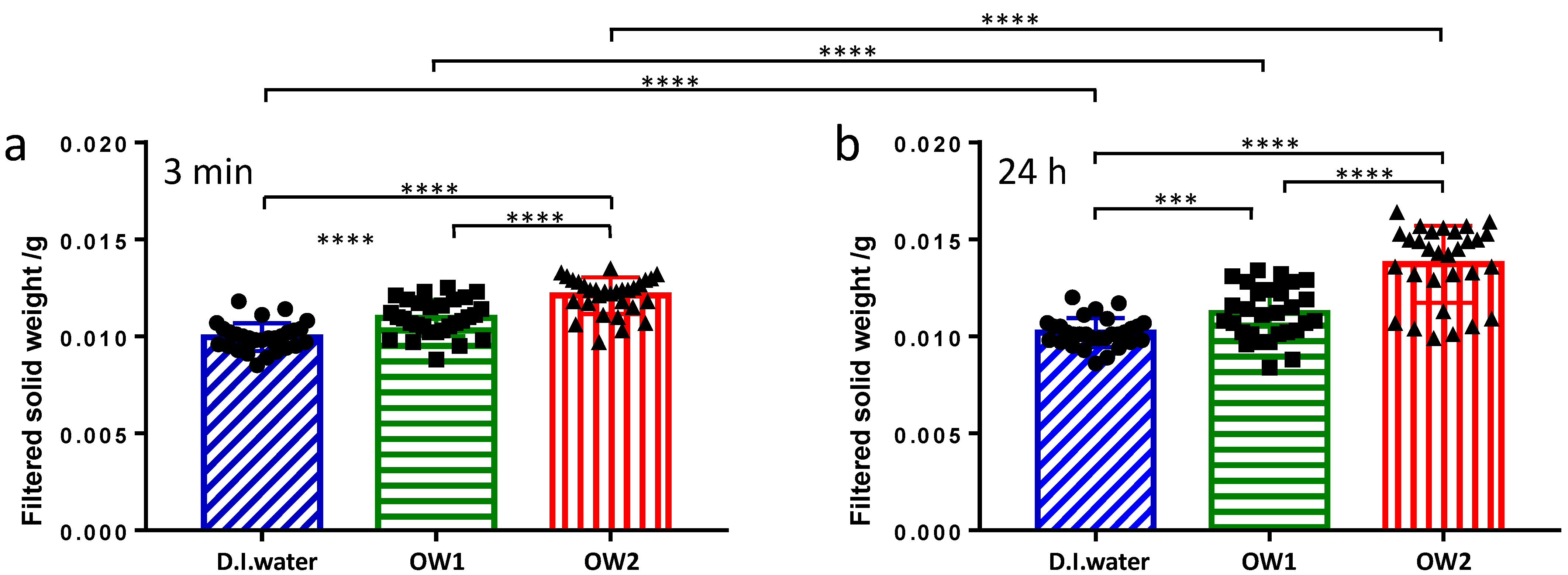
3.1. CaOx Precipitation and Gravimetric Analysis

With the aim to have information about the effect of the ions dissolved in the bottled mineral water on the formation of CaOx stones, we set up a protocol for controlled CaOx precipitation. Our method is based on the mixing of two solutions, the former containing calcium chloride and the latter containing sodium oxalate. The resulting mixture presented a calcium to oxalate ion ratio equal to 6.25. Based on the thermodynamic solubility product of calcium oxalate trihydrate, which is the initial precipitating phase at 25 °C (KSP = 5.3 × 10⁻⁹ mol2 L−6 [18]), a high apparent relative supersaturation S = ([Ca2+][Ox2−]/KSP)1/2 ≈ 30) can be computed [19,20]. For this reason, as soon as the two solutions were mixed, solid precipitation occurred from the homogeneous solution [21], with no need for additional seed crystals, as it would be required in the protocol of Ryal et al., which used 10-fold lower ion concentrations [17].
The CaOx precipitation procedure was preliminary conducted using deionized water as solvent (30 samples). The results of these blank experiments allow the effects of ion composition of the mineral waters to be highlighted. Experimental values of the filtered solid precipitate are shown in Figure 1. The average values are reported in Table 2.
The results obtained for the samples left to rest for 3 min clearly show a higher weight of filtrate precipitate for samples prepared with bottled mineral water compared to deionized water. We found an increment of about 9% for OW1 and 21% for OW2. Regarding the samples left to rest for 24 h, a further increment of the observed values was observed. Its limited extent implies that the nucleation and growth rates are fast, and consequently, the supersaturation decreases fast. In detail, this further increment is lower for the samples prepared with deionized water or OW1 (2–3%), and more pronounced for OW2 (13%). All these differences are well above the standard deviations, and the results are statistically significant.
Overall, the results indicate that the presence of additional salts in the water used to prepare the samples increases the amount of filtered precipitate, as if the solubility of CaOx in OW2 was lower than those in deionized water and OW1. This effect is different for the two considered mineral waters; thus, it does not depend on the total ion content (which is similar for the two water brands). OW1 presents a lower weight increment and, moreover, the precipitate weight shows only minor variation with increasing time.

3.2. Optical Microscopy Results

Optical microscopy was employed to further investigate the difference of the CaOx precipitation in the two oligomineral waters with similar total ion content and different ion composition. Samples were prepared following the same procedure used for the gravimetric assays. The aqueous CaOx suspensions were prepared using either OW1 or OW2 as solvent and left to rest for 1 h or 24 h. Successively, the suspensions were poured into a glass Petri dish and left to decant for 30 min in order to allow the visualization at the microscope of solid particles laid down on the glass surface. For this reason, resting times of the prepared suspensions shorter than 1 h were not considered.
Examples of the micrographs acquired on these samples are shown in Figure 2.
CaOx suspensions could contain single crystals (see Figure 2, blue circles), crystals twinned by contact (green circles) or by penetration (red circles), and complex twins. The single crystals show a tabular habit. They mainly present the pinacoid {100}, {010}, and prism forms. The predominant form is the pinacoid {100}, which shows a rhomboid shape. Contact and penetration twins are relatively rare. The twins for contact usually involve the {101} faces, but twins for contact on the {121} faces are also present. The penetration twins are cross-shaped and can involve two single crystals, two crystals with contact twinning, as well as one single crystal and one contact twin. Moreover, complex twins, which can involve multiple crystals with both single and with contact twinning, are found.
CaOx crystallizes in different polymorphs [22]: the monoclinic CaOx monohydrate (COM), which is the thermodynamically stable polymorph at room temperature and pressure; the tetragonal CaOx dihydrate (COD), which is metastable; and the triclinic CaOx trihydrate (COT), which is unstable but has been proposed to contain the polymorphs that initially precipitate at 25 °C [18] and are quickly converted in the more stable forms. COM is generally observed to be the dominant precipitated hydrate [22,23,24,25]. This is fully confirmed by the analysis of our micrographs, which predominantly show the typical COM “coffin” structures. The formation of penetration and complex twins, which are clearly detectable in the images, is favored by the presence of the ions present in the considered mineral waters [25].
In the samples obtained using OW1 as solvent, after 1 h, most of the particles are single crystals, and a few twins are also visible (Figure 2a). No evidence of crystal aggregation is found. Single crystals are also present in the sample prepared using the OW2 and analyzed after 1 h (Figure 2b); in this sample, the number of visible crystals is much lower, and no twin is found. Micrographs of the samples kept at rest for 24 h show both a higher number of crystals and an increase in their size, with the prevailing effect depending on the solvent medium. In the sample prepared using OW1 as solvent, the increase in the number of CaOx crystals is more evident than that of their size, and a large number of crystals with similar size is visualized (Figure 2c). In contrast, in the sample prepared using OW2, the presence of large crystals, coexisting with smaller ones, is found (Figure 2d). In both samples, the number of contact and penetration twins increases, and evidence of crystal aggregation is detected.
The distributions of the crystal area for the various samples are shown in Figure 3, and the results of their quantitative analysis are summarized in Table 3. In the sample prepared with OW1 and left to rest for 1 h, the crystal area ranges between 9 and 175 μm2 with an average of 63 μm2. After 24 h, the distribution shifts to higher values (from 11 and 218 μm2, mean value 84 μm2). In the sample prepared using the OW2 as solvent, after 1 h, only small single crystals are detected, whose area ranges between 11 and 62 μm2, with an average of 25 μm2. After 24 h, the distribution shows a relevant increase in the area (which ranges from 81 to 338 μm2, mean value 64 μm2). Even though the mean value observed in this case is lower than that observed with OW1 (64 vs. 84 μm2), it has to be highlighted that the distribution shown when the OW2 is used as solvent presents a tail at high area values, indicating the presence of a significant number of large crystals and thus confirming what was directly observed from the micrographs.

4. Discussion

The physicochemical investigation of the effects of trace solutes on the crystallization of calcium oxalate (CaOx), the main component of kidney stones, contributes to furnish a reliable scientific basis for the interpretation of nephrolithiasis occurring in vivo.
In the present work, we have proposed a method based on the gravimetric and microscopic study of CaOx crystal formation artificially induced in two oligomineral waters. The presence of a very similar solid residue between the two waters permits the relevance of the different salt composition at almost constant total amount to be highlighted in the comparative analysis of the results.
Our approach is inspired by the fact that the large majority of urinary stones (about 75% of the total) are composed by CaOx with a small percentage of calcium phosphate [4]. CaOx stone formation is caused by the precipitation of calcium and oxalate dissolved ions, which is driven by supersaturation in urine. We have set up a robust and reproducible protocol to induce CaOx precipitation in aqueous mixtures, using the oligomineral waters under investigation as solvent. Being the method based on the crystallization and precipitation of an insoluble solid, gravimetry (the measurement of the precipitate weight) has been chosen as an obvious technique of analysis [26,27]. The gravimetric approach is simpler and can be used for a larger number of samples compared to other methods reported in the literature, based on conductivity [28], turbidimetry [22,27,29,30], or on resistive pulse sensing (i.e., on the use of a Coulter counter [17]).
The gravimetric method we optimized is based on the separate preparation of a calcium chloride and a sodium oxalate solution. When these two solutions are mixed, CaOx precipitates. After pre-determined resting times (3 min or 24 h), the solid is filtered and weighted. This protocol can be repeated using an oligomineral water as solvent, instead of deionized water, thus highlighting the effects of the additional ions or molecules present in the medium on the solid CaOx formation. The results of our gravimetric investigation demonstrate that the considered oligomineral waters induce a higher weight of the CaOx precipitate compared to deionized water, and that this effect is more pronounced for OW2 than for OW1. The amount of filtered precipitate increases only slightly with prolonging the resting time of the samples after the precipitation has started, indicating that, because of the fast nucleation and growth, the supersaturation rapidly decreases. Particularly, the increment is lower for the samples prepared using OW1 as solvent and more relevant for the samples prepared with the OW2.
The different behavior observed for the two bottled waters shows that CaOx precipitation is not simply affected by their total ion content (which is similar), but it is specifically related to the different ion composition. A first consideration is that the increased precipitate is not related to the calcium content (which is higher in OW1). Indeed, even though it is generally believed that Ca2+ concentration directly correlates with kidney stone formation [13], the effect of calcium content is controversial, and other studies report negligible or even protective effects on nephrolithiasis [31], or even a reduction of it. In other words, the increments of the CaOx precipitate are not related to an increased concentration of reactants (calcium).
Consequently, the effect of other ions, which could affect the kinetics of the nucleation and the growth of CaOx crystals, has to be analyzed. To allow this analysis, we found optical microscopy to be perfectly suited to complement the gravimetric assays, giving information on the dimension, number, and morphology of the particles formed during solid precipitation [23,28,29,32]. Our samples contain single COM crystals along with contact, penetration, and complex twins. The number of twins increases with time and is accompanied by crystal aggregation. After 1 h from the CaOx precipitation, a lower number of crystals is visualized in the micrographs of the sample obtained using the OW2 as solvent, with respect to the sample prepared using OW1. The discrepancy between gravimetry and microscopy results could probably arise from the low dimension of the particles present in the former sample, which are sufficiently large to be filtered and weighted but too small to be clearly detected in the micrographs. Micrographs acquired after 24 h show that when OW1 is used as solvent, there is an increase in the number of crystals with no change in the distribution of the dimension and a negligible effect on the average value. In contrast, if OW2 is used as the solvent, large crystals coexist with smaller ones; consequently, even though the average dimension remains quite low, the distribution shows an evident tail at high values. Thus, optical microscopy results indicate that in OW1, crystals quickly form and grow, maintaining an almost uniform dimension with time. In OW2, the growth of crystals seems slower but proceeds with time, leading to the formation of larger crystals.
Summarizing, we can conclude that OW1, with respect to OW2, leads to the formation of particles with a more uniform distribution and a lower total weight, with limited changes over time. Particularly, this aqueous medium avoids the formation of large and heavy solid crystals. This peculiarity of OW1 should depend on its chemical and physical features. An inspection of Table 1 reveals that OW1 contains more sodium and bicarbonate, while the magnesium and sulfate content is lower compared to OW2. It could be hypothesized that any of these ions could directly affect CaOx precipitation. For example, magnesium was initially included among the ions and molecules that can inhibit solid formation. Indeed, it has been reported that concentrations higher than saturating ones can be reached and maintained with no precipitation in the presence of proteins (e.g., osteopontin and Tamm–Horsfall glycoprotein [33]), glycosaminoglycans (e.g., chondroitin sulfate and heparan sulfate [34]), and other ions (e.g., citrate, hydroxycitrate, and last but not least, magnesium [35]). However, recent results have definitely demonstrated that magnesium has little to no effect on CaOx crystallization [23]. Even sulfate has been proposed to reduce CaOx precipitation by binding with calcium [36]. However, the sulfate effect has been successively demonstrated to be very low, with only thiosulfate exerting a measurable inhibitory action [37]. Concerning sodium, its direct effect on CaOx precipitation is usually considered negligible, and in vivo experiments only show an indirect effect of a dietary NaCl increased due to the significant increase in water intake and urine dilution [38]. Bicarbonate has been reported to exert a favorable effect in reducing oxalate precipitation [39]; however, this effect has been detected at ion concentrations much higher than those of the mineral water considered by us. Overall, it seems unlikely that the differences in CaOx precipitation seen in our study could be ascribed to the specific direct effect of a single ion.
In a previous work, we studied the surface tension of the same oligomineral waters considered in the present work, finding the surface tension of OW2 to be slightly but significantly higher than that of OW1 [40]. We interpreted this difference as due to the effect of specific ions on the water structure at the interfaces. Kosmotropic ions, such as magnesium and sulfate, are able to induce ordering in the structure of the aqueous medium [41], which is reflected in a higher interfacial energy. Chaotropic ions, such as bicarbonate, reduce the water structure and exert a minor effect on the interfacial energy [42]. The interfacial energy between the aqueous medium and a solid plays a key role in the nucleation and growth of crystals of stone-forming minerals [43]. The increased interfacial tension could explain the initially limited growth of CaOx crystals in the samples prepared with OW2, in which a high amount of small crystals not detectable by optical microscopy is present. Since the interfacial tension is a decreasing function of the particle surface [44], in this sample, the formation of a few large particles is favored, which is in line with our experimental evidence. As a result, the CaOx crystallization in the mineral waters investigated in the present study seems to be affected by a “collective” physicochemical property of the aqueous medium, such as surface tension, rather than by the direct action of specific ions.
In a recent work, Li et al. have demonstrated that water whose microstructure, based on the natural network of transient hydrogen bonds, has been weakened by exposure to magnetic field, reduces solid precipitation [45]. Here, we propose that trace solutes, able to perturb water structure in a concerted way, could affect CaOx crystal formation (including nucleation and growth rates as well as supersaturation). To what extent these effects reflect on nephrolithiasis modulation in vivo remains to be investigated in future studies.

5. Conclusions

The chemical and physical properties of drinking water are associated with the probability of urinary stone formation [9,10]. These effects have long been ascribed to the direct action of specific ions/molecules (e.g., complexation of calcium ions, adsorption on the crystal surface, thus avoiding stone formation).
Our work indicates that the “collective” physicochemical properties of the aqueous medium, indirectly deriving from the ion composition and combination, could also be of importance. Particularly, the complex ion composition determines the structuring of the water molecules, which in turn determines the interfacial energy of the aqueous medium. Lastly, interfacial energy modulates the formation and growth of CaOx crystals. By combining gravimetry and optical microscopy investigation, we have shown that an oligomineral water rich in magnesium and sulfate, which enhances the water structure and increases its interfacial energy, induces the formation of a large amount of small crystals; with time, a few of them grow, leading to the formation of large crystals and particle aggregates. The overall result is an increase in the weight of the CaOx precipitate. On the other hand, an oligomineral water with relatively high bicarbonate and low magnesium and sulfate content is less structured and presents a lower interfacial tension. In this case, a quite narrow distribution of small crystals with similar size is observed. Overall, the weight of the CaOx precipitate remains low.
The present preliminary study highlights some indirect, solvent-mediated effects of water ion composition on the CaOx crystallization. Direct extrapolation of these results to the effect of drinking water on nephrolithiasis is premature. Further studies are needed to confirm the effects of each ion in combination with the others and to assess the relation between the ion compositions of drinking water and urine. A deep and wide physicochemical understanding of these phenomena constitutes the basis for a definitive trial in kidney stone patients to be performed in the future.

Author Contributions

Conceptualization, D.D.D. and G.D.; methodology, G.D. and M.R.; investigation, D.D.D., R.E. and A.F.; data curation, B.B., M.R. and R.E.; writing—original draft preparation, G.D.; writing—review and editing, B.B., M.R and G.D.; supervision, G.D.; project administration, D.D.D. and G.D.; funding acquisition, D.P. All authors have read and agreed to the published version of the manuscript.

Funding

The authors wish to thank European Union (FSE, POC Ricerca e Innovazione 2014–2020, Azione I.1 “Dottorati Innovativi con caratterizzazione Industriale”), for funding XXXV Cycle Ph.D. grant to Rodolfo Esposito. Moreover, the authors thank the “Club della Litiasi Urinaria” (CLU), for financial support.

Data Availability Statement

The data presented in this study are available on request from the corresponding author.

Conflicts of Interest

The authors declare no conflict of interest.

References

  1. Edvardsson, V.O.; Indridason, O.S.; Haraldsson, G.; Kjartansson, O.; Palsson, R. Temporal trends in the incidence of kidney stone disease. Kidney Int. 2013, 83, 146–152. [Google Scholar] [CrossRef] [Green Version]
  2. Sorokin, I.; Mamoulakis, C.; Miyazawa, K.; Rodgers, A.; Talati, J.; Lotan, Y. Epidemiology of stone disease across the world. World J. Urol. 2017, 35, 1301–1320. [Google Scholar] [CrossRef]
  3. Pak, C.Y.C. Kidney stones. Lancet 1998, 351, 1797–1801. [Google Scholar] [CrossRef]
  4. Coe, F.L.; Evan, A.; Worcester, E. Kidney stone disease. J. Clin. Investig. 2005, 115, 2598–2608. [Google Scholar] [CrossRef] [PubMed] [Green Version]
  5. Jabbar, F.; Asif, M.; Dutani, H.; Hussain, A.; Malik, A.; Kamal, M.A.; Rasool, M. Assessment of the role of general, biochemical and family history characteristics in kidney stone formation. Saudi J. Biol. Sci. 2015, 22, 65–68. [Google Scholar] [CrossRef] [Green Version]
  6. Baatiah, N.Y.; Alhazmi, R.B.; Albathi, F.A.; Albogami, E.G.; Mohammedkhalil, A.K.; Alsaywid, B.S. Urolithiasis: Prevalence, risk factors, and public awareness regarding dietary and lifestyle habits in Jeddah, Saudi Arabia in 2017. Urol. Ann. 2020, 12, 57–62. [Google Scholar] [CrossRef]
  7. Daudon, M.; Jungers, P. Drug-induced renal calculi: Epidemiology, prevention and management. Drugs 2004, 64, 245–275. [Google Scholar] [CrossRef]
  8. Xu, C.; Zhang, C.; Wang, X.-L.; Liu, T.-Z.; Zeng, X.-T.; Li, S.; Duan, X.-W. Self-fluid management in prevention of kidney stones: A PRISMA-compliant systematic review and dose–response meta-analysis of observational studies. Medicine 2015, 94, e1042. [Google Scholar] [CrossRef]
  9. Borghi, L.; Meschi, T.; Schianchi, T.; Briganti, A.; Guerra, A.; Allegri, F.; Novarini, A. Urine volume: Stone risk factor and preventive measure. Nephron 1999, 81, 31–37. [Google Scholar] [CrossRef]
  10. Mirzazadeh, M.; Nouran, M.G.; Richards, K.A.; Zare, M. Effects of drinking water quality on urinary parameters in men with and without urinary tract stones. Urology 2012, 79, 501–507. [Google Scholar] [CrossRef] [PubMed]
  11. Haddock, R.L.; Olson, D.R.; Backer, L.; Malilay, J. Urolithiasis, urinary Cancer, and home drinking water source in the united states territory of Guam, 2006–2010. Int. J. Environ. Res. Public Health 2016, 13, 523. [Google Scholar] [CrossRef] [Green Version]
  12. Chatterjee, D.; Verma, V.; Amiti; Rohil, S.; Arunai Nambi Raj, N.; Vidya, R. Demographic study of water hardness with potential to predict formation of renal crystals. Adv. Mater. Res. 2012, 584, 504–508. [Google Scholar] [CrossRef]
  13. Panhwar, A.H.; Kazi, T.G.; Afridi, H.I.; Shaikh, H.R.; Arain, S.A.; Arain, S.S.; Brahman, K.D. Evaluation of calcium and magnesium in scalp hair samples of population consuming different drinking water: Risk of kidney stone. Biol. Trace Elem. Res. 2013, 156, 67–73. [Google Scholar] [CrossRef] [PubMed]
  14. Domrongkitchaiporn, S.; Stitchantrakul, W.; Kochakarn, W. Causes of hypocitraturia in recurrent calcium stone formers: Focusing on urinary potassium excretion. Am. J. Kidney Dis. 2006, 48, 546–554. [Google Scholar] [CrossRef] [PubMed]
  15. Davydova, N.K.; Sergeev, V.N.; Girbul, E. The role of humous acids in Acqua di Fiuggi mineral water in degrading stones formed in the urinary tract. Pharm. Chem. J. 2014, 48, 589–594. [Google Scholar] [CrossRef]
  16. Kershaw, K.N.; Klikuszowian, E.; Schrader, L.; Siddique, J.; Van Horn, L.; Womack, V.Y.; Zenk, S.N. Assessment of the influence of food attributes on meal choice selection by socioeconomic status and race/ethnicity among women living in Chicago, USA: A discrete choice experiment. Appetite 2019, 139, 19–25. [Google Scholar] [CrossRef]
  17. Ryal, R.L.; Bagley, C.J.; Marshall, V.R. Independent assessment of the growth and aggregation of calcium oxalate crystals using the coulter counter. Investig. Urol. 1981, 18, 401–405. [Google Scholar]
  18. Gardner, G.L. Nucleation and crystal growth of calcium oxalate trihydrate. J. Cryst. Growth 1975, 30, 158–168. [Google Scholar] [CrossRef]
  19. Bramley, A.S.; Hounslow, M.J.; Ryall, R.L. Aggregation during precipitation from solution. Kinetics for calcium oxalate monohydrate. Chem. Eng. Sci. 1997, 52, 747–757. [Google Scholar] [CrossRef]
  20. Finlayson, B. Calcium stones: Some physical and clinical aspects, Chapter 10. In Calcium Metabolism in Renal Failure and Nephrolithiasis; David, D.S., Ed.; JohnWiley & Sons: New York, NY, USA, 1977. [Google Scholar]
  21. Bashar, A.; Townshend, A. Comparative study of methods for precipitating calcium oxalate from homogeneous solution. Talanta 1966, 13, 1123–1128. [Google Scholar] [CrossRef]
  22. Hsu, Y.-C.; Lin, Y.-H.; Shiau, L.-D. Effects of various inhibitors on the nucleation of calcium oxalate in synthetic urine. Crystals 2020, 10, 333. [Google Scholar] [CrossRef] [Green Version]
  23. Kuliasha, C.A.; Rodriguez, D.; Lovett, A.; Gower, L.B. In situ flow cell platform for examining calcium. Oxalate and calcium phosphate crystallization on films of basement membrane extract in the presence of urinary ‘inhibitors’. CrystEngComm 2020, 22, 1448–1458. [Google Scholar] [CrossRef]
  24. Stanković, A.; Šafranko, S.; Jurišić, K.; Balić, I.; Bijelić, J.; Jokić, S.; Medvidović-Kosanović, M. Investigation of system complexity and addition of vitamin C on calcium oxalate precipitation. Chem. Pap. 2020, 74, 3279–3291. [Google Scholar] [CrossRef]
  25. Stanković, A.; Šafranko, S.; Kontrec, J.; Njegić DžakulaJurišić, B.; Lyons, D.M.; Marković, B.; Kralj, D. Calcium oxalate precipitation in model systems mimicking the conditions of hyperoxaluria. Cryst. Res. Technol. 2019, 54, 1800210. [Google Scholar] [CrossRef]
  26. Benalia, H.; Djeridane, A.; Bensafieddine, F.; Yousfi, M. High in vitro antiurolithiatic effect of Pituranthos scoparius roots extracts. Pharmacologyonline 2016, 1, 31–43. [Google Scholar]
  27. Bensatal, A.; Rahmoun, D.; Ardja, S.A.; Cheikh, M.; Kahouadji, A.; Bekhit, M. In vitro antilithiasic activity of saponins rich fraction from the leaves of Zizyphus lotus. Int. J. Green Pharm. 2020, 14, 280–285. [Google Scholar] [CrossRef]
  28. Liu, Y.; Mao, H.; Liu, X.; Qiao, L.; Guo, R. Calcium oxalate crystallization in the presence of amphiphilic phosphoproteins. CrystEngComm 2014, 16, 8841–8851. [Google Scholar] [CrossRef]
  29. Khan, A.; Khan, S.R.; Gilani, A.H. Studies on the in vitro and in vivo antiurolithic activity of Holarrhena antidysenterica. Urol. Res. 2012, 40, 671–681. [Google Scholar] [CrossRef] [PubMed] [Green Version]
  30. Siddiqui, W.A.; Shahzad, M.; Shabbir, A.; Ahmad, A. Evaluation of anti-urolithiatic and diuretic activities of watermelon (Citrullus lanatus) using in vivo and in vitro experiments. Biomed. Pharmacother. 2018, 97, 1212–1221. [Google Scholar] [CrossRef]
  31. Marangella, M.; Vitale, C.; Petrarulo, M.; Rovera, L.; Dutto, F. Effects of mineral composition of drinking water on risk for stone formation and bone metabolism in idiopathic calcium nephrolithiasis. Clin. Sci. 1996, 91, 313–318. [Google Scholar] [CrossRef] [Green Version]
  32. Hsu, Y.-C.; Pan, L.-C.; Shiau, L.-D. A Photomicroscopic study on the growth rates of calcium oxalate crystals in a new synthetic urine without inhibitors and with various inhibitors. Crystals 2021, 11, 223. [Google Scholar] [CrossRef]
  33. Mohamaden, W.I.; Wang, H.; Guan, H.; Meng, X.; Li, J. Osteopontin and Tamma-Horsefall proteins–Macromolecules of myriad values. J. Basic Appl. Zool. 2014, 67, 158–163. [Google Scholar] [CrossRef] [Green Version]
  34. Turudic, D.; Batinic, D.; Golubic, A.T.; Lovric, M.; Milosevic, D. Calcium oxalate urolithiasis in children: Urinary promoters/inhibitors and role of their ratios. Eur. J. Pediatr. 2016, 175, 1959–1965. [Google Scholar] [CrossRef] [PubMed]
  35. Kim, D.; Rimer, J.D.; Asplin, J.R. Hydroxycitrate: A potential new therapy for calcium urolithiasis. Urolithiasis 2019, 47, 311–320. [Google Scholar] [CrossRef]
  36. Rodgers, A.; Gauvin, D.; Edeh, S.; Allie-Hamdulay, S.; Jackson, G.; Lieske, J.C. Sulfate but not thiosulfate reduces calculated and measured urinary ionized calcium and supersaturation: Implications for the treatment of calcium renal stones. PLoS ONE 2014, 9, e103602. [Google Scholar] [CrossRef] [Green Version]
  37. Landry, G.M.; Hirata, T.; Anderson, J.B.; Cabrero, P.; Gallo, C.J.R.; Dow, J.A.T.; Romero, M.F. Sulfate and thiosulfate inhibit oxalate transport via a dPrestin (Slc26a6)-dependent mechanism in an insect model of calcium oxalate nephrolithiasis. Am. J. Physiol. Renal. Physiol. 2016, 310, F152–F159. [Google Scholar] [CrossRef] [Green Version]
  38. Queau, Y.; Bijsmans, E.S.; Feugier, A.; Biourge, V.C. Increasing dietary sodium chloride promotes urine dilution and decreases struvite and calcium oxalate relative supersaturation in healthy dogs and cats. J. Anim. Physiol. Anim. Nutr. 2020, 104, 1524–1530. [Google Scholar] [CrossRef] [Green Version]
  39. Ferraro, P.M.; Baccaro, R.; Baroni, S.; D’Alessandri, L.; Carpenito, C.; Di Daniele, N.; Urbani, A.; Gambaro, G. Effect of water composition and timing of ingestion on urinary lithogenic profile in healthy volunteers: A randomized crossover trial. J. Nephrol. 2021, 34, 875–881. [Google Scholar] [CrossRef]
  40. Prezioso, D.; Di Domenico, D.; Pane, M.; Ciccarelli, D.; D’Errico, G. Ion specificity in determining physico-chemical properties of drinking water. Food Sci. Technol. 2019, 39, 485–490. [Google Scholar] [CrossRef] [Green Version]
  41. Tobias, D.J.; Hemminger, J.C. Getting specific about specific ion effects. Science 2008, 319, 1197–1198. [Google Scholar] [CrossRef]
  42. Du, H.; Liu, J.; Ozdemir, O.; Nguyen, A.V.; Miller, J.D. Molecular features of the air/carbonate solution interface. J. Colloid Interface Sci. 2008, 318, 271–277. [Google Scholar] [CrossRef] [PubMed]
  43. Wu, W.; Gerard, D.E.; Nancollas, G.H. Nucleation at surfaces: The importance of interfacial energy. J. Am. Soc. Nephrol. 1999, 10, S355–S358. [Google Scholar] [PubMed]
  44. Goujon, F.; Dequidt, A.; Ghoufi, A.; Malfreyt, P. How does the surface tension depend on the surface area with coarse-grained models? J. Chem. Theory Comput. 2018, 14, 2644–2651. [Google Scholar] [CrossRef]
  45. Li, Y.; Pan, J.; Zhang, Y.; Chang, Y.; Yang, X.; Yang, B.; Mao, X.; Wang, Z.; Gao, B.; Lu, X. Effects of small molecules water that may retard kidney stone formation. Int. Urol. Nephrol. 2018, 50, 225–230. [Google Scholar] [CrossRef]
Figure 1. The weight of the precipitate recovered by filtration of the solid CaOx aqueous suspensions obtained after 3 min (a) or 24 h (b) by mixing calcium chloride and sodium oxalate solutions. The different aqueous media used as solvent to prepare the solutions are indicated (D.I. water stands for deionized water). Bars represent the mean value ± standard deviation (**** p < 0.0001; *** p < 0.001).
Crystals 11 01507 g001
Figure 2. Micrographs of the CaOx suspensions obtained by mixing calcium chloride and sodium oxalate solution and prepared using OW1 (a,c) or OW2 (b,d) as solvent. Samples were left to rest for 1 h (a,b) or 24 h (c,d). The different crystal forms are highlighted with colored circles (blue circles for single crystals, green circles for contact twins, red circles for penetration twins).
Crystals 11 01507 g002
Figure 3. Distributions of the crystal dimension in CaOx suspensions obtained by mixing calcium chloride and sodium oxalate solution and prepared using OW1 or OW2 as solvent. Samples were left at rest for 1 h or 24 h.
Crystals 11 01507 g003
Table 1. Values of the physicochemical parameters reported on the labels of the bottled mineral waters considered in this study.
Physicochemical ParameterUnitsOW1OW2
Solid residue at 180 °Cmg L−1174.1162
Specific electric conductivity at 20 °CμS cm−1276.3251
pH at the spring temperature-7.567.9
Ca++mg L−157.3635.9
Na+mg L−14.132.0
Mg++mg L−13.2312.6
K+mg L−10.350.5
HCO3mg L−1182.1148
SO4mg L−16.7519.3
NO3mg L−11.104.4
Table 2. Average weight (± standard deviation) of the precipitate recovered by filtration of the solid CaOx aqueous suspensions obtained by mixing the calcium chloride and sodium oxalate solutions, which were prepared using the water indicated.
Resting Time after MixingDeionized WaterOW1OW2
Filtered Precipitate Weight/mg3 min9.96 ± 0.1310.9 ± 0.212.1 ± 0.2
24 h10.19 ± 0.1511.2 ± 0.313.7 ± 0.4
Table 3. Quantitative analysis of the area of the CaOx crystals visualized in the micrographs of the aqueous suspensions obtained by mixing the calcium chloride and sodium oxalate solutions, prepared using the oligomineral water indicated.
Resting Time after MixingNumber of Analyzed CrystalsDistribution Range of Crystal Area/μm2Mean Crystal Area/μm2
minmax
OW11 h738917563
24 h10321121884
OW21 h72116225
24 h991833864
Publisher’s Note: MDPI stays neutral with regard to jurisdictional claims in published maps and institutional affiliations.

