Article

Rapid Microwave Synthesis of β-SnWO4 Nanoparticles: An Efficient Anode Material for Lithium Ion Batteries

by
H. N. Sumedha
1,
Mabkhoot A. Alsaiari
2,3,
Mohammed S. Jalalah
2,4,
M. Shashank
1,5,
Fahad A. Alharthi
6,*,
Naushad Ahmad
6,
Jari S. Algethami
3,
Vishwanth Vishwanth
7 and
Nagaraju Ganganagappa
1,*
1
Energy Materials Research Laboratory, Department of Chemistry, Siddaganga Institute of Technology, Tumakuru 572103, India
2
Promising Center for Sensors and Electronic Devices (PCSED), Advanced Materials and Nano Research Center, Najran University, P.O. Box 1988, Najran 11001, Saudi Arabia
3
Chemistry Department, Faculty of Science and Art at Sharurah, Najran University, Sharurah 68342, Saudi Arabia
4
Department of Electrical Engineering, Faculty of Engineering, Najran University, Najran, P.O. Box 1998, Najran 11001, Saudi Arabia
5
Department of Studies and Research in Industrial Chemistry, School of Chemical Sciences, Kuvempu University, Shankaragatta 577451, India
6
Department of Chemistry, College of Science, King Saud University, P.O. Box 2455, Riyadh 11451, Saudi Arabia
7
Department of Chemistry, Jyothi Institute of Technology, Bangalore 560082, India
*
Authors to whom correspondence should be addressed.
Crystals 2021, 11(4), 334; https://doi.org/10.3390/cryst11040334
Submission received: 26 February 2021 / Revised: 23 March 2021 / Accepted: 23 March 2021 / Published: 26 March 2021
(This article belongs to the Special Issue Functional Nanomaterials for Advanced Applications)

Abstract

:
We report a facile synthesis of β-SnWO4 nanoparticles via a microwave heat treatment using SnCl2 and H2WO4 in the presence of tamarind seed powder. An X-ray diffraction analysis confirmed a crystalline nature revealing a cubic structure of β-SnWO4 nanoparticles. The morphological features were visualized using a scanning electron microscope that exhibited homogenously distributed clusters of nanoparticles, which were further confirmed using a transmission electron microscope. The micrographs also displayed some porosity. Energy dispersive X-ray spectroscopy confirmed the elemental contents such as tin, oxygen and tungsten in the same stoichiometric ratio as expected by the respective empirical formula. A high-resolution transmission electron microscope was used to find the d-spacing, which was ultimately used to analyze the structural parameters. The spectrum obtained using Fourier transform infrared spectroscopy illuminated different stretching vibrations. Additionally, a Barrett–Joyner–Halenda analysis was carried out to investigate the N2 adsorption-desorption isotherm as well as to govern the pore size distribution. Cyclic voltammetry measurements were implemented to analyze the ongoing electrode reactions throughout the charge/discharge for the β-SnWO4 nanostructures. The galvanometric charge/discharge curves for β-SnWO4 are also discussed. A high specific capacitance (600 mAhg–1 at 0.1 C) and excellent columbic efficiency (~100%) were achieved.

1. Introduction

The demand of energy storage devices with enhanced capabilities has increased manifold in the 21st century. In this context, researchers worldwide are looking for such devices and materials that might lead the scientific community to cope with energy storage needs by designing and developing innovative resources. In the current era, energy renovation is at the forefront of research [1]. Lithium-ion batteries (LIBs) are considered to be promising because of their high power density, life cycle and gravimetric and volumetric energy along with long self-discharge properties. Therefore, LIBs are still unbeaten in the energy storage industry [2]. Meanwhile, the earth’s crust has a limited availability of lithium resources and their use is beyond the limits. Consequently, they are becoming costly [3,4]. Alternatively, batteries are facing various problems including storage capacity and chemical stability [5]. Additionally, its specific capacity is extremely low, i.e., 372 mAhg–1. To resolve such issues, there is a need to investigate other favorable electrode materials exhibiting promising specific capacities and chemical stabilities. In the realm of rechargeable LIBs, various nanomaterials are under discussion as potential electrode materials.
Historically, LIBs have shown a massive intensification through two decades in specific energy values ranging from 90 to 250 mAhg–1 [6]. In this regime, there have been many breakthroughs in energy storage devices (as electrode materials) due to the advancement in the field of nanomaterials [7,8,9,10,11]. Nanomaterials have exceptionally attractive physical and chemical properties; hence, they are reflected as being suitable for energy storage devices. The use of single metal oxides including TiO2 [12], SnO2 [13] and Ta2O5 [14] has led to a loss in capacity with cycling. A literature survey reveals a good electrochemical performance of β-SnWO4 nanoparticles. These nanoparticles also offer an excellent diffusion of Li+ ions along exceptional cyclic stability as well as a high capacity. Furthermore, they are widely utilized as anode materials, for water splitting, for photodynamic tumor therapy, for humidity sensing and photocatalytic degradation, for example. Various routes have been adopted for the synthesis of SnWO4 including hydrothermal synthesis [15], a combustion method and a microwave method [16]. Initially, the gas sensing properties as well as the phase structure of α- and β-SnWO4 were reported by Solis and Lantoo by employing different spectroscopic techniques [16,17]. Ede et al. adopted microwave synthesis to prepare SnWO4 [16]. Warmuth et al. employed a microwave assisted method to synthesize β-SnWO4 nanoparticles [18]. Furthermore, the photoelectrochemical properties of β-SnWO4 nanomaterials were analyzed by Cho et al. [19]. ZnWO4 nanocrystals/reduced graphene oxide hybrids were prepared by Wang Xiao et al. via a facile hydrothermal route that revealed a performance of 477 mAhg−1 after 40 cycles [6]. α-SnWO4/reduced graphene oxide nanoparticles were synthesized by Renkun Huang et al. through a facile single step hydrothermal reaction exhibiting a capacity of 500 mAhg–1 after 20 cycles [20].
The search for the finest electrode material for LIBs with a high capacity and that can easily be diffused into Li+ ions along with an increased life cycle is still under investigation. Owing to the promising features associated with β-SnWO4 nanoparticles and their wide utilization in energy storage applications, here we report a facile synthesis based on a rapid microwave heat treatment by the reaction of SnCl2 and H2WO4 in the presence of tamarind seed powder. We present a detailed analysis of the electrochemical performance for potential use as an electrode material in LIBs. The material owed an exceptional specific capacitance (600 mAhg−1 at 0.1 C) and a remarkable columbic efficiency nearly approaching 100% was achieved.

2. Experiment

2.1. Synthesis of β-SnWO4 Nanoparticles

β-SnWO4 nanoparticles were prepared through a rapid microwave heat treatment by the reaction of tin (II) chloride (SnCl2) and tungstic acid (H2WO4) in the presence of tamarind seed powder. A typical procedure involved the addition of 220 mg of SnCl2 and 249 mg of H2WO4 in a clean and dried crucible. In the meantime, 220 mg of tamarind seed powder was also added followed by the addition of 10 mL of distilled water. The obtained blend was stirred for about 10 min. At that time, the crucible was kept in a microwave oven at 900 W for 5 min. The resultant product was taken out of the oven and allowed to cool to room temperature naturally. It was then ground in a clean dried mortar and calcined at 700 °C for 4 h under static air. A schematic of the sample synthesis is presented in Figure 1.

2.2. Characterization

A Rigaku’s (Tokyo, Japan) Smart Lab X-ray diffractometer (XRD) with Cu-Kα radiations (λ = 1.5406 Å) was used to determine the crystalline nature and the confirmation of the phase. The molecular vibrations and structures of the materials were analyzed from 350–4000 cm−1 using a Bruker (Billerica, Massachusetts, United States) Alpha-P Fourier transforms infrared (FTIR) spectrometer by the KBr pellet method. JEOL(Tokyo, Japan)-JCM-6000 Plus scanning electron microscopy (SEM) and JEOL-JEM-2100 transmission electron microscopy (TEM) were utilized to observe the morphological features, particle size and structural parameters of β-SnWO4 nanoparticles, respectively. Using a Quanta chrome Nova 2200E-BET (surface area analyzer), the pore size and surface area were determined.

2.3. Electrode Fabrication for the Lithium Ion Battery

A coin cell was made to examine the electrochemical performance of β-SnWO4 nanomaterials by fabricating it on a copper foil. The slurry was prepared by mixing the active materials (β-SnWO4 nanoparticles), PVDF (polyvinylidene fluoride) and acetylene black using a weight ratio of 80:5:15, respectively, using a few drops of N-methyl pyrrolidine as a solvent. The prepared slurry was uniformly coated on a Cu foil and subsequently dried at 100 °C for 12 h in a vacuum oven. During the coil cell fabrication, a polypropylene membrane (Celgard-2400) was utilized as a separator. LiPF6 (1M) dissolved in ethylene carbonate, dimethyl carbonate and diethyl carbonate (2:2:1) was used as an electrolyte. Cyclic voltammetry (CV) and galvanostatic charge-discharge (GCD) studies were recorded using a Bio-logic BCS 805 battery system. The CV measurements were performed in the voltage window from 0.01 to 3 V at a 50 mV/s scan rate. The voltage range of 0.01–3 V was also applied during the GCD analysis.

3. Results and Discussion

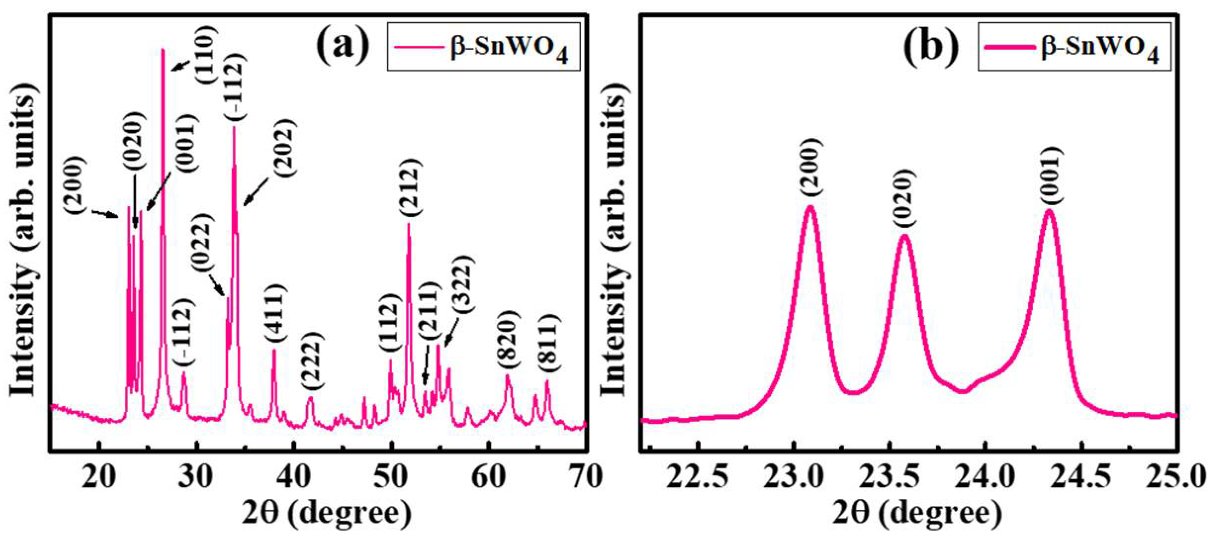
The XRD pattern of the sample was recorded in the 2θ range of 15–70°, as shown in Figure 2a. The existence of intensity peaks corresponding to the (200), (020), (001), (100), (−112), (202), (411), (222), (112), (212), (211), (322), (820) and (811) planes was perfectly matched with JCPDS (Joint Committee on Powder Diffraction Standards) reference no. 01-070-1497 [16], a characteristic reference pattern of β-SnWO4. It confirmed the wolframite-type cubic crystal structure of the sample, having space group no. 198 and a space group category of P-213 [21]. There were no extra peaks signifying the phase purity of the sample [18]. Further, the magnified image of a few peaks was illustrated to differentiate between these peaks (Figure 2b). The lattice parameter (a) was calculated from following equation:
a   =   λ h 2 + k 2 + l 2 2 s i n θ B .
The lattice parameter was measured to be a = b = c = 7.298 Å, resulting in a unit cell volume of 388.697 Å3. The average crystallite size (Dhkl) was determined using Scherrer’s formula as described by Equation (2) [22]:
D   =   k λ β c o s θ B
where β was line broadening termed as full width at half maximum (FWHM), θB represented Bragg’s angle, λ was the wavelength with a value of 1.54 Å and k = 0.9, the shape factor. The crystallite size as evaluated by this formula was 28.63 nm.
The crystallite size of the as-synthesized samples was also calculated using the Williamson–Hall (W–H) method as given by Equation (3) [23]:
β cos θ   =   k λ / D   +   4 ε sin θ
where ε represented the lattice strain. Figure 3 indicates the W–H plot of β-SnWO4 plotted between 4sinθ at the x-axis and βcosθ along the y-axis. The D was measured from the y-intercept and depicted to be 24.32 nm while ε measured the slope of the linear fit and was measured to be 7.2 × 10⁻². The linear fit of the W–H plot was expected to be a straight line parallel to x-axis but in the manifestation of ε it gave a non-zero slope [24]. The measured crystallite size (24.32 nm) from the W–H plot was in close agreement with Scherrer’s formula (28.63 nm).
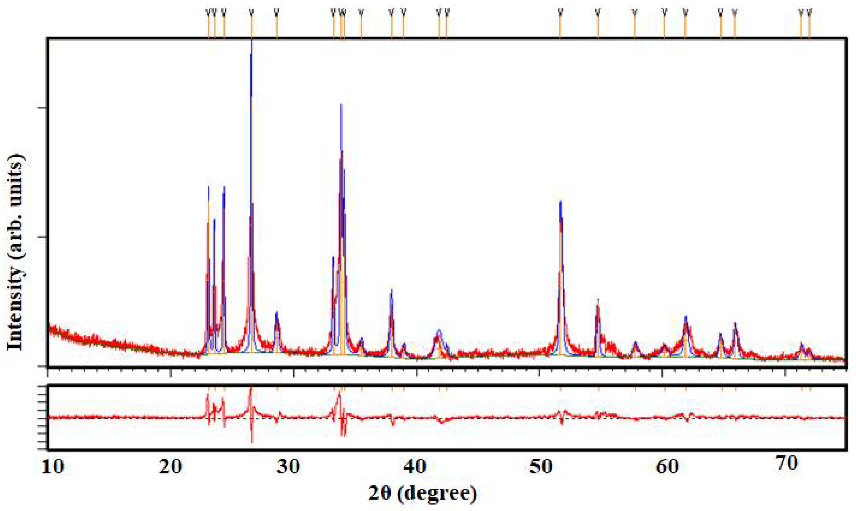
The accurate phase analysis and quantitative phase measurement of β-SnWO4 were determined through a Rietveld refinement analysis via X’pert HighScore (Worcestershire, UK) Plus software. Figure 4 shows the well fitted XRD pattern of β-SnWO4 at the same positions as prescribed in Figure 2. No significant phase transformation was perceived attributing the pure phase formation of the specimen. The structural reliability was evaluated by measuring R-factors such as the R-profile (Rp), the R-expected (Rexp), the R-profile (Rwp) and the R-Bragg (RB) as well through the following Equations [25]:
R w p   =   [ Ʃ i w i   ( I c I 0 ) Ʃ i w i   I 0 2 ] 1 / 2 .
R e x p   =   [ N P Ʃ i w i   I 0 2 ] 1 / 2 .
R B r a g g   =   Ʃ   | I ( 0 ) I ( c ) | Ʃ   | I ( 0 ) | .
R p   =   Ʃ   | I ( 0 i ) I ( c i ) | Ʃ   | I ( 0 i ) | .
Here, P and N represented the number of refined parameters and the number of experimentally observed points and Io and Ic presented the observed and calculated integrated intensities, respectively. Accordingly, wi was the illustrating weight factor at i point of the diffraction profile and the goodness of fit (χ2) was measured using Equation (8), given as [25]:
χ 2   =   R w p R e x p .
The measured parameters from a Rietveld refinement analysis are summarized in Table 1. The obtained smaller value of χ2 depicted the pure phase development of β-SnWO4.
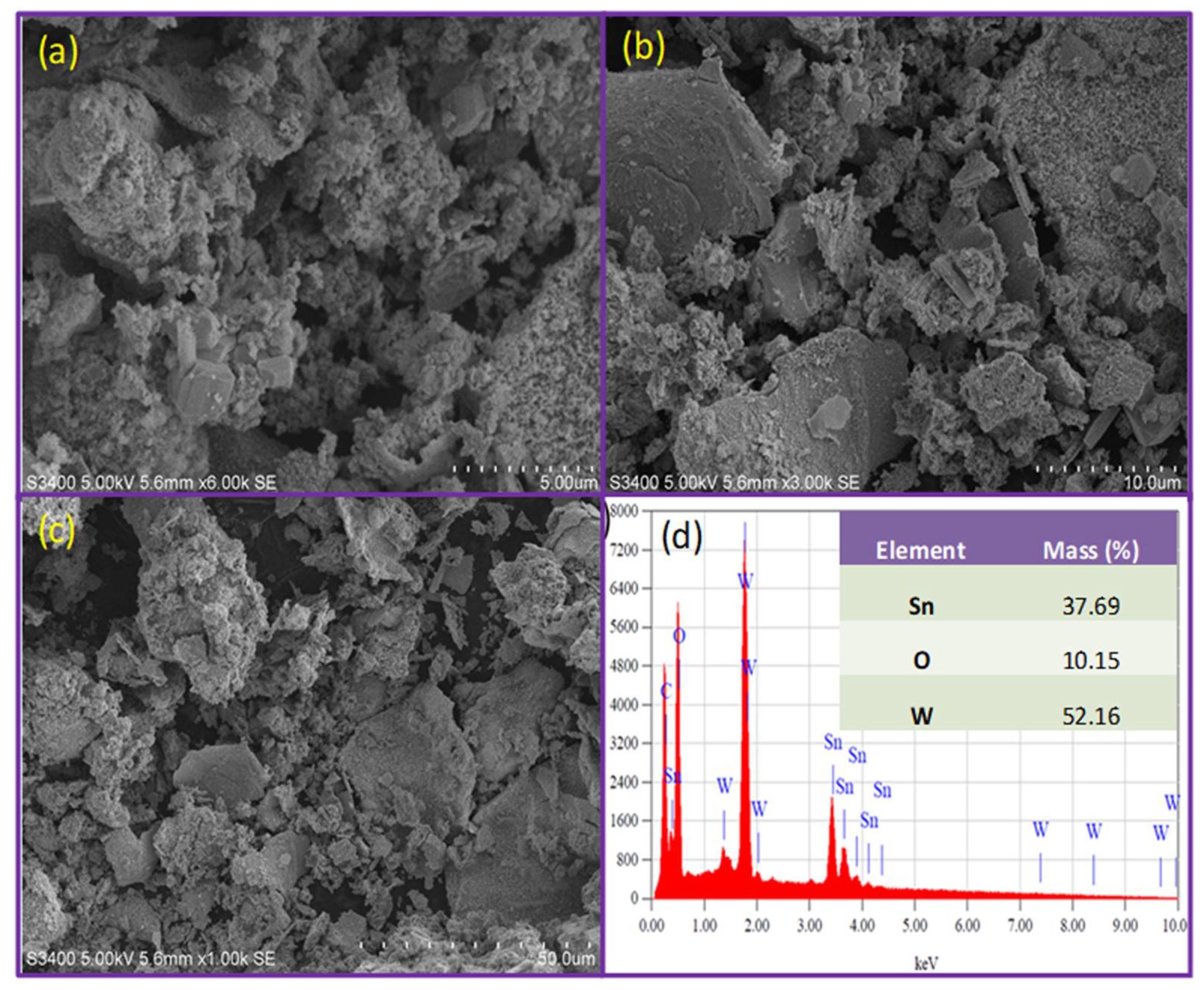
The SEM images for β-SnWO4 are illustrated in Figure 5a–c. The micrographs were obtained at diverse magnifications revealing the formation of β-SnWO4 nanoclusters along with a few sorts of layered and rough structures as well. Stone-like arbitrary and amalgamated particles of various sizes and shapes could be perceived in the micrographs. Figure 5b,c illustrates some rock-like structures along with very small particles. Furthermore, the images also revealed the porosity present in the samples.
The EDX spectrum of β-SnWO4 was profiled (Figure 5d). It confirmed the existence of all of the constituent elements such as tin, oxygen and tungsten in the sample. The table shown in an inset of Figure 5d demonstrates the mass percentages of all of the elements.
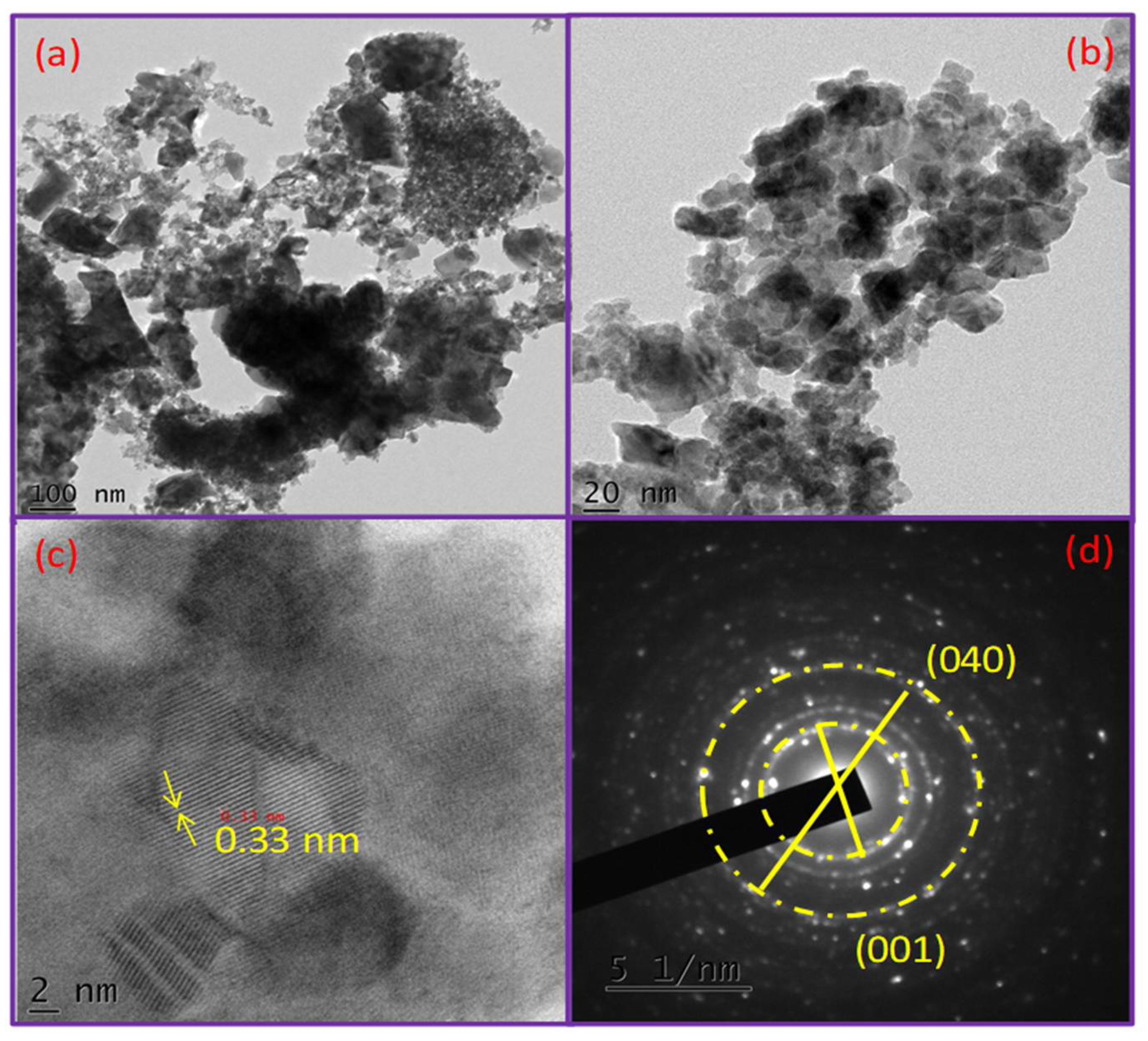
The morphology of β-SnWO4 was also examined using TEM and high resolution transmission electron microscope (HR-TEM) to obtain a detailed breakdown of the structures and to determine the interlayer d-spacing. Figure 6a,b illustrates the TEM images obtained at different magnifications. The micrographs profiled almost spherical shaped particles accompanied by some amalgamation. Further, a few pores could also be professed in the micrographs. The interlayer d-spacing was examined through HR-TEM and found to be 0.33 nm [24] and can be seen in Figure 6c. The lattice fringes of the (001) lattice plane of a wolframite-type cubic crystal structure β-SnWO4 could be perceived (Figure 5d) as sketched by a selected area electron diffraction (SEAD) pattern. The occurrences of bright rings in the concerning pattern illustrated the company of highly crystallite particles. The existence of the lattice plane was in good agreement with the XRD pattern.
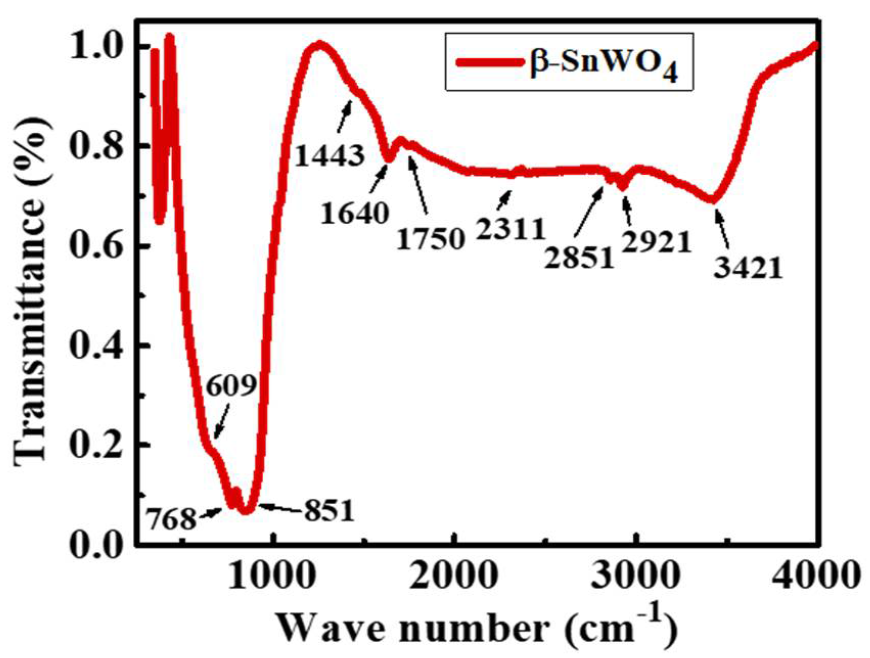
The FTIR spectrum for β-SnWO4 was recorded in the range of 350–4000 cm−1, as shown in Figure 7. The peak appearing around 609 cm–1 demonstrated an asymmetric stretching vibration of the W O 6 6 group [24,26]. In the same way, the peaks emerging at 768 and 851 cm−1 illuminated the stretching vibrations of the W–W and W–O–W bridging bonds [26]. The structural vibrations of the C–C or C–O–C bonds were in agreement with the peak occurring at 1443 cm−1. Further, the existence of a strong peak at 1640 cm−1 and a broad peak at 3421 corresponded with the stretching and bending vibrations of the hydroxyl (–OH) group. The symmetric stretching vibration of the C–H bond could be observed from the peak befalling at 2921 cm−1. The co-existence of all of the stretching and vibrational peaks confirmed the formation of the β-SnWO4 structure [27].
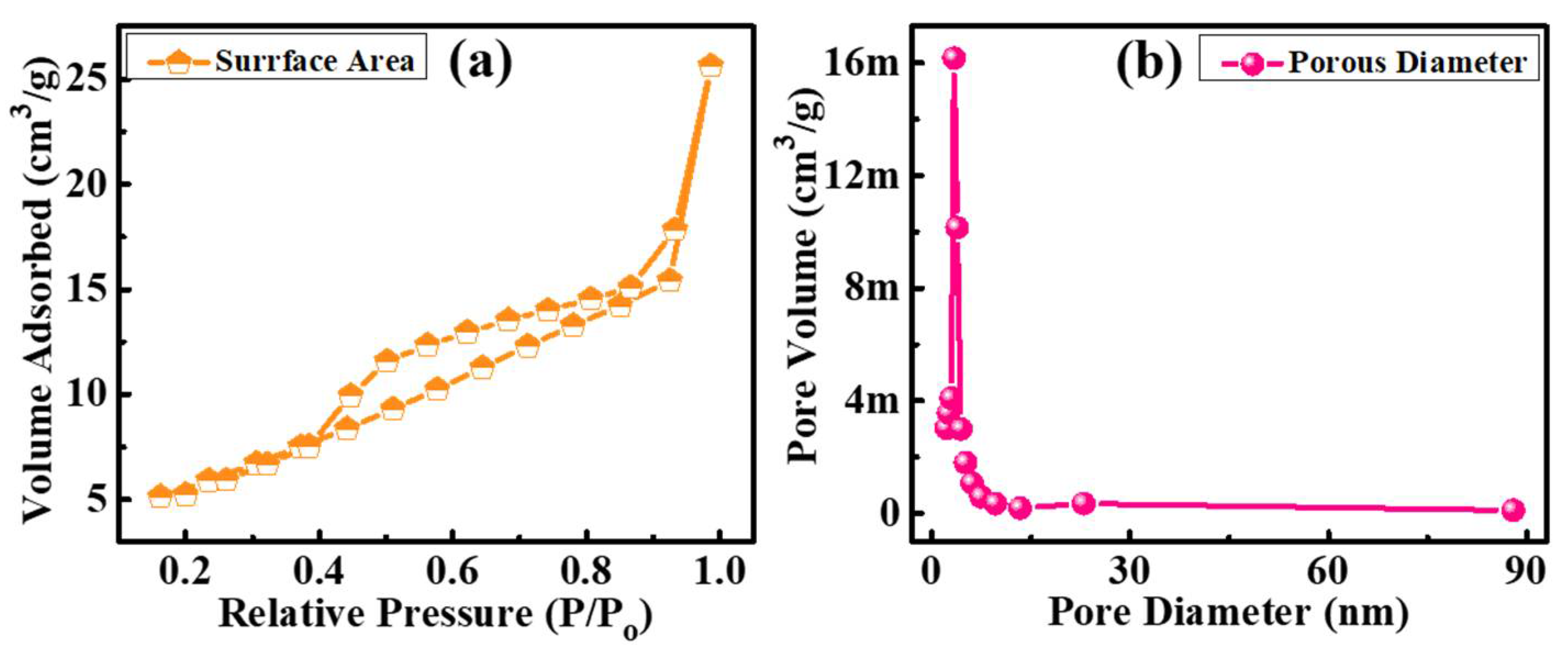
A Barrett–Joyner–Halenda (BJH) analysis was carried out to investigate the N2 adsorption-desorption isotherm as well as to determine the pore size distribution. The curve sketched in Figure 8a clearly offered a desorption hysteresis of a type-IV isotherm curve [28,29] obtained under a relative pressure (P/Po) of 0.1–1.0. The characteristics of the mesostructured materials could be well explained on the basis of sharp capillary condensation steps occurring between the relative pressure intervals of 0.40–0.95. It also perceived an irregularity of mesopores in the product. The BJH pore size distribution is profiled in Figure 8b. A sharp peak could be depicted in the beginning of the distribution curve demonstrating an average pore diameter of 3.35 nm. The pore volume and surface area of the particles in the sample were determined using a Brunauer–Emmett–Teller (BET) analysis. Accordingly, due to its plentiful range of pore diameters (2–80 nm), it could accumulate a higher number of charge carriers thus improving the electrolyte performance of the current specimen. Hence, it can offer its utilization in a wide collection of potential applications.
CV measurements were implemented to analyze the ongoing electrode reactions throughout the charge/discharge for the β-SnWO4 nanostructures. The CV profile was mapped under various current cycles and a potential window of 0.01 to 3.0 V and is demonstrated in Figure 9a. The composition clearly described the actuality of the anodic/cathodic peaks. Similar current humps could be perceived from all of the current cycles. During lithiation, three peaks could be depicted specifying a multi-lithiation progression [21]. The reduction of β-SnWO4 to Sn and W was noticed from the first reduction peak and appeared around 2.31 V as prescribed in Equation (9). Another reduction peak occurred at 1.59 V justifying the Sn-metal reaction in the excess vicinity of the Li+ ions (alloying reaction) [30]. The following reaction could be perceived in Equation (10). In addition, the W-metal reaction with LiSn and excessive Li+ ions could be observed in Equation (11) that resulted from the existence of a third reduction peak at around 0.77 V. The reverse of the reduction known as oxidation also occurred. The anodic peak that appeared around 0.53 V was attributed to the de-alloying of LixSn while the peak that appeared at 2.40 V identified the oxidation of Sn and W to β-SnWO4. The probable electrochemical reactions were as follows [21]:
β-SnWO4 + xLi+ + xe → Sn + W + xLi2O.
Sn + Li+ + e ↔ LiSn.
LiSn + W + xLi2O ↔ Sn + WO3 + xLi+ + xe + Li2O.
The galvanometric charge/discharge (GCD) curves for β-SnWO4 at a scan rate of 0.1 C and in the voltage range of 0.01–3.0 V can be seen in Figure 9b. The first cycle exhibited a discharge capacity of 1180 mAhg−1 and the second cycle displayed charge and discharge capacities of 608 mAhg−1 and 638 mAhg−1, respectively. Theoretically, β-SnWO4 demonstrates a capacity of 850 mAhg−1 [26]. The excessive discharge capacity is mainly due to interfacial storage [31]. As the curves depicted a higher discharge capacity than charge capacity, this could be attributable to factors such as electrolyte decomposition, unavoidable solid electrolyte interface (SEI) formation and the generation of Li2O owing to irreversible Li-ion interactions [12]. A decrease in the charge/discharge capacities with an increase in the current rate represented an excellent ionic and electrical conductivity and an exceptional rate capability.
The rate capability of β-SnWO4 was examined at numerous current rates (0.1–3 C) and is shown in Figure 9c. A specific capacity of 54 mAhg–1 was attained at a current rate of 3 C. Further, an excessive specific capacitance of 600 mAhg–1 was noticeable when the reverse current was brought at 0.1 C. The contemporary extreme electrochemical performance of β-SnWO4 indicates a highly stable electrode material towards energy storage applications. Table 2 illustrates a comparative study of the specific capacities of pure β-SnWO4 and for various dopants.
The specific charge-discharge capacities and columbic efficiency of β-SnWO4 were performed at a C/10 rate and are profiled in Figure 9d. From the graph it was evident that almost constant and stabilized charge-discharge capacities of 105 and 58 mAhg−1 were obtained. In addition, a coulombic efficiency of nearly 100% was professed.
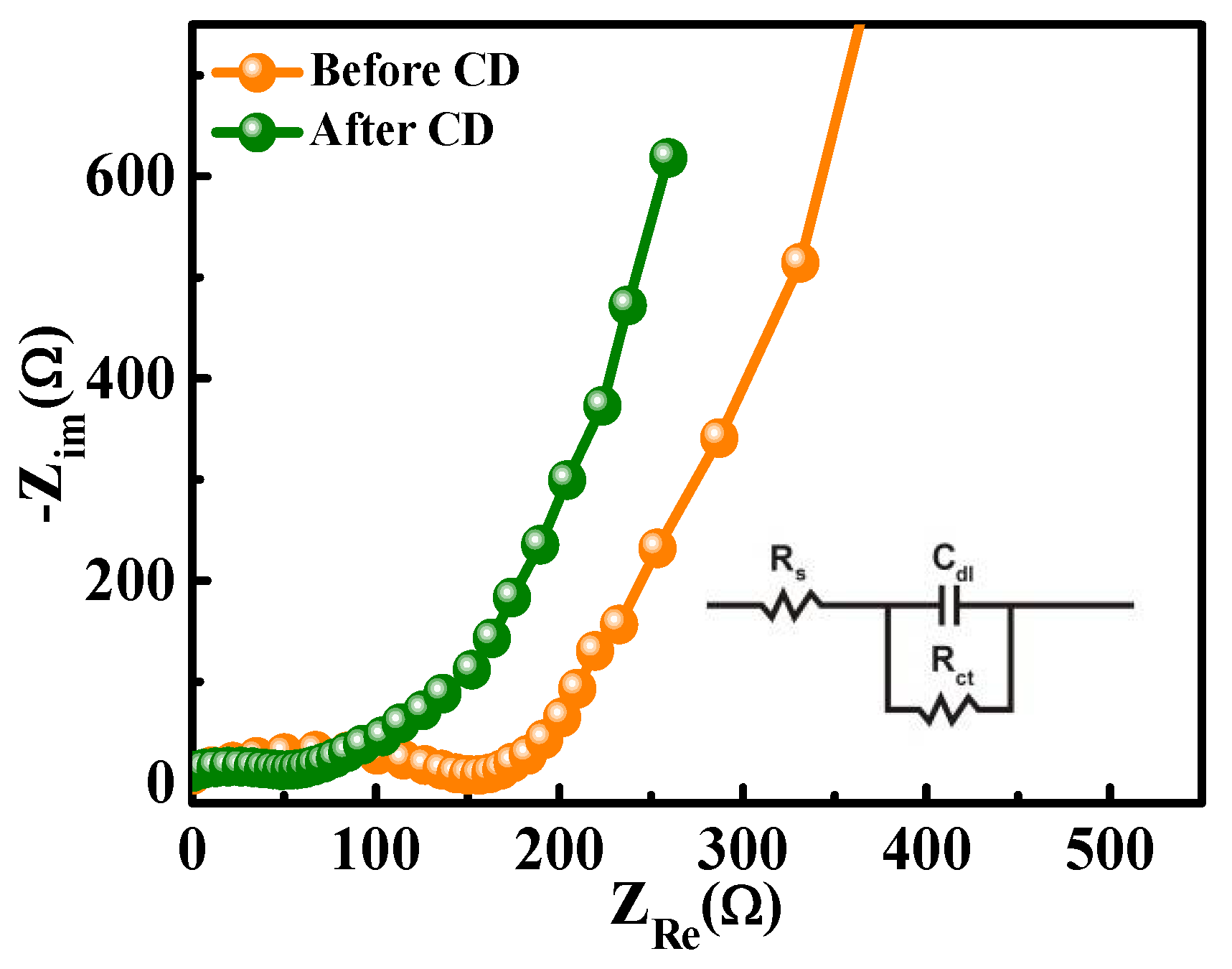
The kinetic behaviors specifying the charge transfer properties of the as-synthesized electrodes were examined by means of a powerful electrochemical impedance spectroscopy (EIS) tool. The EIS measurements were obtained before and after CD cycles and a Nyquist plot was sketched (illustrated in Figure 10) using a Randles circuit model (inset Figure 10). The outcome of the graph was a slope line in the low frequency region signifying an electron diffusion process and a semicircle arc in the high frequency region attributing the charge transfer resistance (Rct) [32]. The Rct could be indicated from the radius of the semicircle at the electrolyte/electrode interface. The formation of semicircles of two different diameters indicated different Rct values. The Rct value for β-SnWO4 after CD was much lower (108 Ω) than that of before CD (163 Ω) characterizing the presence of negative charges on the β-SnWO4 surface. It thus hosted an electrostatic repulsion between the electrolyte/electrode boundaries leading to a low rate charge transfer [32].

4. Conclusions

β-SnWO4 nanoparticles were synthesized by a microwave heat treatment method facilitated by the reaction of SnCl2 and H2WO4 in the presence of tamarind seed powder. An XRD analysis confirmed the crystalline nature of β-SnWO4 nanoparticles, which were found to be cubic in nature. A SEM analysis revealed the formation of β-SnWO4 stone-like arbitrary and amalgamated nanoclusters with some traces of porosity. An EDX analysis confirmed the existence of all constituent elements, i.e., tin, oxygen and tungsten, in the desired stoichiometric ratio. Spherical shaped particles were perceived by TEM analysis and an interlayer d-spacing was examined through HR-TEM, which was found to be 0.33 nm corresponding to a (001) plane. The occurrence of bright rings in the pattern illustrated the highly crystallite nature of the sample. The peaks emerging in the FTIR spectrum at about 609, 768, 851and 1443 cm−1 illuminated the stretching vibrations of the W O 6 6 group, the W–W and W–O–W bridging bonds and the C–C or C–O–C bonds, respectively. The irregularity of the mesopores in the product was perceived by a BJH analysis. A sharp peak as evident in the beginning of distribution curve demonstrated an average pore diameter of 3.35 nm. The CV profile was mapped under various current cycles, which confirmed the composition by describing the actuality of the anodic/cathodic peaks (multi-lithiation and de-lithiation processes). The reduction peak appearing around 2.31, 1.59 and 0.77 V demonstrated the reduction of β-SnWO4 to Sn and W and the Sn-metal reaction and W-metal reaction, respectively. In the GCD analysis, the first cycle exhibited a discharge capacity of 1180 mAhg–1 and the second cycle displayed charge and discharge capacities of 608 and 638 mAhg–1, respectively. Moreover, a decrease in charge/discharge capacities was observed with an increase in the current rate that represented excellent ionic and electrical conductivity and an exceptional rate capability of about 600 mAhg−1 at 0.1 C (reverse current). In addition, it also exhibited ~100% columbic efficiency. The EIS measurements owed a high resistance for β-SnWO4 before CD (163 Ω) than that of after CD (108 Ω) cycles. Thus, β-SnWO4 is highly favorable to be utilized as an electrode material for energy storage devices.

Author Contributions

Data curation, V.V.; Formal analysis, M.S.; Investigation, V.V.; Methodology, H.N.S.; Project administration, F.A.A.; Resources, M.A.A.; Software, J.S.A.; Supervision, N.G.; Validation, M.S.J.; Writing—original draft, N.A.; Writing—review & editing, F.A.A. and N.G. All authors have read and agreed to the published version of the manuscript.

Funding

The authors thank the financial support from the Kingdom of Saudi Arabia, Ministry of Education, Najran University, Promising Centre for Sensors and Electronic Devices (PCSED) sanction on 2019 (Ref. No: PCSED-021-18). The researchers supporting project number (RSP-2020/160), King Saud University, Saudi Arabia. Nagaraju Ganganagappa thank DST Nanomission (Project No. SR/NM/NS-1262/2013 (G), New Delhi, Govt. of India for financial support to carry out the research work.

Institutional Review Board Statement

Not applicable.

Informed Consent Statement

Not applicable.

Data Availability Statement

Not applicable.

Acknowledgments

The authors thank the financial support from the Kingdom of Saudi Arabia, Ministry of Education, Najran University, Promising Centre for Sensors and Electronic Devices (PCSED) sanction on 2019 (Ref. No: PCSED-021-18). The researchers supporting project number (RSP-2020/160), King Saud University, Saudi Arabia. Nagaraju Ganganagappa thank DST Nanomission (Project No. SR/NM/NS-1262/2013 (G), New Delhi, Govt. of India for financial support to carry out the research work.

Conflicts of Interest

The authors declare no conflict of interest.

References

  1. Shinde, P.A.; Jun, S.C. Review on recent progress in the development of tungsten oxide based electrodes for electrochemical energy storage. ChemSusChem 2020, 13, 11–38. [Google Scholar] [CrossRef] [Green Version]
  2. Hu, X.; Zhang, W.; Liu, X.; Mei, Y.; Huang, Y. Nanostructured Mo-based electrode materials for electrochemical energy storage. Chem. Soc. Rev. 2015, 44, 2376–2404. [Google Scholar] [CrossRef] [PubMed]
  3. Zhang, N.; Xiao, X.; Pang, H. Transition metal (Fe, Co, Ni) fluoride-based materials for electrochemical energy storage. Nanoscale Horizons 2019, 4, 99–116. [Google Scholar] [CrossRef]
  4. Borzutzki, K.; Thienenkamp, J.; Diehl, M.; Winter, M.; Brunklaus, G. Fluorinated polysulfonamide based single ion conducting room temperature applicable gel-type polymer electrolytes for lithium ion batteries. J. Mater. Chem. A 2019, 7, 188–201. [Google Scholar] [CrossRef] [Green Version]
  5. Gao, B.; Li, X.; Ding, K.; Huang, C.; Li, Q.; Chu, P.K.; Huo, K. Recent progress in nanostructured transition metal nitrides for advanced electrochemical energy storage. J. Mater. Chem. A 2019, 7, 14–37. [Google Scholar] [CrossRef]
  6. Wang, X.; Li, B.; Liu, D.; Xiong, H. ZnWO4 nanocrystals/reduced graphene oxide hybrids: Synthesis and their application for Li ion batteries. Sci. China Ser. B Chem. 2013, 57, 122–126. [Google Scholar] [CrossRef]
  7. Wang, P.; Liu, H.; Tan, Q.; Yang, J. Ruthenium oxide-based nanocomposites with high specific surface area and improved capacitance as a supercapacitor. RSC Adv. 2014, 4, 42839–42845. [Google Scholar] [CrossRef]
  8. Shinde, P.A.; Lokhande, V.C.; Ji, T.; Lokhande, C.D. Facile synthesis of hierarchical mesoporous weirds-like morphological MnO2 thin films on carbon cloth for high performance supercapacitor application. J. Colloid Interface Sci. 2017, 498, 202–209. [Google Scholar] [CrossRef]
  9. Zhang, J.; Ding, J.; Li, C.; Li, B.; Li, D.; Liu, Z.; Liu, Y. Fabrication of novel ternary three-dimensional RuO2/graphitic-C3N4@ reduced graphene oxide aerogel composites for supercapacitors. ACS Sustain. Chem. Eng. 2017, 5, 4982–4991. [Google Scholar] [CrossRef]
  10. Prabakar, S.R.; Han, S.C.; Jeong, J.; Sohn, K.S.; Pyo, M. CoSn (OH)6 hybridized with anionic and cationic graphenes as a new high-capacity anode for lithium ion batteries. Mater. Des. 2017, 118, 294–303. [Google Scholar] [CrossRef]
  11. Prabakar, S.R.; Babu, R.S.; Oh, M.; Lah, M.S.; Han, S.C.; Jeong, J.; Pyo, M. Dense CoO/graphene stacks via self-assembly for improved reversibility as high performance anode in lithium ion batteries. J. Power Sources 2014, 272, 1037–1045. [Google Scholar] [CrossRef]
  12. Manukumar, K.N.; Kishore, B.; Manjunath, K.; Nagaraju, G. Mesoporous Ta2O5 nanoparticles as an anode material for lithium ion battery and an efficient photocatalyst for hydrogen evolution. Int. J. Hydrogen Energy 2018, 43, 18125–18135. [Google Scholar] [CrossRef]
  13. Huang, B.; Li, X.; Pei, Y.; Li, S.; Cao, X.; Masse, R.C.; Cao, G. Novel carbon-encapsulated porous SnO2 anode for lithiumion batteries with much improved cyclic stability. Small 2016, 12, 1945–1955. [Google Scholar] [CrossRef]
  14. Liu, H.; Li, W.; Shen, D.; Zhao, D.; Wang, G. Graphitic carbon conformal coating of mesoporous TiO2 hollow spheres for high-performance lithium ion battery anodes. J. Am. Chem. Soc. 2015, 137, 13161–13166. [Google Scholar] [CrossRef] [PubMed]
  15. Zhu, G.; Que, W.; Zhang, J.; Zhong, P. Photocatalytic activity of SnWO4 and SnW3O9 nanostructures prepared by a surfactant-assisted hydrothermal process. Mater. Sci. Eng. B 2011, 176, 1448–1455. [Google Scholar] [CrossRef]
  16. Ede, S.R.; Kundu, S. Microwave synthesis of SnWO4 nanoassemblies on DNA scaffold: A novel material for high performance supercapacitor and as catalyst for butanol oxidation. ACS Sustain. Chem. Eng. 2015, 3, 2321–2336. [Google Scholar] [CrossRef]
  17. Solis, J.L.; Lantto, V. Gas-sensing properties of different α-SnWO4-based thick films. Phys. Scripta 1997, 1997, 281. [Google Scholar] [CrossRef]
  18. Warmuth, L.; Feldmann, C. β-SnWO4 with morphology-controlled synthesis and facet-depending photocatalysis. ACS Omega 2019, 4, 13400–13407. [Google Scholar] [CrossRef] [PubMed] [Green Version]
  19. Cho, I.S.; Kwak, C.H.; Kim, D.W.; Lee, S.; Hong, K.S. Photophysical, photoelectrochemical, and photocatalytic properties of novel SnWO4 oxide semiconductors with narrow band gaps. J. Phys. Chem. C 2009, 113, 10647–10653. [Google Scholar] [CrossRef]
  20. Huang, R.; Ge, H.; Lin, X.; Guo, Y.; Yuan, R.; Fu, X.; Li, Z. Facile one-pot preparation of α-SnWO4/reduced graphene oxide (RGO) nanocomposite with improved visible light photocatalytic activity and anode performance for Li-ion batteries. RSC Adv. 2012, 3, 1235–1242. [Google Scholar] [CrossRef]
  21. Alharthi, F.A.; Alsaiari, M.A.; Jalalah, M.S.; Shashank, M.; Alghamdi, A.A.; Algethami, J.S.; Ganganagappa, N. Combustion synthesis of β-SnWO4-rGO: Anode material for Li-ion battery and photocatalytic dye degradation. Ceram. Int. 2020, 47, 10291–10300. [Google Scholar] [CrossRef]
  22. Ansar, M.T.; Ali, A.; Mustafa, G.M.; Afzal, F.; Ishaq, S.; Kanwal, F.; Atiq, S. Polypyrrole-based nanocomposites architecture as multifunctional material for futuristic energy storage applications. J. Alloy. Compd. 2021, 855, 157341. [Google Scholar] [CrossRef]
  23. Mustafa, G.M.; Atiq, S.; Abbas, S.K.; Riaz, S.; Naseem, S. Tunable structural and electrical impedance properties of pyrochlores based Nd doped lanthanum zirconate nanoparticles for capacitive applications. Ceram. Int. 2018, 44, 2170–2177. [Google Scholar] [CrossRef]
  24. Pavithra, N.S.; Patil, S.B.; Kumar, S.K.; Alharthi, F.A.; Nagaraju, G. Facile synthesis of nanocrystalline β-SnWO4: As a photocatalyst, biosensor and anode for Li-ion battery. SN Appl. Sci. 2019, 1, 1123. [Google Scholar] [CrossRef] [Green Version]
  25. Quader, A.; Mustafa, G.M.; Abbas, S.K.; Ahmad, H.; Riaz, S.; Naseem, S.; Atiq, S. Efficient energy storage and fast switching capabilities in Nd-substituted La2Sn2O7 pyrochlores. Chem. Eng. J. 2020, 396, 125198. [Google Scholar] [CrossRef]
  26. Garadkar, K.M.; Ghule, L.A.; Sapnar, K.B.; Dhole, S.D. A facile synthesis of ZnWO4 nanoparticles by microwave assisted technique and its application in photocatalysis. Mater. Res. Bull. 2013, 48, 1105–1109. [Google Scholar] [CrossRef]
  27. Han, L.; Zhou, X.; Wan, L.; Deng, Y.; Zhan, S. Synthesis of ZnFe2O4 nanoplates by succinic acid-assisted hydrothermal route and their photocatalytic degradation of rhodamine B under visible light. J. Environ. Chem. Eng. 2014, 2, 123–130. [Google Scholar] [CrossRef]
  28. Mani, S.; Vediyappan, V.; Chen, S.M.; Madhu, R.; Pitchaimani, V.; Chang, J.Y.; Liu, S.B. Hydrothermal synthesis of NiWO4 crystals for high performance non-enzymatic glucose biosensors. Sci. Rep. 2016, 6, 24128. [Google Scholar] [CrossRef] [PubMed] [Green Version]
  29. Ding, S.; Chen, J.S.; Lou, X.W. One-dimensional hierarchical structures composed of novel metal oxide nanosheets on a carbon nanotube backbone and their lithium-storage properties. Adv. Funct. Mater. 2011, 21, 4120–4125. [Google Scholar] [CrossRef]
  30. Shim, H.W.; Cho, I.S.; Hong, K.S.; Lim, A.H.; Kim, D.W. Wolframite-type ZnWO4 nanorods as new anodes for Li-ion batteries. J. Phys. Chem. C 2011, 115, 16228–16233. [Google Scholar] [CrossRef]
  31. Patil, S.B.; Kishore, B.; Nagaraju, G.; Dupont, J. High capacity MoO3/rGO nanocomposite anode for lithium ion batteries: An intuition into the conversion mechanism of MoO3. New J. Chem. 2018, 42, 18569–18577. [Google Scholar] [CrossRef]
  32. Kokulnathan, T.; Kumar, J.V.; Chen, S.M.; Karthik, R.; Elangovan, A.; Muthuraj, V. One-step sonochemical synthesis of 1D β-stannous tungstate nanorods: An efficient and excellent electrocatalyst for the selective electrochemical detection of antipsychotic drug chlorpromazine. Ultrason. Sonochem. 2018, 44, 231–239. [Google Scholar] [CrossRef] [PubMed]
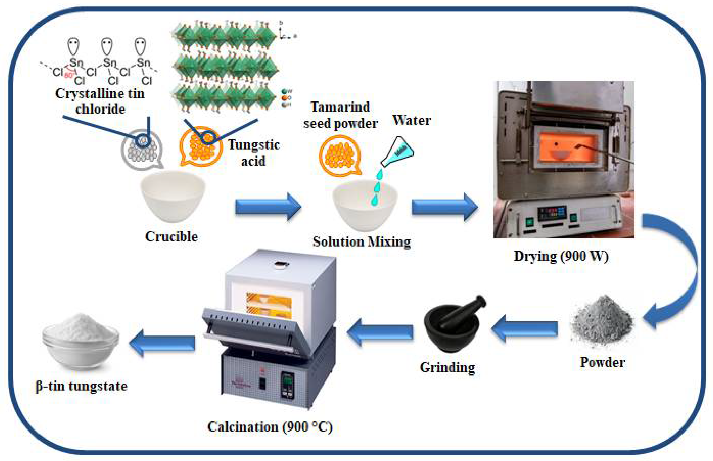
Figure 1. Schematic illustration of the sample synthesis.
Crystals 11 00334 g001
Figure 2. (a) Indexed X-ray diffractometer (XRD) pattern and (b) magnified image of certain peaks of β-SnWO4 nanoparticles.
Crystals 11 00334 g002
Figure 3. Williamson–Hall (W–H) plot of β-SnWO4 nanoparticles.
Crystals 11 00334 g003
Figure 4. Rietveld refined XRD pattern of β-SnWO4 nanostructures.
Crystals 11 00334 g004
Figure 5. (ac) SEM images and (d) EDX spectrum (inset: elemental composition) of β-SnWO4 nanoparticles.
Crystals 11 00334 g005
Figure 6. (a,b) TEM, (c) HR-TEM and (d) selected area electron diffraction (SEAD) pattern of β-SnWO4 nanoparticles.
Crystals 11 00334 g006
Figure 7. Fourier transforms infrared (FTIR) spectrum of β-SnWO4 nanoparticles.
Crystals 11 00334 g007
Figure 8. (a) Barrett–Joyner–Halenda (BJH) adsorption-desorption isotherm and (b) pore size distribution of β-SnWO4 nanostructures.
Crystals 11 00334 g008
Figure 9. (a) Cyclic voltammogram, (b) galvanostatic charge-discharge, (c) rate capability at different current density and (d) cycling performance and columbic efficiency of β-SnWO4 nanoparticles.
Crystals 11 00334 g009
Figure 10. Nyquist plot for electrochemical impedance spectroscopy for β-SnWO4. CD = charge-discharge.
Crystals 11 00334 g010
Table 1. Data obtained through Rietveld refinement of the diffraction data of β-SnWO4.
RexpRpRwpDstaticsWt. Dstaticsχ2
10.8413.7318.040.440.562.77
Table 2. Comparative study of specific capacities along different C-rates for pure β-SnWO4 and for other dopants.
CompositionSynthesis MethodC-RatesSpecific Capacity (mAhg−1)References
β-SnWO4–rGOSolution combustion method followed by a low temperature hydrothermal method0.1 C578[21]
β-SnWO4~0.1 C271[21]
β-SnWO4–rGO~3 C165[21]
β-SnWO4~3 C35[21]
β-SnWO4Rapid microwave heat treatment0.1 C600Current work
β-SnWO4~3 C54Current work
Publisher’s Note: MDPI stays neutral with regard to jurisdictional claims in published maps and institutional affiliations.