Article

Magnetic Properties and Local Structure of the (La, Co) Co-doped Bi1−xLaxFe0.95Co0.05O3

1
College of Science, Nanjing University of Posts and Telecommunications, Nanjing 210023, China
2
New Energy Technology Engineering Laboratory of Jiangsu Province, Nanjing 210023, China
3
College of Electronic Science and Engineering, Nanjing University of Posts and Telecommunications, Nanjing 210023, China
*
Author to whom correspondence should be addressed.
Crystals 2021, 11(9), 1059; https://doi.org/10.3390/cryst11091059
Submission received: 9 August 2021 / Revised: 31 August 2021 / Accepted: 31 August 2021 / Published: 2 September 2021

Abstract

:
In this study, the structural, morphological, and magnetic properties and the local structure of Bi1−xLaxFe0.95Co0.05O3 (x = 0.05, 0.10, 0.15, 0.20) nanoparticles were systematically investigated. In the (La, Co) co-doped BiFeO3 samples, a structural transition from a rhombohedral to orthorhombic structure was observed via X-ray diffraction. This structural phase transition resulted in significantly improved magnetic properties (Ms = 1.706 emu/g at 60 kOe). SEM analysis revealed that grain size decreased with increasing La concentration. The magnetic hysteresis loops confirmed that the significant enhancement of magnetization for all samples. Finally, combining the experimental synchrotron radiation techniques, we studied the samples’ local structure in order to analyze the reasons why the samples’ magnetic properties were enhanced.

1. Introduction

In recent years, multiferroic materials have drawn much attention due to the coexistence of ferroelectricity, ferromagnetism, and ferroelasticity in a single phase. These physical properties can couple each other under a magnetoelectric effect (ME), meaning such materials have applications in sensors, information storage, and spintronic devices [1,2,3,4]. BiFeO3 (BFO) is one of the most well-studied ME multiferroic materials so far due to it showing both ferroelectricity and antiferromagnetism concurrently at room temperature (Curie temperature TC = 1103 K and Neel temperature TN = 643 K) [5]; however, BFO has some inherent defects. For example, it is quite difficult to simultaneously obtain the ferromagnetic and ferroelectric ordering above room temperature owing to its incommensurate cycloid magnetic spin structure [6,7]. In order to solve these problems, various attempts have been made involving techniques such as synthesis of pure single-phase crystals, suitable modifications at the Bi or Fe sites, or the fabrication of composites. The doping or co-doping of rare earth and transition metal ions has been found to improve the electrical and magnetic properties of BFO materials.
Recent reports have confirmed that La-doped BFO shows improved magnetic properties. Sen et al. proved that the enhancement of magnetization for Bi1−xLaxFeO3 ceramics did not result from Fe2+ or the variation of the oxygen stoichiometry using Mössbauer spectroscopy [8]. It has also been demonstrated that La doping in Bi1−xLaxFeO3 suppresses the spiral magnetic structure of BFO, while the (BiLa)FeO3 compounds show weak ferromagnetism [9]. Kuang et al. reported that the ferromagnetic behavior of (Y, Co) co-doped thin films was enhanced with increasing Co ion concentration due to the different magnetic moments of Co3+ and Fe3+ [10]. Xu et al. reported that Co3+ is a magnetic ion intrinsically; thus, substitution at the Fe-site can efficiently enhance the ferromagnetism [11].
To our current knowledge, there are few studies on the co-doping effect of La and Co in BFO, focusing on the local structure and magnetic behaviors. In this study, we report on the preparation of Bi1-xLaxFe0.95Co0.05O3 multiferroic nanoparticles and the influence of co-doping on the structural, morphological, and magnetic properties and on the local structure.

2. Experiments

The polycrystalline samples of Bi1xLaxFe0.95Co0.05O3 (x = 0.05, 0.10, 0.15, and 0.20) series were synthesized via a sol–gel method. The analytically pure Bi2O3, Fe(NO3)3, Co(NO3)2, and La(NO3)3 powders were used as starting materials and dissolved in dilute nitric (analytically pure) acid and distilled water. An appropriate amount of tartaric acid was added as a chelating agent, then the solution was mixed thoroughly using the magnetic stirrer at 130 °C for 4 h and baked at 80 °C for 8 h. The dry ground powders were then conducted by heat treatment at 250 °C for 2 h and sintered at 600 °C for 2 h. Finally, the samples were slowly cooled down to room temperature.
The structure of the samples was detected by X-ray powder diffraction (XRD) at room temperature. The Cu (Kα, 1.5405 Å) target was used as the ray source and the light was monochromated using a graphite monochromator. The sample morphology and grain size were observed using scanning electron microscopy (SEM). The magnetization hysteresis loops were evaluated at room temperature using a vibrating sample magnetometer (VSM) integrated into a physical property measurement system (PPMS-9, Quantum Design). The local structure of the samples and X-ray absorption near the edge structure (XANES) spectra of the Fe K-edge were measured using an XAFS experimental station at Beijing Synchrotron Radiation Facility (BSRF).

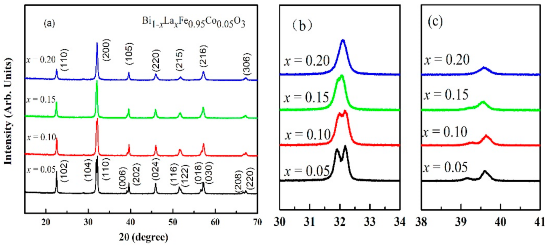
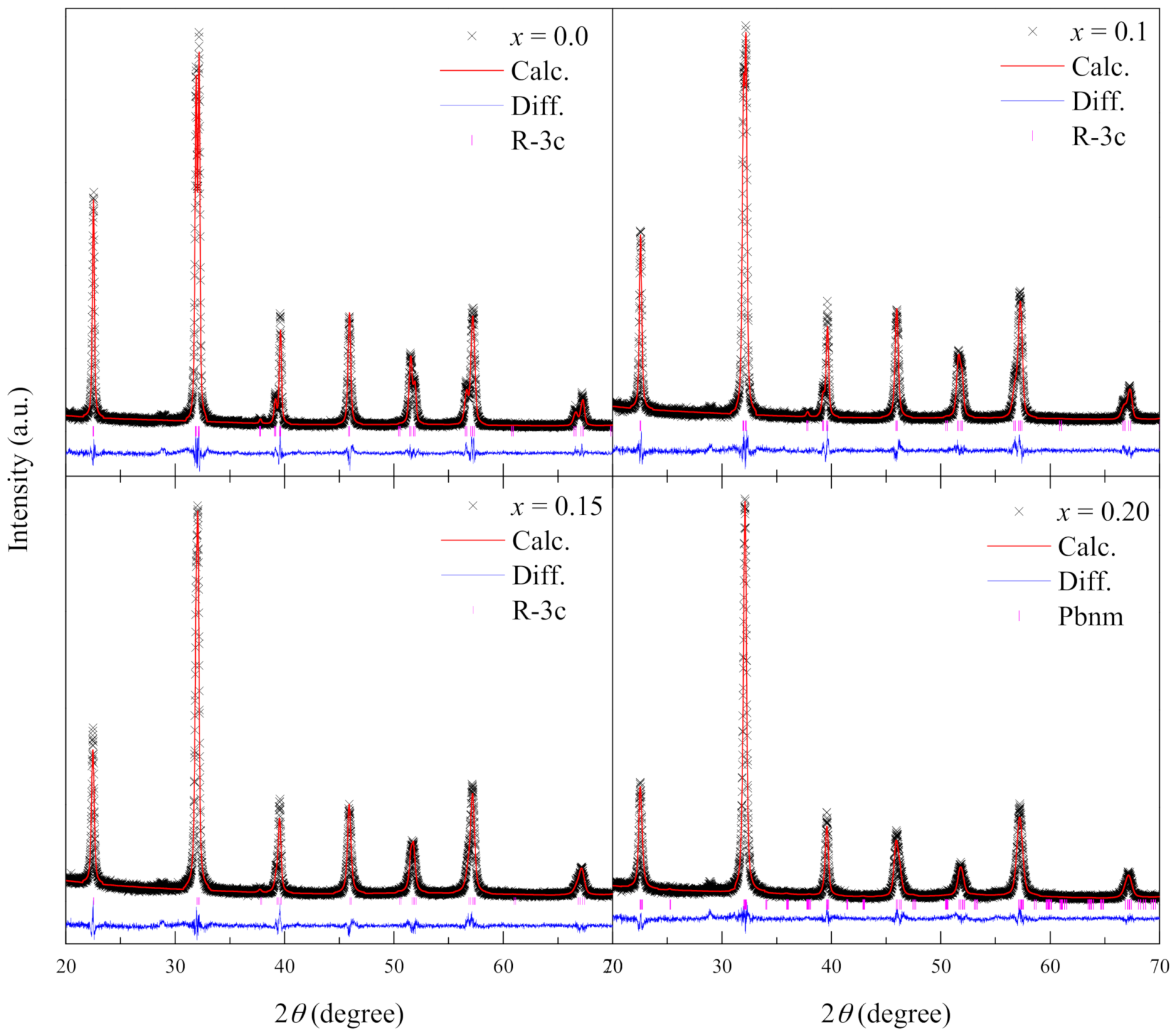
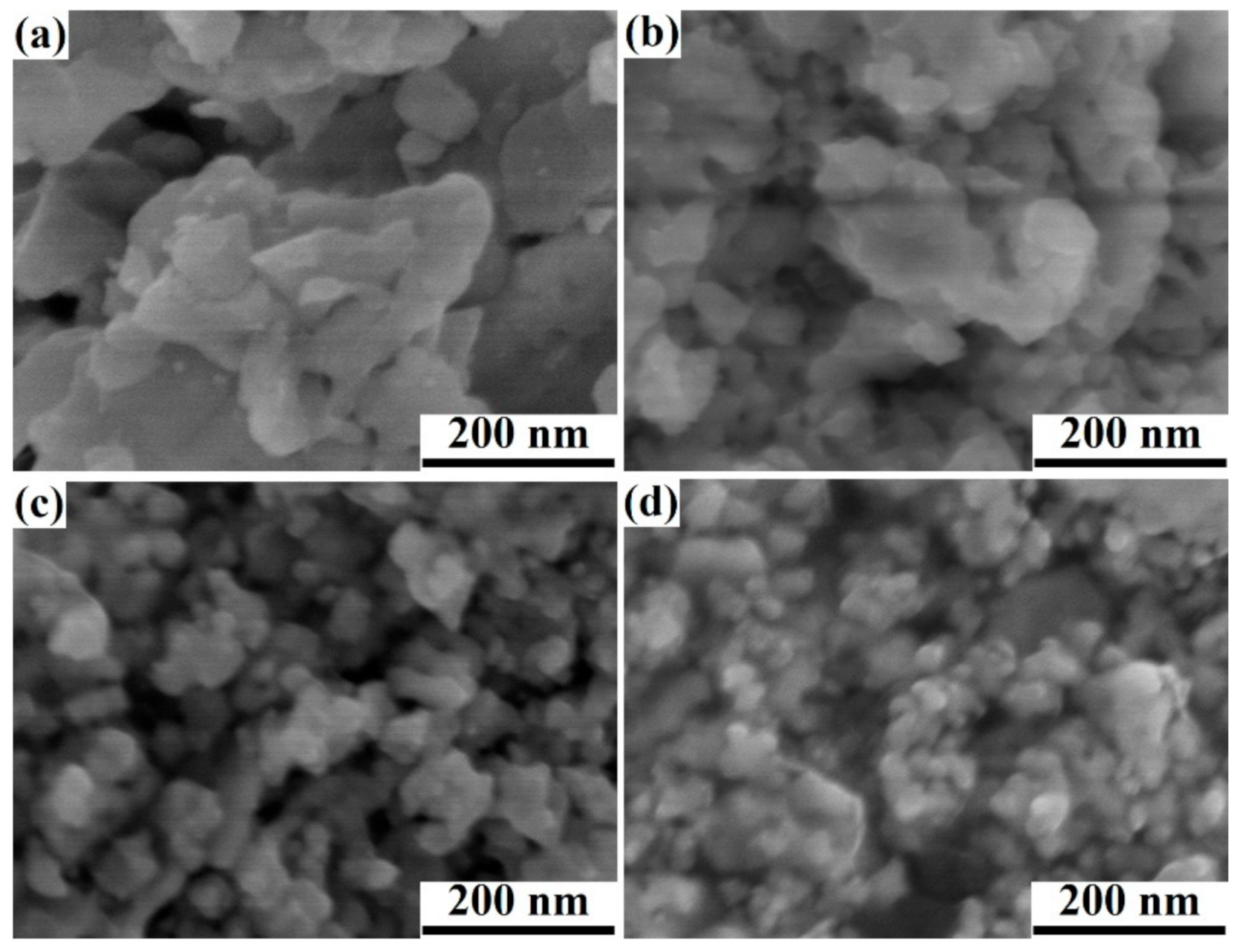
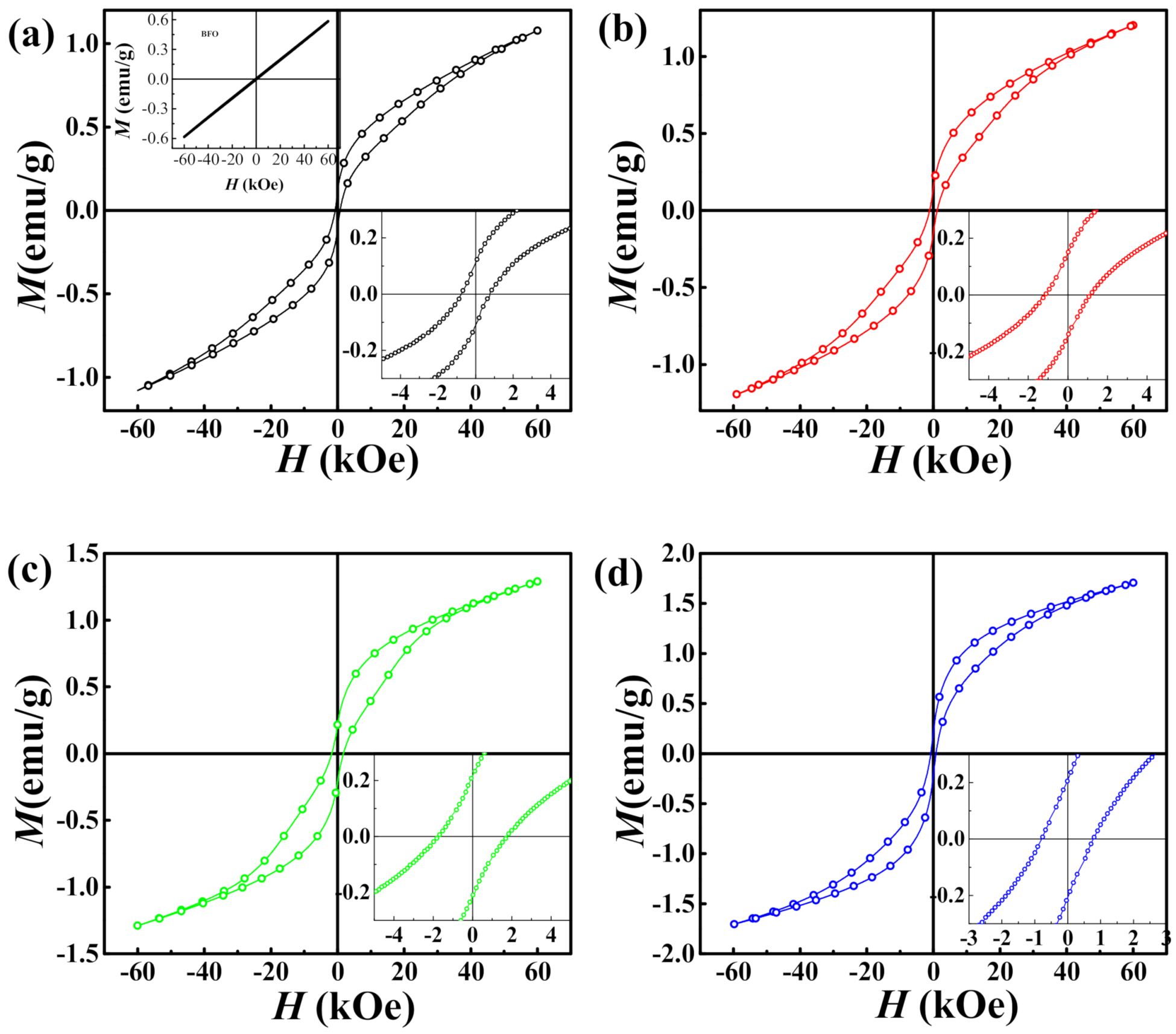
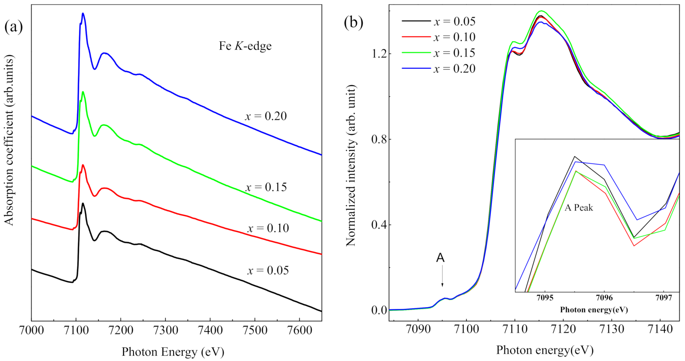
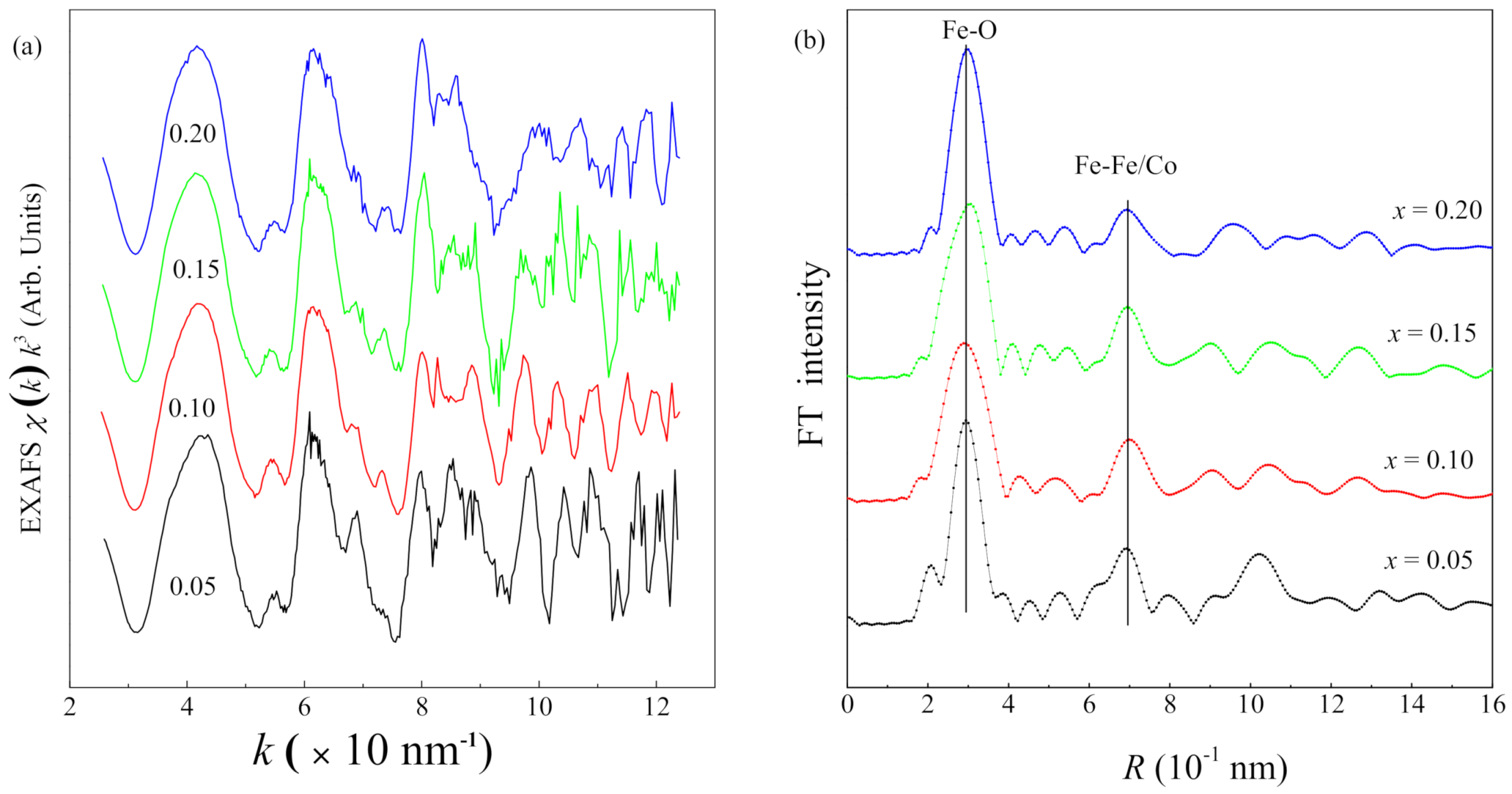
3. Results and Discussion

Figure 1a shows the XRD patterns of Bi1xLaxFe0.95Co0.05O3 (x = 0.05, 0.10, 0.15, 0.20). Mao et al. [12] reported that a single crystal Co at the B-site will create an impurity phase, although all of the samples achieved here contained a single phase without an impurity phase, which indicates that La doping can suppress the impurity phase, coinciding with the report in [13]. In addition, the XRD patterns clearly revealed the gradual variations in the shape and positions of peaks, indicating a structural phase transition with increasing doping concentration of La ions. Magnified images in the vicinity of 2θ~32° and 39° show are shown in Figure 1b,c. For x = 0.05, reflections (104) and (110) are totally split. Upon increasing the La content from 5% to 10%, the two reflections become weak and tend to merge into a single reflection. Meanwhile, the peak position of the two reflections shifts toward a higher angle. It can be seen that doublets (104) and (110) merged into a single peak. The peaks (006) and (202) at about 2θ = 39° become broad and peak (006) almost disappears at x = 0.20. These results indicate a transition of the crystal structure from the rhombohedral to orthorhombic phase.
The Rietveld refinement was performed to further manifest this structural transition. The samples with x ≤ 0.15 were well refined by the rhombohedral structure with the space group of R-3c, while the XRD pattern of sample x = 0.2 fit well with the orthorhombic structure with the space group of Pbnm, as shown shown in Figure 2. Similar behavior was also observed for Pr/Co co-doped Ba, Dy, and Mn-doped BFO [14,15,16,17]. In Figure 1c, the slight shift of the diffraction peak to a high angle results from the smaller ion radius of La3+ than that of Bi3+, which corresponds to the decreased lattice parameter (listed in Table 1).
In order to research the microstructure and particle size distribution of Bi1xLaxFe0.95Co0.05O3 (x = 0.05, 0.10, 0.15, 0.20), the morphologies of all the samples were examined by SEM, as shown in Figure 3. For x = 0.05 and 0.10, the microstructure is rather irregular and agglomerate [18]. The morphology of samples x = 0.15 and 0.20 is different from the former samples, and these samples are dense and uniform with a nearly spherical shape. Furthermore, the average particle size decreases with the increasing La concentration, which is similar to Zheng et al.’s report [13]. The reason for this decrease may be interpreted due to the suppression of the oxygen vacancy concentration. Less oxygen vacancies imply slower oxygen ion motion and consequently a lower grain growth rate [19,20]. The change in particle morphologies proves that the La ion doping has an impact on the crystallization of BFO particles prepared by sol–gel method.
The magnetization (M) versus applied magnetic field (H) loops for all samples collected at room temperature are presented in Figure 4. It is known that pristine BFO is a G-type antiferromagnet that does not possess any spontaneous magnetization [21]. The measured M-H curve for BFO shows the linear relationship, proving the antiferromagnetism (the higher left panel in Figure 4a). Compared to the shape of the M-H curve for BiFe0.95Co0.05O3 [11,22,23], the M-H shape for the La-doped BiFe0.95Co0.05O3 shows a wasp-waisted hysteresis loop. This wasp-waisted hysteresis loop was often observed in the doped BFO, which is considered to indicate the presence of inhomogeneity [24]. Obviously, the (La, Co)-co-doped samples show remarkably enhanced magnetization with clear magnetization hysteresis loops compared to the pristine BFO sample (Figure 4). The saturation magnetization levels of all samples were obtained from the magnetization hysteresis loops, with the values ranging from 1.078 emu/g to 1.706 emu/g at an applied field of 60 kOe. Figure 4 clearly shows that all doped samples display slight remnant magnetization (Mr) and a small coercive field (Hc). From x = 0.05 to x = 0.15, both the Mr and Hc increase with increasing content of x; however, the value of Mr for x = 0.2 is smaller than that of x = 0.15, while the value of Hc for x = 0.2 is smaller than that of x = 0.1 as well. The detailed magnetic parameters are listed in Table 1. It is noteworthy that the saturation magnetization of x = 0.2 is much bigger than for the other three samples. A similar phenomenon was reported in [12].
The Fe-K edge XAFS data were measured and are shown in Figure 5a. The X-ray absorption near-edge structure (XANES) features give useful information about the electronic state of the absorber and the atomic structure around it. Figure 5b presents the Fe K-edge XANES spectra of Bi1xLaxFe0.95Co0.05O3 (x = 0.05, 0.10, 0.15, and 0.20), clearly showing that all spectra have similar shapes and that there is no obvious shift of the absorption energy position. This suggests that the valence state of Fe ions does not change with increasing x. As shown in the inset of Figure 5b, a weak pre-edge peak appears in the range of 7094–7097 eV in all samples. This is due to the 1s–3d quadrupole transition caused by the deformation away from the regular octahedron [25,26].
The obtained EXAFS signals were converted into the k space of the photoelectron wave vector, defined as k = [ ( 2 m / η 2 ) ( E E 0 ) ] 1 / 2 , where (EE0) is the photoelectron kinetic energy measured. The experimental XAFS signal χ(k) was multiplied by factor k3 to compensate for the decrease in its amplitude with the increase in the wave vector value (Figure 6a). Figure 6b shows the radial distribution function, as obtained by Fe K-edge Fourier transformation (FT) of the Extend XAFS function by analyzing the data with USTCXAFS2.0 software. The FTs of the experimental XAFS signals were carried out with a Kaiser–Bessel window in the range of 2.6–12.4 Å−1. The FTs reveal that all the samples exhibit two main coordination peaks. The first peak has the highest intensity of all peaks, which is assigned to the Fe-O coordination shell, while the peak with an R value region of 0.30–0.38 nm arises from the next closest Fe-Fe and Fe-Co coordination shells. It can be seen that the Fe-O peak shifts to a higher R value region and the width of the peak becomes broader with increasing La, which indicates a change of the local structure and distortion of the lattice structure. These results are in good agreement with the XRD analysis. This significant structural change may result in a modification of the canted G-type antiferromagnetic order of Fe3+ in the rhombohedral structure, then resulting in net magnetization [27,28]; however, the Fe-Fe and Fe-Co peaks show almost no change.
From the above analysis, we believe that the change of the sample magnetism is related to the structural distortion and structural phase transition of the sample. As the XAFS data show, the changes of the pre-edge peaks and Fe-O peaks are related to the local structure distortion with the increasing La concentration (x = 0.05~0.15), which induces a small and slow improvement in the magnetic properties. The significant enhancement in magnetic properties for La concentrations up to 0.20 compared to the other three samples’ magnetization curves may be caused by a structural phase transition from a distorted rhombohedral structure to an orthorhombic structure, as the XRD model displays above; therefore, the La substitution phase transformation to the orthorhombic phase is expected to suppress the cycloid spin structure and release the latent magnetization locked within the cycloid, resulting in greatly enhanced magnetism [29].

4. Conclusions

In summary, we have successfully prepared single-phase Bi1−xLaxFe0.95Co0.05O3 (x = 0.05, 0.10, 0.15, 0.20) samples via the sol–gel method. The structural, morphological, and magnetic properties and the local structure of the nanoparticles were investigated in detail. Room temperature magnetic hysteresis loops of the samples indicated substantially increased magnetization in the co-substituted samples. Based on the XRD and XAFS results, we concluded that the observed magnetic properties were due to the change of the local structure and the structural phase transition.

Author Contributions

Data curation, X.H. and L.L.; Formal analysis, C.S.; Investigation, L.L.; Project administration, Y.L.; Writing—original draft, Y.L.; Writing—review & editing, H.Z. All authors have read and agreed to the published version of the manuscript.

Funding

This research was funded by National Natural Science Foundation of China, grant number 51772157, and Nanjing University of Posts and Telecommunications, grant number NY217096.

Acknowledgments

This work is supported by NSFC under contract No. 51772157 and by the research project of Nanjing University of Posts and Telecommunications under contract No. NY217096. Moreover, we thank BSRF for its beamtime.

Conflicts of Interest

The authors declare no conflict of interest.

References

  1. Schmid, H. Multi-ferroic magnetoelectrics. Ferroelectrics 1994, 162, 317–338. [Google Scholar] [CrossRef]
  2. Wang, J.; Neaton, J.B.; Zheng, H.; Nagarajan, V.; Ogale, S.B.; Liu, B.; Viehland, D.; Vaithyanathan, V.; Schlom, D.G.; Waghmare, U.V.; et al. Epitaxial BiFeO3 multiferroic thin film heterostructures. Science 2003, 299, 1719–1722. [Google Scholar] [CrossRef] [PubMed]
  3. Ma, J.; Hu, J.; Li, Z.; Nan, C.W. Recent progress in multiferroic magnetoelectric composites: From bulk to thin films. Adv. Mater. 2011, 23, 1062–1087. [Google Scholar] [CrossRef] [PubMed]
  4. Ramesh, R.; Spaldin, N.A. Multiferroics: Progress and prospects in thin films. Nat. Mater. 2007, 6, 9–21. [Google Scholar] [CrossRef] [PubMed]
  5. Fiebig, M. Revival of the magnetoelectric effect. J. Phys. D Appl. Phys. 2005, 38, R123–R152. [Google Scholar] [CrossRef]
  6. Marzouki, A.; Loyau, V.; Gemeiner, P.; Bessais, L.; Dkhil, B.; Megriche, A. Increase of magnetic and magnetoelectric properties in Co/Mn co-doped BiFeO3 multiferroic. J. Magn. Magn. Mater. 2020, 498, 166137. [Google Scholar] [CrossRef] [Green Version]
  7. Zeng, J.; Tang, Z.H.; Tang, M.H.; Xu, D.L.; Xiao, Y.G.; Zeng, B.W.; Li, L.Q.; Zhou, Y.C. Enhanced ferroelectric, dielectric and leakage properties in Ce and Ti co-doping BiFeO3 thin films. J. Sol.-Gel. Sci. Techn. 2014, 72, 587–592. [Google Scholar] [CrossRef]
  8. Sen, K.; Thakur, S.; Singh, K.; Gautam, A.; Singh, M. Room-temperature magnetic studies of La-modified BiFeO3 ceramic. Mater. Lett. 2011, 65, 1963–1965. [Google Scholar] [CrossRef]
  9. Kumar, P.; Kar, M. Effect of structural transition on magnetic and dielectric properties of La and Mn co-substituted BiFeO3 ceramics. Mater. Chem. Phys. 2014, 148, 968–977. [Google Scholar] [CrossRef] [Green Version]
  10. Kuang, D.; Tang, P.; Wu, X.; Ding, X.D.; Yang, S.H.; Zhang, Y.L. Structural, optical and magnetic studies of (Y, Co) co-substituted BiFeO3 thin films. J. Alloy. Compd. 2016, 671, 192–199. [Google Scholar] [CrossRef]
  11. Xu, Q.; Zai, H.; Wu, D.; Qiu, T.; Xu, M.X. The magnetic properties of Bi (Fe0.95Co0.05) O3 ceramics. Appl. Phys. Lett. 2009, 95, 112510. [Google Scholar] [CrossRef]
  12. Mao, W.W.; Li, X.A.; Li, Y.T.; Li, P.; Bao, G.; Yang, T.; Yang, J.P. Structural and magnetic properties of single-phase Bi0.9Eu0.1Fe0.95Co0.05O3 and Bi0.9Eu0.05La0.05Fe0.95Co0.05O3 nanoparticles. Mater. Lett. 2012, 76, 135–137. [Google Scholar] [CrossRef]
  13. Zheng, X.H.; Xu, Q.Y.; Wen, Z.; Lang, X.Z.; Wu, D.; Qiu, T.; Xu, M.X. The magnetic properties of La doped and codoped BiFeO3. J. Alloy. Compd. 2010, 499, 108–112. [Google Scholar] [CrossRef]
  14. Xi, X.J.; Wang, S.Y.; Liu, W.F.; Wang, H.J.; Guo, F.; Wang, X.; Gao, J.; Li, D.J. Enhanced magnetic and conductive properties of Ba and Co co-doped BiFeO3 ceramics. J. Magn. Magn. Mater. 2014, 355, 259–264. [Google Scholar] [CrossRef]
  15. Li, Y.B.; Yu, J.; Li, J.J.; Zheng, C.D.; Wu, Y.Y.; Zhao, Y.A.; Wang, M.; Wang, Y.B. Influence of Dy-doping on ferroelectric and dielectric properties in Bi1.05-xDyxFeO3 ceramics. J. Mater. Sci.-Mater. Electron. 2011, 22, 323–327. [Google Scholar] [CrossRef]
  16. Chauhan, S.; Kumar, M.; Chhoker, S.; Katyal, S.C.; Singh, H.; Jewariya, M.; Yadav, K.L. Multiferroic, magnetoelectric and optical properties of Mn doped BiFeO3 nanoparticles. Solid State Commun. 2012, 152, 525–529. [Google Scholar] [CrossRef]
  17. Li, X.A.; Wang, X.W.; Li, Y.T.; Mao, W.W.; Li, P.; Yang, T.; Yang, J.P. Structural, morphological and multiferroic properties of Pr and Co co-substituted BiFeO3 nanoparticles. Mater. Lett. 2013, 90, 152–155. [Google Scholar] [CrossRef]
  18. Dai, H.Y.; Xue, R.Z.; Chen, Z.P.; Li, T.; Chen, J.; Xiang, H.W. Effect of Eu, Ti co-doping on the structural and multiferroic properties of BiFeO3 ceramics. Ceram. Int. 2014, 40, 15617–15622. [Google Scholar] [CrossRef]
  19. Lan, C.Y.; Jiang, Y.W.; Yang, S.G. Magnetic properties of La and (La, Zr) doped BiFeO3 ceramics. J. Mater. Sci. 2011, 46, 734–738. [Google Scholar] [CrossRef]
  20. Priyadharsini, P.; Pradeep, A.; Sathyamoorthy, B.; Chandrasekaran, G. Enhanced multiferroic properties in La and Ce co-doped BiFeO3, nanoparticles. J. Phys. Chem. Solids 2014, 75, 797–802. [Google Scholar] [CrossRef]
  21. Wang, Y.; Guo, Z.Y.; Jia, Q.J.; Dong, J.C.; Zhang, J.; Chen, D.L. Effect of Nd/Mn substitution on the structure and magnetic properties of nano-BiFeO3. J. Alloy. Compd. 2019, 786, 385–393. [Google Scholar] [CrossRef]
  22. Hojo, H.; Kawabe, R.; Shimizu, K.; Yamamoto, H.; Mibu, K.; Samanta, K.; Saha-Dasgupta, T.; Azuma, M. Ferromagnetism at room temperature induced by spin structure change in BiFe1−xCoxO3 thin films. Adv. Mater. 2017, 29, 1603131. [Google Scholar] [CrossRef] [PubMed]
  23. Naganuma, H.; Yasui, S.; Nishida, K.; Iijima, T.; Funakubo, H.; Okamura, S. Enhancement of magnetization at morphotropic phase boundary in epitaxial BiCoO3-BiFeO3 solid solution films grown on SrTiO3(100) substrates. J. Appl. Phys. 2011, 109, 07D917. [Google Scholar] [CrossRef]
  24. Liu, J.; Li, M.; Pei, L.; Wang, J.; Yu, B.; Wang, X.; Zhao, X. Structural and multiferroic properties of the Ce-doped BiFeO3 thin films. J. Alloy. Compd. 2010, 493, 544–548. [Google Scholar] [CrossRef]
  25. Lee, J.H.; Choi, H.J.; Lee, D.; Kim, M.G.; Bark, C.W.; Ryu, S.; Oak, M.A.; Jang, H.M. Variations of ferroelectric off-centering distortion and 3d-4p orbital mixing in La-doped BiFeO3 multiferroics. Phys. Rev. B 2010, 82, 045113. [Google Scholar] [CrossRef] [Green Version]
  26. Westre, T.E.; Kennepohl, P.; DeWitt, J.G.; Hedman, B.; Hodgson, K.O.; Solomon, E.I. A multiplet analysis of Fe K-edge 1s→3d pre-edge features of iron complexes. J. Am. Chem. Soc. 1997, 119, 6297–6314. [Google Scholar] [CrossRef]
  27. Wen, Z.; Shen, X.A.; Wu, D.; Xu, Q.Y.; Wang, J.L.; Li, A.D. Enhanced ferromagnetism at the rhombohedral–tetragonal phase boundary in Pr and Mn co-substituted BiFeO3 powders. Solid State Commun. 2010, 150, 2081–2084. [Google Scholar] [CrossRef]
  28. Yang, C.; Jiang, J.S.; Qian, F.Z.; Jiang, D.M.; Wang, C.M.; Zhang, W.G. Effect of Ba doping on magnetic and dielectric properties of nanocrystalline BiFeO3 at room temperature. J. Alloy. Compd. 2010, 507, 29–32. [Google Scholar] [CrossRef]
  29. Hussain, S.; Hasanain, S.K.; Jaffari, G.H.; Ali, N.Z.; Siddique, M.; Shah, S.I. Correlation between structure, oxygen content and the multiferroic properties of Sr doped BiFeO3. J. Alloy. Compd. 2015, 622, 8–16. [Google Scholar] [CrossRef]
Figure 1. (a) X-ray diffraction patterns of Bi1xLaxFe0.95Co0.05O3 (x = 0.05, 0.10, 0.15, and 0.20) nanoparticles. Enlarged pattern of the diffraction peaks at (b) 2θ = 32° and (c) 2θ = 39°.
Crystals 11 01059 g001
Figure 2. The Rietveld refinement of the XRD patterns for Bi1xLaxFe0.95Co0.05O3 samples (x = 0.05, 0.10, 0.15, and 0.20).
Crystals 11 01059 g002
Figure 3. SEM micrographs of fracture surfaces of Bi1xLaxFe0.95Co0.05O3: (a) x = 0.05; (b) x = 0.10; (c) x = 0.15; (d) x = 0.20.
Crystals 11 01059 g003
Figure 4. Room temperature M-H curves for LaxBi1−xFe0.95Co0.05O3: (a) x = 0.05; (b) x = 0.10; (c) x = 0.15; (d) x = 0.20. The inset on the lower right is the magnified view. The inset on the high left in Figure 3a is the M-H curve for the parent BiFeO3.
Crystals 11 01059 g004
Figure 5. Raw data for the XAFS spectra (a) and Fe K-edge XANES spectra (b) of LaxBi1−xFe0.95Co0.05O3.
Crystals 11 01059 g005
Figure 6. (a) Experimental XAFS spectra χ(k)k3 of the Fe K-edge and (b) Fourier transformation of the Fe K-edge EXAFS oscillation functions for the LaxBi1−xFe0.95Co0.05O3 (x = 0.05, 0.10, 0.15, and 0.20) samples.
Crystals 11 01059 g006
Table 1. The refined lattice parameters (a, b, and c), goodness of fit (χ2), and magnetic parameters (saturation magnetization Ms, remenant magnetization Mr, and coercivity Hc) for all samples.
x = 0.0x = 0.10x = 0.15x = 0.20
a (Å)5.5795.5705.5645.574
b (Å)5.5795.5705.5645.591
c (Å)13.84113.79013.7407.842
χ21.2341.1701.1411.155
Ms (emu/g)1.0781.2001.2891.706
Mr (emu/g)0.1140.1510.2160.205
Hc (kOe)0.7671.0971.7660.813
Publisher’s Note: MDPI stays neutral with regard to jurisdictional claims in published maps and institutional affiliations.

