Article

Assessment of the Physical Properties of an Experimental Adhesive Dentin Bonding Agent with Carbon Nanoparticles

1
Department of Restorative Dentistry, Operative Division, College of Dentistry, King Saud University, P.O. Box 21069, Riyadh 11475, Saudi Arabia
2
Department of General Dentistry, College of Dentistry, King Saud University, P.O. Box 21069, Riyadh 11475, Saudi Arabia
3
Faculty of Dentistry, University of Toronto, Toronto, ON M5G 1G6, Canada
4
Department of Prosthetic Dental Sciences, College of Dentistry, King Saud University, P.O. Box 21069, Riyadh 11475, Saudi Arabia
*
Authors to whom correspondence should be addressed.
Crystals 2022, 12(10), 1441; https://doi.org/10.3390/cryst12101441
Submission received: 7 July 2022 / Revised: 26 September 2022 / Accepted: 28 September 2022 / Published: 12 October 2022

Abstract

:
The present study was aimed at reinforcing the control adhesive (CA) with two concentrations (2.5% & 5%) of carbon nanoparticles (CNPs) and evaluating the impact of these additions on the adhesive’s properties. Scanning electron microscopy (SEM) and energy dispersive X-Ray (EDX) spectroscopy were utilized to examine the morphological characteristics and elemental mapping of the filler CNPs. To investigate the adhesive’s properties, rheological assessment, shear bond strength (SBS) testing, analysis of the adhesive–dentin interface, degree of conversion (DC) analysis, and failure mode investigations were carried out. The SEM micrographs of CNPs verified roughly hexagonal-shaped cylindrical particles. The EDX plotting established the presence of carbon (C), oxygen (O), and zirconia (Zr). Upon rheological assessment, a gradual reduction in the viscosity was observed for all the adhesives at higher angular frequencies. The SBS testing revealed the highest values for 2.5% CNP adhesive group (25.15 ± 3.08 MPa) followed by 5% CNP adhesive group (24.25 ± 3.05 MPa). Adhesive type interfacial failures were most commonly found in this study. The 5% CNP containing adhesive revealed thicker resin tags and a uniform hybrid layer without any gaps (compared with 2.5% CNP adhesive and CA). The reinforcement of the CA with 2.5% and 5% CNPs augmented the adhesive’s bond strength. Nevertheless, a diminished viscosity (at higher angular frequencies) and reduced DC were observed for the two CNP reinforced adhesives. CNP reinforced dentin adhesives are effective in enhancing the adhesive bond integrity of resin to dentin.

1. Introduction

Adhesion with dental hard tissues (enamel and dentin) forms the basis of aesthetic dentistry [1]. The clinical success of dental composite restorations is dependent upon the formation of a micromechanical bond of adhesives with dentinal collagen [2]. Although it has been claimed that dental adhesives have developed to the eighth generation [3], the bond strength of these adhesives is insufficient [4]. A reasonable strategy is to reinforce the dentin adhesive’s structure with nanofiller particles that could improve its mechanical properties resulting in improved adhesive-dentin bond strength [5]. In the past, researchers have incorporated various filler nanoparticles (bioactive glasses, calcium phosphate, hydroxyapatite) in the adhesive and observed an enhancement in its mechanical properties and remineralization capabilities [6]. However, an ideal filler group that could optimally enhance an adhesive’s mechanical properties is under continued investigation.
Carbon is one of the most common natural elements. Its nanoparticles (carbon nanoparticles, CNPs) contain unique characteristics including low toxicity, high inertness, greater surface area, and abundant edge site, which are suitable for various clinical applications [7,8,9]. In the field of medicine, the CNPs-reinforced biomaterials have been used as a bone substitute and to augment bone regeneration [10]. In dentistry, the CNPs have been particularly researched due to their superior properties (high mechanical strength, acceptable biocompatibility, and high absorption) [11,12]. A former study has shown that the incorporation of only 1 wt.% of CNPs can increase the tensile strength and stiffness of composites by 36–42% and 25%, respectively [13]. The impact of CNPs resembles that of steel that is used to boost the hardness of composites, cements, and concrete [14]. Therefore, the CNPs are useful for the fabrication of various dental materials, as they possess high tensile strength and stiffness. Apart from their use to reinforce dental materials, they have been found to be suitable as a synthesis material for scaffolds and for targeted drug delivery [11,15]. Khan et al. previously showed that composites reinforced with CNPs demonstrated improved mechanical properties and hence can be utilized for load-bearing clinical tasks [16].
An earlier study demonstrated that CNPs could selectively adhere with the dentin (possibly with the exposed collagen fibers) [17], implying that their inclusion in adhesives could potentially improve their bond strength. Another study reported that CNPs could interact with the collagen and form a stable coating on the surface of dentin [18]. The aforementioned studies strengthen the rationale of incorporating CNPs as a filler in dentin adhesives, as their incorporation in adhesive could possibly boost its mechanical properties. However, in these studies [17,18], CNPs were directly applied to the dentin surface as a coating material. In the current study, we aim to present novel findings by including CNPs in the adhesive and then assess various properties of CNP-reinforced adhesives in comparison to the control adhesive (CA). We hypothesize that incorporation of 2.5% CNPs and 5% CNPs into the CA (without CNPs as a filler) would improve its mechanical properties. To the best of the authors’ knowledge, this is the first study to attempt to study the performance of dentin adhesives after incorporating 2.5% and 5% CNPs as fillers. Our results could present the first evidence concerning the performance of CNPs inside dentin adhesives and can positively influence research in dental materials science by contributing essential information for the development of multifunctional NPs-reinforced adhesives with promising properties. Therefore, the present study aimed to reinforce a dentin adhesive with two concentrations of CNPs (2.5% and 5%) to see the impact of this compositional change on the rheological properties, adhesive’s bond strength, and their interaction with the dentin.

2. Materials and Methods

The present study was reviewed and approved by the College of Dentistry, Ethics and research committee with Reference No. FR-0611. The study was performed in accordance with the Declaration of Helsinki (2013). Teeth were obtained from the orthodontic department after formal consent from the patients.

2.1. Filler Nanoparticles and Their Inclusion in Dentin Adhesives

CNPs were obtained commercially (Carbon NPs- CAS- 7440-44-0) Sigma Aldrich-Merck, Darmstadt, Germany). Two concentrations of these CNPs (2.5% and 5% m/m) were added separately to the CA (Prime and bond- Dentsply, Caulk, Tulsa, OK, USA) [di- and tri-methacrylate resins, PENTA (dipentaerythritol penta acrylate monophosphate), and acetone]. This addition resulted in yielding 2.5 wt.% CNP and 5 wt.% CNP adhesives, while the adhesive without CNPs acted as a control. The precise ratios of the adhesive and nanoparticles were established by measuring portions, using a digital scale (Precisa 1600C Scale—electronic weighing scale, Precisa, CH-8953, Dietikon, Switzerland) and mixing was performed with a speed mixer (Hauschild speed mixer, DAC 150, Hamm, Germany). The particles were added to the solvent with respect to the monomer, and probe sonication was performed at 20% amplitude for 2 min and 10 s (on-off) homogenizer (Sonics and materials, Inc. Newton, CT, USA). This resulted in a well-dispersed nanoparticle-solvent-adhesive monomer solution. All the adhesives (CA, 2.5% CNP, and 5% CNP) were then kept at 4 °C and consumed for our tests within 21 days of post-formulation.

2.2. Morphological and Mapping Analysis of Filler CNPs

In the present study, different methods were used to study various characteristics of the filler CNPs. First, morphological characteristics were evaluated via scanning electron microscopy (SEM). For SEM, a minute amount of CNPs were dispersed over the aluminum stubs and then covered with a layer of gold. For this assessment, SEM (FEI Inspect-F, Eindhoven, The Netherlands) was used with a 30 kV accelerating voltage. Based on suitability, several magnifications were utilized to visualize the morphology of CNPs. Next, we employed energy-dispersive X-ray (EDX) spectroscopy to inspect the elemental distribution inside CNPs.

2.3. Rheological Assessment

The adhesives in our study (CA, 2.5% CNP, and 5% CNP) were analyzed for their rheological properties by means of an MCR-72 rheometer (Anton Paar, Graz, Austria). The rheological assessment was carried out using a rotation method in a frequency sweep pre-set state of 8 mm (parallel plate) and 0.25 mm (opening). Utilizing angular frequencies encompassing between 0.1–100 rad/s at 25 °C, we assessed the adhesives in the present study, as previously recommended by Al-Saleh et al. [19].

2.4. Cutting of Tooth Samples and Adhesive Bonding Protocol

Seventy-five (n = 75) teeth were inserted in acrylic resin blocks such that only the anatomical crown was exposed with a 15 mm (height) section of polyvinyl pipes (4 mm) and then positioned in deionized water. Utilizing a high-speed handpiece (NSK-Nakanishi International, Tokyo, Japan) with a 0.15 mm diamond disc (D943-080, Kerr-Rotary, IL, USA), we exposed the dentin of these teeth. These teeth were randomly allocated such that each adhesive group (CA, 2.5% CNP, and 5% CNP) acquired 25 teeth (n = 25). The exposed dentin was then washed with distilled water and then etched with 35% phosphoric acid for 15 s, trailed by another washing with distilled water and drying. The adhesives were smeared onto the dentin surfaces with the help of a micro-brush. This was followed by a 3 s air thinning and another application of adhesive. The adhesive layer was cured with a light-emitting diode (LED; Elipar S10, 3M ESPE, St Paul, MN, USA). Curing was performed for 20 s, and the curing light was held at a 10 mm distance from the dentinal surface. A layer of dental resin composite (Z 250, shade A2; 3M ESPE, St Paul, MN, USA) was applied to each tooth and then cured using the curing light. The bonded samples were then stowed at 37 °C for 24 h in deionized water. Of the 25 bonded samples in each group, 20 were utilized for shear-bond strength (SBS) testing, and the remaining five were used for adhesive−dentin interface analysis.

2.5. SBS Testing and Analysis of the Failure Modes

The teeth for SBS testing were sectioned (in a similar manner as explained for dentinal exposure previously) to develop a 5 × 5 mm even surface. These samples were then polished for 30 s using #240, #300, and #400 grit sandpaper. The resin build-ups were performed on the dentin surfaces using a metal mold with 3 mm diameter and 3 mm height. For this testing, an Instron universal testing machine (Instron 5965—Material testing system) was utilized with a knife-shaped probe at a 5 mm/min crosshead speed with a standard load cell until specimen fracture. The SBS values were calculated in mega-Pascals (MPa) (Figure 1). The formula applied for shear bond strength calculation was
S = F/A
where S is the shear bond strength, F is the force applied and A is the bonded surface area.
Pre-SBS testing, 10 samples each from CA, 2.5% CNP, and 5% CNP adhesive groups were thermocycled (TC), and the remaining 10 remained non-TC (NTC). Thermocycling was carried out in a thermocycler (THE-1100, SD Mechatronik GmbH, Berlin, Germany). The thermocycling (10,000 cycles) was carried out inside water baths with temperatures between 5 and 55 °C. The time spent by each sample inside a water bath was 30 s, and the dwell time was 5 s. Post-TC, all SBS samples were kept in deionized water for three weeks.
Failure mode analysis (FMA) was carried out to determine if the observed fractures were adhesive, cohesive, or mixed in nature. The FMA was accomplished using a digital microscope (Hirox KH 7700, Tokyo, Japan). Failure was recognized as adhesive when no signs of fractures were seen on the dentin or remnants of resin were seen on the tooth. The failure was categorized as cohesive when complete fracture of dentin or resin was observed, and failure of the tooth substrate or failure of the resin composite was seen. The failure was called as mixed, when it demonstrated signs of both adhesive and cohesive failure(s).

2.6. Analysis of the Adhesive−Dentin Interface

The SEM was again utilized to examine the bonded adhesive−dentin interface. Five tooth samples from each adhesive group were used for the adhesive−dentin interface analysis. The teeth were bucco-lingually sectioned with a water-cooled diamond saw (Isomet® 5000 Linear Precision Saw, Buehler Ltd., Lombart, IL, USA). This aided the formation of 1 × 1 mm beams. Wet polishing of the beams was then accomplished with a polisher (Beuhler Polisher, Lake Bluff, lombart, IL, USA). They were then washed and positioned in an ultrasonic bath (Bandelin Digital-Sigma-Aldrich Darmstadt, Berlin, Germany) which contained distilled water for 5 min. The samples were then conditioned with 36% phosphoric acid (DeTrey conditioner, Dentsply, Plymouth, PA, USA) and washed with distilled water. This was then followed by another washing with sodium hypochlorite (5.25%) and solution immersion (for 15 min). The samples were cleaned with distilled water, and then with the use of ethanol solutions (concentrations ranging between 80 and 100%), they were dehydrated. Finally, samples were sputter-coated with gold and analyzed in an SEM (FEI Inspect-F, Eindhoven, The Netherlands). Similar to the CNPs morphological characterization, 30 kV accelerating voltage was used, and various magnifications were utilized.

2.7. FTIR Spectroscopy for the Calculation of Degree of Conversion (DC) Values

The DC of the adhesives belonging to different groups (CA, 2.5% CNP, and 5% CNP) was computed using Fourier transform infrared (FTIR) spectroscopy. The absorbance peaks of the C-C double bonds were gathered for the unpolymerized resin first, followed by curing of the resin for 20 s and gathering of FTIR spectra again. Following the suggestions of a previous study [20], C-C aromatic peaks and aliphatic absorbance peaks of C=C were collected at 1607 and 1638 cm−1, respectively. To calculate DC, the conversion rates of the adhesives were estimated with the ratios of C=C and C-C absorbance strengths (the percentage of unreacted double bonds) pre- and post-curing using the following equation [21].
DC = [1 − (C aliphatic/C aromatic)/(U aliphatic/U aromatic)] × 100%
where C aliphatic is the 1638 cm−1 absorption peak of the cured resin, C aromatic is the 1607 cm−1 absorption peak of the cured resin, Ualiphatic is the 1638 cm−1 absorption peak of the uncured resin, and Uaromatic is the 1607 cm−1 absorption peak of the uncured resin.

2.8. Statistical Analysis

Using the Statistical Package for Social Science (SPSS), SPSS-22 (IBM, Chicago, IL, USA), the SBS testing results and DC values were evaluated statistically. The ANOVA and post hoc multiple comparison tests were used for the analysis. The p-value was considered to be significant at <0.01.

3. Results

3.1. Morphological and Mapping Analysis of Filler CNPs

Morphologically, the CNPs revealed irregularly shaped flat discs on the SEM micrograph (Figure 2A). The size of these filler nanoparticles was nano to <5 µm. The nanoparticles formed an agglomeration resulting in micron-sized agglomerates, resulting in an increased-sized cluster of particles. On higher magnification SEM micrographs, the CNPs demonstrated a typical cylindrical and hexagonal shape that is usually associated with these nanoparticles (Figure 2B). On an EDX spectrum, a sharp peak was observed due to C, while oxygen (O) and zirconia (Zr) were also found in trace quantities (Figure 2C).

3.2. Rheological Assessment

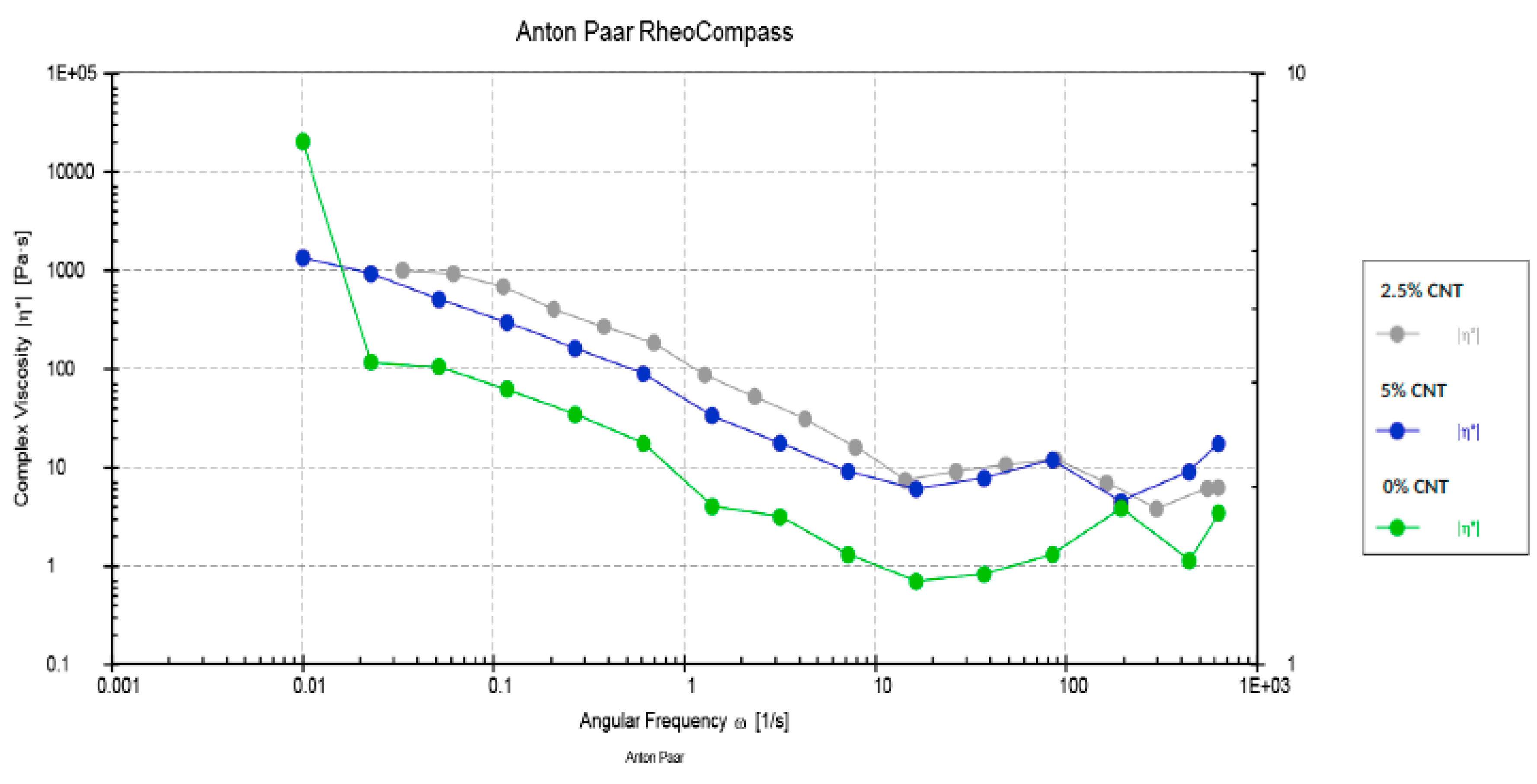
The rheological analysis of the adhesives demonstrated that at greater angular frequencies, a steady decrease in the viscosity was witnessed (Figure 3). Considering these outcomes, it can be recognized that our adhesives displayed non-Newtonian behavior (shear-thinning or pseudo-plasticity). In the current study, CA showed greater viscosity than CNP containing adhesives at 0.01 angular frequency; however, the complex viscosity of 2.5 and 5% CNP adhesive groups showed a smaller decrease than CA adhesives at higher angular frequencies (Figure 3).

3.3. SBS Testing and FMA

The SBS testing results for the three adhesive groups (CA, 2.5 CNP, and 5% CNP) are shown in Table 1. The highest SBS (25.15 ± 3.0 MPa) was observed for the NTC-2.5% CNP adhesive followed by the NTC-5% CNP adhesive group (24.25 ± 3.0 MPa). The next highest SBS (22.43 ± 3.4 MPa) was perceived for the TC-2.5% CNP group followed by the TC-5% CNP adhesive group (19.75 ± 2.7 MPa), while the CA displayed the lowest SBS values for both NTC and TC samples (19.71 ± 2.8 MPa and 15.11 ± 3.8 MPa, respectively (Table 1). The intergroup comparisons were significant (p < 0.01) when the SBS values of CA were matched with both the other groups for both TC and NTC samples. The intra-group comparisons were significant (p < 0.01) for all the groups when the NTC and TC values were compared within the same group (Table 1).
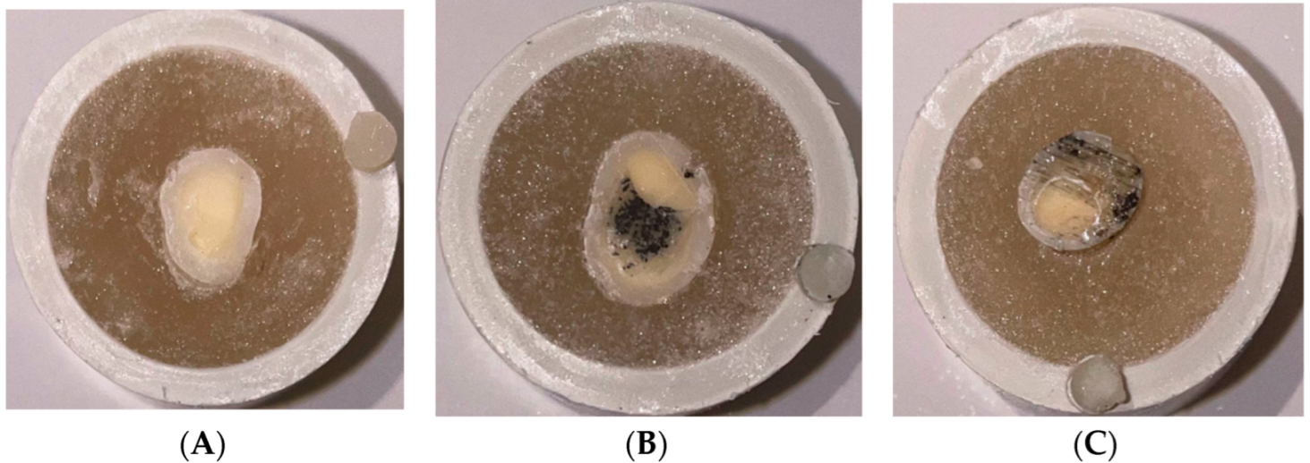
The failure modes analysis results are displayed in Table 1 (Figure 4). The adhesive-type interfacial failures ranging between 70–100% were most common. The mixed-type failures were found to be the next most common type of failures (ranging between 10 and 30%), while cohesive-type failures were not observed for any of the adhesive groups.

3.4. Analysis of the Adhesive–Dentin Interface

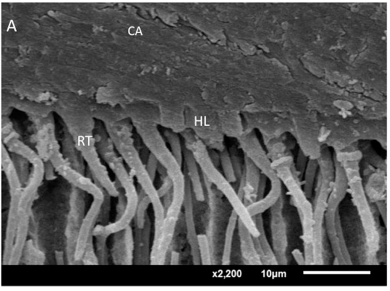
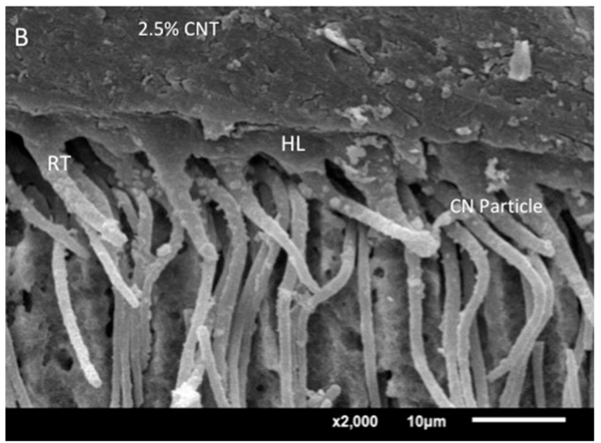
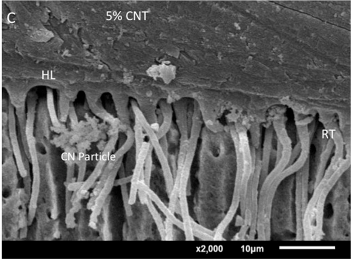
The demonstrative SEM micrographs exhibiting the interface between CA, 2.5% CNP and 5% CNP adhesive groups and dentin are presented in Figure 5A–C. The CA (control) demonstrated an appropriate resin tag formation and hybrid layer (with gaps) (Figure 5A). For the 2.5% CNP adhesive group, thick resin tags and hybrid layer formation (with few gaps) were seen (Figure 5B). For the 5% CNP group, comparable resin tags to that of the 2.5% CNP group were seen, but the hybrid layer was uniform with no to minimum gaps (Figure 5C).

3.5. FTIR and DC Analysis

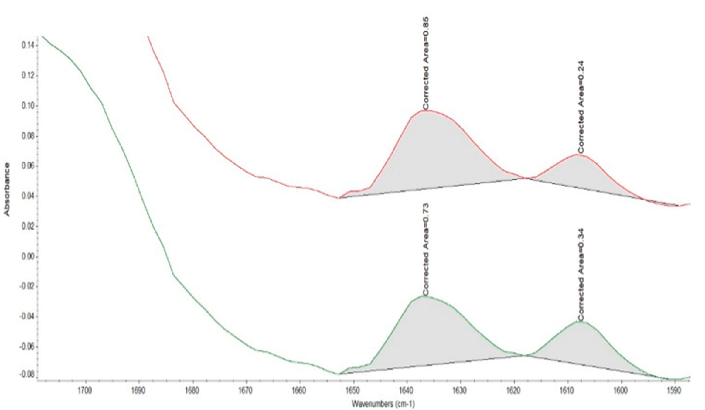
For all the adhesive groups, representative FTIR spectra were recorded. The DC was calculated by approximating the disparities in the peak height ratio of the absorbance intensities of the aliphatic C=C peak at 1638 cm−1 and that of a standard inner peak of aromatic C=C at 1608 cm−1 during polymerization, as opposed to the uncured adhesive. The highest DC was shown by the 0% CNPs (CA) (68.24 ± 4.88) followed by 2.5% CNP group (42.91 ± 5.43), while the 5% CNP (Figure 6) group demonstrated the lowest DC values (34.32 ± 4.60) (Table 2). Statistically significant results (p < 0.01) were detected when the DC values of the CA group were matched with the other two groups and when both CNP adhesive groups were compared with each other (Table 2).

4. Discussion

Based on the outcomes of the present study, the hypothesis was partially accepted as we detected that CNP-reinforced adhesives displayed better SBS values compared to the CA (2.5% CNP group showed superior SBS values to all the other groups). The hypothesis was partly rejected as the CNP-reinforced adhesives displayed lower DC values compared to the CA. The literature shows that the adhesives’ reinforcement with filler nanoparticles improves their mechanical properties [21,22,23]. The CNPs have been primarily studied in dentistry as potential contenders for drug delivery systems and as fillers for the osteogenic scaffolds [11]. However, there is a scarcity of studies that have merged these nanoparticles as fillers in the adhesive to reinforce their mechanical properties. Therefore, the present study aimed to incorporate CNPs as a filler in the composition of the CA and then assess the impact of their insertion on its various properties.
On SEM micrographs, the filler CNPs demonstrated cylindrical and roughly hexagonal shapes (Figure 1A,B). This finding matches several previously published studies that have confirmed the cylindrical/hexagonal shape of the nanoparticles form [24,25]. Nevertheless, this cylindrical or hexagonal shape could be attributed to the hexagonal organization of the carbon atoms as these atoms are entirely sp2-hybridized while being organized in a hexagonal lattice [11]. The EDX mapping of the CNPs showed the presence of C as the primary element in the CNPs (Figure 1C). This conforms to previously published literature where C was also found to be the principal element of CNPs upon EDX analysis [26,27].
Concerning the rheological assessment, it was noticed that the viscosity of the CNP adhesives in our study decreased at higher angular frequencies (Figure 2). A previous similar study reported identical results and demonstrated that when the adhesives are reinforced with nanoparticles, this results in a decreased viscosity at higher angular frequencies [19]. It is difficult to ascertain an exact cause of this finding as the rheological properties of adhesives are volatile, and they are altered with a slight modification in the material’s handling or any experimental protocol [28]. Therefore, future studies directed at determining the exact cause of the decrease in viscosity at higher angular frequencies are warranted. The flow property of an adhesive is critical for its penetration in the dentinal tubules. It has been speculated that increased infiltration of low viscosity adhesive will allow for superior bond strength, as observed in the present study.
SBS testing was performed in the present study. SBS testing allows for ease of specimen preparation, simple testing protocol, and a lower incidence of pretest failure [29]. To complement the SBS testing, failure mode analysis was included to evaluate if the outcomes are based on adhesive or cohesive failures. The SBS results demonstrated that NTC-2.5% CNP followed by NTC-5% demonstrated the highest bond strengths. These findings are consistent with previously published studies where the NTC adhesives containing filler nanoparticles demonstrated higher SBS as compared to their TC counterparts and controls [19,30]. One probable reason for this outcome could be that due to a bigger length to diameter ratio, CNPs make the inorganic matrix of dental tissues thicker and stronger [12,31]. This could have resulted in the formation of a stronger adhesive–dentin bond in this study which was verified by improved SBS values seen for the CNPs-reinforced adhesives. Another credible reason for these results could be that it has been shown in the past that CNPs in the adhesives have the capability to bond with the exposed collagen surfaces of the dentin [17]. Therefore, this bonding of CNPs with the dentinal collagen fibers, which were exposed by the prior application of phosphoric acid in our study, could have resulted in the increased SBS observed for CNPs-reinforced adhesives. The majority of the fractures in the current study were adhesive in nature. This result of our study is consistent with a prior study, which also reported that adhesive failure could occur when the adhesives are reinforced with nanoparticles [32]. It should be noted that dentin is moist and collagenous in nature, and bonding with it is challenging [33]. This could possibly have influenced an adhesive type of fracture seen in our study. In addition, mixed failures were also observed in the present study. The failure interface (mixed) included an adhesive interface and dentin structure, subsequently resulting in high bond strength. Nonetheless, the SBS was increased after CNPs incorporation, and this is an encouraging finding.
Upon evaluation of the adhesive–dentin interface, it was observed that all adhesives demonstrated an appropriate resin tag formation and hybrid layer (Figure 4A–C). However, in the CA, the hybrid layer was seen with the presence of gaps, while for the 2.5% and 5% CNP adhesive group, few to no gaps were seen. During the bonding process, while placing the aesthetic restoration, the collagen fibers of dentin become exposed, thus allowing the adhesive monomers to penetrate between the fibers and form resin tags. Regrettably, the dentinal collagen fibers are only placed 20–30 nm apart, and monomers of a general adhesive could not penetrate these gaps sufficiently [18,34]. On the other hand, CNPs are nanometer-sized and can penetrate inter-collagen gaps competently [35]. This could have resulted in thicker resin tags and a hybrid layer with few to no gaps, as seen for the 2.5% and 5% CNP adhesive groups.
DC is an extremely desirable property for an adhesive as it guarantees that an adequate number of monomers is photo-polymerized [36]. This then reduces the probability of micro-leakage and the development of secondary caries around the restoration. The highest DC in this study was perceived for the CA shadowed by 2.5% and 5% CNP adhesives, respectively. Previously conducted studies have verified that while the addition of filler nanoparticles reinforces the adhesive’s bond strength, it can lower its DC [22,37]. Our results agree with these formerly published studies as we also detected a lower DC for both CNP reinforced adhesives, compared with the CA. A likely reason for this outcome could be that the integration of filler caused a blockage in the pathway of the curing light resulting in the incomplete conversion of the adhesive monomers to polymers, leading to a lower DC [38]. However, it is pertinent to mention that, although the DC reduced, the formation of a hybrid layer was well established in the SEM observations, therefore showing good SBS and adhesive bond integrity.
There are certain limitations associated with the current study. First, the study was conducted in-vitro and this experimental setting is different from the in-vivo environment. The real in-vivo setting is vibrant and vigorous and offers numerous challenges to the foreign material. Hence, these displayed properties of our adhesives could be modified when tested under in-vivo conditions. Additionally, we observed a lower DC for our CNP-reinforced adhesives. Future studies with filler concentrations of <5% are warranted to understand the association between the filler concentration and DC.

5. Conclusions

The reinforcing of the CA in the present study with CNPs (2.5% and 5%) resulted in an increase in SBS values. The 2.5% CNP adhesive presented with the highest SBS values followed by 5% CNP adhesive. The increase in CNP concentration (2.5% to 5%) resulted in the formation of appropriate resin tags and an uniform hybrid layer. Nevertheless, both 2.5% and 5% CNP adhesives demonstrated lower viscosity and DC as matched with the CA (increasing filler content decreased the DC and vice versa). Further studies inspecting the influence of various other filler concentrations (0.5%, 1%, 2%, 3%, 3.5%, and 4%) inside the adhesive on their interaction with dentin, bond strength, rheological properties, degree of conversion and failure mode are suggested.

Author Contributions

Conceptualization, M.B., A.S.A., K.M.A.-H., I.F., F.V., Y.F.A., T.A.A. and T.A.; methodology, M.B., A.S.A., T.A.A. and T.A.; validation, T.A.A., I.F., F.V. and T.A.; formal analysis, M.B., I.F., K.M.A.-H. and T.A.; investigation, M.B., A.S.A. and T.A.; data curation, M.B. and A.S.A.; writing—original draft preparation, I.F., F.V. and T.A.; writing—review and editing, I.F., T.A.A., K.M.A.-H., T.A., Y.F.A. and F.V.; supervision, F.V., T.A. and I.F.; funding acquisition, T.A. All authors have read and agreed to the published version of the manuscript.

Funding

The authors are grateful to the Researchers supporting project at King Saud University for funding through Researchers supporting project No. (RSP-2021-44).

Institutional Review Board Statement

The research ethics review committee of King Saud University approved the study protocol with Reference No. FR-0611. All the recommendations of the Helsinki Declaration and its later amendments were strictly followed. The teeth extracted for orthodontic treatments, which were free from any apparent defects, were gathered after attaining the patients’ written informed consent and were utilized for the experiments in our study.

Informed Consent Statement

All the recommendations of the Helsinki Declaration and its later amendments were strictly followed. The teeth extracted for orthodontic treatments, which were free from any apparent defects, were gathered after attaining the patients’ written informed consent and were utilized for the experiments in our study.

Data Availability Statement

Data of the study are available on request from the corresponding author.

Conflicts of Interest

The authors declare no conflict of interest.

References

  1. Zhao, S.; Hua, F.; Yan, J.; Yang, H.; Huang, C. Effects of Plant Extracts on Dentin Bonding Strength: A Systematic Review and Meta-Analysis. Front. Bioeng. Biotechnol. 2022, 10, 836042. [Google Scholar] [CrossRef]
  2. Sofan, E.; Sofan, A.; Palaia, G.; Tenore, G.; Romeo, U.; Migliau, G. Classification review of dental adhesive systems: From the IV generation to the universal type. Ann. Stomatol. 2017, 8, 1–17. [Google Scholar]
  3. Taneja, S.; Kumari, M.; Bansal, S. Effect of saliva and blood contamination on the shear bond strength of fifth-, seventh-, and eighth-generation bonding agents: An in vitro study. J. Conserv. Dent. 2017, 20, 157–160. [Google Scholar] [CrossRef]
  4. Hass, V.; Luque-Martinez, I.; Munoz, M.A.; Reyes, M.F.; Abuna, G.; Sinhoreti, M.A.; Liu, A.Y.; Loguercio, A.D.; Wang, Y.; Reis, A. The effect of proanthocyanidin-containing 10% phosphoric acid on bonding properties and MMP inhibition. Dent. Mater. 2016, 32, 468–475. [Google Scholar] [CrossRef]
  5. Farooq, I.; Ali, S.; Al-Saleh, S.; AlHamdan, E.M.; AlRefeai, M.H.; Abduljabbar, T.; Vohra, F. Synergistic Effect of Bioactive Inorganic Fillers in Enhancing Properties of Dentin Adhesives-A Review. Polymers 2021, 13, 2169. [Google Scholar] [CrossRef]
  6. Zhou, W.; Liu, S.; Zhou, X.; Hannig, M.; Rupf, S.; Feng, J.; Peng, X.; Cheng, L. Modifying Adhesive Materials to Improve the Longevity of Resinous Restorations. Int. J. Mol. Sci. 2019, 20, 723. [Google Scholar] [CrossRef] [Green Version]
  7. Ghahramani, Y.; Javanmardi, N. Graphene oxide quantum dots and their applications via stem cells: A mini-review. Adv. Appl. NanoBio-Technol. 2021, 2, 54–56. [Google Scholar]
  8. Vijay, R.; Mendhi, J.; Prasad, K.; Xiao, Y.; MacLeod, J.; Ostrikov, K.K.; Zhou, Y. Carbon Nanomaterials Modified Biomimetic Dental Implants for Diabetic Patients. Nanomaterials 2021, 11, 2977. [Google Scholar] [CrossRef]
  9. Bonilla-Represa, V.; Abalos-Labruzzi, C.; Herrera-Martinez, M.; Guerrero-Perez, M.O. Nanomaterials in Dentistry: State of the Art and Future Challenges. Nanomaterials 2020, 10, 1770. [Google Scholar] [CrossRef]
  10. De Vasconcellos, L.M.R.; do Prado, R.F.; Sartori, E.M.; Mendonca, D.B.S.; Mendonca, G.; Marciano, F.R.; Lobo, A.O. In vitro osteogenesis process induced by hybrid nanohydroxyapatite/graphene nanoribbons composites. J. Mater. Sci. Mater. Med. 2019, 30, 81. [Google Scholar] [CrossRef] [PubMed]
  11. Castro-Rojas, M.A.; Vega-Cantu, Y.I.; Cordell, G.A.; Rodriguez-Garcia, A. Dental Applications of Carbon Nanotubes. Molecules 2021, 26, 4423. [Google Scholar] [CrossRef]
  12. Mousavi, S.M.; Yousefi, K.; Hashemi, S.A.; Afsa, M.; Bahran, I.S.; Gholami, A.; Ghahramani, Y.; Alizadeh, A.; Chiang, W.H. Renewable Carbon Nanomaterials: Novel Resources for Dental Tissue Engineering. Nanomaterials 2021, 11, 2800. [Google Scholar] [CrossRef]
  13. Thess, A.; Lee, R.; Nikolaev, P.; Dai, H.; Petit, P.; Robert, J.; Xu, C.; Lee, Y.H.; Kim, S.G.; Rinzler, A.G.; et al. Crystalline Ropes of Metallic Carbon Nanotubes. Science 1996, 273, 483–487. [Google Scholar] [CrossRef] [Green Version]
  14. Li, Q.L.J.; Xu, S. Progress in Research on Carbon Nanotubes Reinforced Cementitious Composites. Adv. Mater. Sci. Eng. 2015, 2015, 307435. [Google Scholar] [CrossRef] [Green Version]
  15. Kechagioglou, P.; Andriotis, E.; Papagerakis, P.; Papagerakis, S. Multiwalled Carbon Nanotubes for Dental Applications. Methods Mol. Biol. 2019, 1922, 121–128. [Google Scholar]
  16. Khan, A.S.; Hussain, A.N.; Sidra, L.; Sarfraz, Z.; Khalid, H.; Khan, M.; Manzoor, F.; Shahzadi, L.; Yar, M.; Rehman, I.U. Fabrication and in vivo evaluation of hydroxyapatite/carbon nanotube electrospun fibers for biomedical/dental application. Mater. Sci. Eng. C Mater. Biol. Appl. 2017, 80, 387–396. [Google Scholar] [CrossRef]
  17. Akasaka, T.; Nakata, K.; Uo, M.; Watari, F. Modification of the dentin surface by using carbon nanotubes. Biomed. Mater. Eng. 2009, 19, 179–185. [Google Scholar] [CrossRef] [Green Version]
  18. Suo, L.; Li, Z.; Luo, F.; Chen, J.; Jia, L.; Wang, T.; Pei, X.; Wan, Q. Effect of dentin surface modification using carbon nanotubes on dental bonding and antibacterial ability. Dent. Mater. J. 2018, 37, 229–236. [Google Scholar] [CrossRef] [Green Version]
  19. Al-Saleh, S.; Alateeq, A.; Alshaya, A.H.; Al-Qahtani, A.S.; Tulbah, H.I.; Binhasan, M.; Shabib, S.; Farooq, I.; Vohra, F.; Abduljabbar, T. Influence of TiO2 and ZrO2 Nanoparticles on Adhesive Bond Strength and Viscosity of Dentin Polymer: A Physical and Chemical Evaluation. Polymers 2021, 13, 3794. [Google Scholar] [CrossRef]
  20. Yoshida, T.; Yamaguchi, K.; Tsubota, K.; Takamizawa, T.; Kurokawa, H.; Rikuta, A.; Ando, S.; Miyazaki, M. Effect of metal conditioners on polymerization behavior of bonding agents. J. Oral Sci. 2005, 47, 171–175. [Google Scholar] [CrossRef] [PubMed] [Green Version]
  21. Al-Hamdan, R.S.; Almutairi, B.; Kattan, H.F.; Alsuwailem, N.A.; Farooq, I.; Vohra, F.; Abduljabbar, T. Influence of Hydroxyapatite Nanospheres in Dentin Adhesive on the Dentin Bond Integrity and Degree of Conversion: A Scanning Electron Microscopy (SEM), Raman, Fourier Transform-Infrared (FTIR), and Microtensile Study. Polymers 2020, 12, 2948. [Google Scholar] [CrossRef] [PubMed]
  22. AlFawaz, Y.F.; Almutairi, B.; Kattan, H.F.; Zafar, M.S.; Farooq, I.; Naseem, M.; Vohra, F.; Abduljabbar, T. Dentin Bond Integrity of Hydroxyapatite Containing Resin Adhesive Enhanced with Graphene Oxide Nano-Particles-An SEM, EDX, Micro-Raman, and Microtensile Bond Strength Study. Polymers 2020, 12, 2978. [Google Scholar] [CrossRef] [PubMed]
  23. Almutairi, B.; Kattan, H.F.; BinMahfooz, A.M.; Qutub, O.A.; Basunbul, G.; ArRejaie, A.S.; Farooq, I.; Vohra, F.; Abduljabbar, T. Synergistic effect of graphene oxide/calcium phosphate nanofiller in a dentin adhesive on its dentin bond integrity and degree of conversion. A scanning electron microscopy, energy dispersive X-ray spectroscopy, Fourier transform infrared, micro-Raman, and bond strength study. Microsc. Res. Tech. 2021, 84, 2082–2094. [Google Scholar] [PubMed]
  24. Dyatlova, O.A.; Gomis-Bresco, J.; Malic, E.; Telg, H.; Maultzsch, J.; Zhong, G.; Geng, J.; Woggon, U. Dielectric screening effects on transition energies in aligned carbon nanotubes. Phys. Rev. B 2012, 85, 245449. [Google Scholar] [CrossRef]
  25. Williams, J.; Broughton, W.; Koukoulas, T.; Rahatekar, S.S. Plasma treatment as a method for functionalising and improving dispersion of carbon nanotubes in epoxy resins. J. Mater. Sci. 2012, 48, 1005–1013. [Google Scholar] [CrossRef]
  26. Osikoya, A.O.; Wankasi, D.; Vala, R.M.K.; Dikio, C.W.; Afolabi, A.O.; Ayawei, N.; Dikio, E.D. Synthesis, characterization and sorption studies of nitrogen–doped carbon nanotubes. Digest. J. Nanomat. Biostruct. 2015, 10, 125–134. [Google Scholar]
  27. Yue, N.; Wang, L.; He, X.; Liu, H.; Zhang, W. Optimizing the SEM specimen preparation method for accurate microanalysis of carbon nanotube/nanocluster hybrids. J. Microsc. 2021, 282, 267–273. [Google Scholar] [CrossRef]
  28. Lee, J.H.; Um, C.M.; Lee, I.B. Rheological properties of resin composites according to variations in monomer and filler composition. Dent. Mater. 2006, 22, 515–526. [Google Scholar] [CrossRef] [PubMed]
  29. Sirisha, K.; Rambabu, T.; Ravishankar, Y.; Ravikumar, P. Validity of bond strength tests: A critical review-Part II. J. Conserv. Dent. 2014, 17, 420. [Google Scholar] [CrossRef] [PubMed] [Green Version]
  30. Al-Qahtani, A.S.; Tulbah, H.I.; Binhasan, M.; Shabib, S.; Al-Aali, K.A.; Alhamdan, M.M.; Abduljabbar, T. Influence of Concentration Levels of beta-Tricalcium Phosphate on the Physical Properties of a Dental Adhesive. Nanomaterials 2022, 12, 125–134. [Google Scholar] [CrossRef]
  31. Pei, B.; Wang, W.; Dunne, N.; Li, X. Applications of Carbon Nanotubes in Bone Tissue Regeneration and Engineering: Superiority, Concerns, Current Advancements, and Prospects. Nanomaterials 2019, 9, 1501. [Google Scholar] [CrossRef] [PubMed] [Green Version]
  32. Alqarawi, F.K.; Alkahtany, M.F.; Almadi, K.H.; Ben Gassem, A.A.; Alshahrani, F.A.; AlRefeai, M.H.; Farooq, I.; Vohra, F.; Abduljabbar, T. Influence of Different Conditioning Treatments on the Bond Integrity of Root Dentin to rGO Infiltrated Dentin Adhesive. SEM, EDX, FTIR and MicroRaman Study. Polymers 2021, 13, 1555. [Google Scholar] [CrossRef] [PubMed]
  33. Peumans, M.; Kanumilli, P.; De Munck, J.; Van Landuyt, K.; Lambrechts, P.; Van Meerbeek, B. Clinical effectiveness of contemporary adhesives: A systematic review of current clinical trials. Dent. Mater. 2005, 21, 864–881. [Google Scholar] [CrossRef]
  34. Sachdeva, P.; Goswami, M.; Singh, D. Comparative evaluation of shear bond strength and nanoleakage of conventional and self-adhering flowable composites to primary teeth dentin. Contemp. Clin. Dent. 2016, 7, 326–331. [Google Scholar]
  35. Terada, M.; Abe, S.; Akasaka, T.; Uo, M.; Kitagawa, Y.; Watari, F. Development of a multiwalled carbon nanotube coated collagen dish. Dent. Mater. J. 2009, 28, 82–88. [Google Scholar] [CrossRef]
  36. Moldovan, M.; Balazsi, R.; Soanca, A.; Roman, A.; Sarosi, C.; Prodan, D.; Vlassa, M.; Cojocaru, I.; Saceleanu, V.; Cristescu, I. Evaluation of the Degree of Conversion, Residual Monomers and Mechanical Properties of Some Light-Cured Dental Resin Composites. Materials 2019, 12, 2109. [Google Scholar] [CrossRef] [Green Version]
  37. Aguiar, T.R.; de Oliveira, M.; Arrais, C.A.; Ambrosano, G.M.; Rueggeberg, F.; Giannini, M. The effect of photopolymerization on the degree of conversion, polymerization kinetic, biaxial flexure strength, and modulus of self-adhesive resin cements. J. Prosthet. Dent. 2015, 113, 128–134. [Google Scholar] [CrossRef] [PubMed]
  38. Utneja, S.; Talwar, S.; Nawal, R.R.; Sapra, S.; Mittal, M.; Rajain, A.; Verma, M. Evaluation of remineralization potential and mechanical properties of pit and fissure sealants fortified with nano-hydroxyapatite and nano-amorphous calcium phosphate fillers: An in vitro study. J. Conserv. Dent. 2018, 21, 681–690. [Google Scholar] [CrossRef] [PubMed]
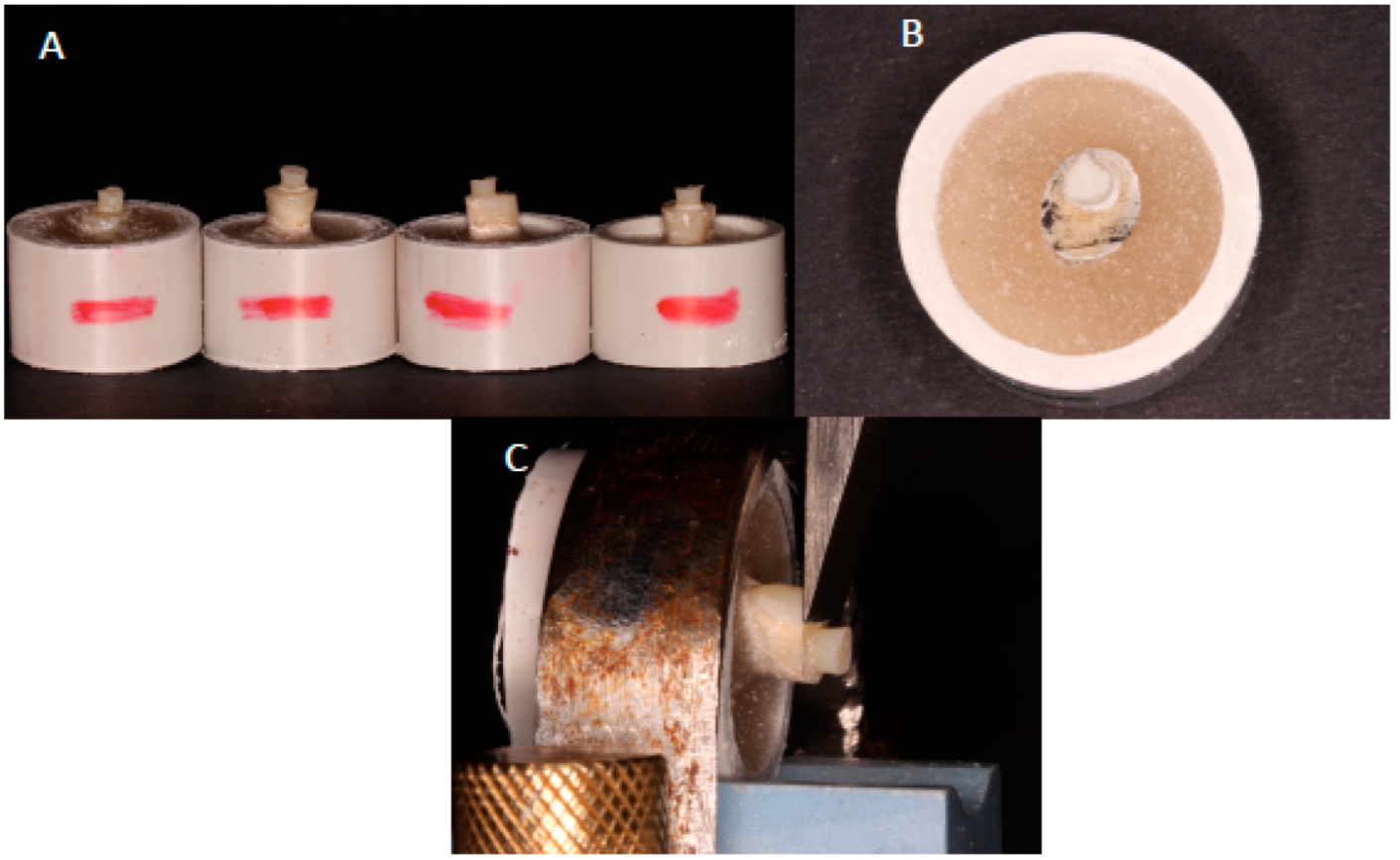
Figure 1. Shear bond strength (SBS) assembly. (A,B) Bonded resin-adhesive-dentin samples.(C) Assembly of the customized metal probe with mounted bonded samples for SBS in universal testing machine.
Crystals 12 01441 g001
Figure 2. (A) SEM image of carbon nanoparticles of variable sizes and orientation, mostly flat-disc shaped, ranging from less than 5 μm to nano size. (B) Showing carbon nanoparticles of flat long dimensions ranging from nano to micron size. (C) EDX analysis showing more than 80% of carbon content of the analyzed powder, with traces of other elements (Zr and O).
Crystals 12 01441 g002
Figure 3. Rheological properties of control adhesive (CA-0% CNP), 2.5% CNP and 5% CNP. Complex viscosities with angular frequency at 0.001 to 1000 rads/s can be observed.
Crystals 12 01441 g003
Figure 4. Shear bond strength samples among groups after failure. (A) 0% CNP; (B) 2.5 wt.% CNP; (C) 5 wt.% CNP.
Crystals 12 01441 g004
Figure 5. SEM images of the bonded resin and dentin interface using (A) 0% carbon nanoparticle adhesive, (B) 2.5 wt.% and (C) 5 wt.% CNP dental adhesive. All specimens formed an adhesive layer of uniform thickness. All group specimens showed development of standard hybrid layer (HL). Resin tags (RT) with uniform length and dimensions were formed within HL among all group specimens (0% CNP, 2.5% CNP and 5% CNP adhesives). (B) 2.5 wt.% CNP adhesive resulted in well-formed and dense RT with few gap formations within the hybrid layer and presence of CNPs. (C) The 5 wt.% CNP adhesive also resulted in a uniform hybrid layer incorporating RTs with minimum gaps at resin–dentin interface and presence of CNPs.
Crystals 12 01441 g005aCrystals 12 01441 g005bCrystals 12 01441 g005c
Figure 6. Typical double-bond conversion FTIR peaks before and after curing for 5% CNP adhesive.
Crystals 12 01441 g006
Table 1. Means and Standard deviations for Shear bond strength (MPa) and failure modes among the study groups.
SBS (MPa) (Mean ± SD)Failure Mode Analysis, FMA (%)
Group (n = 10)NTCTCp-Value *AdhesiveCohesiveMixed
0% CNP-adhesive19.71 ± 2.8 a,A-< 0.0110000
-15.11 ± 3.8 b,A10000
2.5 wt.% CNP- adhesive25.15 ± 3.0 a,B-70030
-22.43 ± 3.4 b,B90010
5.0 wt.% CNP- adhesive 24.25 ± 3.0 a,B-90010
-19.75 ± 2.7 b,B80020
CNP: Carbon nanoparticles, NTC: No thermocycling, TC: Thermocycling, * ANOVA. SBS: Shear bond strength, SD: standard deviation. Dissimilar upper-case superscript alphabets in same column denote statistical significance (p < 0.01); dissimilar lower-case superscript alphabets in same material denote statistical significance (p < 0.01).
Table 2. Showing degree of conversion (DC) values in mean (SD) among 0% CNP adhesive (CA), 2.5% CNP adhesive and 5% CNP adhesive.
Serial No.Group NameDC (%)
10% CNP (CA)68.24 (4.88) A
22.5 % CNP adhesive42.91 (5.43) B
35% CNP adhesive34.32 (4.60) C
Different upper-case alphabets in same column represent statistically significant difference, (p < 0.01). CNP: Carbon nanoparticle.
Publisher’s Note: MDPI stays neutral with regard to jurisdictional claims in published maps and institutional affiliations.