Review

Life from a Snowflake: Diversity and Adaptation of Cold-Loving Bacteria among Ice Crystals

by
Carmen Rizzo
1,2,* and
Angelina Lo Giudice
2
1
Stazione Zoologica Anton Dohrn—Ecosustainable Marine Biotechnology Department, Sicily Marine Centre, Villa Pace, Contrada Porticatello 29, 98167 Messina, Italy
2
Institute of Polar Sciences, National Research Council (CNR-ISP), Spianata S. Raineri 86, 98122 Messina, Italy
*
Author to whom correspondence should be addressed.
Crystals 2022, 12(3), 312; https://doi.org/10.3390/cryst12030312
Submission received: 19 January 2022 / Revised: 12 February 2022 / Accepted: 15 February 2022 / Published: 23 February 2022
(This article belongs to the Section Biomolecular Crystals)

Abstract

:
Incredible as it is, researchers have now the awareness that even the most extreme environment includes special habitats that host several forms of life. Cold environments cover different compartments of the cryosphere, as sea and freshwater ice, glaciers, snow, and permafrost. Although these are very particular environmental compartments in which various stressors coexist (i.e., freeze–thaw cycles, scarce water availability, irradiance conditions, and poorness of nutrients), diverse specialized microbial communities are harbored. This raises many intriguing questions, many of which are still unresolved. For instance, a challenging focus is to understand if microorganisms survive trapped frozen among ice crystals for long periods of time or if they indeed remain metabolically active. Likewise, a look at their site-specific diversity and at their putative geochemical activity is demanded, as well as at the equally interesting microbial activity at subzero temperatures. The production of special molecules such as strategy of adaptations, cryoprotectants, and ice crystal-controlling molecules is even more intriguing. This paper aims at reviewing all these aspects with the intent of providing a thorough overview of the main contributors in investigating the microbial life in the cryosphere, touching on the themes of diversity, adaptation, and metabolic potential.

1. The Cryosphere

Although apparently the same, the extreme cold environments host a great diversity of habitats and a wide variety of living organisms. Cold environments comprise both aquatic and terrestrial ecosystems, including deep sea, sea ice, lake ice, snow, glacial habitats, permafrost, cold lakes, cold soils and deserts, atmosphere, and clouds [1]. Contrary to what one might think, a large percentage of the Earth’s biosphere is dominated by environments at temperatures below 5 °C, having very different features, all contributing to form the cryosphere. The term “cryosphere” indicates those regions characterized by temperature so cold that water turns into ice. The icy habitats are characterized by solid form of water, as such as ice and snow, which are not available for cellular processes. Several studies, reviewed by Margesin and Collins [1], carried out on the cryosphere have shown that water is the basis of life even when it assumes conformations hostile to life, as these habitats are generally represented by porous matrices in which liquids may occur, thus allowing survival of cold-adapted organisms. Morita et al. [2] distinguished cold-adapted bacteria in psychrophiles and psychrotrophs. The first prefer cold temperature and are more strictly related to it, while the second ones tolerate low temperatures but are also able to grow until a temperature of 20 °C. This means also that their distribution is different, with psychrotrophs that are more widespread distributed, and also in environments with thermal fluctuations [3,4].
The presence of ice crystals can cause damages to cells, by disrupting cell membranes, and the low temperature affects the metabolic processes and the chemical folding of molecules. Despite this, it has been proved that several icy habitats are inhabited by microorganisms, which shape their physiological and metabolic features according to the external conditions, by making the subzero temperatures a driving force for microbial diversification. If photoautotrophy dominates habitats where sunlight penetrates the ice, chemoautotrophy is the strategy adopted by microorganisms living in dark cold environments. If the membrane integrity and biochemical processes are threatened by the freezing point, flexibility and homeostasis are implemented thanks to special cold lipids and enzymes or by the production of anti-freeze proteins and extracellular polymeric substances that act as cryoprotectants [5].
The comprehension of the ecological role played by the microbial fraction in such environments is pivotal due to its sensitivity to climate change effects [6,7,8], dangerously highlighted by the retreat of glaciers and ice sheets and by the permafrost thawing. Moreover, frozen environments are suitable models for study in an astrobiology perspective, since they are recognized as analogues of extraterrestrial habitats [9,10,11] and are an untapped source of relevance in the biotechnological field [12,13]. Today, the cryosphere covers about one-fifth of the surface of the Earth, with substantial seasonal variations and a long-term trend of losses in its area and volume due to climate warming. It comprises highly diversified habitats, with unique features to be considered in all studies addressed at their deeper comprehension.

2. Convergent and Divergent Aspects of Frozen Habitats at the Poles

Although they may all look the same, polar environments have many differences and host many different habitats. Each of these cryo-matrices possess peculiar features in terms of temperature, consistency, ice morphology, salinity, availability of oxygen, and light. This means that, despite having a common basic background in physico-chemical terms, the organisms that live there have several morphologies and physiological capacities. In the following sections the main cryoenvironments will be described, outlining their characteristics, formation processes, distinctive characteristics, and their representative microbial inhabitants.

2.1. Snow

Snow is a special kind of precipitation that falls to the ground in form of ice crystals, formed in case of low temperatures accompanied by high humidity rates in the atmosphere. It can be found in every point of the planet, reflects sunlight, affects the global climate, represents a habitat for animals and plants. The first step is the formation of an ice crystal that absorbs and freezes additional water vapor from the surrounding air, forming a snow crystal or snow pellet, which then precipitates to Earth. It is surprising to realize that even the snow, which seems homogeneous and always the same, comes in different forms [14]. Snow pellets, as an example, are opaque ice particles formed when ice crystals fall through supercooled cloud droplets, which then freeze to the crystals resulting in a final lumpy mass. Differently, sleet is a sort of mix of rain and drizzle that freezes into little ice balls of about 0.76 cm in diameter. Sometimes we refer to sleet for a mixture of ice pellets and freezing rain. Hail is not considered snow, because it tends to assume bigger dimensions than snow and it is generated during thunderstorms, when drops of supercooled water freeze to graupel and became too heavy to be supported due to updrafts or upward moving air, thus falling as hailstones. Interestingly, snow could be classified as a mineral, as it is solid, homogenous, and with an ordered arrangement granted by the hydrogen bonds between water molecules [15]. The biotic component possibly occurring in snow habitats (mainly constituted by bacteria, viruses, fungi, small protists, seeds, and insects) is carried by winds from different areas of the globe and deposited on snow surface. The composition of this biotic component is variable, depending on the local features of snow ecological systems [16], and it is naturally exposed to high UV radiation and seasonal fluctuations.

2.2. Glaciers, Icebergs, and Ice Platforms

When temperatures are below the freezing point and water naturally turns to solid form, glaciers sea ice, ice shelves, icebergs, and frozen ground are formed. These special forms of solid water could exist all over the world but are more easily observable at high latitudes or elevation, or during the nights when the temperature is colder. They could be affected by seasonal changes and, in some cases, they can be so thick on the surface of lakes and oceans that only icebreakers can navigate. The different forms of ice are scientifically significative; in the form of ice cores they can provide pivotal information about the past conditions of Earth. Glaciers, which store the 69% of world’s fresh water, slowly exhibit modelling effects on mountains and carved valleys, by changing actively the landscape configuration. They possess an important ecological role, as a nutrient deliverer through melting into aquatic basins, i.e., lakes, rivers, and oceans. The nutrient inputs influence phytoplankton blooms and regulate the aquatic food chains, with direct and indirect effects on the entire ecosystem. Among all forms in which water freezes by contributing to form the cryosphere, glaciers are the biggest contributor to sea level rises and represent also an important water supply for native people of some countries. Glaciers are constituted by big masses of ice on land, deriving from many seasons of snowfall and covering about 10% of the global land. Their recent regression represents a worrying point for scientists as most impactful effects of climate change [17].
Ice shelves are platforms of ice that form when ice sheets and glaciers move towards the oceans, whereas icebergs are chunks of ice released from glaciers and ice shelves and drift in the oceans. Ice shelves are mainly distributed in Antarctica and Greenland, as well as in the Arctic region of Canada and Alaska. These great ice giants offer shelter for polar animals and they represent habitats of microbic forms of life. Contrary to what is commonly thought, they raise the sea level only when they detach from the earth to pour into the water basins, but not when they melt in the water [17].

2.3. Sea Ice

The sea ice is a special kind of ice, occurring when water in the oceans achieves temperatures below freezing, mainly in the Arctic and Antarctic Oceans. It creates a unique habitat for polar organisms even if with different features in the two polar areas, and in the Arctic regions, it also influences the customs of native people. Sea ice tends to increase during the colder months and melts during the warmer season, but a little fraction of it remains all year in some regions [18,19]. The main difference between sea ice and other ice blocks (such as icebergs, glaciers, or lake ice) is that sea ice forms from salty ocean water [20]. This implies some additional differences in the formation process: we should remember that freshwater tends to become less dense near the freezing points, resulting in an ice layer on the top of lakes and rivers; the presence of salts in ocean water affects its density and decrease the freezing point level. As a results, the occurrence of sea ice is very slow compared to freshwater ice [21]. Sea ice affects the movement of ocean waters, since when it is formed, salts are pushed into the ocean by forming masses of denser water below the ice, which sinks and move along the ocean bottom toward the equator, while warm water circulates from mid-depth to the surface toward the poles. Changes occurring in the distribution and amount of sea ice could be disrupting for the normal ocean circulation, and thus on the global climate conditions. Moreover, the extrusion of salts from ice crystals generates a porous matrix inside the ice in which brines are contained, whose salinity and volume strongly depend on the temperature values of the surrounding ice. The brines are interconnected with the ice, with a complex system of permeable channels and pockets [17].
Even if sea ice occurs mostly in polar areas, it is closely related to the planet’s climate and for this reason it is particularly interesting for researchers who are focused on the effects of climate changes. The regions covered by sea ice do not absorb much solar energy, due to the sea ice surface reflection of sunlight in the space. This means that polar regions remain relatively cool, but the melting of sea ice caused by warming temperatures reduces bright surfaces available to reflects sunlight, thus determining a major absorption of solar energy and a resulting rise of temperature, in a cascade process. Even though this cycle stops during the polar dark seasons, even a little increase in the temperature level can lead to a greater warming effect. For this reason, polar areas are the most sensitive regions to climate change. Interestingly, new ice is generally really salty because it contains concentrated droplets called brine, trapped in pockets between the ice crystals.
Several microorganisms find suitable habitat in sea ice [22,23,24]. Sea ice microbial communities are dominated by phytoplanktonic organisms, mostly represented by diatoms, and heterotrophic bacteria, which regulate the algal blooms and the organic matter flux from the ice to the seawater [25,26]. The heterotrophic production and bacterial growth act as fertilizers during the cold season, and the secondary production represents a constant source of organic matter in the sea ice and water [27]. The growth of bacteria in sea ice is regulated be numerous factors, i.e., salinity, temperature, nutrient availability [28], and trophic interactions [29]. Despite an initial transient reduction in activity during the formation of sea ice, they are metabolically active after its consolidation, and play a central role in the circulation of organic matter even inside the ice matrix [30]. A shift from psychrotrophic to psychrophilic bacteria (see below for differences) occurs with maturation of sea ice, and the development of bacterial communities proceeds in relation to the substrate concentration and attachment sites [31].
A strong temperature gradient occurs between winter and early spring sea ice, with a surface temperature of about 4 °C and the bottom at 1.86 °C [20]. The upper layer of sea ice is also influenced by more intense UV radiations and higher oxidation stress [32]. Interestingly, in the microhabitat created by the brines, the microorganisms find refuge in a certain sense from the most external hostile conditions, with higher temperatures, lower salinity, increased volume, lower UV radiation and oxygen radical occurrence [33]. In the Arctic region wide fluctuations in sea ice levels are observed during summer and winter, and it is possible to recognize a multilayer ice, a perennial form of ice characterized by persistence more durable than one summer, and the seasonal ice, also called first-year ice. During spring, the perennial ice is constituted by an old layer that was not affected by previous melting season and a new layer added at the bottom of the ice structure during the freeze period. The old layer undergoes a desalination process and fresh water flushing from snowmelt [20,34], which together with the growth of new ice leads to a strong vertical stratification in terms of solute and organic matter content.
Snow and glacier systems are strongly correlated because glacier ice derives from gradual and long-time deposition and compression of snow.

2.4. Brines

Brines are hypersaline systems observed within permafrost, glaciers, and polar lakes [35,36,37,38,39,40]. Despite their unfrozen state, they are characterized by high salt content, which maintains them in unfrozen conditions and also at temperatures below 0 °C, and they are therefore counted here among cryosystems. The formation and mobilization of these systems is scarcely known [41], but geochemical studies depicted brines as dynamic systems in which fluxes and interconnections with lakes and ponds can occur [42,43]. The occurrence of two distinct pressurized hypersaline brine pockets, separated by a thin ice layer, has recently been discovered in an ice-covered Antarctic Lake in Tarn Flat (Northern Victoria Land), fed by an underground talik external to the lake basin [44]. According to Forte et al. [44], while one of the brines had possible exchange with atmosphere, the second one was supposed to be isolated since 12,000 cal years BP. In the Boulder Clay area, the presence of brine pockets of possible hydrostatic origin in two perennially ice-covered Antarctic lakes has also been detected [45,46,47,48]. Similarly, a peculiar briny system, located at Lake Vida in Antarctica, has also been described. Anoxic brine had no gas exchanges with the atmosphere for the 2800-year-estimated period [49]. The brine has a salinity range of 188–210 psu, temperature of −13.4 °C, and is rich in nutrients (ammonia, iron, and nitrates) and organic carbon [38].
The interest in such extreme habitats has been powered by the numerous common features with extraterrestrial environments, leading to suppose they could be used as model study to improve our knowledge about the existence of life on other planets.

2.5. Permafrost

When soil or rock are partially or totally formed by frozen water, frozen ground occurs and if it is frozen all year long it is called permafrost, as permanently frozen ground. Specifically, permafrost is defined as a ground that could be constituted by soil, sediment, or rocks permanently exposed to temperatures below 0 °C and that have remained frozen for at least two consecutive years [50]. It is mainly formed by ice, 20–70%, and unfrozen water, 1–7%. It is mainly distributed in the polar areas, but it can also be observed at high elevations, as for example in the Alps, Andes, and Himalayas. This implies different features, not only between polar and high elevation permafrost, characterized by higher spatial variability and mean temperatures [51,52], but also between the two poles. Indeed, Arctic permafrost is subjected to mean temperature of −10 °C and low organic carbon and nitrogen content, while Antarctic permafrost presents temperatures ranging between −18.5 and −27 °C, and an alkaline pH [1,11,53]. Within permafrost systems is included submarine permafrost, which is influenced by the salt penetration from sea water in addition to the phenomena of heat transfer [54].
Generally, the most superficial layer is considered the active component, where living organisms can occur due to the thawing for part of the year. Interestingly, it stores greenhouse gases such as carbon and methane, and scientists are studying how these gases will affect climate as temperatures warm and permafrost thaws.
The greater part of the bacteria living in permafrost habitats are small in size and are viable but non culturable [1], probably in a dormant state, despite metabolic activity under frozen conditions having also been demonstrated [55,56]. The active layer hosts very diverse microbial communities, well adapted to the conditions of temperature changes, and ice and water availability. To date, an in-depth knowledge of these populations in terms of ecological role and structure is lacking and poorly understood [57,58].

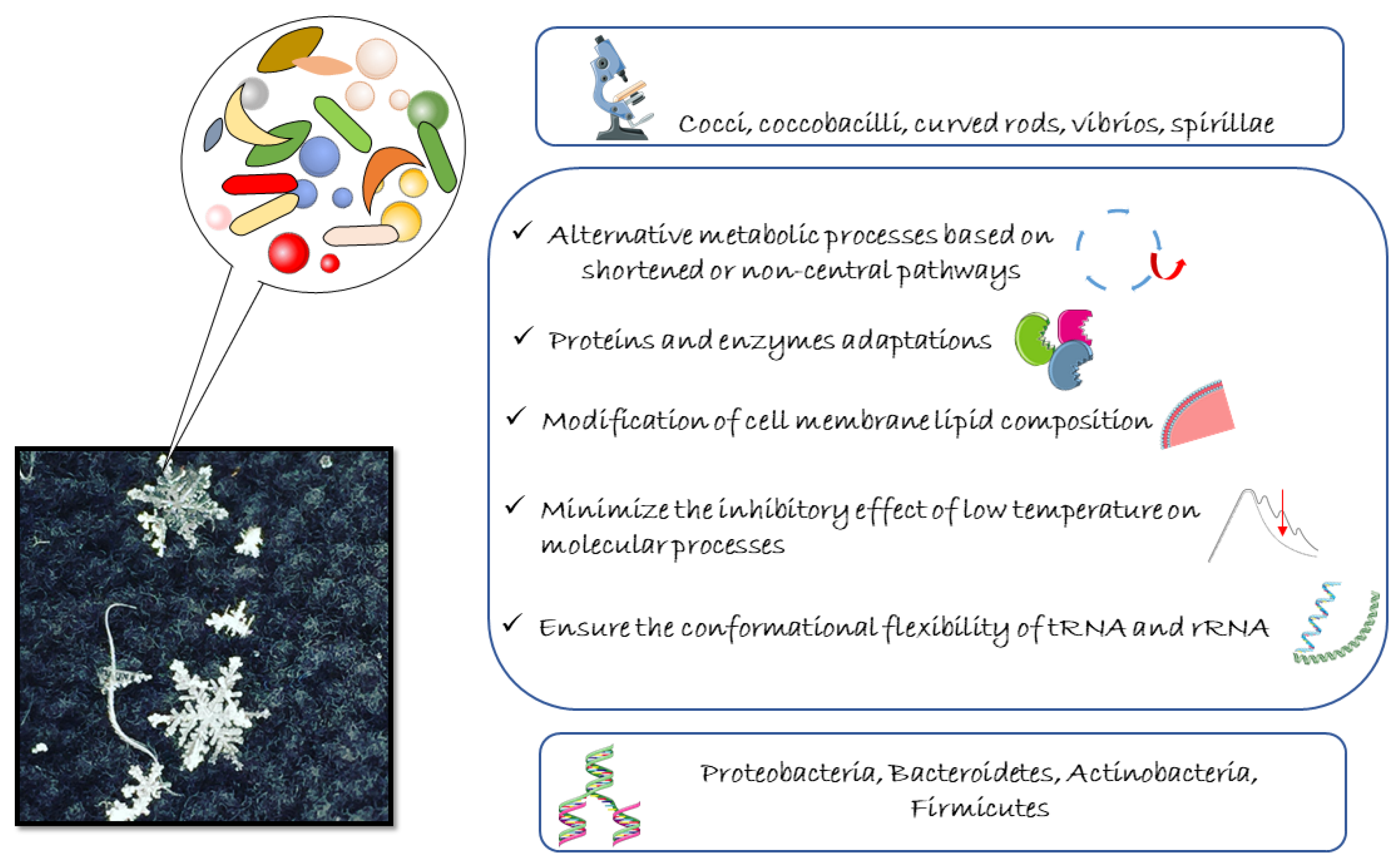
3. Microbial Life at Subzero Temperatures

3.1. Cryoinjury on Bacterial Life

The adversities to which organisms living in polar environments are subjected are not limited to the extreme conditions themselves, but also to the extreme variability over time and depending on the type of habitat.
The low temperature is clearly the first adverse parameter that acts on multiple levels, affecting water availability, thermal energy, and molecular stability. The availability of water in liquid form is substantial for all metabolic processes, as it is the main reaction environment as a solvent system for enzymes and other organic substrates. The freezing process is influenced by several factors, which are limited not only to the external temperature value, but also include freezing point depression, ordering effects, supercooling, and pressure. If generally liquid water freezes at 0 °C, the presence of solutes, e.g., in seawater, could induce the lowering of freezing point, by also maintaining liquid water several degrees below the zero. In a chain process, freezing a first fraction of water leaves a more concentrated solution, which further lowers the freezing point and so on until complete saturation and consequent crystallization of both solute and solvent. This generates the creation of thin films of water in contact with solid matrices, i.e., soils and ice [59]. It remains questionable whether these limited quantities of liquid water are useful for biological activities [60].
The low temperature also decreases thermal energy levels, thus affecting reaction rates and consequently metabolic activities. Low thermal energy also acts at molecular levels by affecting fluidity and flexibility of biological molecules, i.e., DNA, RNA, and proteins. The limit for active metabolism of bacteria is determined by the process of vitrification in the cytoplasm, due to the high internal viscosity. When temperature decreases, lipids forming biological membranes move from liquid crystal to gel phase. The increasing rigidity of membranes hinders the nutrient transport, by determining cellular starvation, while bacteria stop functioning when 50–90% of membrane lipids are in gel phase [61]. Differently from intracellular freezing, vitrification is not lethal, and metabolic activities could restart after rehydration processes [61,62]. Despite this clearly representing a strong stressful event, microbial metabolism can still support low rates for long periods of time, as observed in several cold environments [63,64,65].
Beside cold temperature, other adverse conditions are represented by low nutrient, high salinity, dryness, UV irradiation, and oxidative stress. Indeed, at low temperature oxygen solubility increases, thus generating a high amount of reactive oxygen species, cause of oxidative stress. This condition tends to modify the active metabolic pathways, inhibiting the most common, i.e., glycolysis, the phosphate pathway, the tricarboxylic acid cycle, and the electron transport chain [66]. Light availability is also a strong influencing factor for microbial life. It generally decreases with depth and can also achieve zero in shallow water if the top of ice is covered by snow [67]. This selects microorganisms living in the ice to be mostly photosynthetic forms, adapted to cope with a lower amount of light.

3.2. Cold-Adapted Bacteria: Dormant or Trapped?

Cold-adapted bacteria are mainly distinguished on the base of the optimal temperature of growth. In line with this, we can have strains strictly related to temperatures of 15 °C or below, but not able to grow above 20 °C, called psychrophiles or obligate psychrophiles, and strains with a wider range of temperature tolerance, called psychrotrophs or facultative psychrophiles [2,68]. This implies a different distribution of them, with obligate psychrophiles generally occurring in permanently cold habitats, such as polar regions, mountains, glaciers, and the deep sea, while facultative psychrophiles live in environments with periodic and seasonal temperature fluctuations. Of the two, psychrophilic bacteria are certainly the ones most exposed to multiple stressors, which, in addition to temperature and water paucity, are represented by UV, salinity, pressure, hypoxia, and low nutrient availability.
Cold-adapted microorganisms possess an undisputable ecological role, being at the base of nutrient fluxes and organic matter biodegradation in polar and subpolar regions [69]. One of the most fascinating aspects of cryomicrobiology is to understand if the microorganisms that live in frozen environments, live trapped in a state of dormancy or if they are metabolically active. The topic has not yet been explored extensively, but some authors believe that possible variations may exist in relation to the specific site or to the microorganism [69]. It has been ascertained that at least some cells are able to survive, limiting metabolic functions to a minimum, without reproduction and preserving the molecular processes necessary for defense against damage. The studies of these microbial communities are made difficult not only by the harsh recovery of the samples in remote areas but also by the concrete reliability of the results obtained. It was underlined how microbiological skills are essential to ensure non-contamination of the samples, to evaluate the possible permeability of external microorganisms inside the ice, and to be able to refer exclusively to those considered indigenous. Several suggestions and methodic proposals (aseptic sub-coring, gradual melting, or use of chemical washing) have been put forward by experienced researchers in the field to enhance current and future research, and they should be strictly considered [70,71,72,73]. Price et al. [74] was the first to highlight the occurrence of microhabitats constituted from thin liquid veins between ice crystals hosting microbial communities by providing water, energy, and carbon sources. First observations, assuming the maintenance of small populations for up to 400,000 years, were further supported by microscopic observation of cells inside the ice veins and retrieved metabolic activity at subfreezing temperatures [22,23,75,76,77,78]. These first studies supported by microscopic analyses made it possible to confirm the presence of cells in the thin channels inside the ice, and later more sophisticated techniques also allowed the delineation of the shape and size of these cells, their distribution in relation to the chemical composition of ice mineral particles and, more importantly, to distinguish live and dormant microbial cells [79,80,81]. Indeed, several parameters can also affect cell size and shape with implications at the evolutionary scale in favoring evolution to a smaller or a larger size or to different shape variations. In ice samples, significant morphological diversity has been observed, mainly represented by micrococci and very short rods [82]. Several studies retrieved very small cells in different ice samples, ranging from 0.1 to 2 µm [83,84,85]. Mader et al. [86] established more detailed correlations between cell size and partitioning in the ice thin channels, reporting that only microorganisms with dimensions under 2 µm would be released in the veins and become active, while larger cells remained entrapped within ice crystals.
The term ultrasmall microorganisms refers to organisms small enough to pass through 0.2 µm pore-size filters [87], which were reported as inhabiting glacial environments. They include ultramicrobacteria, bacterial cells of 0.1 µm3 volume able to maintain their morphology independently from their growth condition, and ultramicrocells, which are cells with reduced size and volume as consequence of harsh environmental conditions or with life cycle changes from rod to cocci [88]. These latter are generally stressed, starved, or dormant and the shape change is used as survival strategy to preserve cell integrity and function in extreme environments [89].
The features of shape and size of the cells are substantial because they can provide important conclusions about the real state of metabolic activity in the microorganisms. Investigations on viability of ice microorganisms have high significance as cells may be dead, dormant, in viable but not culturable state, or also damaged but still partially active [90]. Papale et al. [91] who studied microbial communities of permafrost samples reported mean cell specific volume ranging 0.054 ± 0.094 μm3, with cell morphologies mainly represented by rods and cocci and in lower percentages coccobacilli, curved rods, vibrios, and spirillae. Moreover, the metabolic activities of microorganisms were estimated by the enzymatic activity measurements, whose results confirmed their role in the degradation of high molecular weight substrates.
Rods and coccobacilli were also confirmed as predominant morphotypes for brine from Antarctic lakes, accounting for 50 and 27%, respectively. However, while in a brine sample, five morphotypes were observed, namely, rods, cocci (sizes smaller than 0.1 µm3) filamentous forms, curved rods, and vibrios, less morphotypes were detected in the other brine samples mainly dominated by rods and coccobacilli. A great variability in size was estimated between brine samples, with volume values ranging globally from 0.01 to 3.170 µm3 [92]. Electron microscope techniques revealed ultrasmall cells of 0.192 ± 0.065 µm in the anoxic and freezing brine located in Lake Vida (Antarctica), mainly shaped as coccoid and diplococcic bacterial cells, often surrounded by iron-rich capsular structures [87].
The issue of viability could be currently explored with success by coupling innovative molecular methods and a cultivable approach based on the use of low nutrient media, and long-time incubation at low temperatures [92,93]. Indeed, cells from these peculiar samples need more time for the initial recovery of their growth abilities, after which faster growth generally occurs. Moreover, liquid media significantly increase the isolation of cold-adapted bacteria as well as the initial anaerobic incubation, which preserves dormant or damaged cells from oxidative stress [94]. Another successful approach is the amendment of ice samples with cultivation media by maintaining the presence of microelements or substrates possibly essential for microbial life of which cultivation media may be lacking [95,96].
More recently the adoption of more specific molecular and next-generation sequencing (NGS) approaches was applied on permafrost samples from Edmonson Point and brine samples from Antarctic lakes in the Tarn Flat and Boulder Clay areas [91,92]. In addition to the diversity profile of bacterial communities obtained by NGS, Papale et al. [91] report interesting results about live/dead cells ratio, with alive cells and respiring cells accounting, respectively, for the 39% and 41% of the total count accounting. The community appeared to be composed of stressed cells of small dimensions, but metabolically active (as testified by enzymatic activity rates) and with a pivotal role in the decomposition of high molecular weight substrates and organic matter turnover. A lower prokaryotic viability was retrieved in Boulder Clay brines compared to that observed for permafrost samples [92].
The metabolic activity of Antarctic brine bacterial communities was further assessed in a comparative study by adopting innovative omics approaches: next generation sequencing and prediction of metabolic functions reconstruction of unobserved states (PICRUSt2) tool for samples from two Antarctic brine systems (Tarn Flat and Boulder Clay) [97], and the use of metagenomics analysis on bulk rRNA as marker of metabolic activity for brines from Boulder Clay [98]. With the first approach the predictive analysis revealed a predicted microbial metabolism mainly based on biosynthesis and biodegradation of organic molecules [97]. The study of rRNA allowed the recovery within the metabolically active communities of a high number of sequences related to Flavobacterium, Pseudorhodobacter, members of the hyperthermophilic genus Ferroglobus strictly anaerobic methanogens, with significant insights on astrobiological perspectives, suggesting a long-term cryopreservation role of microorganisms and nucleic acids from brine systems [98].
All these aspects embrace a very broad and crucial issue, which also falls within the recent considerations regarding climate change and the melting of frozen water reserves. Global warming could influence the mobilization of trapped compounds and microorganisms, among which potential or actual human pathogens, mostly dormant but viable, could occur [99]. Coliforms and viruses have been detected in ice cores or meltwater runoff in different regions [100,101] and emerging pathogens have been isolated from Arctic environments in Greenland and Svalbard [102]. In conclusion, only a polyphasic strategy could be sufficiently exhaustive to address all aspects related to cold-adapted bacteria, specifically those associated to frozen matrices.

3.3. Molecular and Physiological Adaptations of Cold-Adapted Bacteria

To front cryoinjuries, cold-adapted microorganisms have developed over time a plethora of survival strategies, extensively reviewed [103,104] (Figure 1).
At physiological levels, for oxidative stress to thrive in cold environments, microorganisms use alternative metabolic processes based on shortened or non-central pathways [103]. This has been reported for several cold-adapted bacteria, which promote alternative metabolism based on acetate or other oxidized carbon sources (Psychrobacter arcticus 273-4) [105], acetyl-CoA metabolism (Psychrobacter sp. PAMC 21119) [106], and glyoxylate shunt (Nesterenkonia sp. AN1) [107]. Peculiar enzymes for metabolic pathways alternative to glycolysis were detected in the permafrost strain Exiguobacterium sibiricum and in the Arctic permafrost strain Planococcus halocryophilus Or1 [108,109]. The composition and structure of envelopes are also recognized as important for cold adaptation as they constitute a dynamic interface between cell and environment, enabling them to cope with environmental challenges. Among the strategies used by cold-adapted microorganisms, a general well-known mechanism consists in the modification of cell membrane lipid composition, favoring shorter chains and decreasing lipid saturation [110,111] to maintain membrane fluidity while avoiding stiffness at low temperatures. Although the modifications of the internal membrane are well studied, little is known about the changes that occur in other envelope components.
In addition to this, proteins and enzymes (referred to as cold-active enzymes or psychrozymes) are characterized by a more flexible 3D structure and high catalytic efficiency, necessary to avoid damages in the conformational state and gain better access to the substrate [112]. The functional properties of cold enzymes are due to several structural adaptations, including a peculiar aminoacidic composition (mainly a higher Glu+Asp/Arg+Lys ratio than mesophilic enzymes), reduced amount of some salt bridge-forming amino acids [113,114], presence of weak intramolecular bound interactions (including hydrogen bonds, electrostatic and salt bridges), higher surface hydrophobicity level and negative charge, and reduction in metal binding affinity [115]. All parameters distinguishing psychroenzymes from mesophilic or thermophilic enzymes have been widely reviewed by Mangiagalli and Lotti [115], who substantially argued that their true hallmark is the ability to maintain high activity at low temperature, and to adapt their oligomerization, flexibility, and plasticity to ensure catalyzation of reactions at low temperature and substrate promiscuity. Special helicases and chaperones are used by cold-adapted microorganisms to minimize the inhibitory effect of low temperature on molecular processes, i.e., transcription, translation, and DNA replication [116,117]. The dihydrouridine content of tRNA and the GC content of 16S rRNA are adjusted (increased and decreased, respectively) to ensure the conformational flexibility of tRNA and rRNA [118,119,120]. Only some contributes are more closely focused on the identification and elucidation of specific cold-enzyme families, as in the case of the β-galactosidases identified in microorganisms endemic to permanently low-temperature environments whose production was detected in Alteromonas, Alkalilactibacillus, Marinomonas, and Pseudoalteromonas species [121,122,123,124,125,126,127,128,129,130]. As was reviewed by Mangiagalli and Lotti [115], it is surprising how much diversity there is within only one family of cold enzymes, and how many possible mechanisms of adaptation to the cold at molecular level can be developed at a structural level. Just think that behind each cold variation in fatty acid composition there is one specific enzyme whose activity is properly thermoregulated [131].
Microbial communities of permanently frozen habitats remain scarcely investigated. One of the first screening of ice-associated bacteria was reported on bacteria inhabiting glacier cryoconite holes in the High Arctic [132], which showed temperature-dependent catalase, amylase, cellulose, lipase, urease, and protease activities. Similar results were obtained for bacteria from a glacial fjord in the Kongsfjorden, showing a wide range of enzymatic activities, i.e., amylase, lipase, caseinase, urease, gelatinase, and DNase at different temperatures (4 and 18 °C) [133], as well as for strains isolated from cryopeg samples from the Kolyma Lowland, Yamal Peninsula, and Point Barrow (Alaska), showing lipolytic enzymes [134].
Interesting insights were reported for a number of strains isolated from brines of perennially ice-covered Antarctic lakes, in the areas of Tarn Flat and Boulder Clay [135]. Overall, the cold-enzyme-producing strains were affiliated to different genera, i.e., Psudomonas, Psychrobacter, Carnobacterium, Shewanella, Sporosarcina, Aeromicrobium, Planococcus, Aeromicrobium, Cryobacterium, and Rhodobacter, and the tests on cold-enzymatic activities showed different results on the base phylogenetic affiliations. This finding provided to the authors intriguing insights from ecological perspective—as the degradation of extracellular DNA provides C, N, and P sources [135].
The efforts in this research area are still inconsistent, as they are limited to the isolation of bacterial producers of cold-enzymes or to the detection of genes encoding for such type of proteins. However, all the aspects related to the optimal conditions for the optimal functioning of cold enzymes, as well as the deep chemical elucidation of their structure, remain mostly untapped and needs to be improved [5].

4. Microbial Diversity in Cryoenvironments

The study of microbial diversity in cryo-environments has been mainly focused on glaciers, ice sheets, sea ice, lake ice, and frozen ground (permafrost) [11,101,136,137,138]. The biological component of ice cores is considered important for retrospective evaluations aimed at reconstructing the conditions of past environments [139]. Indeed, microorganisms trapped in the snow and ice sheets from the ancient atmosphere could provide meaningful insights concerning the global environmental conditions at that time. As described by Bowman [140], the bacterial communities of the sea ice have been studied in more detail only in recent times. In fact, for many years, despite their recognized relevant ecological role, the difficulties encountered in logistical activities and scientific analysis of the type of sample prevented the carrying out of complete and exhaustive studies. They were initially based on microscopic observations, or on experiments performed on the few isolates obtained, or finally on the detection of enzymatic rates from ice samples [140]. With the advent of the 16S rRNA gene sequence techniques, more insights on the taxonomical composition of the sea ice bacterial population have been obtained, but functional roles remain quite unknown. The more advanced -omics techniques have been less employed for the study of ice core samples, and only applied to unique samples of young sea ice [141] or in more specific applications [142].

4.1. Ice

The available studies on microbial population inside the ice systems are generally focused on differences between different kinds of ice or on the determination of vertical profiles along the ice layers. Generally, Proteobacteria dominate the icy bacterial communities, with several differences in their composition depending on the local origin. Bacteroidetes and Gammaproteobacteria are known to encompass a wide range of ecological roles, and both include taxa often associated with phytoplankton communities [143,144,145]. Interestingly, archaeal and cyanobacterial sequences were detected at low abundances in old Arctic and Antarctic ice samples [146,147,148,149].
In support of the possible use of ice samples as markers of past events, some bacterial taxa not considered to be autochthonous of polar environments have been also detected. For instance, Segawa et al. [139] used 16S rRNA gene sequencing to examine the bacterial diversity in a Holocene interglacial age sample (Mizuho Base in Enderby Land, 2000–4000-years old) and in a glacial age sample (Yamato Mountains in Dronning Maud Land, 55,000–60,000-years old). The glacial and interglacial samples differed in terms of abundance and diversity, with higher bacterial cell density and diversity in Mizuho ice core. While the Mizuho sample was dominated by Firmicutes, Gammaproteobacteria were more abundant in the Yamato sample. Moreover, the first ice core sample contained a higher number of taxonomic bacterial groups from aquatic and snow–ice environments, while the Yamato ice core sample housed a higher number of bacterial species of animal origin. In addition to this, to further support the possible use of ice cores as markers of past environmental conditions, the authors reported twelve Operational Taxonomic Units (OTUs) correlated to bacterial species typical of temperate or tropical regions, so therefore are unlikely to come from Antarctica. The cell density values detected by Segawa et al. [139] were higher than those reported by Christner et al. [150] in the Vostok ice core, thus reflecting probable differences in the local features. Similarly, Hatam et al. [151] reported a stronger variability of the taxonomic structure in bacterial communities from first-year ice than in multi-year ice samples of north Ellesmere Island, Canada. Despite an overall similar profile in terms of membership and OTU composition in first-year ice from different locations and years, the composition of bacterial populations strongly varied, suggesting that the ongoing transition from multi-years ice to first-year ice could increase variability, with consequent effects on nutrient dynamics in cryoenvironments.
Studies focusing on the vertical profile of microbial communities in the ice core were aimed at determining niche differentiation due to physical and chemical stratification [152], as the ice cores are generally composed of different layers, with distinctive features. The old fresh ice (with low permeability), the new saline ice, and the occasional surface melt pond layer. In two Arctic ice cores, bacterial populations characterized by 16S rRNA gene pyrotag sequencing, showed clustering in three groups, respectively located in the top (0–30 cm), middle (30–150 cm), and bottom (150–236 cm) layers, corresponding to the refrozen melt pond ice (2-year old) and newly grown first-year ice. The stratification of the microbial community showed more correlation to the age and conditions of ice than to the in situ conditions, as the salinity profile and crystal texture reflected the separation in three layers [152]. A top-to-bottom increase in taxon richness and diversity was retrieved, with one-third of the observed taxa exclusively present in the melt pond and old ice, while resulted lost in the free Arctic Ocean. The predominance of Alphaproteobacteria, Gammaproteobacteria, Bacteroidetes, and Verrucomicrobia was detected, in line with other reports on microbial communities of sea ice [146,153,154,155,156]. Differently from previous data, Hataam et al. [152] also found prevalence of Betaproteoacteria and Actinobacteria members. Vertical stratification of bacterial species distribution and a layer-specific composition was also evidenced in samples of sea ice from the Western Baltic Sea by using double gradient denaturing gradient gel electrophoretic technique (DG-DGGE) [154]. They firstly reported the presence of phototrophic purple sulfur bacteria in sea ice, suggesting the occurrence of oxygen-deficient and anoxic zones in sea ice matrix.
Predominance of Gammaproteobacteria, Alphaproteobacteria and members of the division Cytophaga–Flavobacteria–Bacteroides in the sea ice bacterial communities with typical seawater phylotypes was also assessed by meta-analysis performed by Bowman [140] on previous studies [145,155,156], including marine Archaea and the ubiquitous SAR11 clade, more abundant in the young and winter sea ice samples than in mature sea ice [146,157]. Other predominant taxa included members affiliated to Alphaproteobacteria, Actinobacteria, and Verrucomicrobia.
Brinkmeyer et al. [156] performed a bipolar study, by comparing the bacterial community structure in the Arctic (north of Svalbard and the Fram Strait) and Antarctic (Weddell and Lazarev Seas in the Southern Ocean) pack ice samples. The study demonstrated a higher diversity degree for the Arctic environment, despite a considerable overlap of phylotypes detected in the two regions. The most abundant groups were Proteobacteria (alpha and gamma) and the Cytophaga–Flavobacterium group, while Actinobacteria were less represented. In both environments, Gammaproteobacteria were best represented by Colwellia spp. and Glaciecola spp., while Roseobacter members were predominant among Alphaproteobacteria. The higher diversity level in the Arctic area, expressed as more phylotypes including diverse limnic groups, suggests a strong terrestrial influence on the sea ice bacterial communities in the Arctic, while Antarctica was strongly affected by seasonal fluctuations and temperature [156]. An interesting approach was that applied by Eronen-Rasimus et al. [158], who studied the relationship between the ice structure and formation and the succession of bacterial communities. Pancake ice floes are formed by deposit of ice crystals under the action of waves and ocean swell, which cause grinding of them with consequent formation of a continuous solid ice sheet [159]. The authors reported a higher bacterial abundance in new ice than in pancake ice, with a decrease in bacterial abundance from new ice to pancake ice, by suggesting an initial state of suffering of bacteria after the ice formation. The subsequent consolidation of ice led to an active bacterial growth and biomass turnover only in the thick ice, while they remained low in the young ice. The taxonomic composition showed a predominance of Alphaproteobacteria and Actinobacteria in open-water, under-ice water and pancake ice bacterial communities, with representation at genus level by Candidatus Pelagibacter sp. and Ilumatobacter coccineus, respectively. The young ice was instead dominated by Gammaproteobacteria, with sequences closely related to Serratia marcescens and cyanobacteria-associated Acinetobacter johnsonii. The thick ice hosted a very diversified community, mainly dominated by the Flavobacteria class, followed by Gammaproteobacteria and Betaproteobacteria. The results suggested a shift in bacterial population during the development of sea ice, which shape to a more typical polar sea ice community.
As was mentioned above, only recently have greater efforts been spent in the study of microbial ice communities, and much has been directed towards defining the biodiversity levels of bacterial communities associated with different forms of ice. However, some early evidence not only reporting a total community, but also an active one, is available, thus providing key information on the evolution over time of external environmental conditions, even if not ever related to polar environment. Paun et al. [160] reported the total and the active fraction of bacterial assemblages associated to a 13,000-year-old ice core from Scarisoara cave (Romania). The taxonomical profiles were different, with dominance of Actinobacteria and Proteobacteria in the total community (represented by psychrotrophic and psychrophilic phylotypes, i.e., Cryobacterium, Lysinomonas, Pedobacter, and Aeromicrobium), while the potentially active bacterial fraction was equally represented by Proteobacteria and Firmicutes (prevalence of Clostridium, Pseudomonas, Janthinobacterium, Stenotrophomonas, and Massilia). Interestingly, the shifts in community compositions have been suggested to be correlated to the fluctuations of dissolved organic carbon (DOC) content, as higher abundance of Beta- and Deltaproteobacteria members in the potentially active community was recorded in response to an increase in DOC and chemical elements (4500–5000-year-old ice). According to the authors, the results suggest the occurrence of an environmental event at 4500–5000-years BP, shaping the existent microbial communities.

4.2. Brines

Recently, interesting insights have been achieved concerning the structure of microbial communities associated with brine systems, which greatly contributed to the knowledge of ecological implications and detection of possible exobiological niches, thanks to the similarity of these systems with extraterrestrial environments. As pointed out by recent evidence, prokaryotic assemblages of Antarctic brines from perennially ice-covered lakes (Tarn Flat and Boulder Clay areas) presented similar diversity range with the predominance of Gammaproteobacteria and Bacteroidetes [98,161]. Due to the high salinity content of such habitats, they often host halotolerant and moderately halophilic bacteria and Archaea, both aerobes and anaerobes [162,163]. Moreover, the surprising detection of thermophilic bacteria and hyperthermophilic Crenarchaeota and Euryarchaeota suggested a possible deep circulation of saline brines [44,161]. The presence of thermophilic RNA sequences in the active microbial fraction detected in the brines from Boulder Clay confirmed first evidence, and strongly supported the role of thermophilic microorganisms as active members of briny communities [98].
Point differences in terms of identification of specific taxonomic groups have been highlighted based on the peculiar characteristics of the brine sample. This is the case of the Deltaproteobacteria group, mainly represented by sulphate-reducing bacteria, and of methane-generating archaeal groups identified in a brine coming from Tarn Flat, which was probably affected by anoxic conditions or upward movement of saline brine from a sub-surface anoxic system, and reflected the presence of a syntrophic consortium, cycling carbon compounds in anaerobic conditions [91]. Similarly, RNA sequences related to the hyperthermophilic genus Ferroglobus and a high abundance of the strictly anaerobic methanogens were detected in brine systems from Boulder Clay area (Antarctica). The metabolic profile of bacterial communities subsequently studied disclosed that the biosynthesis of amino acids as most predicted pattern, and the carbon metabolism, methane metabolism, and oxidative phosphorylation as most prominent energy-generating metabolic pathways, suggesting ATP production by electron transfer to terminal acceptor, i.e., oxygen, nitrate, or sulfate [97]. The cultivable bacteria partially reflected the information gained by molecular approach applied on Tarn Flat and Boulder Clay brine systems, as Proteobacteria was confirmed as the dominant taxonomic group, followed by Actinobacteria, Firmicutes, and Bacteroidetes [5]. The occurrence of other taxonomic groups, namely, Epsilonproteobacteria and Verrucomicrobia, was highlighted firstly by Murray et al. [39] in brine system of Lake Vida, Antarctica. The authors revealed the presence of bacterial assemblages living at high levels of reduced metals, hydrogen and oxidized species of nitrogen and sulfur.
At genus level, cultivable bacterial isolates in cryogenic environments, with special regards to water brine lenses, were affiliated with Psychrobacter and Marinobacter [164,165], detected in brines from Tarn Flat area and Lake Vida [5,30,166,167], Devosia and Rhodobacter, well represented in Tarn Flat brines but detected also in alpine glacier and saline systems [168], Carnobacterium and Sporosarcina, retrieved in Antarctic lake systems and Arctic permafrost ice [169,170,171].

4.3. Permafrost

Most of the studies concerning the microbial diversity in permafrost-affected soils are focused on the Arctic area and Alpine regions [172,173,174,175,176,177,178], while less information is available in relation to similar systems in Antarctica. Generally, taxonomic groups in permafrost samples (including Actinobacteria, Acidobacteria, Bacteroidetes, Planctomycetes Chloroflexi, and Verrucomicrobia) were detected by culture-independent techniques, while Proteobacteria, Actinobacteria, Firmicutes, and Bacteroidetes members were detected by culture-dependent approach [174,175,176,177,178,179,180,181].
Martineau et al. [182] focused on methanotrophic bacterial communities in the active layer soils from the Canadian High Arctic by sequencing the 16S rRNA and pmoA genes. They found a low diversity of the active methanotrophic bacteria, with all methanotrophs mainly related to type I methanotrophs in the genera Methylobacter and Methylosarcina. Interestingly, the detection of Methylobacter tundripaludum in all samples suggested a pivotal role of this bacterium in the reduction in methane emissions in melting permafrost. Similar conclusions were obtained also by Yergeau et al. [183,184], who retrieved genes related to methane generation and oxidation, and nitrogen fixation and ammonia oxidation in metagenomic libraries of Arctic active layer soil and permafrost samples. Microbial communities were mainly represented by Actinobacteria, but also by Betaproteobacteria and Firmicutes; all taxonomic groups well suited to resist to long-term exposure to subzero temperature. These findings were in line with other reports, which support the predominance of non-spore forming bacteria in ancient permafrost thank to their ability to remain metabolically active at low temperature and the presence of DNA-repair mechanisms [185]. Methanogenic community composition, structure, and activity were also investigated in the permafrost samples of the Tibetan Plateau via deep sequencing on functional genes involved in methanogenesis. Overall, the communities were dominated by hydrogenotrophic methanogens, in particular, Methanoregula, and H2-dependent methanogens. The genus Methanoregula was the most abundant and diverse with 11 assigned OTUs, followed by Methanomassiliicoccus, the acetoclastic genus Methanosarcina, Methanobacterium, Methanomassiliicoccus, and Methanosaeta [186].
Goordial et al. [57] reported the microbial community profiles for permafrost samples collected at McMurdo Dry Valleys in Antarctica. The predominance of Proteobacteria, including Gammaproteobacteria, Betaproteobacteria, followed by Firmicutes, Actinobacteria, and Bacteroidetes was observed. At genus level, mainly taxonomic groups related to soils or aquatic/marine environments were detected, i.e., Burkholderia, Ralstonia, Sphingomonas, Bradyrhizobium and Alcanivorax, Pelagibacter, and Gillisia. Interestingly, the authors detected specific taxa related to fermentation, sulfate reduction processes, sulfur, sulfite, and nitrite oxidation. Anyway, possible allochthonous sources of microorganisms were suggested, a function of the absence of metabolic activity and the presence of obligate anaerobes in aerobic surface soils, together with the detection of halophilic and phototrophic organisms. This shows how important it is to not only rely on just one experimental approach, but also to try to identify a combination of procedures whose results can give a reliable overview. In terms of taxonomic composition, similar findings were obtained for the microbial communities inhabiting the active layer at Edmonson Point (Northern Victoria Land, Antarctica), recently proved to be mainly composed of Proteobacteria, Actinobacteria, and Firmicutes representatives [91].

4.4. Snow

Only in recent times has the study of snow bacteria gained major attention, and it is now understood that snow harbors a very diversified microbial community, in terms of abundance, structure, and diversity level [187,188,189,190,191,192,193,194,195,196]. Despite snow systems now being recognized as integral component of the cryosphere, the microbial ecology of such compartment is still poorly known. Several assumptions have been suggested by researchers to explain the occurrence and permanence of microbial life in snow. Some authors have suggested the establishment of a specific microbial community in post-depositional processes [197,198,199], while another has suggested the maintenance of dormant microbes until release in other environments with melting processes [200]. Interestingly, the communities inhabiting snow systems are dynamic and strictly correlated to the snow chemistry [197].
The bacterial populations living in snow systems seem to derive from mixed sources, marine and terrestrial, as suggested by the detection of taxonomic group related to soil and atmosphere, i.e., Segetibacter strains [201] in different snow samples [197]. The community structure of microbes inhabiting snow environments is strongly dependent on the atmosphere intake and on the meteorological conditions. The contribution deriving from diverse sources can change depending on the seasonal fluxes and on the atmospheric conditions. As was demonstrated by Maccario et al. [195], snow bacterial community is more similar to the atmospheric one than to the microbial assemblages of sea ice or seawater, with occurrences of Alternomonodales, Actinomycetales, Saccharomycota, Acidobacteriales, Nostocales, and Sphingobacteriales members along with less abundant groups such as Cytophagales and Bacillales. The snow bacterial populations are characterized by the occurrence of genes involved in photochemical defense strategies, primary production, and metabolism of carbon sources. The inflow of sea ice brine is also a factor influencing the snow bacterial community structure, with incidence on the basal snow, which receive the nutrient-rich sea ice brine, and an observed increase in copiotrophic psychro- and halotolerant strains. The snow surface system at Dome C, on the Antarctic Plateau, hosted a low abundance of microbiome, mainly represented by Alphaproteobacteria (class Kiloniellaceae and Rhodobacteraceae), Bacteroidetes (class Cryomorphaceae and Flavobacteriaceae), and Cyanobacteria, considered an active fraction of the total microbial community [196]. Overall, the snow microbiome at Dome C was similar to that retrieved in other cryoenvironments, i.e., glaciers, snow, and sea ice. Here, a marine influence was revealed by the presence of Proteobacteria members. Similar conclusions were drawn by Lopatina et al. [194] who detected in the snow systems surrounding two Russian Antarctic stations bacterial genera (e.g., Variovorax, Janthinobacterium, Pseudomonas, and Sphingomonas) that could be considered endogenous of Antarctica.

5. Cryoprotection in an Icy World

The cryodamage caused by freezing processes at cellular level is correlated to the speed of the ice formation and to the location of ice crystals [202]. Generally, a slow rate is more detrimental than a flash freezing rate [203]. Moreover, while the cytoplasmatic ice crystals can act as solutes able to draw water inside the cells until lysis, the extracellular ice could affect membrane integrity or osmotic regulation [204], and the water solidification could also take away available liquid water and concentrated extracellular solutes. As was extensively reviewed [5], cold adaptation is exerted at a molecular level by acting on the fluidity of cellular membrane [205], through reduction in growth factors [206], metabolic adaptation [60], and by producing special molecules [68]. Indeed, one of the strategies adopted by cold-adapted microorganisms to cope the low temperature is the production of cold-active enzymes [5,207], chaperonins [208], and molecules acting as cryoprotectants [202,209]. By cryoprotective agent, we mean any molecule that acts on the colligative property of the freezing point, lowering it. This is made possible by the regulating action on the osmotic pressure and by the modulation of the solute concentration. Cryoprotectants include extracellular polymeric substances, alcohol sugars, sugars, and amines but also proteins such as cold shock proteins, anti-freeze proteins, and ice-binding proteins.
With special regard to the bacterial species living inside the ice, recent studies have demonstrated a strong influence of the possible interactions with other organisms in their resistance to the freezing conditions. Indeed, microalgae are another important living component of ice communities, and very often their occurrence and distribution is related to those of bacteria. Bacterial populations have been proven to be strictly related to the algal blooms, with a role in their evolution but also in the consumption of algal exudates and therefore in the nutrient recycle processes [210,211]. Within these interactions, the production of extracellular polymers and cold enzymes can occur, by enhancing the coexistence of the microbial population [212,213]. In general, it is ensured by a mutual exchange between bacteria and microalgae, in which microalgae provide the carbon source for the bacterial symbionts and in turn these latter remobilize nutrients. Moreover, the production of enzymes involved in the oxidative chemical reactions with a defensive role for the microalgae has also been hypothesized [214].
Here, considering the vastness of the literature, the focus will be on the macro groups of extracellular polymeric substances and ice-binding proteins (Table 1).

5.1. Exopolymeric Substances (EPSs)

EPSs are complex organic molecules mainly composed of polysaccharides with high-molecular weight. The chemical composition is very varied, generally made of hexose (D-glucose, D-galactose, and D- and L-mannose) and pentoses (D-ribose, D-arabinose, and D-xylose), and in a few cases of heptoses and sugars with side chains and sometimes significant amounts of protein, up to 50% [215]. Some specific constituents can be present, i.e., uronic acids, amino sugars, lipids, pyruvates, sulfate esters, and nucleic acids, which confer unique properties to the molecules, as, for example, binding or adsorbing capacity [202]. EPSs could be arranged in linear structure, i.e., dextrans are composed of glucose polymers with α-1-4 bonds or branched molecules resulting from the repetition of oligosaccharide subunits, as in the case of xanthan or colanic acid [216]. They are also distinguished in homo- or hetero-polysaccharides and may contain several different organic and inorganic substituents (e.g., sulfate, phosphate, acetic acid, and acetylate) [104]. Generally, EPSs are highly hydrated molecules, with water content that exceeds 99%, due to the presence of hydroxyl and carboxy groups, which provide hydrophilic feature in aqueous solutions [217]. Two different forms have been described, namely the capsular EPS covalently bound to the cell surface and the slime EPS loosely bound to the cell surface [204]. The roles covered by EPSs for microorganisms are numerous and range from the biofilm formation and cellular aggregation to the entrapment of nutrients and other suspended particles in the water, to the protection against enzyme cold denaturation and autolysis [218]. The production of EPSs by microorganisms generally occurs during the late logarithmic phase or in the stationary phase of growth [219,220], but could be influenced by a wide range of parameters, i.e., physiological state of the organism, nutrient availability and concentration, carbon source, pH level, oxygen availability, temperature, and salinity [220]. In most cases, an increase in productivity is related to limited conditions of nitrogen, phosphorous, sulfur, and potassium or to high concentrations of a sugar carbon source. The presence of precursors such as acetyl CoA, isoprenoids, nucleoside diphosphates, and phosphophenol pyruvates is also a favorable factor for EPS production [221].
The biosynthesis processes are divided into two types, namely, the extracellular and intracellular process. During the extracellular biosynthesis external precursors are used to produce EPSs through the action of glycosyltransferases to assemble monomers of sugar without the need of intermediaries such as sugar–nucleosides–diphosphates [220]. In ATP-dependent intracellular production, heteropolysaccharides are produced by assembling components in proximity of the membrane [222], requiring activated precursors and carrier molecules [220]. In this case the polymerization is supported by a specific nucleosyl transferases that move the sugar unit from the nucleotides to the nascent chain of the polysaccharide. A lipid carrier links the polysaccharide to the cell membrane. During the last step of the biosynthesis the neo-synthesized polysaccharide could be released outside the cell (slime-EPS) or remain attached on the surface (capsular-EPS).
The production of EPSs by polar microbial strains is still little explored, with studies involving Arctic and Antarctic producers [217,222] and several cold-adapted bacteria isolated from abiotic matrices [223,224,225,226,227]. Both Arctic and Antarctic environmental samples (i.e., seawater, sediments, and ice) have been widely reported as source for the isolation of EPS producers, even if the Arctic samples have been less investigated and their potential is therefore underestimated. Several Pseudoalteromonas spp. producers isolated from Antarctic seawater and ice samples from the Southern Ocean have been reported by Mancuso Nichols et al. [215,227] as being able to produce extracellular polymeric substances during growth at 2 and 10 °C by evidencing an influence of temperature on the final chemical structure of polymers. Differences in chemical composition was highlighted also within the same Pseudoalteromonas taxonomic group on several affiliates, and other producers have been retrieved among Shewanella, Polaribacter, and Flavobacterium members. The cryoprotection of EPSs produced from polar bacteria has been explored by Caruso and coauthors [212,228,229], who reported a role of preservation for strains subjected to freeze–thaw cycles, being still able to maintain viability after several freezing steps. The producers were phylogenetically related to the genera Pseudoalteromonas (strain MER144), Marinobacter (strain W1-16), Winogradskyella (strains CAL384 and CAL396), Colwellia (strain GW185), and Shewanella (strain CAL606). In addition to the EPS role in the improvement of the freeze–thaw survival ratio, the EPS production was also investigated in relation to the pollutant tolerance and/or removal. Indeed, the presence of EPSs enhanced the heavy metal tolerance of bacterial producers, and in the case of Pseudoalteromonas sp. MER 144, a removal of cadmium from an aqueous solution with a percentage of 48% was achieved. Similarly, an EPS with cryoprotective role has also been reported from the Arctic strain Colwellia psychrerythraea 34H, whose production was strongly stimulated by extreme conditions of temperature, pressure, and salinity [230]. Pseudoalteromonas members from the Arctic area have been revealed as the most profitable in terms of EPS production. This evidence has also been provided from an extensive screening performed on 110 bacteria isolated from sea-ice [231]. Moreover, the production of a 1.7 × 10⁷ Da EPS composed of mannose and galacturonic acid from the strain Pseudoalteromonas sp. ArcPo 1 from the Arctic Ocean was reported [232].

5.2. Ice-Binding Proteins (IBPs)

A freezing process needs a nucleus or a crystal to start. Microorganisms that have to confront subzero temperatures take advantage of this step, by preventing nucleation or by producing antifreeze proteins [233,234]. As was exhaustively described by Białkowska et al. [68], the main strategies used to cope with the cold temperatures could be summarized in freezing avoiding and freezing tolerance. Both approaches include the production of ice-binding proteins (IBPs) also called ice-controlling proteins [210], comprising of anti-freeze proteins (AFPs) able to decrease the freezing point of fluids, and of ice-nucleating proteins (INPs) that initiate the formation of ice crystals. Both kind of proteins act through similar mechanisms in the ice crystal development, differing only in molecular size and concentration [235,236,237,238,239] (Figure 2).
The water solidification process occurs through the achievement of a three-dimensional structure granted by hydrogen bonds between water molecules. The IBPs act by interfering with the processes involved in the ice crystals formation, which follows two main steps, namely, the ice nucleation and the ice growth [238]. While ice-nucleating proteins trigger the ice nucleation process at high subzero temperatures by stimulating the formation of embryonic ice crystals, the anti-freeze proteins inhibit the growth of embryonic ice crystals, or prevent their formation, avoiding the further association of water molecules to ice nucleators, and thus preventing the ice growth keeping a small dimension [239]. The three-dimensional structure is a result of the intrinsic asymmetry of the water molecules, due to the distance between the oxygen atoms that varies between the different surfaces, called ice planes, site of ice interactions. The primary prism is the plane lying perpendicular to the α-axes and parallel to the c-axis, with a distance between oxygen atoms in region 7.35 of 4.5 Å, while the basal plane is perpendicular to the c-axis and parallel to the α-axes with oxygen atoms spaced in regions 7.8 by 4.5 Å. IBPs interfere with the common ice growth pattern, which should imply the addition of water molecules on these planes by creating another layer perpendicularly [202,240], by binding the aforementioned planes or other unspecified planes [241,242].

5.2.1. Anti-Freeze Proteins

The AFPs act on ice growth by controlling the size and shape of ice crystals through two main activities, namely, the thermal hysteresis (TH) and the ice recrystallization inhibition (IRI), which together make the ice crystals non-dangerous for the cells. The TH activity consists in the decreasing of the freezing temperature (freezing hysteresis) concurrently with an increase in melting temperature (melting hysteresis) [243]. The combination of these factors creates a thermal hysteresis gap between the two temperatures, and when the surrounding temperatures falls within this gap the water in liquid and crystal form enter a supercooled state where ice crystals neither melt nor freeze [244]. Despite both the THs and IRI being low in IBPs of microbial origin [199], they were still observed. The first evidence of IRI activity in bacteria from Arctic lakes dates back to relatively recent times, when Gilbert and coauthors [241] detected 19 bacterial species exhibiting IRI activity, while the sea ice Colwellia sp. strain SLW05 was reported as an AFP producer [245]. After these first attempts, more detailed information was provided from studies revealing the production of AFPs from Antarctic lake bacteria, and the protein structure endowed of a binding site for water molecules perfectly matching in the ice crystal network was elucidated [227,231,246]. More recently, Do et al. [247] reported a preliminary X-ray crystallographic analysis of an ice-binding protein from the Arctic strain Flavobacterium frigoris PS1, similar to 56% of AFP from Leucosporidium yeast but much more active in terms of TH activity. During the recrystallization process smaller ice crystals recrystallize into larger crystals, by stabilizing ice to reduce the surface area exposed to melting process [248]. The AFPs inhibit ice recrystallization by preventing the recombination of ice crystals in favor of smaller ice crystals by reducing rapid water movement between ice [248]. The ice recrystallization protein, differently from thermal hysteresis, can also occur when AFPs are in a low concentration [202]. Despite having been mostly reported for a wide variety of eukaryotic multicellular organisms, it has been confirmed that microorganisms are also capable of synthetizing these kinds of molecules. This finding was highlighted especially for a microbial population living in water microchannels within sea ice, which, thanks to IBPs, has access to nutrients and oxygen. The variety of mechanisms by which these proteins allow for a life-compatible ice-bacterial interaction is truly fascinating. As an example, the Arctic strain Marinomonas primoryensis was reported as producer of extracellular, multidomain Ca2+-dependent adhesion proteins, which possess one moiety suitable for the anchorage of bacteria and another for ice binding [240,244,249]. This sort of bridge that is formed guarantees the access to nutrients and oxygen, maintaining an optimal distance of cell–ice to prevent bacteria incorporation into the ice. Specifically, bacterial production of AFPs was found in many cold-adapted bacteria, even if only basic chemical characterization was performed [202,241,250,251,252]. The common feature among bacterial producers was a low thermal hysteresis in favor of a higher ice recrystallization inhibiting activity, so that most of the reports are focused on this activity and on ice morphology shaping [253]. The first antifreeze activity attributed to a protein source was reported for the strains Micrococcus cryophilus and Rhodococcus erythropolis [252], with a thermal hysteresis activity of 0.29 °C and 0.35 °C. The strain Pseudomonas putida GR12-2 from the Canadian Arctic [254], able to survive at −20 and −50 °C, was reported as a producer of an anti-freeze protein of 164 kDa with lipidic and sugar content, AfpA (72 kDa of the original 164 kDa antifreeze protein was further shown to be composed of glycans) [69]. Further studies also detected the genes encoding for the protein [246], by predicting 7 N-glycosylation, 2O-glycosylation, and 20myristolation sites. Similarly, a lipoprotein with a mass of 52 kDa produced by an Antarctic Moraxella sp. strain altered ice crystals into a hexagonal shape [251]. An intensive screening for IRI activity, through a qualitative assay based on opaque solution observation containing small and dense ice crystals as indicator of activity, was performed on 186 bacterial strains isolated from Antarctic lakes, among which 19 strains possessed thermal hysteresis activity. The strain Marinomonas protea, reclassified as Marinomonas primoryensis [244], expressed a hyperactive protein >1000 kDa with high thermal hysteresis activity of 2 °C at 0.1 mg mL−1 [246], by suggesting a freeze avoidance survival strategy adopted by the strain, in line with the external temperature conditions, ranging from −1 to +1 °C [241]. The protein is calcium-dependent [244,245,246], and it is supposed to be located on the cell surface [244], a localization considered suitable for the transient binding of bacterium to ice and its access to nutritional sources.
More recently, a cytoplasmatic AFP of 59 kDa was isolated from the cell-free extract of Flavobacterium xanthum [250]. Several bacterial taxonomic groups were reported in relation to IRI activity, such as Sphingomonas sp., Halomonas sp., Pseudoalteromonas sp., Stenotrophomonas maltophilia, Psychrobacter sp., Enterobacter agglomerans, Pseudomonas fluorescens, Rahnella sp., Duganella zoogloeoides, Erwinia billingiae, and Sphingobacterium kitahiroshimense [248]. Many other genera were reported despite there being no certainty of a proteinaceous compound at the base of it, as in the case of Acinetobacter, Bacillus, Buttiauxella, Chryseobacterium, and Idiomarina [204,253].

5.2.2. Ice Nucleation Proteins

Ice nucleation proteins use their binding site to adjust the free water molecules in an ice lattice template leading to the formation of ice crystal nuclei [69]. Commonly, ice nucleation active bacteria are Gram negative, epiphytic, and pathogenic, and could be psychrophilic or mesophilic. Moreover, in this case, the ice nucleation activity was not always attributed to protein molecules, with some exceptions such as for Pseudomonas syringae, which produced several variants of ice nucleation proteins [255]. The functioning of this molecules seems to rely in the regulation of ice formation in the extracellular space, thus permitting the organisms to adapt to freezing stress [69]. There is still no conventionally proved method for reporting ice nucleation activity; although, several approaches have been proposed [202,256]. One of them refers to ice nucleation activity as temperature necessary to freeze 50% of water droplets added to a thermoelectric cold plate (T50) [202]. The protein composition greatly affects the effectiveness of ice nucleation activity, so that three main INPs classes are recognized [257], according to the weaker or stronger activity. INPs Class C (Type III), constituted by protein aggregates with overall molecular weight higher than 1000 kDa, possess the lower activity (less than −8 °C), and were reported for proteins obtained by Pseudomonas fluorescens KUAF-68 and Flavobacterium sp. GL7 (T50 −10.6 and −8 °C, respectively) [258,259]. INPs of Class B (Type II), of glycoproteic composition, have a moderate activity with T50 values around −4.5 °C [258]. Finally, Class A (Type I) are lipoglycoprotein aggregates linked to cell membranes via phosphatidylinositol, with T50 values up to −2 °C [247,248]. One INP Class A was reported for Pseudomonas syringae (T50 −2 °C) and Pseudomonas borealis DL7 (T50 −3.7 °C). Several studies demonstrated that post-translational modifications, such as glycosylation and lipidation, can act as enhancers of ice nucleating activity [242,260,261] and play a role in the process.
In addition to specific proteins, some other compounds could regulate the ice nucleating activity as well, generally mentioned as extracellular ice nucleating material [261]. As was observed by Kawahara et al. [262] for the ice strain Erwinia uredovora, some bacteria can produce ice nucleation complexes (in the case of E. uredovora they were composed of 43% protein, 35% polysaccharide, 12% polyamine, and 10% lipid), described as liposomes [263,264,265].
Interestingly, in some cases both activities, ice nucleation and antifreeze, could be exhibited by the same strain [254,255,256,257,258,259]. This finding was reported for the strains Pseudomonas fluorescens KUAF-68 and Pseudomonas borealis DL7 [258,259]. Various theories have been proposed to explain the individual role of the two activities for the enhancement of the bacterial survival. According to Xu et al. [69], while antifreeze proteins maintain small dimension of ice crystals, the ice recrystallization inhibition protects from the freeze–thaw stress. On the other hand, Kawahara et al. [258] suggested that AFPs with minimal TH activity stabilize the outer membrane, while INPs decrease the supercooling point. Further investigations are necessary. Examples of coupled activity have been detected for Pseudomonas putida GR12-2, which expresses both an AFP and INP [69,253], and for Pseudomonas fluorescens KUAF-68, which expresses an 80 kDa AFP and a >120 kDa INP. A correlation seems to occur between the molecular size of protein complex and the mechanism of functioning [69,236,240]. Indeed, different studies demonstrated that large protein complexes (protein aggregates) usually exhibit ice nucleation activity, while smaller proteins (>50 kDa) mostly show antifreeze activity [240].
Table 1. Production of extracellular polymeric substances and ice-binding proteins by cold-adapted bacterial isolates.
StrainOriginBiomoleculeFunctionFunctioning/Production Temperature (°C)Reference(s)
Pseudoalteromonas spp.AntarcticEPSEmulsifying activity, heavy metal chelation, cryoprotection2–10[215,227]
Pseudoalteromonas sp. MER144AntarcticEPSEmulsifying activity, heavy metal chelation, Cryoprotection4–15[228]
Colwellia sp. GW185 Shewanella sp. CAL606 Winogradskyella sp. CAL384, CAL396AntarcticEPSEmulsifying activity, heavy metal chelation, Cryoprotection4–15[222]
Marinobacter sp. W1-16AntarcticEPSEmulsifying activity, heavy metal chelation, Cryoprotection4–15[229]
Colwellia psychrerythraea 34HAntarcticEPSCryoprotection−4 to 10[230]
Pseudoalteromonas sp. ArcPo 1ArcticEPSCryoprotection, ice nucleation-[245]
Colwellia sp. SLW05AntarcticAFPCryoprotection0–3[245]
Flavobacterium frigoris PS1AntarcticIBPCryoprotectionNot specified[247]
Marinomonas primoryensisAntarcticIBPCryoprotection4[240,244,261]
Micrococcus cryophilus Rhodococcus erythropolis AFPCryoprotection3[252]
Pseudomonas putida GR12-2ArcticAFPCryoprotection5[69]
Moraxella sp.AntarcticAFPCryoprotection5[251]
Flavobacterium xanthumAntarcticAFPCryoprotection4[248]
Pseudomonas fluorescens KUAF-68 and AntarcticINPCryoprotection−10.6 to −8[259]
Flavobacterium sp. GL7, Chryseobacterium sp. GL8, Pseudomonas borealis DL7, Acinetobacter radioresistens DL5Temperate regionsINPCryoprotection4[260]

6. Conclusions

The review is an attempt to depict the life of microorganisms in environment mainly represented by water in a solid form. After a first description of the main cryohabitats, the review aims at reporting the bacterial adaptation to thrive in stressful conditions, and the diversity and the molecules that are involved in the cryoadaptation. The life of a microorganism inside the cryoenvironments is not crystallized. Growth rates are slower, sometimes quiescent, and difficult, but the extreme conditions of the icy habitats hide a world apart that contains great biodiversity and peculiar adaptations that are not found elsewhere. These microworlds deserve to be explored further because they reveal fascinating ecological (untapped taxonomic groups, response to the climate change processes and future effects of it), and astrobiological potentials (deeper comprehension about limits of life and extraterrestrial living forms). Technological and logistical development has now brought researchers closer to extreme cold environments, facilitating sample recovery activities. The parallel improvement and advancement of laboratory analysis techniques now makes it possible to validate hypotheses that were once impossible to propose. Researchers are therefore called upon to make greater efforts to explore the aspects not yet elucidated, to reveal all the secrets still hidden in this fascinating icy world.

Author Contributions

Conceptualization, C.R. and A.L.G.; writing—original draft preparation, C.R.; writing—review and editing, C.R. and A.L.G.; supervision, A.L.G. All authors have read and agreed to the published version of the manuscript.

Funding

This research received no external funding.

Data Availability Statement

Not applicable.

Acknowledgments

Authors thank Servier Medical Art (SMART) website (https://smart.servier.com/; 18 January 2022) for the elements of Figures in this manuscript.

Conflicts of Interest

The authors declare no conflict of interest.

References

  1. Margesin, R.; Collins, T. Microbial ecology of the cryosphere (glacial and permafrost habitats): Current knowledge. Appl. Microbiol. Biotechnol. 2019, 103, 2537–2549. [Google Scholar] [CrossRef] [PubMed] [Green Version]
  2. Morita, R.Y. Psychrophilic bacteria. Bacteriol. Rev. 1975, 39, 144–167. [Google Scholar] [CrossRef] [PubMed]
  3. Pearce, D.A. Extremophiles in Antarctica: Life at low temperatures. In Adaption of Microbial Life to Environmental Extremes; Stan-Lotter, H., Fendrihan, S., Eds.; Springer: Vienna, Austria, 2012; pp. 87–218. [Google Scholar]
  4. Margesin, R.; Neuner, G.; Storey, K.B. Cold-loving microbes, plants, and animals—Fundamental and applied aspects. Naturwissenschaften 2007, 94, 77–99. [Google Scholar] [CrossRef]
  5. Rizzo, C.; Lo Giudice, A. The Variety and Inscrutability of Polar Environments as a Resource of Biotechnologically Relevant Molecules. Microorganisms 2020, 8, 1422. [Google Scholar] [CrossRef] [PubMed]
  6. Beniston, M.; Farinotti, D.; Stoffel, M.; Andreassen, L.M.; Coppola, E.; Eckert, N.; Fantini, A.; Giacona, F.; Hauck, C.; Huss, M.; et al. The European mountain cryosphere: A review of its current state, trends, and future challenges. Cryosphere 2018, 12, 759–794. [Google Scholar] [CrossRef] [Green Version]
  7. Bibi, S.; Wang, L.; Li, X.; Zhou, J.; Chen, D.; Yao, T. Climatic and associated cryospheric, biospheric, and hydrological changes on the Tibetan Plateau: A review. Int. J. Climatol. 2018, 38, e1–e17. [Google Scholar] [CrossRef] [Green Version]
  8. Huss, M.; Bookhagen, B.; Huggel, C.; Jacobsen, D.; Bradley, R.S.; Clague, J.J.; Vuille, M.; Buytaert, W.; Cayan, D.R.; Greenwood, G.; et al. Toward mountains without permanent snow and ice. Earths Future 2017, 5, 418–435. [Google Scholar] [CrossRef]
  9. Cheptsov, V.S.; Vorobyova, E.A.; Manucharova, N.A.; Gorlenko, M.V.; Pavlov, A.K.; Vdovina, M.A.; Lomasov, V.N.; Bulat, S.A. 100 kGy gamma-affected microbial communities within the ancient Arctic permafrost under simulated Martian conditions. Extremophiles 2017, 21, 1057–1067. [Google Scholar] [CrossRef]
  10. Garcia-Lopez, E.; Cid, C. Glaciers and ice sheets as analog environments of potentially habitable icy worlds. Front. Microbiol. 2017, 8, 1407. [Google Scholar] [CrossRef]
  11. Jansson, J.K.; Tas, N. The microbial ecology of permafrost. Nat. Rev. Microbiol. 2014, 12, 414–425. [Google Scholar] [CrossRef]
  12. Margesin, R. Psychrophiles: From Biodiversity to Biotechnology, 2nd ed.; Springer: Berlin/Heidelberg, Germany, 2017. [Google Scholar]
  13. Collins, T.; Margesin, R. Psychrophilic lifestyles: Mechanisms of adaptation and biotechnological tools. Appl. Microbiol. Biotechnol. 2019, 103, 2857–2871. [Google Scholar] [CrossRef] [PubMed]
  14. Doesken, N.J.; Judson, A. The Snow Booklet: A Guide to the Science, Climatology, and Measurement of Snow in the United States; Colorado State University: Fort Collins, CO, USA, 2000. [Google Scholar]
  15. Steele, P. Snow: Causes and Effects; Franklin Watts: New York, NY, USA, 1991. [Google Scholar]
  16. Jones, H.G. The ecology of snow-covered systems: A brief overview of nutrient cycling and life in the cold. Hydrol. Process. 1999, 13, 2135–2147. [Google Scholar] [CrossRef]
  17. Bragdon, B.; Breault, A.; Bush, B.; Gamble, J.; Hudak, R.; Johnston, E.; Kennedy, K.; Makucewicz, A.; Maynard, S.; Mohr, E.; et al. Life in the Cold: An Investigation of Polar Regions. Available online: https://dune.une.edu/marinesci_studproj/2/ (accessed on 18 January 2022).
  18. Haas, C. Dynamics versus thermodynamics. In Sea Ice—An Introduction to Its Physics, Chemistry, Biology and Geology; Thomas, D.N., Dieckmann, G.S., Eds.; Blackwell: Oxford, UK, 2003; pp. 82–111. [Google Scholar]
  19. Dieckmann, G.S.; Hellmer, H.H. The importance of sea ice: An overview. In Sea Ice—An Introduction to Its Physics, Chemistry, Biology and Geology; Thomas, D.N., Dieckmann, G.S., Eds.; Blackwell: Oxford, UK, 2003; pp. 1–21. [Google Scholar]
  20. Eicken, H. From the microscopic, to the macroscopic, to the regional scale: Growth, microstructure, and properties of sea ice. In Sea Ice—An Introduction to Its Physics, Chemistry, Biology and Geology; Thomas, D.N., Dieckmann, G.S., Eds.; Blackwell: Oxford, UK, 2003; pp. 22–81. [Google Scholar]
  21. Kirst, G.O.; Wiencke, C. Ecophysiology of polar algae. J. Phycol. 1995, 31, 181–199. [Google Scholar] [CrossRef]
  22. Junge, K.; Eicken, H.; Deming, J.W. Bacterial activity at −2 to −20 °C in Arctic wintertime sea ice. Appl. Environ. Microbiol. 2004, 70, 550–557. [Google Scholar] [CrossRef] [PubMed] [Green Version]
  23. Junge, K.; Eicken, H.; Swanson, B.D.; Deming, J.W. Bacterial incorporation of leucine into protein down to −20 °C with evidence for potential activity in sub-eutectic saline ice formations. Cryobiology 2006, 52, 417–429. [Google Scholar] [CrossRef]
  24. Mock, T.; Thomas, D.N. Sea ice—Recent advances in microbial studies. Environ. Microbiol. 2005, 7, 605–619. [Google Scholar] [CrossRef]
  25. Deming, J.W. Life in ice formations at very cold temperatures. In Physiology and Biochemistry of Extremophiles; Gerday, C., Glansdorff, N., Eds.; ASM Press: Washington, DC, USA, 2007; pp. 133–145. [Google Scholar]
  26. Poltermann, M. Arctic sea ice as feeding ground for amphipods—Food sources and strategies. Polar Biol. 2001, 24, 89–96. [Google Scholar] [CrossRef]
  27. Wing, S.; McLeod, R.; Leichter, J.; Frew, R.; Lamare, M. Sea ice microbial production supports Ross Sea benthic communities: Influence of a small but stable subsidy. Ecology 2012, 93, 314–323. [Google Scholar] [CrossRef]
  28. Kuosa, H.; Kaartokallio, H. Experimental evidence on nutrient and substrate limitation of Baltic Sea sea-ice algae and bacteria. Hydrobiologia 2006, 554, 1–10. [Google Scholar] [CrossRef]
  29. Riedel, A.; Michel, C.; Gosselin, M. Grazing of large-sized bacteria by sea-ice heterotrophic protists on the Mackenzie shelf during the winter-spring transition. Aquat. Microb. Ecol. 2007, 74, 3–4. [Google Scholar] [CrossRef] [Green Version]
  30. Deming, J.W. Sea ice bacteria and viruses. In Sea Ice, 2nd ed.; Thomas, D.N., Dieckmann, G.S., Eds.; Wiley-Blackwell Publishing: Oxford, UK, 2010; pp. 247–282. [Google Scholar]
  31. Eronen-Rasimus, E.; Lyra, C.; Rintala, J.-M.; Jürgens, K.; Ikonen, V.; Kaartokallio, H. Ice formation and growth shape bacterial community structure in Baltic Sea drift ice. FEMS Microbiol. Ecol. 2015, 91, 1–13. [Google Scholar] [CrossRef] [PubMed] [Green Version]
  32. King, M.D.; France, J.L.; Fisher, F.N.; Beine, H.J. Measurement and modelling of UV radiation penetration and photolysis rates of nitrate and hydrogen peroxide in Antarctic sea ice: An estimate of the production rate of hydroxyl radicals in first-year sea ice. J. Photochem. Photobiol. A 2005, 176, 39–49. [Google Scholar] [CrossRef]
  33. Junge, K.; Krembs, C.; Deming, J.; Stierler, A.; Eicken, H. A microscopic approach to investigate bacteria under in situ conditions in sea-ice samples. Ann. Glaciol. 2001, 33, 304–310. [Google Scholar] [CrossRef] [Green Version]
  34. Wadhams, P. Ice in the Ocean; Gordon and Breach Science Publishers: London, UK, 2000. [Google Scholar]
  35. Dickson, J.; Head, J.; Levy, J.S.; Marchant, D.R. Don Juan Pond, Antarctica: Near-surface CaCl2-brine feeding Earth’s most saline lake and implications for Mars. Sci. Rep. 2013, 3, 1166. [Google Scholar] [CrossRef] [PubMed] [Green Version]
  36. Toner, J.D.; Sletten, R.S. The formation of Ca-Cl-rich groundwaters in the Dry Valleys of Antarctica: Field measurements and modelling of reactive transport. Geochim. Cosmochim. Acta 2013, 110, 84–105. [Google Scholar] [CrossRef]
  37. Mikucki, J.; Auken, E.; Tulaczyk, S.; Virginia, R.A.; Schamper, C.; Sørensen, K.I.; Doran, P.T.; Dugan, H.; Foley, N. Deep groundwater and potential subsurface habitats beneath an Antarctic dry valley. Nat. Commun. 2015, 6, 6831. [Google Scholar] [CrossRef] [Green Version]
  38. Badgeley, J.A.; Pettit, E.C.; Carr, C.G.; Tulaczyk, S.; Mikucki, J.A.; Lyons, W.B.; MIDGE Science Team. An englacial hydrologic system of brine within a cold glacier: Blood Falls, McMurdo Dry Valleys, Antarctica. J. Glaciol. 2017, 63, 387–400. [Google Scholar] [CrossRef] [Green Version]
  39. Murray, A.E.; Kenig, F.; Fritsen, C.H.; McKay, C.P.; Cawley, K.M.; Edwards, R.; Kuhn, E.; McKnight, D.M.; Ostrom, N.E.; Peng, V.; et al. Microbial life at −13 °C in the brine of an ice-sealed Antarctic lake. Proc. Natl. Acad. Sci. USA 2012, 109, 20626–20631. [Google Scholar] [CrossRef] [Green Version]
  40. Dugan, H.A.; Doran, P.T.; Wagner, B.; Kenig, F.; Fritsen, C.H.; Arcone, S.A.; Kuhn, E.; Ostrom, N.E.; Warnock, J.P.; Murray, A.E. Stratigraphy of Lake Vida, Antarctica: Hydrologic implications of 27m of ice. Cryosphere 2015, 9, 439–450. [Google Scholar] [CrossRef] [Green Version]
  41. Chan, K.; Grima, C.; Blankenship, D.; Young, D.A.; Soderlund, K.M. Mobilization of Near-Surface Brine on Europa. In Proceedings of the Conference Held, Europa Deep Dive 1: Ice-Shell Exchange Processes, Houston, TX, USA, 1–2 November 2017. [Google Scholar]
  42. Mikucki, J.A.; Priscu, J.C. Bacterial diversity associated with blood falls, a subglacial outflow from the Taylor Glacier, Antarctica. Appl. Environ. Microbiol. 2007, 73, 4029–4039. [Google Scholar] [CrossRef] [Green Version]
  43. Siegert, M.J.; Ross, N.; Le Brocq, A.M. Recent advances in understanding Antarctic subglacial lakes and hydrology. Philos. Trans. R. Soc. A Math. Phys. Eng. Sci. 2016, 374, 20140306. [Google Scholar] [CrossRef] [PubMed] [Green Version]
  44. Forte, E.; Dalle Fratte, M.; Azzaro, M.; Guglielmin, M. Pressurized brines in continental Antarctica as a possible analogue of Mars. Sci. Rep. 2016, 6, 33158. [Google Scholar] [CrossRef] [Green Version]
  45. Guglielmin, M.; Biasini, A.; Smiraglia, C. The contribution of geoelectrical investigations in the analysis of periglacialand glacial landforms in ice free areas of the Northern Foothills (Northern Victoria Land, Antarctica). Geogr. Ann. Ser. A Phys. Geogr. 1997, 79, 17–24. [Google Scholar] [CrossRef]
  46. French, H.M.; Guglielmin, M. Observations on the ice marginal periglacial geomorphology of Terra Nova Bay, Northern Victoria Land, Antarctica. Permafr. Periglac. Process. 1999, 10, 331–348. [Google Scholar] [CrossRef]
  47. French, H.M.; Guglielmin, M. Frozen ground phenomena in the vicinity of Terra Nova Bay, Northern Victoria Land, Antarctica: A preliminary report. Geogr. Ann. Ser. A Phys. Geogr. 2000, 82, 513–526. [Google Scholar] [CrossRef]
  48. Guglielmin, M.; Lewkowicz, A.; French, H.M.; Strini, A. Lakeice blisters, Terra Nova Bay area, Northern Victoria Land, Antarctica. Geogr. Ann. Ser. A Phys. Geogr. 2009, 91, 99–111. [Google Scholar] [CrossRef]
  49. Doran, P.T.; Fritsen, C.H.; McKay, C.P.; Priscu, J.C.; Adams, E.E. Formation and character of an ancient 19-m ice cover and underlying trapped brine in an “ice-sealed” east Antarctic lake. Proc. Natl. Acad. Sci. USA 2003, 100, 26–31. [Google Scholar] [CrossRef] [Green Version]
  50. Margesin, R. Permafrost Soils; Springer: Berlin/Heidelberg, Germany, 2009; p. 16. [Google Scholar]
  51. Frey, B.; Rime, T.; Phillips, M.; Stierli, B.; Hajdas, I.; Widmer, F.; Hartmann, M. Microbial diversity in European alpine permafrost and active layers. FEMS Microbiol. Ecol. 2016, 92, fiw018. [Google Scholar] [CrossRef] [Green Version]
  52. Hu, W.; Zhang, Q.; Tian, T.; Cheng, G.; An, L.; Feng, H. The microbial diversity, distribution, and ecology of permafrost in China: A review. Extremophiles 2015, 19, 693–705. [Google Scholar] [CrossRef]
  53. Jansson, J.K.; Baker, E.S. Multi-omic future for microbiome studies. Nat. Microbiol. 2016, 1, 16049. [Google Scholar] [CrossRef]
  54. Mitzscherling, J.; Winkel, M.; Winterfeld, M.; Horn, F.; Yang, S.Z.; Grigoriev, M.N.; Wagner, D.; Overduin, P.P.; Liebner, S. The development of permafrost bacterial communities under submarine conditions. J. Geophys. Res. Biogeosci. 2017, 122, 1689–1704. [Google Scholar] [CrossRef]
  55. Nikrad, M.P.; Kerkhof, L.J.; Haggblom, M.M. The subzero microbiome: Microbial activity in frozen and thawing soils. FEMS Microbiol. Ecol. 2016, 92, fiw081. [Google Scholar] [CrossRef] [PubMed]
  56. Vishnivetskaya, T.A.; Buongiorno, J.; Bird, J.; Krivushin, K.; Spirina, E.V.; Oshurkova, V.; Shcherbakova, V.A.; Wilson, G.; Lloyd, K.G.; Rivkina, E.M. Methanogens in the Antarctic Dry Valley permafrost. FEMS Microbiol. Ecol. 2018, 94, fiy109. [Google Scholar] [CrossRef] [PubMed] [Green Version]
  57. Goordial, J.; Davila, A.; Lacelle, D.; Pollard, W.; Marinova, M.M.; Greer, C.W.; DiRuggiero, J.; McKay, C.P.; Whyte, L.G. Nearing the cold-arid limits of microbial life in permafrost of an upper dry valley, Antarctica. ISME J. 2016, 10, 1613–1624. [Google Scholar] [CrossRef]
  58. Goordial, J.; Raymond-Bouchard, I.; Zolotarov, Y.; de Bethencourt, L.; Ronholm, J.; Shapiro, N.; Woyke, T.; Stromvik, M.; Greer, C.W.; Bakermans, C.; et al. Cold adaptive traits revealed by comparative genomic analysis of the eurypsychrophile Rhodococcus sp. JG3 isolated from high elevation McMurdo Dry Valley permafrost, Antarctica. FEMS Microbiol. Ecol. 2016, 92, fiv154. [Google Scholar] [CrossRef] [Green Version]
  59. Bock, C.; Eicken, H. A magnetic resonance study of temperature-dependent microstructural evolution and self-diffusion of water in Arctic first-year sea ice. Ann. Glaciol. 2005, 40, 179–184. [Google Scholar] [CrossRef]
  60. Bakermans, C. Limits for microbial life at subzero temperatures. In Psychrophiles: From Biodiversity to Biotechnology; Margesin, R., Schinner, F., Marx, J.C., Gerday, C., Eds.; Springer: Berlin/Heidelberg, Germany, 2008. [Google Scholar]
  61. Clarke, A. The thermal limits to life on Earth. Int. J. Astrobiol. 2014, 13, 141–154. [Google Scholar] [CrossRef] [Green Version]
  62. Bakermans, C. Determining the limits of microbial life at subzero temperatures. In Psychrophiles: From Biodiversity to Biotechnology; Margesin, R., Ed.; Springer: Berlin/Heidelberg, Germany, 2017; pp. 21–38. [Google Scholar]
  63. Price, P.B.; Sowers, T. Temperature dependence of metabolic rates for microbial growth, maintenance, and survival. Proc. Natl. Acad. Sci. USA 2004, 101, 4631–4636. [Google Scholar] [CrossRef] [Green Version]
  64. Tung, H.C.; Bramall, N.E.; Price, P.B. Microbial origin of excess methane in glacial ice and implications for life on Mars. Proc. Natl. Acad. Sci. USA 2005, 102, 18292–18296. [Google Scholar] [CrossRef] [Green Version]
  65. Tung, H.C.; Price, P.B.; Bramall, N.E.; Vrdoljak, G. Microorganisms metabolizing on clay grains in 3-km-deep Greenland basal ice. Astrobiology 2006, 6, 69–86. [Google Scholar] [CrossRef]
  66. Piette, F.; Leprince, P.; Feller, G. Is there a cold shock response in the Antarctic psychrophile Pseudoalteromonas haloplanktis? Extremophiles 2012, 16, 681–683. [Google Scholar] [CrossRef] [PubMed]
  67. Thomas, D.N.; Dieckmann, G.S. Sea Ice—An Introduction to Its Physics, Chemistry, Biology and Geology; Blackwell: Oxford, UK, 2002; pp. 112–142. [Google Scholar]
  68. Białkowska, A.; Majewska, E.; Olczak, A.; Twarda-Clapa, A. Ice Binding Proteins: Diverse Biological Roles and Applications in Different Types of Industry. Biomolecules 2020, 10, 274. [Google Scholar] [CrossRef] [PubMed] [Green Version]
  69. Xu, H.; Griffith, M.; Patten, C.L.; Galick, B.R. Isolation and characterization of an antifreeze protein with ice nucleation activity from the plant growth promoting rhizobacterium Pseudomonas putida GR12-2. Can. J. Microbiol. 1998, 44, 64–73. [Google Scholar]
  70. Christner, B.C.; Mikucki, J.A.; Foreman, C.M.; Denson, J.; Priscu, J.C. Glacial ice cores: A model system for developing extraterrestrial decontamination protocols. Icarus 2005, 174, 572–584. [Google Scholar]
  71. Rogers, S.O.; Theraisnathan, V.; Ma, L.J.; Zhao, Y.; Zhang, G.; Shin, S.G.; Castelo, J.D.; Starmer, W.T. Comparisons of protocols for decontamination of environmental ice samples for biological and molecular examinations. Appl. Environ. Microbiol. 2004, 70, 2540–2544. [Google Scholar] [CrossRef] [Green Version]
  72. Rogers, S.O.; Ma, L.J.; Zhao, Y.; Theraisnathan, V.; Shin, S.G.; Zhang, G.; Catranis, C.M.; Starmer, W.T.; Castelo, J.D. Recommendations for elimination of contaminants and authentication of isolates in ancient ice cores. In Life in Ancient Ice; Castello, J.D., Rogers, S.O., Eds.; Princeton University Press: Princeton, NJ, USA, 2005; pp. 5–21. [Google Scholar]
  73. Jepsen, S.M.; Adams, E.E.; Priscu, J.C. Fuel movement along grain boundaries in ice. Cold Reg. Sci. Technol. 2006, 45, 158–165. [Google Scholar]
  74. Price, P.B. A habitat for psychrophiles in deep Antarctic ice. Proc. Natl. Acad. Sci. USA 2000, 97, 1247–1251. [Google Scholar]
  75. Christner, B.C. Incorporation of DNA and protein precursors into macromolecules by bacteria at −15 °C. Appl. Environ. Microbiol. 2002, 68, 6435–6438. [Google Scholar]
  76. Bakermans, C.; Tsapin, A.I.; Souza-Egipsy, V.; Gilichinsky, D.A.; Nealson, K.H. Reproduction and metabolism at −10 °C of bacteria isolated from Siberian permafrost. Environ. Microbiol. 2003, 5, 321–326. [Google Scholar]
  77. Junge, K.; Eicken, H.; Deming, J.W. Motility of Colwellia psychrerythraea strain 34H at subzero temperatures. Appl. Environ. Microbiol. 2003, 69, 4282–4284. [Google Scholar] [CrossRef] [Green Version]
  78. Panikov, N.S.; Flanagan, P.W.; Oechel, W.C.; Mastepanov, M.A.; Christensen, T.R. Microbial activity in soils frozen to below −39 °C. Soil Biol. Biochem. 2006, 38, 785–794. [Google Scholar] [CrossRef]
  79. Bay, R.; Bramall, N.; Price, P.B. Search for microbes and biogenic compounds in polar ice using flourescence. In Life in Ancient Ice; Castello, J.D., Rogers, S.O., Eds.; Princeton University Press: Princeton, NJ, USA, 2005; pp. 268–276. [Google Scholar]
  80. Royston-Bishop, G.; Priscu, J.C.; Tranter, M.; Christner, B.C.; Siegert, M.J.; Lee, V. Incorporation of particulates into accreted ice above subglacial Vostok lake, Antarktica. Ann. Glaciol. 2005, 40, 145–150. [Google Scholar] [CrossRef] [Green Version]
  81. Vorobyova, E.A.; Soina, V.S.; Manukelashvili, A.G.; Bolshakova, A.; Yaminski, I.V.; Mulyukin, A.L. Living cells in permafrost as models for astrobiology research. In Life in Ancient Ice; Castello, J.D., Rogers, S.O., Eds.; Princeton University Press: Princeton, NJ, USA; Oxford, UK, 2005; pp. 277–288. [Google Scholar]
  82. Abyzov, S.S.; Poglazova, M.N.; Mitskevich, I.N.; Ivanov, M.V. Common features of microorganisms in ancient layers of the Antarctic ice sheet. In Life in Ancient Ice; Castello, J.D., Rogers, S.O., Eds.; Princeton University Press: Princeton, NJ, USA, 2005; pp. 240–250. [Google Scholar]
  83. Karl, D.M.; Bird, D.F.; Bjõrkman, K.; Houlihan, T.; Shackelford, R.; Tupas, L. Microorganisms in the accreted ice of Lake Vostok, Antarctica. Science 1999, 286, 2144–2147. [Google Scholar] [CrossRef] [Green Version]
  84. Priscu, J.C.; Adams, E.E.; Lyons, W.B.; Voytek, M.A.; Mogk, D.W.; Brown, R.L.; McKay, C.P.; Takacs, C.D.; Welch, K.A.; Wolf, C.F.; et al. Geomicrobiology of subglacial ice above Lake Vostok, Antarctica. Science 1999, 286, 2141–2143. [Google Scholar] [CrossRef] [PubMed] [Green Version]
  85. Sheridan, P.P.; Miteva, V.I.; Brenchley, J.B. Phylogenetic analysis of anaerobic psychrophilc enrichment cultures obtained from a Greenland ice core. Appl. Environ. Microbiol. 2003, 69, 2153–2160. [Google Scholar] [CrossRef] [PubMed] [Green Version]
  86. Mader, H.M.; Pettitt, M.E.; Wadham, J.L.; Wolff, E.W.; Parkes, R.J. Subsurface ice as a microbial habitat. Geology 2006, 34, 169–172. [Google Scholar] [CrossRef]
  87. Kuhn, E.; Ichimura, A.S.; Peng, V.; Fritsen, C.H.; Trubl, G.; Doran, P.T.; Murray, A.E. Brine assemblages of ultrasmall microbial cells within the ice cover of Lake Vida, Antarctica. Appl. Environ. Microbiol. 2014, 80, 3687. [Google Scholar] [CrossRef] [PubMed] [Green Version]
  88. Cavicchioli, R.; Ostrowski, M. Ultramicrobacteria. In Encyclopedia of Life Sciences; Battista, J., Ed.; John Wiley & Sons: Chichester, UK, 2003; pp. 1–8. [Google Scholar]
  89. Ponder, M.A.; Thomashow, M.F.; Tiedje, J.M. Metabolic activity of Siberian permafrost isolates, Psychrobacter arcticus and Exiguobacterium sibiricum, at low water activities. Extremophiles 2008, 12, 481–490. [Google Scholar] [CrossRef]
  90. Miteva, V. Bacteria in Snow and Glacier Ice. In Psychrophiles: From Biodiversity to Biotechnology; Margesin, R., Schinner, F., Marx, J.-C., Gerday, C., Eds.; Springer: Berlin/Heidelberg, Germany, 2008; pp. 31–50. [Google Scholar]
  91. Papale, M.; Conte, A.; Mikkonen, A.; Michaud, L.; La Ferla, R.; Azzaro, M.; Caruso, G.; Paranhos, R.; Anderson, S.-C.; Maimone, G.; et al. Prokaryotic assemblages within permafrost active layer at Edmonson Point (Northern Victoria Land, Antarctica). Soil Biol. Biochem. 2018, 123, 165–179. [Google Scholar] [CrossRef]
  92. Azzaro, M.; Maimone, G.; La Ferla, R.; Cosenza, A.; Rappazzo, A.C.; Caruso, G.; Paranhos, R.; Anderson, S.-C.; Forte, E.; Guglielmin, M. The prokaryotic community in an extreme Antarctic environment: The brines of Boulder Clay lakes (Northern Victoria Land). Hydrobiologia 2021, 848, 1837–1857. [Google Scholar] [CrossRef]
  93. Miteva, V.I.; Sheridan, P.P.; Brenchley, J.E. Phylogenetic and physiological diversity of microorganisms isolated from a deep Greenland glacier ice core. Appl. Environ. Microbiol. 2004, 70, 202–213. [Google Scholar] [CrossRef] [PubMed] [Green Version]
  94. Xiang, S.; Yao, T.; An, L.; Xu, B.; Wang, J. 16S rRNA sequences and difference in bacteria isolated Muztag Ata Glacier at increasing depths. Appl. Environ. Microbiol. 2005, 71, 4619–4627. [Google Scholar] [CrossRef] [PubMed] [Green Version]
  95. Skidmore, M.L.; Foght, J.M.; Sharp, M.J. Microbial life beneath a high Arctic glacier. Appl. Environ. Microbiol. 2000, 66, 3214–3220. [Google Scholar] [CrossRef] [Green Version]
  96. Zhang, X.J.; Yao, T.D.; Ma, X.J.; Wang, N.L. Microorganisms in a high altitude glacier ice in Tibet. Folia Microbiol. 2002, 47, 241–245. [Google Scholar] [CrossRef] [PubMed]
  97. Papale, M.; Rizzo, C.; Caruso, G.; La Ferla, R.; Maimone, G.; Lo Giudice, A.; Azzaro, M.; Guglielmin, M. First Insights into the Microbiology of Three Antarctic Briny Systems of the Northern Victoria Land. Diversity 2021, 13, 323. [Google Scholar] [CrossRef]
  98. Lo Giudice, A.; Conte, A.; Papale, M.; Rizzo, C.; Azzaro, M.; Guglielmin, M. Prokaryotic Diversity and Metabolically Active Communities in Brines from Two Perennially Ice-Covered Antarctic Lakes. Astrobiology 2021, 21, 9. [Google Scholar] [CrossRef]
  99. Tesán Onrubia, J.A.; Petrova, M.V.; Puigcorbe, V.; Black, E.E.; Valk, O.; Dufour, A.; Hamelin, B.; Buesseler, K.O.; Masque, P.; Le Moigne, F.A.C.; et al. Mercury Export Flux in the Arctic Ocean Estimated from 234 Th/238 U Disequilibria. ACS Earth Space Chem. 2020, 4, 795–801. [Google Scholar] [CrossRef]
  100. Petäjä, T.; Duplissy, E.-M.; Tabakova, K.; Schmale, J.; Altstadter, B.; Ancellet, G.; Arshinov, M.; Balin, Y.; Baltensperger, U.; Bange, J.; et al. Overview: Integrative and Comprehensive Understanding on Polar Environments (iCUPE)—Concept and initial results. Atmos. Chem. Phys. 2020, 20, 8551–8592. [Google Scholar] [CrossRef]
  101. Anesio, A.M.; Lutz, S.; Chrismas, N.A.M.; Benning, L.G. The microbiome of glaciers and ice sheets. NPJ Biofilms Microbiomes 2017, 3, 10. [Google Scholar] [CrossRef] [Green Version]
  102. Perini, L.; Gostinčar, C.; Gunde-Cimerman, N. Fungal and bacterial diversity of Svalbard subglacial ice. Sci. Rep. 2019, 9, 20230. [Google Scholar] [CrossRef]
  103. Tribelli, P.M.; López, N.I. Reporting key features in cold-adapted bacteria. Life 2018, 13, 8. [Google Scholar] [CrossRef] [PubMed] [Green Version]
  104. Lo Giudice, A.; Rizzo, C. Bacteria associated with marine benthic invertebrates from polar environments: Unexplored frontiers for biodiscovery? Diversity 2018, 10, 80. [Google Scholar] [CrossRef] [Green Version]
  105. Ayala-Del-Río, H.; Chain, P.S.; Grzymski, J.J.; Ponder, M.A.; Ivanova, N.; Bergholz, P.W.; Di Bartolo, G.; Hauser, L.; Land, M.; Bakermans, C.; et al. The genome sequence of Psychrobacter arcticus 273-4, a psychroactive siberian permafrost bacterium, reveals mechanisms for adaptation to low-temperature growth. Appl. Environ. Microbiol. 2010, 76, 2304–2312. [Google Scholar] [CrossRef] [PubMed] [Green Version]
  106. Koh, H.Y.; Park, H.; Lee, J.H.; Han, S.J.; Sohn, Y.C.; Lee, S.G. Proteomic and transcriptomic investigations on cold-responsive properties of the psychrophilic Antarctic bacterium Psychrobacter sp. PAMC 21119 at subzero temperatures. Environ. Microbiol. 2017, 19, 628–644. [Google Scholar] [CrossRef] [PubMed]
  107. Aliyu, H.; De Maayer, P.; Cowan, D. The genome of the Antarctic polyextremophile Nesterenkonia sp. AN1 reveals adaptive strategies for survival under multiple stress conditions. FEMS Microbiol. Ecol. 2016, 92, fiw032. [Google Scholar] [CrossRef] [Green Version]
  108. Rodrigues, D.F.; Ivanova, N.; He, Z.; Huebner, M.; Zhou, J.; Tiedje, J.M. Architecture of thermal adaptation in an Exiguobacterium sibiricum strain isolated from 3 million year old permafrost: A genome and transcriptome approach. BMC Genom. 2008, 9, 547. [Google Scholar] [CrossRef] [Green Version]
  109. Mykytczuk, N.C.; Foote, S.J.; Omelon, C.R.; Southam, G.; Greer, C.W.; Whyte, L.G. Bacterial growth at −15 °C; molecular insights from the permafrost bacterium Planococcus halocryophilus Or1. ISME J. 2013, 7, 1211–1226. [Google Scholar] [CrossRef] [PubMed] [Green Version]
  110. De Maayer, P.; Anderson, D.; Cary, C.; Cowan, D.A. Some like it cold: Understanding the survival strategies of psychrophiles. EMBO Rep. 2014, 15, 508–517. [Google Scholar] [CrossRef]
  111. Chattopadhyay, M.K.; Jagannadham, M.V.; Vairamani, M.; Shivaji, S. Carotenoid pigments of an Antarctic psychrotrophic bacterium Micrococcus roseus: Temperature dependent biosynthesis, structure and interaction with synthetic membranes. Biochem. Biophys. Res. Commun. 1997, 239, 85–90. [Google Scholar] [CrossRef] [PubMed]
  112. Santos, A.F.; Pires, F.; Jesus, H.E.; Dos Santos, A.L.S.; Peixoto, R.; Rosado, A.S.; D’Avila-Levy, C.M.; Branquinha, M.H. Detection of proteases from Sporosarcina aquimarina and Algoriphagus antarcticus isolated from Antarctic soil. An. Acad. Bras. Ciências 2015, 87, 109–119. [Google Scholar] [CrossRef] [Green Version]
  113. D’Amico, S.; Collins, T.; Marx, J.C.; Feller, G.; Gerday, C. Psychrophilic microorganisms: Challenges for life. EMBO Rep. 2006, 7, 385–389. [Google Scholar] [CrossRef] [PubMed]
  114. Grzymski, J.J.; Carter, B.J.; DeLong, E.F.; Feldman, R.A.; Ghadiri, A.; Murray, A.E. Comparative genomics of DNA fragments from six Antarctic marine planktonic bacteria. Appl. Environ. Microbiol. 2006, 72, 1532–1541. [Google Scholar] [CrossRef] [PubMed] [Green Version]
  115. Mangiagalli, M.; Lotti, M. Cold-active β-galactosidases: Insight into cold adaptationn mechanisms and biotechnological exploitation. Mar. Drugs 2021, 19, 43. [Google Scholar] [CrossRef]
  116. Chamot, D.; Owttrim, G.W. Regulation of cold shock-induced RNA helicase gene expression in the cyanobacterium Anabaena sp. strain PCC 7120. J. Bacteriol. 2000, 182, 1251–1256. [Google Scholar] [CrossRef] [PubMed] [Green Version]
  117. Phadtare, S.; Inouye, M.; Severinov, K. The nucleic acid melting activity of Escherichia coli CspE is critical for transcription antitermination and cold acclimation of cells. J. Biol. Chem. 2002, 277, 7239–7245. [Google Scholar] [CrossRef] [PubMed] [Green Version]
  118. Dalluge, J.J.; Hamamoto, T.; Horikoshi, K.; Morita, R.Y.; Stetter, K.O.; McCloskey, J.A. Posttransciptional modification of tRNA in psychrophilic bacteria. J. Bacteriol. 1997, 179, 1918–1923. [Google Scholar] [CrossRef] [Green Version]
  119. Noon, K.R.; Guymon, R.; Crain, P.F.; McCloskey, J.A.; Thomm, M.; Lim, J.; Cavicchioli, R. Influence of temperature on tRNA modification in Archaea: Methanococcoides burtonii (optimum growth temperature, 23 °C) and Stetteria hydrogenophila (Topt, 95 °C). J. Bacteriol. 2003, 185, 5483–5490. [Google Scholar] [CrossRef] [Green Version]
  120. Khachane, A.N.; Timmis, K.N.; dos Santos, V.A.P.M. Uracil content of 16S rRNA of thermophilic and psychrophilic prokaryotes correlates inversely with their optimal growth temperatures. Nucleic Acids Res. 2005, 33, 4016–4022. [Google Scholar] [CrossRef] [Green Version]
  121. Mangiagalli, M.; Lapi, M.; Maione, S.; Orlando, M.; Brocca, S.; Pesce, A.; Barbiroli, A.; Camilloni, C.; Pucciarelli, S.; Lotti, M. The co-existence of cold activity and thermal stability in an Antarctic GH42 β-galactosidase relies on its hexameric quaternary arrangement. FEBS J. 2020, 288, 546–565. [Google Scholar] [CrossRef]
  122. Li, S.; Zhu, X.; Xing, M. A New β-Galactosidase from the Antarctic Bacterium Alteromonas sp. ANT48 and Its Potential in Formation of Prebiotic Galacto-Oligosaccharides. Mar. Drugs 2019, 17, 599. [Google Scholar] [CrossRef] [Green Version]
  123. Hoyoux, A.; Jennes, I.; Dubois, P.; Genicot, S.; Dubail, F.; François, J.-M.; Baise, E.; Feller, G.; Gerday, C. Cold-adapted βgalactosidase from the Antarctic psychrophile Pseudoalteromonas haloplanktis. Appl. Environ. Microbiol. 2001, 67, 1529–1535. [Google Scholar] [CrossRef] [PubMed] [Green Version]
  124. Turkiewicz, M.; Kur, J.; Białkowska, A.; Cieśliński, H.; Kalinowska, H.; Bielecki, S. Antarctic marine bacterium Pseudoalteromonas sp. 22b as a source of cold-adapted β-galactosidase. Biomol. Eng. 2003, 20, 317–324. [Google Scholar] [CrossRef]
  125. Ding, H.; Zeng, Q.; Zhou, L.; Yu, Y.; Chen, B. Biochemical and structural insights into a novel thermostable β-1, 3-galactosidase from Marinomonas sp. BSi20414. Mar. Drugs 2017, 15, 13. [Google Scholar] [CrossRef] [Green Version]
  126. Schmidt, M.; Stougaard, P. Identification, cloning and expression of a cold-active β-galactosidase from a novel Arctic bacterium, Alkalilactibacillus ikkense. Environ. Technol. 2010, 31, 1107–1114. [Google Scholar] [CrossRef] [PubMed]
  127. Wierzbicka-Woś, A.; Bartasun, P.; Cieśliński, H.; Kur, J. Cloning and characterization of a novel cold-active glycoside hydrolase family 1 enzyme with β-glucosidase, β-fucosidase and β-galactosidase activities. BMC Biotechnol. 2013, 13, 22. [Google Scholar] [CrossRef] [PubMed] [Green Version]
  128. Yao, C.; Sun, J.; Wang, W.; Zhuang, Z.; Liu, J.; Hao, J. A novel cold-adapted β-galactosidase from Alteromonas sp. ML117 cleaves milk lactose effectively at low temperature. Process Biochem. 2019, 82, 94–101. [Google Scholar] [CrossRef]
  129. Sun, J.; Yao, C.; Wang, W.; Zhuang, Z.; Liu, J.; Dai, F.; Hao, J. Cloning, Expression and Characterization of a Novel Cold-adapted β-galactosidase from the Deep-sea Bacterium Alteromonas sp. ML52. Mar. Drugs 2018, 16, 469. [Google Scholar] [CrossRef] [PubMed] [Green Version]
  130. Sun, J.; Wang, W.; Yao, C.; Dai, F.; Zhu, X.; Liu, J.; Hao, J. Overexpression and characterization of a novel cold-adapted and salt-tolerant GH1 β-glucosidase from the marine bacterium Alteromonas sp. L82. J. Microbiol. 2018, 56, 656–664. [Google Scholar] [CrossRef]
  131. Russell, N.J. Toward a molecular understanding of cold activity of enzymes from psychrophiles. Extremophiles 2000, 4, 83–90. [Google Scholar] [CrossRef]
  132. Singh, P.; Singh, S.M.; Dhakephalkar, P. Diversity, cold active enzymes and adaptation strategies of bacteria inhabiting glacier cryoconite holes of High Arctic. Extremophiles 2014, 18, 229–242. [Google Scholar] [CrossRef]
  133. Prasad, S.; Manasa, P.; Buddhi, S.; Tirunagari, P.; Begum, Z.; Rajan, S.; Shivaji, S. Diversity and Bioprospective Potential (Cold-Active Enzymes) of Cultivable Marine Bacteria from the Subarctic Glacial Fjord, Kongsfjorden. Curr. Microbiol. 2013, 68, 233–238. [Google Scholar] [CrossRef] [PubMed]
  134. Petrovskaya, L.E.; Novototskaya-Vlasova, K.A.; Spirina, E.V.; Khokhlova, G.V.; Rivkina, E.; Gilichinsky, D.A.; Dolgikh, D.; Kirpichnikov, M.P. Lipolytic enzymes of microorganisms from permafrost cryopegs. Dokl. Biol. Sci. 2012, 445, 279–282. [Google Scholar] [CrossRef] [PubMed]
  135. Rizzo, C.; Conte, A.; Azzaro, M.; Papale, M.; Rappazzo, A.C.; Battistel, D.; Roman, M.; Lo Giudice, A.; Guglielmin, M. Cultivable Bacterial Communities in Brines from Perennially Ice-Covered and Pristine Antarctic Lakes: Ecological and Biotechnological Implications. Microorganisms 2020, 8, 819. [Google Scholar] [CrossRef] [PubMed]
  136. Boetius, A.; Anesio, A.M.; Deming, J.W.; Mikucki, J.A.; Rapp, J.Z. Microbial ecology of the cryosphere: Sea ice and glacial habitats. Nat. Rev. Microbiol. 2015, 13, 677–690. [Google Scholar] [CrossRef]
  137. Hotaling, S.; Finn, D.S.; Giersch, J.J.; Weisrock, D.W.; Jacobsen, D. Climate change and alpine stream biology: Progress, challenges, and opportunities for the future. Biol. Rev. 2017, 92, 2024–2045. [Google Scholar] [CrossRef] [Green Version]
  138. Martin, A.; McMinn, A. Sea ice, extremophiles and life on extraterrestrial ocean worlds. Int. J. Astrobiol. 2018, 17, 1–16. [Google Scholar] [CrossRef] [Green Version]
  139. Segawa, T.; Ushida, K.; Narita, H.; Kanda, H.; Kohshima, S. Bacterial communities in two Antarctic ice cores analysed by 16S rRNA gene sequencing analysis. Polar Sci. 2010, 4, 215–227. [Google Scholar] [CrossRef] [Green Version]
  140. Bowman, J.S. The relationship between sea ice bacterial community structure and biogeochemistry: A synthesis of current knowledge and known unknowns. Elem. Sci. Anthr. 2015, 3, 000072. [Google Scholar] [CrossRef] [Green Version]
  141. Bowman, J.S.; Berthiaume, C.T.; Armbrust, E.V.; Deming, J.W. The genetic potential for key biogeochemical processes in Arctic frost flowers and young sea ice revealed by metagenomic analysis. FEMS Microbiol. Ecol. 2014, 89, 376–387. [Google Scholar] [CrossRef]
  142. Koh, E.Y.; Atamna-Ismaeel, N.; Martin, A.; Cowie, R.O.M.; Beja, O.; Davy, S.K.; Maas, E.W.; Ryan, K.G. Proteorhodopsin-bearing bacteria in Antarctic sea ice. Appl. Environ. Microbiol. 2010, 76, 5918–5925. [Google Scholar] [CrossRef] [Green Version]
  143. Schäfer, H.; Abbas, B.; Witte, H.; Muyzer, G. Genetic diversity of “satellite” bacteria present in cultures of marine diatoms. FEMS Microbiol. Ecol. 2002, 42, 25–35. [Google Scholar] [CrossRef]
  144. Jasti, S.; Sieracki, M.E.; Poulton, N.J.; Giewat, W.; Rooney-Varga, J.N. Phylogenetic diversity and specificity of bacteria closely associated with Alexandrium spp. and other phytoplankton. Appl. Environ. Microbiol. 2002, 71, 3483–3494. [Google Scholar] [CrossRef] [PubMed] [Green Version]
  145. Brown, M.V.; Bowman, J.P. A molecular phylogenetic survey of sea-ice microbial communities (SIMCO). FEMS Microbiol. Ecol. 2001, 35, 267–275. [Google Scholar] [CrossRef] [PubMed]
  146. Collins, R.E.; Rocap, G.; Deming, J.W. Persistence of bacterial and archaeal communities in sea ice through an Arctic winter. Environ. Microbiol. 2010, 12, 1828–1841. [Google Scholar] [CrossRef] [Green Version]
  147. Cowie, R.; Maas, E.; Ryan, K. Archaeal diversity revealed in Antarctic sea ice. Antarct. Sci. 2011, 23, 531–536. [Google Scholar] [CrossRef] [Green Version]
  148. Cowie, R.; Williams, G.; Maas, E.; Voyles, K.; Ryan, K. Antarctic sea-ice microbial communities show distinct patterns of zonation in response to algal-derived substrates. Aquat. Microb. Ecol. 2014, 73, 123–134. [Google Scholar] [CrossRef]
  149. Baer, S.E.; Connelly, T.L.; Bronk, D.A. Nitrogen uptake dynamics in landfast sea ice of the Chukchi Sea. Polar Biol. 2015, 38, 781–797. [Google Scholar] [CrossRef]
  150. Christner, B.C.; Royston-Bishop, G.; Foreman, C.M.; Arnold, B.R.; Tranter, M.; Welch, K.A.; Lyons, W.B.; Tsapin, A.I.; Studinger, M.; Priscu, J.C. Limnological conditions in subglacial Lake Vostok, Antarctica. Limnol. Oceanogr. 2006, 51, 2485–2501. [Google Scholar] [CrossRef]
  151. Hatam, I.; Lange, B.; Beckers, J.; Haas, C.; Lanoil, B. Bacterial communities from Arctic seasonal sea ice are more compositionally variable than those from multi-year sea ice. ISME J. 2016, 10, 2543–2552. [Google Scholar] [CrossRef] [Green Version]
  152. Hatam, I.; Charchuk, R.; Lange, B.; Beckers, J.; Haas, C.; Lanoil, B. Distinct bacterial assemblages reside at different depths in Arctic multilayer sea ice. FEMS Microbiol. Ecol. 2014, 90, 115–125. [Google Scholar] [CrossRef] [Green Version]
  153. Bowman, J.S.; Rasmussen, S.; Blom, N.; Deming, J.W.; Rysgaard, S.; Sicheritz-Ponten, T. Microbial community structure of Arctic multiyear sea ice and surface seawater by 454 sequencing of the 16S RNA gene. ISME J. 2012, 6, 11–20. [Google Scholar] [CrossRef]
  154. Petri, R.; Imhoff, J. Genetic analysis of sea-ice bacterial communities of the Western Baltic Sea using an improved double gradient method. Polar Biol. 2001, 24, 252–257. [Google Scholar] [CrossRef]
  155. Junge, K.; Imhoff, F.; Staley, T.; Deming, J.W. Phylogenetic diversity of numerically important Arctic sea-ice bacteria cultured at subzero temperature. Microb. Ecol. 2002, 43, 315–328. [Google Scholar] [CrossRef] [PubMed]
  156. Brinkmeyer, R.; Knittel, K.; Jürgens, J.; Weyland, H.; Amann, R.; Helmke, E. Diversity and structure of bacterial communities in Arctic versus Antarctic Pack Ice. Appl. Environ. Microbiol. 2003, 69, 6610–6619. [Google Scholar] [CrossRef] [PubMed] [Green Version]
  157. Barber, D.G.; Ehn, J.K.; Pućko, M.; Rysgaard, S.; Deming, J.W.; Bowman, J.S.; Papakyriakou, T.; Galley, R.J.; Søgaard, D.H. Frost flowers on young Arctic sea ice: The climatic, chemical and microbial significance of an emerging ice type. J. Geophys. Res. Atmos. 2014, 119, 11593–11612. [Google Scholar] [CrossRef]
  158. Eronen-Rasimus, E.; Kaartokallio, H.; Lyra, C.; Autio, R.; Kuosa, H.; Dieckmann, G.S.; Thomas, D.N. Bacterial community dynamics and activity in relation to dissolved organic matter availability during sea-ice formation in a mesocosm experiment. MicrobiologyOpen 2014, 3, 139–156. [Google Scholar] [CrossRef] [Green Version]
  159. Petrich, C.; Eicken, H. Growth, structure and properties of sea ice. In Sea Ice, 2nd ed.; Thomas, D.N., Dieckmann, G.S., Eds.; Wiley-Blackwell Publishing: Oxford, UK, 2010; pp. 23–77. [Google Scholar]
  160. Paun, V.I.; Icaza, G.; Lavin, P.; Marin, C.; Tudorache, A.; Persoiu, A.; Dorador, C.; Purcarea, C. Total and Potentially Active Bacterial Communities Entrapped in a Late Glacial through Holocene Ice Core from Scarisoara Ice Cave, Romania. Front. Microbiol. 2019, 10, 1193. [Google Scholar] [CrossRef]
  161. Papale, M.; Lo Giudice, A.; Conte, A.; Rizzo, C.; Rappazzo, A.C.; Maimone, G.; Caruso, G.; La Ferla, R.; Azzaro, M.; Gugliandolo, C.; et al. Microbial Assemblages in Pressurized Antarctic Brine Pockets (Tarn Flat, Northern Victoria Land): A Hotspot of Biodiversity and Activity. Microorganisms 2019, 7, 333. [Google Scholar] [CrossRef] [Green Version]
  162. Bakermans, C.; Ayala-del-Río, H.L.; Ponder, M.A. Psychrobacter cryohalolentis sp. nov. and Psychrobacter arcticus sp. nov., isolated from Siberian permafrost. Int. J. Syst. Evol. Microbiol. 2006, 56, 1285–1291. [Google Scholar] [CrossRef]
  163. Pecheritsyna, S.A.; Rivkina, E.M.; Akimov, V.N. Desulfovibrio arcticus sp. nov., a psychrotolerant sulfate-reducing bacterium from a cryopeg. Int. J. Syst. Evol. Microbiol. 2012, 62, 33–37. [Google Scholar] [CrossRef] [Green Version]
  164. Vishnivetskaya, T.; Kathariou, S.; McGrath, J.; Gilichinsky, D.; Tiedje, J.M. Low-temperature recovery strategies for the isolation of bacteria from ancient permafrost sediments. Extremophiles 2000, 4, 165–173. [Google Scholar] [CrossRef]
  165. Gilichinsky, D.A.; Rivkina, E.; Shcherbakova, V.; Laurinavichuis, K.; Tiedje, J.M. Supercooled water brines within permafrost—An unknown ecological niche for microorganisms: A model for astrobiology. Astrobiology 2003, 3, 331–341. [Google Scholar] [CrossRef]
  166. Mondino, L.J.; Asao, M.; Madigan, M.T. Cold-active halophilic bacteria from the ice-sealed Lake Vida, Antarctica. Arch. Microbiol. 2009, 191, 785–790. [Google Scholar] [CrossRef]
  167. Peeters, K.; Hodgson, D.A.; Convey, P.; Willems, A. Culturable diversity of heterotrophic bacteria in Forlidas Pond (Pensacola Mountains) and Lundström Lake (Shackleton Range), Antarctica. Microb. Ecol. 2011, 62, 399. [Google Scholar] [CrossRef]
  168. Franzmann, P.D.; Hopfl, P.; Weiss, N.; Tindall, B.J. Psychrotrophic, lactic acid producing bacteria from anoxic waters in Ace Lake, Antarctica: Carnobacterium funditum sp. nov. and Carnobacterium aherfunditum sp. nov. Arch. Microbiol. 1991, 156, 255–262. [Google Scholar] [CrossRef]
  169. Bratina, B.J.; Stevenson, B.S.; Green, W.J.; Schmidt, T.M. Manganese reduction by microbes from oxic regions of the Lake Vanda (Antarctica) water column. Appl. Environ. Microbiol. 1998, 64, 3791–3797. [Google Scholar] [CrossRef] [Green Version]
  170. Reddy, G.S.; Matsumoto, G.I.; Shivaji, S. Sporosarcina macmurdoensis sp. nov., from a cyanobacterial mat sample from a pond in the McMurdo Dry Valleys, Antarctica. Int. J. Syst. Evol. Microbiol. 2003, 53, 1363–1367. [Google Scholar] [CrossRef]
  171. Pikuta, E.V.; Marsic, D.; Bej, A.; Tang, J.; Krader, P.; Hoover, R.B. Carnobacterium pleistocenium sp. nov., a novel psychrotolerant, facultative anaerobe isolated from permafrost of the Fox tunnel in Alaska. Int. J. Syst. Evol. Microbiol. 2005, 55, 473–478. [Google Scholar] [CrossRef] [Green Version]
  172. Wagner, D.; Kobabe, S.; Liebner, S. Bacterial community structure and carbon turnover in permafrost-affected soils of the Lena Delta, northeastern Siberia. Can. J. Microbiol. 2009, 55, 73–83. [Google Scholar] [CrossRef] [Green Version]
  173. Wilhelm, R.C.; Niederberger, T.D.; Greer, C.; Whyte, L.G. Microbial diversity of active layer and permafrost in an acidic wetland from the Canadian High Arctic. Can. J. Microbiol. 2011, 57, 303–315. [Google Scholar] [CrossRef] [Green Version]
  174. Frank-Fahle, B.A.; Yergeau, È.; Greer, C.W.; Lantuit, H.; Wagner, D. Microbial functional potential and community composition inpermafrost-affected soils of the NW Canadian Arctic. PLoS ONE 2014, 9, e84761. [Google Scholar] [CrossRef] [Green Version]
  175. Ganzert, L.; Bajerski, F.; Wagner, D. Bacterial community composition and diversity of five different permafrost-affected soils of Northeast Greenland. FEMS Microbiol. Ecol. 2014, 89, 426–441. [Google Scholar] [CrossRef]
  176. Deng, J.; Gu, Y.; Zhang, J.; Xue, K.; Qin, Y.; Yuan, M.; Yin, H.; He, Z.; Wu, L.; Schuur, E.A.; et al. Shifts of tundra bacterial and archaeal communities along a permafrost thaw gradient in Alaska. Mol. Ecol. 2015, 24, 222–234. [Google Scholar] [CrossRef]
  177. Hultman, J.; Waldrop, M.P.; Mackelprang, R.; David, M.M.; McFarland, J.; Blazewicz, S.J.; Harden, J.; Turetsky, M.R.; McGuire, A.D.; Shah, M.B. Multi-omics of permafrost, active layer and thermokarst bogsoil microbiomes. Nature 2015, 521, 208–212. [Google Scholar] [CrossRef]
  178. Shivaji, S.; Reddy, G.S.; Aduri, R.P.; Kutty, R.; Ravenschlag, K. Bacterial diversity of a soil sample from Schirmacher Oasis, Antarctica. Cell. Mol. Biol. 2004, 50, 525–536. [Google Scholar]
  179. Aislabie, J.M.; Chhour, K.; Saul, D.J.; Miyauchi, S.; Ayton, J.; Paetzold, R.F.; Balks, M.R. Dominant bacterial groups in soils of marble point and wright valley, Victoria Land, Antarctica. Soil Biol. Biochem. 2006, 38, 3041–3056. [Google Scholar] [CrossRef]
  180. Hansen, A.A.; Herbert, R.A.; Mikkelsen, K.; Jensen, L.L.; Kristoffersen, T.; Tiedje, J.M.; Lomstein, B.A.; Finster, K.W. Viability, diversity and composition of the bacterial community in a high Arctic permafrost soil from Spitsbergen, Northern Norway. Environ. Microbiol. 2007, 9, 2870–2884. [Google Scholar] [CrossRef]
  181. Steven, B.; Briggs, G.; McKay, C.P.; Pollard, W.H.; Greer, C.W.; Whyte, L.G. Characterization of the microbial diversity in a permafrost sample from the Canadian high Arctic using culture-dependent and culture-independent methods. FEMS Microbiol. Ecol. 2007, 59, 513–523. [Google Scholar] [CrossRef] [Green Version]
  182. Martineau, C.; Whyte, L.G.; Greer, C.W. Stable isotope probing analysis of the diversity and activity of methanotrophic bacteria in soils from the Canadian High Arctic. Appl. Environ. Microbiol. 2010, 76, 5773–5784. [Google Scholar] [CrossRef] [Green Version]
  183. Yergeau, E.; Hogues, H.; Whyte, L.G.; Greer, C.W. The functional potential of high Arctic permafrost revealed by metagenomic sequencing, qPCR and microarray analyses. ISME J. 2010, 4, 1206–1214. [Google Scholar] [CrossRef]
  184. Yergeau, E.; Bokhorst, S.; Kang, S.; Zhou, J.; Greer, C.W.; Aerts, R.; Kowalchuk, G.A. Shifts in soil microorganisms in response to warming are consistent across a range of Antarctic environments. ISME J. 2012, 6, 692–702. [Google Scholar] [CrossRef] [PubMed] [Green Version]
  185. Johnson, S.S.; Hebsgaard, M.B.; Christensen, T.R.; Mastepanov, M.; Nielsen, R.; Munch, K.; Brand, T.; Gilbert, M.T.P.; Zuber, M.T.; Bunce, M.; et al. Ancient bacteria show evidence of DNA repair. Proc. Natl. Acad. Sci. USA 2007, 104, 14401–14405. [Google Scholar] [CrossRef] [PubMed] [Green Version]
  186. Yang, S.; Liebner, S.; Winkel, M.; Alawi, M.; Horn, F.; Dorfer, C.; Ollivier, J.; He, J.-S.; Jin, H.; Kuhn, P.; et al. In-depth analysis of core methanogenic communities from high elevation permafrost-affected wetlands. Soil Biol. Biochem. 2017, 111, 66–77. [Google Scholar] [CrossRef]
  187. Harding, T.; Jungblut, A.D.; Lovejoy, C.; Vincent, W.F. Microbes in high arctic snow and implications for the cold biosphere. Appl. Environ. Microbiol. 2011, 77, 3234–3243. [Google Scholar] [CrossRef] [PubMed] [Green Version]
  188. Chuvochina, M.S.; Alekhina, I.A.; Normand, P.; Petit, J.-R.; Bulat, S.A. Three events of Saharan dust deposition on the Mont Blanc glacier associated with different snow-colonizing bacterial phylotypes. Microbiology 2011, 80, 125–131. [Google Scholar] [CrossRef]
  189. Carpenter, E.J.; Lin, S.; Capone, D.G. Bacterial activity in South Pole snow. Appl. Environ. Microbiol. 2000, 66, 4514–4517. [Google Scholar] [CrossRef] [Green Version]
  190. Larose, C.; Berger, S.; Ferrari, C.; Navarro, E.; Dommergue, A.; Schneider, D.; Vogel, T.M. Microbial sequences retrieved from environmental samples from seasonal arctic snow and meltwater from Svalbard, Norway. Extremophiles 2010, 14, 205–212. [Google Scholar] [CrossRef]
  191. Wunderlin, T.; Ferrari, B.; Power, M. Global and local-scale variation in bacterial community structure of snow from the Swiss and Australian Alps. FEMS Microbiol. Ecol. 2016, 92, fiw132. [Google Scholar] [CrossRef] [Green Version]
  192. Zhang, S.; Yang, G.; Wang, Y.; Hou, S. Abundance and community of snow bacteria from three glaciers in the Tibetan Plateau. J. Environ. Sci. 2010, 22, 1418–1424. [Google Scholar] [CrossRef]
  193. Møller, A.K.; Søborg, D.A.; Al-soud, W.A.; Sørensen, S.J.; Kroer, N. Bacterial community structure in High-Arctic snow and freshwater as revealed by pyrosequencing of 16S rRNA genes and cultivation. Polar Res. 2013, 32, 17390. [Google Scholar] [CrossRef]
  194. Lopatina, A.; Krylenkov, V.; Severinov, K. Activity and bacterial diversity of snow around Russian Antarctic stations. Res. Microbiol. 2013, 164, 949–958. [Google Scholar] [CrossRef] [PubMed]
  195. Maccario, L.; Vogel, T.M.; Larose, C. Potential drivers of microbial community structure and function in Arctic spring snow. Front. Microbiol. 2014, 5, 413. [Google Scholar] [CrossRef] [PubMed] [Green Version]
  196. Michaud, L.; Lo Giudice, A.; Mysara, M.; Monsieurs, P.; Raffa, C.; Leys, N.; Amalfitano, S.; Van Houdt, R. Snow surface microbiome on the High Antarctic Plateau (DOME C). PLoS ONE 2014, 9, e104505. [Google Scholar] [CrossRef] [PubMed]
  197. Larose, C.; Prestat, E.; Cecillon, S.; Berger, S.; Malandain, C.; Lyon, D.; Ferrari, C.; Schneider, D.; Dommergue, A.; Vogel, T.M. Interactions between snow chemistry, mercury inputs and microbial population dynamics in an Arctic snowpack. PLoS ONE 2013, 8, e79972. [Google Scholar] [CrossRef] [PubMed] [Green Version]
  198. Segawa, T.; Miyamoto, K.; Ushida, K.; Agata, K.; Okada, N.; Kohshima, S. Seasonal change in bacterial flora and biomass in mountain snow from the Tateyama Mountains. Japan, analyzed by 16S rRNA gene sequencing and real-time PCR. Appl. Environ. Microbiol. 2005, 71, 123–130. [Google Scholar] [CrossRef] [Green Version]
  199. Xiang, S.-R.; Shang, T.-C.; Chen, Y.; Yao, T.-D. Deposition and postdeposition mechanisms as possible drivers of microbial population variability in glacier ice. FEMS Microbiol. Ecol. 2009, 70, 9–20. [Google Scholar] [CrossRef] [Green Version]
  200. Cowan, D.A.; Tow, L.A. Endangered Antarctic environments. Annu. Rev. Microbiol. 2004, 58, 649–690. [Google Scholar] [CrossRef] [Green Version]
  201. Euzebi, J.P. List of Bacterial Names with Standing in Nomenclature: A folder available on the internet. Int. J. Syst. Bacteriol. 1997, 47, 590–592. [Google Scholar] [CrossRef] [Green Version]
  202. Kawahara, H. Cryoprotectants and ice-binding proteins. In Psychrophiles: From Biodiversity to Biotechnology; Springer: Berlin/Heidelberg, Germany, 2008; pp. 229–246. [Google Scholar]
  203. Bouvet, V.; Ben, R.N. Antifreeze glycoproteins: Structure, conformation, and biological applications. Cell Biochem. Biophys. 2003, 39, 133–144. [Google Scholar] [CrossRef]
  204. Wilson, S.L.; Walker, V.K. Selection of low-temperature resistance in bacteria and potential applications. Environ. Technol. 2010, 31, 943–956. [Google Scholar] [CrossRef]
  205. Drake, H.; Ivarsson, M.; Bengtson, S.; Heim, C.; Siljeström, S.; Whitehouse, M.J.; Broman, C.; Belivanova, V.; Åström, M.E. Anaerobic consortia of fungi and sulfate reducing bacteria in deep granite fractures. Nat. Commun. 2017, 8, 55. [Google Scholar] [CrossRef] [Green Version]
  206. Satyanarayana, T.; Kunze, G. Yeast Biotechnology: Diversity and Applications; Springer: Amsterdam, The Netherlands, 2009. [Google Scholar]
  207. Buzzini, P.; Branda, E.; Goretti, M.; Turchetti, B. Psychrophilic yeast from worldwide glacial habitats: Diversity, adaptation strategies and biotechnological potential. FEMS Microbiol. Ecol. 2012, 82, 217–241. [Google Scholar] [CrossRef]
  208. Mason, O.U.; Nakagawa, T.; Rosner, M.; Van Nostrand, J.D.; Zhou, J.; Maruyama, A.; Fisk, M.R.; Giovanonni, S.J. First Investigation of the Microbiology of the Deepest Layer of Ocean Crust. PLoS ONE 2010, 5, e15399. [Google Scholar] [CrossRef] [PubMed] [Green Version]
  209. Fröhlich-Nowoisky, J.; Kampf, C.J.; Weber, B.; Huffman, J.A.; Pöhlker, C.; Andreae, M.O.; Lang-Yona, N.; Burrows, S.M.; Gunthe, S.S.; Elbert, W.; et al. Bioaerosols in the Earth system: Climate, health, and ecosystem interactions. Atmos. Res. 2016, 182, 346–376. [Google Scholar] [CrossRef] [Green Version]
  210. Kirchman, D.L.; Meon, B.; Ducklow, H.W.; Carlson, C.A.; Hansell, D.A.; Steward, G.F. Glucose fluxes and concentrations of dissolved combined neutral sugars (polysaccharides) in the Ross Sea and Polar Front Zone, Antarctica. Deep. Sea Res. Part II Top. Stud. Oceanogr. 2001, 48, 4179–4197. [Google Scholar] [CrossRef]
  211. Lauritano, C.; Rizzo, C.; Lo Giudice, A.; Saggiomo, M. Physiological and Molecular Responses to Main Environmental Stressors of Microalgae and Bacteria in Polar Marine Environments. Microorganisms 2020, 8, 1957. [Google Scholar] [CrossRef] [PubMed]
  212. Caruso, C.; Rizzo, C.; Mangano, S.; Poli, A.; Di Donato, P.; Finore, I.; Nicolaus, B.; Di Marco, G.; Michaud, L.; Giudice, A.L. Production and Biotechnological Potential of Extracellular Polymeric Substances from Sponge-Associated Antarctic Bacteria. Appl. Environ. Microbiol. 2017, 84, e01624-17. [Google Scholar] [CrossRef] [Green Version]
  213. Methé, B.A.; Nelson, K.E.; Deming, J.W.; Momen, B.; Melamud, E.; Zhang, X.; Moult, J.; Madupu, R.; Nelson, W.C.; Dodson, R.J.; et al. The psychrophilic lifestyle as revealed by the genome sequence of Colwellia psychrerythraea 34H through genomic and proteomic analyses. Proc. Natl. Acad. Sci. USA 2005, 102, 10913–10918. [Google Scholar] [CrossRef] [Green Version]
  214. Hünken, M.; Harder, J.; Kirst, G.O. Epiphytic bacteria on the Antarctic ice diatom Amphiprora kuerathii Manguin cleave hydrogen peroxide produced during algal photosynthesis. Plant Biol. 2008, 10, 519–526. [Google Scholar] [CrossRef]
  215. Nichols, C.M.; Bowman, J.P.; Guezennec, J. Effects of incubation temperature on growth and production of exopolysaccharides by an Antarctic sea ice bacterium grown in batch culture. Appl. Environ. Microbiol. 2005, 71, 3519–3523. [Google Scholar] [CrossRef] [Green Version]
  216. Christensen, B.E.; Kjosbakken, J.; Smidsrød, O. Partial chemical and physical characterization of two extracellular polysaccharides produced by marine, periphytic Pseudomonas sp. strain NCMB 2021. Appl. Environ. Microbiol. 1985, 50, 837–845. [Google Scholar] [CrossRef] [PubMed] [Green Version]
  217. Poli, A.; Anzelmo, G.; Nicolaus, B. Bacterial exopolysaccharides from extreme marine habitats: Production, characterization and biological activities. Mar. Drugs 2010, 8, 1779–1802. [Google Scholar] [CrossRef] [PubMed]
  218. Mykytczuk, N.C.S.; Lawrence, J.R.; Omelon, C.R.; Southam, G.; Whyte, L.G. Microscopic characterization of the bacterial cell envelope of Planococcus halocryophilus Or1 during subzero growth at −15 °C. Polar Biol. 2016, 39, 701–712. [Google Scholar] [CrossRef]
  219. Decho, A.W.; Gutierrez, T. Microbial extracellular polymeric substances (EPSs) in Ocean Systems. Front. Microbiol. 2017, 8, 92. [Google Scholar] [CrossRef]
  220. Sutherland, I.W. The biofilm matrix—An immobilized but dynamic microbial environment. Trends Microbiol. 2001, 9, 222–227. [Google Scholar] [CrossRef]
  221. Whitfield, C. Bacterial extracellular polysaccharides. Can. J. Microbiol. 1988, 34, 415–420. [Google Scholar] [CrossRef]
  222. Kumar, A.S.; Mody, K.; Jha, B. Bacterial exopolysaccharides—A perception. J. Basic Microbiol. 2007, 47, 103–117. [Google Scholar] [CrossRef]
  223. Nicolaus, B.; Kambourova, M.; Öner, E.T. Exopolysaccharides from extremophiles: From fundamentals to biotechnology. Environ. Technol. 2010, 31, 1145–1158. [Google Scholar] [CrossRef]
  224. Carrión, O.; Delgado, L.; Mercade, E. New emulsifying and cryoprotective exopolysaccharide from Antarctic Pseudomonas sp. ID1. Carbohydr. Polym. 2015, 117, 1028–1034. [Google Scholar] [CrossRef]
  225. Corsaro, M.M.; Lanzetta, R.; Parrilli, E.; Parrilli, M.; Tutino, M.L.; Ummarino, S. Influence of Growth Temperature on Lipid and Phosphate Contents of Surface Polysaccharides from the Antarctic Bacterium Pseudoalteromonas haloplanktis TAC 125. J. Bacteriol. 2004, 186, 29–34. [Google Scholar] [CrossRef] [Green Version]
  226. Kim, S.-J.; Yim, J.-H. Cryoprotective properties of exopolysaccharide (P-21653) produced by the Antarctic bacterium, Pseudoalteromonas arctica KOPRI 21653. J. Microbiol. 2007, 45, 510–514. [Google Scholar] [PubMed]
  227. Mancuso Nichols, C.A.; Garon, S.; Bowman, J.P.; Raguenes, G.; Guezennec, J. Production of exopolysaccharides by Antarctic marine bacterial isolates. J. Appl. Microbiol. 2004, 96, 1057–1066. [Google Scholar] [CrossRef] [PubMed] [Green Version]
  228. Caruso, C.; Rizzo, C.; Mangano, S.; Poli, A.; Di Donato, P.; Nicolaus, B.; Di Marco, G.; Michaud, L.; Giudice, A.L. Extracellular polymeric substances with metal adsorption capacity produced by Pseudoalteromonas sp. MER144 from Antarctic seawater. Environ. Sci. Pollut. Res. 2017, 25, 4667–4677. [Google Scholar] [CrossRef] [PubMed]
  229. Caruso, C.; Rizzo, C.; Mangano, S.; Poli, A.; Di Donato, P.; Nicolaus, B.; Finore, I.; Di Marco, G.; Michaud, L.; Giudice, A.L. Isolation, characterization and optimization of EPSs produced by a cold-adapted Marinobacter isolate from Antarctic seawater. Antarct. Sci. 2019, 31, 69–79. [Google Scholar] [CrossRef]
  230. Marx, J.G.; Carpenter, S.D.; Deming, J.W. Production of cryoprotectant extracellular polysaccharide substance (EPS) by the marine psychrophilic bacterium Colwellia psychrerythraea strain 34H under extreme conditions. Can. J. Microbiol. 2009, 55, 63–72. [Google Scholar] [CrossRef]
  231. Liu, S.-B.; Chen, X.-L.; He, H.-L.; Zhang, X.-Y.; Xie, B.-B.; Yu, Y.; Chen, B.; Zhou, B.-C.; Zhang, Y.-Z. Structure and Ecological Roles of a Novel Exopolysaccharide from the Arctic Sea Ice Bacterium Pseudoalteromonas sp. Strain SM20310. Appl. Environ. Microbiol. 2012, 79, 224–230. [Google Scholar] [CrossRef] [Green Version]
  232. Kim, S.J.; Kim, B.-G.; Park, H.J.; Yim, J.H. Cryoprotective Properties and Preliminary Characterization of Exopolysaccharide (P-ArcPo 15) Produced by the Arctic Bacterium Pseudoalteromonas elyakovii ArcPo 15. Prep. Biochem. Biotechnol. 2016, 46, 261–266. [Google Scholar] [CrossRef]
  233. Kawahara, H. The structures and functions of ice crystal-controlling proteins from bacteria. J. Biosci. Bioeng. 2002, 94, 492–496. [Google Scholar] [CrossRef]
  234. Scotter, A.J.; Marshall, C.B.; Graham, L.A.; Gilbert, J.A.; Garnham, C.P.; Davies, P.L. The basis for hyperactivity of antifreeze proteins. Cryobiology 2006, 53, 229–239. [Google Scholar] [CrossRef]
  235. Ben, R.N. Antifreeze glycoproteins—Preventing the growth of ice. ChemBioChem 2001, 2, 161–166. [Google Scholar] [CrossRef]
  236. Graether, S.P.; Jia, Z. Modeling Pseudomonas syringae icenucleation protein as a β-helical protein. Biophys. J. 2001, 80, 1169–1173. [Google Scholar] [CrossRef] [Green Version]
  237. Muryoi, N.; Sato, M.; Kaneko, S.; Kawahara, H.; Obata, H.; Yaish, M.W.F.; Griffith, M.; Glick, B.R. Cloning and expression of afpA, a gene encoding an antifreeze protein from the arctic plant Scientifica 17 growth-promoting rhizobacterium Pseudomonas putida GR12-2. J. Bacteriol. 2004, 186, 5661–5671. [Google Scholar] [CrossRef] [PubMed] [Green Version]
  238. Du, N.; Liu, X.Y.; Hew, C.L. Aggregation of antifreeze protein and impact on antifreeze activity. J. Phys. Chem. B 2006, 110, 20562–20567. [Google Scholar] [CrossRef] [PubMed]
  239. Lorv, J.S.H.; Rose, D.R.; Glick, B.R. Bacterial Ice Crystal Controlling Proteins. Scientifica 2014, 2014, 976895. [Google Scholar] [CrossRef] [Green Version]
  240. Garnham, C.P.; Campbell, R.L.; Walker, V.K.; Davies, P.L. Novel dimeric β-helical model of an ice nucleation protein with bridged active sites. BMC Struct. Biol. 2011, 11, 36. [Google Scholar] [CrossRef] [PubMed] [Green Version]
  241. Gilbert, J.A.; Hill, P.J.; Dodd, C.E.R.; Laybourn-Parry, J. Demonstration of antifreeze protein activity in Antarctic lake bacteria. Microbiology 2004, 150, 171–180. [Google Scholar] [CrossRef] [Green Version]
  242. Kondo, H.; Hanada, Y.; Sugimoto, H.; Hoshino, T.; Garnham, C.P.; Davies, P.L.; Tsuda, S. Ice-binding site of snow mold fungus antifreeze protein deviates from structural regularity and high conservation. Proc. Natl. Acad. Sci. USA 2012, 109, 9360–9365. [Google Scholar] [CrossRef] [PubMed] [Green Version]
  243. Celik, Y.; Graham, L.A.; Mok, Y.F.; Bar, M.; Davies, P.L.; Braslavsky, I. Superheating of ice crystals in antifreeze protein solutions. Proc. Natl. Acad. Sci. USA 2010, 107, 5423–5428. [Google Scholar] [CrossRef] [Green Version]
  244. Gilbert, J.; Davies, P.; Laybourn-Parry, J. A hyperactive, Ca2+—Dependent antifreeze protein in an Antarctic bacterium. FEMS Microbiol. Lett. 2005, 145, 67–72. [Google Scholar] [CrossRef] [Green Version]
  245. Raymond, J.A.; Fritsen, C.; Shen, K. An ice-binding protein from an Antarctic sea ice bacterium. FEMS Microbiol. Ecol. 2007, 61, 214–221. [Google Scholar] [CrossRef] [Green Version]
  246. Garnham, C.P.; Gilbert, J.A.; Hartman, C.P.; Campbell, R.L.; Laybourn-Parry, J.; Davies, P.L. A Ca2+—Dependent bacterial antifreeze protein domain has a novel –helical ice–binding fold. Biochem. J. 2008, 411, 171–180. [Google Scholar] [CrossRef] [PubMed] [Green Version]
  247. Do, H.; Lee, J.H.; Lee, S.G.; Kim, H.J. Crystallization and preliminary X-ray crystallographic analysis of an ice-binding protein (FfIBP) from Flavobacterium frigoris PS1. Acta Crystallogr. Sect. F Struct. Biol. Cryst. Commun. 2012, 68, 806–809. [Google Scholar] [CrossRef] [PubMed] [Green Version]
  248. Yu, S.O.; Brown, A.; Middleton, A.J.; Tomczak, M.M.; Walker, V.K.; Davies, P.L. Ice restructuring inhibition activities in antifreeze proteins with distinct differences in thermal hysteresis. Cryobiology 2010, 61, 327–334. [Google Scholar] [CrossRef] [PubMed]
  249. Vance, T.D.R.; Olijve, L.L.C.; Campbell, R.L.; Voets, I.K.; Davies, P.L.; Guo, S. Ca2+—Stabilized adhesin helps an Antarctic bacterium reach out and bind ice. Biosci. Rep. 2014, 34, 357–368. [Google Scholar] [CrossRef] [PubMed]
  250. Kawahara, H.; Iwanaka, Y.; Higa, S.; Muryoi, N.; Sato, M.; Honda, M.; Omura, H.; Obata, H. A novel, intracellular antifreeze protein in an Antarctic bacterium, Flavobacterium xanthum. Cryoletters 2007, 28, 39–49. [Google Scholar] [PubMed]
  251. Yamashita, Y.; Nakamura, N.; Omiya, K.; Nishikawa, K.; Kawahara, H.; Obata, H. Identification of an antifreeze lipoprotein from Moraxella sp. of Antarctic origin. Biosci. Biotechnol. Biochem. 2002, 66, 239–247. [Google Scholar] [CrossRef] [Green Version]
  252. Duman, J.G.; Olsen, T.M. Thermal hysteresis protein activity in bacteria, fungi, and phylogenetically diverse plants. Cryobiology 1993, 30, 322–328. [Google Scholar] [CrossRef]
  253. Wilson, S.L.; Grogan, P.; Walker, V.K. Prospecting for ice association: Characterization of freeze-thaw selected enrichment cultures from latitudinally distant soils. Can. J. Microbiol. 2012, 58, 402–412. [Google Scholar] [CrossRef] [Green Version]
  254. Sun, X.; Griffith, M.; Pasternak, J.J.; Glick, B.R. Low temperature growth, freezing survival, and production of antifreeze protein by the plant growth promoting rhizobacterium Pseudomonas putida GR12-2. Can. J. Microbiol. 1995, 41, 776–784. [Google Scholar] [CrossRef]
  255. Li, Q.; Yan, Q.; Chen, J.; He, Y.; Wang, J.; Zhang, H.; Yu, Z.; Li, L. Molecular characterization of an ice nucleation protein variant (InaQ) from Pseudomonas syringae and the analysis of its transmembrane transport activity in Escherichia coli. Int. J. Biol. Sci. 2012, 8, 1097. [Google Scholar] [CrossRef] [Green Version]
  256. Hartmann, S.; Augustin, S.; Clauss, T.; Wex, H.; Šantl-Temkiv, T.; Voigtländer, J.; Niedermeier, D.; Stratmann, F. Immersion freezing of ice nucleation active protein complexes. Atmos. Chem. Phys. 2013, 13, 5751–5766. [Google Scholar] [CrossRef] [Green Version]
  257. Turner, M.A.; Arellano, F.; Kozloff, L.M. Components of ice nucleation structures of bacteria. J. Bacteriol. 1991, 173, 6515–6527. [Google Scholar] [CrossRef] [PubMed] [Green Version]
  258. Kawahara, H.; Nakano, Y.; Omiya, K.; Muryoi, N.; Nishikawa, J.; Obata, H. Production of two types of ice crystalcontrolling proteins in Antarctic bacterium. J. Biosci. Bioeng. 2004, 98, 220–223. [Google Scholar] [CrossRef]
  259. Wilson, S.L.; Kelley, D.L.; Walker, V.K. Ice-active characteristics of soil bacteria selected by ice-affinity. Environ. Microbiol. 2006, 8, 1816–1824. [Google Scholar] [CrossRef]
  260. Guo, S.; Garnham, C.P.; Whitney, J.C.; Graham, L.A.; Davies, P.L. Re-evaluation of a bacterial antifreeze protein as an adhesin with ice-binding activity. PLoS ONE 2012, 7, e48805. [Google Scholar] [CrossRef] [Green Version]
  261. Kozloff, L.M.; Turner, M.A.; Arellano, F.; Lute, M. Phosphatidylinositol, a phospholipid of ice-nucleating bacteria. J. Bacteriol. 1991, 173, 2053–2060. [Google Scholar] [CrossRef] [Green Version]
  262. Kawahara, H.; Mano, Y.; Obata, H. Purification and characterization of extracellular ice-nucleating matter from Erwinia uredovora KUIN-3. Biosci. Biotechnol. Biochem. 1993, 57, 1429–1432. [Google Scholar] [CrossRef] [Green Version]
  263. Fukuoka, S.; Kamishima, H.; Tamiya, E.; Karube, I. Spontaneous release of outer-membrane vesicles by Erwinia Carotovora. Microbios 1992, 72, 167–173. [Google Scholar]
  264. Obata, H.; Tanaka, T.; Kawahara, H.; Tokuyama, T. Properties of cell-free ice nuclei from ice nucleation-active Pseudomonas fluorescens KUIN-1. J. Ferment. Bioeng. 1993, 76, 19–24. [Google Scholar] [CrossRef]
  265. Muryoi, N.; Matsukawa, K.; Yamade, K.; Kawahara, H.; Obata, H. Purification and properties of an ice-nucleating protein from an ice-nucleating bacterium, Pantoea ananatis KUIN-3. J. Biosci. Bioeng. 2003, 95, 157–163. [Google Scholar] [CrossRef]
Figure 1. Main features of cold-adapted bacteria associated with frozen matrices and main survival strategies to front cryoinjuries.
Crystals 12 00312 g001
Figure 2. Schematization of ice binding proteins (IBPs) and their action toward ice formation. IBPs are distinguished in antifreeze proteins (AFPs), which prevent recrystallization processes, and ice-nucleating proteins (INPs), which regulate the ice formation in the extracellular space. AFPs act by increasing the melting temperature (Mt) and decreasing the freezing temperature (Ft). This triggers a thermal hysteresis (TH) gap and, if the external temperature falls within it, water in liquid and crystal forms enters a supercooled state (no melt nor frozen).
Crystals 12 00312 g002
Publisher’s Note: MDPI stays neutral with regard to jurisdictional claims in published maps and institutional affiliations.

