Article

Synthesis and Characterization of Silver Nanoparticle-Polydimethylsiloxane (Ag-NP-PDMS) Stretchable Conductive Nanocomposites

1
Department of Materials Engineering, NED University of Engineering and Technology, Karachi 75270, Pakistan
2
Department of Chemical Engineering, Quaid-e-Awam University of Engineering and Technology, Nawabshah 67480, Pakistan
3
Department of Chemistry, Government College University, Lahore 54000, Pakistan
4
Department of Production Technology, Faculty of Technology and Education, Helwan University, Cairo 11281, Egypt
5
Department of Chemistry, College of Science, Imam Mohammad ibn Saud Islamic University (IMSIU), Riyadh 11623, Saudi Arabia
*
Author to whom correspondence should be addressed.
Crystals 2022, 12(8), 1098; https://doi.org/10.3390/cryst12081098
Submission received: 28 June 2022 / Revised: 1 August 2022 / Accepted: 2 August 2022 / Published: 5 August 2022

Abstract

:
A number of different research methodologies have been developed to increase the conductivity and mechanical properties of stretchable or flexible conductors. One of the promising techniques recommended for applying metallic nanoparticles (NPs) to PDMS (polydimethylsiloxane) substrate is to develop a thin-film that gives possible conductivity and good mechanical strain. This article discusses the preparation of silver nanoparticles using the chemical reduction method with silver nitrate as the precursor, and uses glucose as a reducing agent. In addition, polyvinyl pyrrolidone (PVP) is used to prevent the nanoparticles’ oxidation and agglomeration once they have been synthesized successfully. Moreover, we utilize the power of diethylamine to accelerate the evolution of nanoparticles, and deionized water is used to prevent any possible contamination. The prepared Ag-NPs are then deposited on the solidified PDMS substrate through sintering. A multimeter is used to measure the electrical resistance. Ag-NPs are confirmed by UV-Vis at a 400-nm peak. Furthermore, we discuss the surface morphologies, particle sizes and thicknesses of the film and substrate when studied using different microscopy techniques. The prepared stretchable conductor is found to be suitable to use in biosensing and electronic devices.

1. Introduction

Stretchable electronics is a thrilling research area that combines numerous engineering fields such as materials, fabrication, electronics and mechanics. In stretchable electronics, the fabricated device maintains its electrical functionality under the application of axial, biaxial and/or repeated stretching loads [1]. This specific feature of the device could open a doorway for sensational and innovative applications of electronics such as artificial skin, strain sensors, artificial muscles or actuators, stretchable and flexible solar cells, and nanogenerators. Physical-sensing electronic devices that provide a sense of touch are receiving increased attention nowadays for futuristic applications [2,3,4].
Patterned structures of flexible, stretchable and electrically conductive materials on soft substrates could lead to the development of unique electronic devices with distinctive mechanical properties allowing them to bend, fold, stretch or fit the environmental conditions. Exploratory research on how we can improve the stretchability of circuits on elastomeric substrates has made outstanding progress. One of the promising advantages of large-area electronic devices is that they are thin and light enough to be placed easily on rooftops and walls. Besides their lightweight properties, the focus has now moved to their bending and rolling [5,6,7]. Large-area flexible sensors and actuator components, such as transistors and diodes, may be embedded in rubber sheets and joined with wavy metal wires by carefully monitoring the strains in thin-films. Their electrical circuits have high mechanical durability and show good electrical performance under stretching conditions as all the circuit components are stretchable [8,9,10,11]. To achieve this, a soft substrate is needed that includes many elastomers, i.e., silicon-based elastomers with versatile properties such as biocompatibility, flexibility, non-toxicity, hydrophobicity, and stretchability, etc. [12]. These samples are coated with thin-films of metals that can conduct electricity.
In this project, we used polydimethylsiloxane, commonly known as PDMS, as a soft elastomeric substrate because of its versatile properties such as stretchability, biocompatibility, bendability, etc. The key aim was to fabricate an elastic conductor that could conduct electricity under an applied strain. To do so, we synthesized silver nanoparticles that acted as a conducting thin-film on the PDMS substrate. A green nanotechnology route taking a chemical reduction method was developed to synthesize Ag-NPs using an environmentally friendly and low-cost method. The chemical reduction method has the advantage of effectively synthesizing nanoparticles both at the laboratory scale and when upscaled for mass production, as well. In our research, the prepared nanoparticles were then deposited on the PDMS substrate. PDMS was used to provide strong elastomeric properties against mechanical strain, in addition to its effect on the conductivity of the produced nanocomposites.

2. Materials and Methods

PDMS in powder form with 97% purity was used as a substrate, purchased from Dow Corning Corporation. Silver nitrate, used for the formation of Ag-NPs, was purchased from Merck (Pvt.) Ltd., Karachi, Pakistan. with a purity of 99.95%. Di-ethyl amine and glucose were purchased from the local market, both of commercial grade and 95% purity. PVP with an average of 35,000 mol. wt. was purchased from Sigma-Aldrich, Burlington, MA, USA.

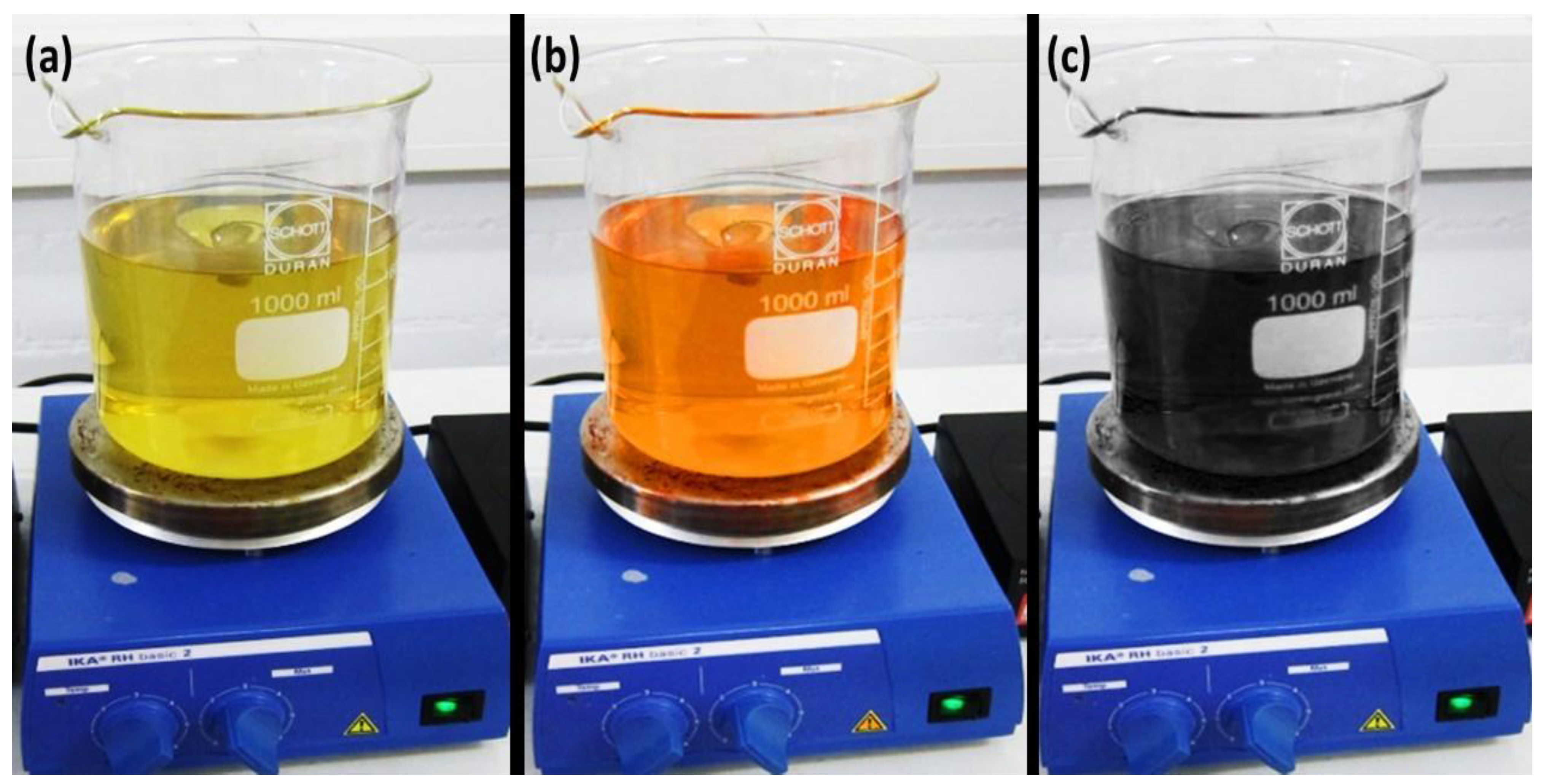
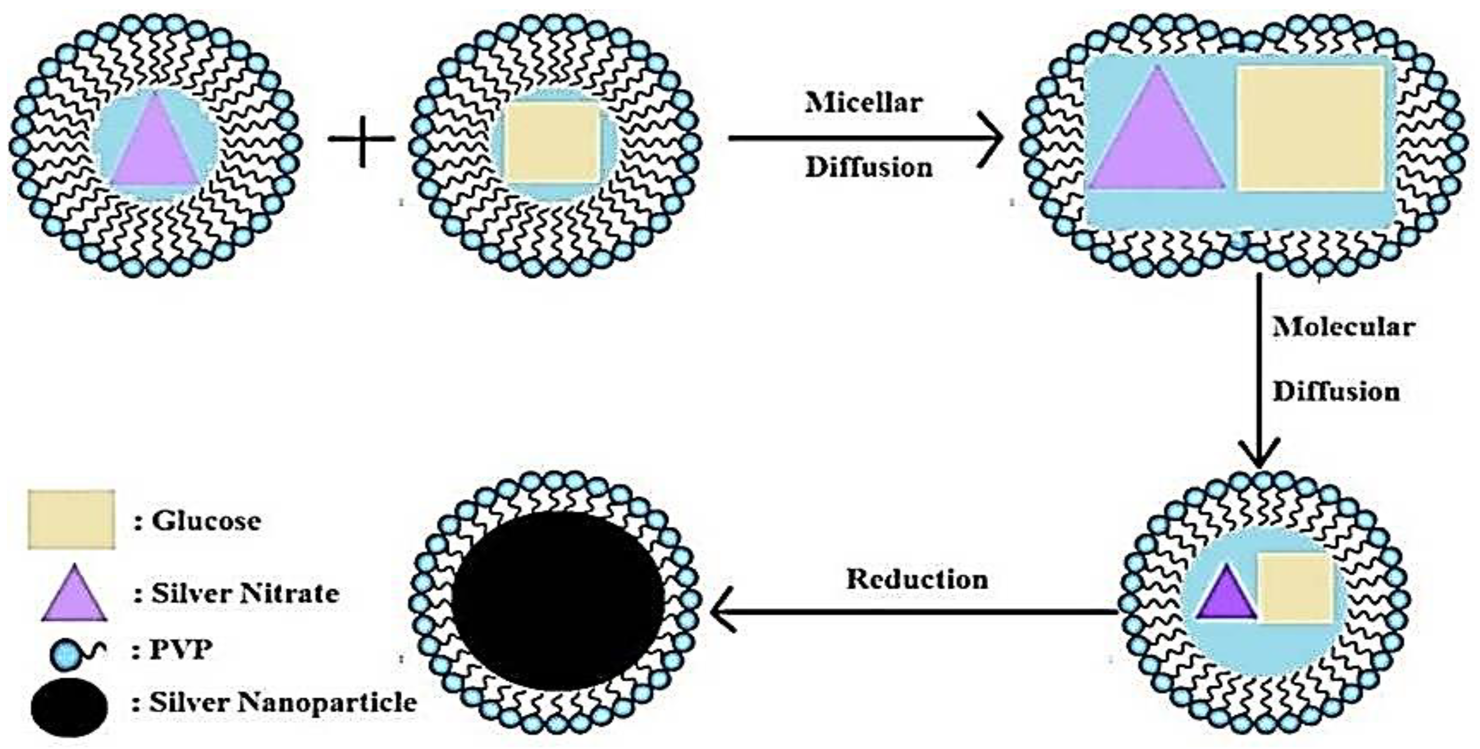
2.1. Synthesis of Ag-NPs

Initially, PDMS substrates were fabricated using the solution processing method. The silicone elastomeric base and silicone elastomeric hardener were mixed with a ratio of 10:1 g. The prepared 2.5 g of PDMS solution was then poured into the mold (5 × 5 cm) to achieve 1 mm-thick PDMS [13,14,15]. The film thickness of the sample was measured by stereomicroscope (MOTIC DMW-143). The Ag-NPs (<100 nm) were then prepared chemically via reduction (a green nanotechnology approach) using the glucose-gluconic acid oxidation-reduction method in diethylamine, with silver nitrate as a source of silver ions. The prepared Ag-NPs were then deposited as a thin-film by inkjet printing and sintering at 100 °C for 30 min. A straightforward in-house technique was used to prepare the thin-film of the nanocomposites via the inkjet printing method. Finally, the samples were characterized through different techniques.
As mentioned, the Ag-NPs solution was prepared by chemical reduction (a green nanotechnology route). An aqueous solution of 100 mM silver nitrate and 50 mM glucose was mixed and stirred using a magnetic stirrer at room temperature for 20 min to obtain a homogeneous solution. An aqueous solution of 115 mM molarity diethylamine (DEA) was added, and the solution was mixed quickly and vigorously [16,17,18]. The color of the solution changed from yellow to brown and finally to black, a possible indication of Ag-NPs [19,20,21], as shown in Figure 1.
The recommended mechanism for this reaction is similar to the silver mirror technique, where the amine is dissolved in water to remove hydrogen ions, leaving hydroxyl ions in the solution. The hydrated amine ion further reacts with silver nitrate to form a complex of silver ions. The remaining hydroxyl ions oxidize the aldehyde groups of the glucose molecules to form gluconic acid, and an electron is released into the solution. This electron reduces the silver ions of the silver complex to get metallic nanoparticles of silver. Diethylamine (DEA) can be used as a catalyst. To prevent agglomeration of the particles, polyvinylpyrrolidone (PVP) is used as a capping agent, as shown in Figure 2. The maximum amount of 3 wt.% PVP is used in the solution, which acts as a stabilizer preventing the agglomeration of the synthesized nanoparticles. Small wt. percentage of PVP was used because the greater the added amount of PVP can makes the particles nonconductive. Nonetheless, when using PVP as the stabilizing agent, producing just small quantities of the synthesized particles is efficient to form the thin-films.

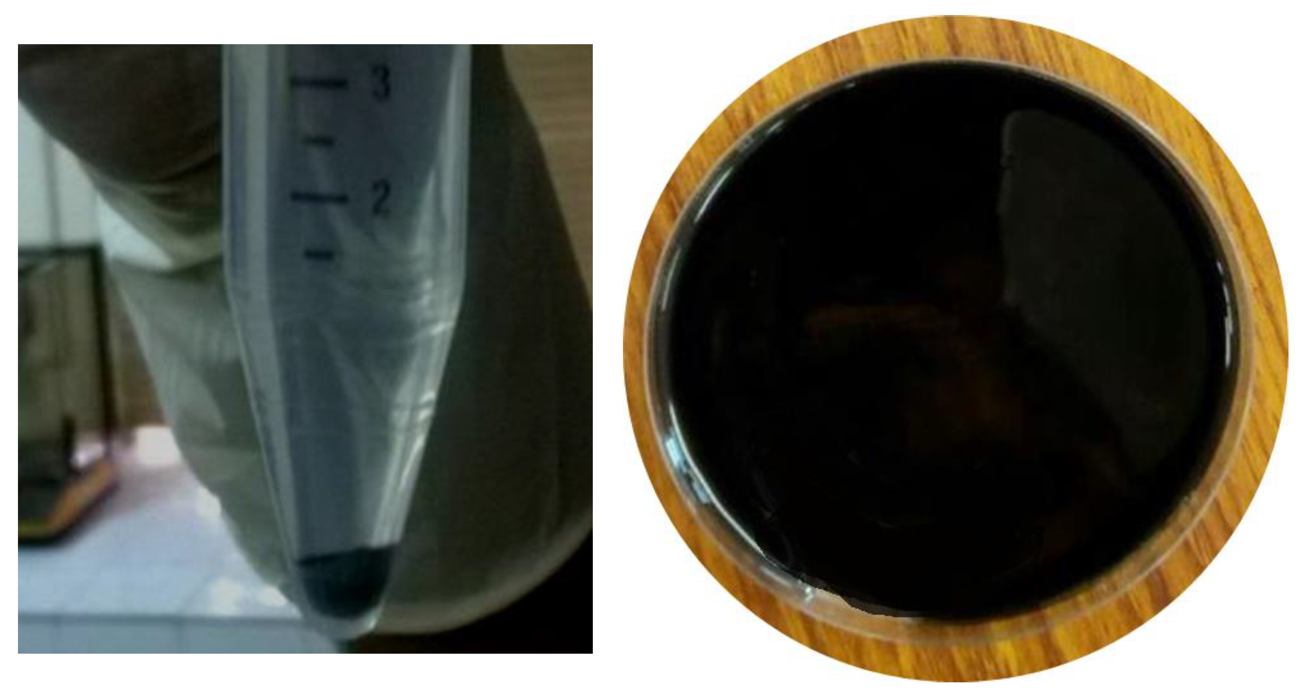
2.2. Preparation of Ag-NPs/Ethylene Glycol Ink

The prepared solution of silver nanoparticles was then used for ink preparation by centrifugation (at a 3000-rpm speed for 10 min at 220 volts), using ethylene glycol as a dispersing agent, as shown in Figure 3.
The silver nanoparticle ink was then placed in a vial and subjected to jet ultrasonication, to break the bonds between agglomerated nanoparticles and thus disperse the nanoparticles in the freshly prepared ink. The nanoparticle ink was stored in a vial that underwent sonication using an ultrasonic cleaning machine.

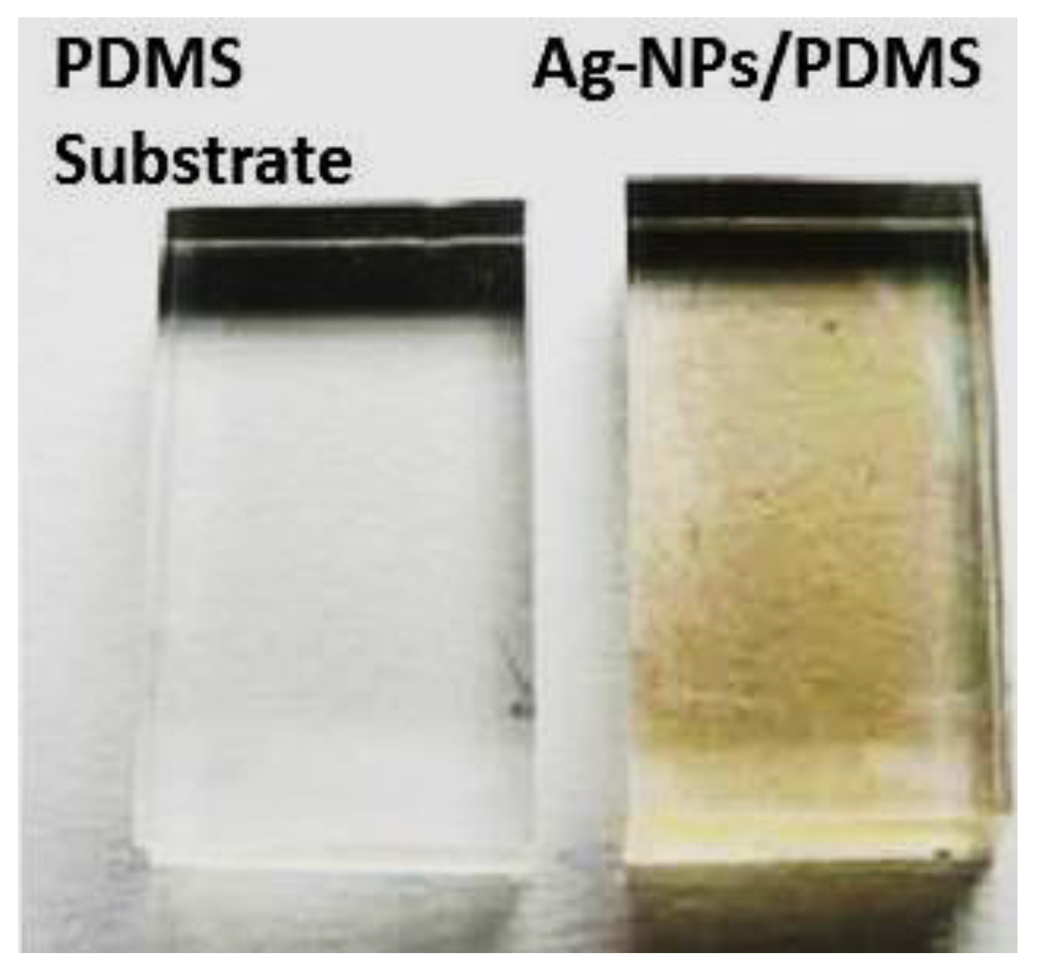
2.3. Fabrication of Ag-NP Thin-Film

The prepared Ag-NPs were then deposited as a thin-film using the inkjet printing technique. The coated film on the substrate (PDMS/Ag-NPS) was then consolidated in the oven for 1 h at 100 °C, and cooled in a dry oven for good adhesion. Figure 4 shows the uncoated and coated substrate samples produced after solidification.

2.4. Characterization of the Ag-NPs and the Ag-NP-PDMS Thin-Film

The surface of the prepared samples was investigated using an Olympus Microscope model GX-51 (Center Valley, PA, USA), while scanning electron microscope (SEM) model Quanta 200 (Hillsboro, OR, USA) was used to determine the surface morphologies and particle sizes and shapes of the prepared Ag-NPs. A UV-Vis spectrophotometer model Spectrumlab 22PC (Shanghai. Lengguang Technology Co. Ltd., Shanghai, China) was used to determine the absorption spectrum of the prepared silver nanoparticles. The laboratory apparatus was designed to measure the strain versus the electrical resistance of the sintered thin-film samples when increasing the applied load, by assessing the electrical resistance of the sintered thin-films at different strain loads using a multimeter. The electrical measurements were recorded before and after applying each load [22,23].

3. Results and Discussion

3.1. Ag-NPs’ Characterization

Investigations of the produced Ag-NPs were carried out using a field emission scanning electron microscope at a standard high voltage of 25–30 kV to study the particle shapes, sizes, and morphologies. Figure 5 shows SEM images with different magnifications of the prepared Ag-NPs. In Figure 5a, the particles are at the nanoscale and we can see some agglomerations. The particle size range is between 108 and 189.6 nm, and some particles are agglomerated in different areas. Undesirable agglomeration of particles results due to the conglomeration and pileup of the particles, resulting in the deterioration of the size range [24,25,26]. Figure 5b–d show SEM images with high magnifications, in which the phenomenon of particle agglomeration can be observed more clearly and effectively. The particle size of one particle was measured as 108.3–189.6 nm, but agglomeration appeared in various regions, resulting in vast increments in the size range. Particle pile-up was obvious in the region of the red box, resulting in a dramatic increase in the sizes of the particles [27].

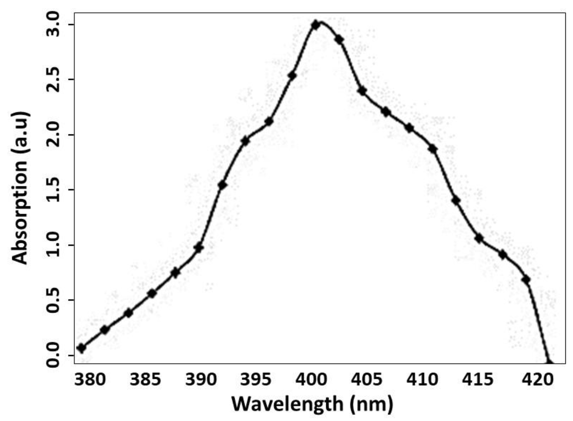
3.2. UV Absorption Spectrum of the Ag-NPs

Figure 6 shows the spectrum of standard UV spectroscopy for the produced silver nanoparticles in the wavelength and absorbance range of UV light. The spectrum was scanned within the wavelength range from 380 to 420 nm. A high-intensity peak was detected at 400 nm, which confirmed the formation of Ag-NPs. It is believed that the UV-Vis absorption peak of Ag-NPs can be detected in the range of 390–470 nm, depending on the particle sizes, shapes, and distribution [28]. Hence, the absorption peak presented in Figure 6 demonstrates the presence of the Ag-NPs, as revealed when investigated by SEM (see Figure 5).

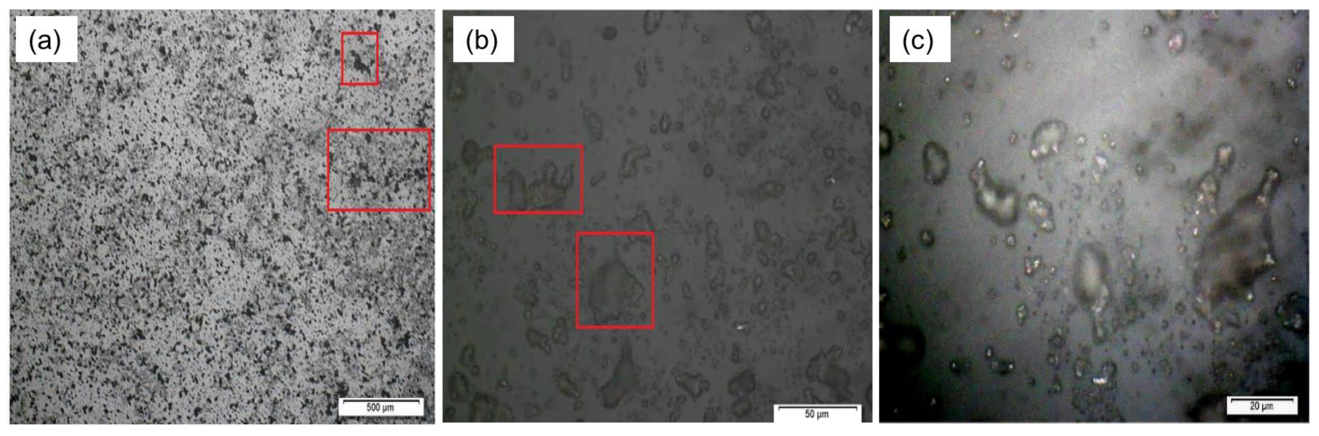
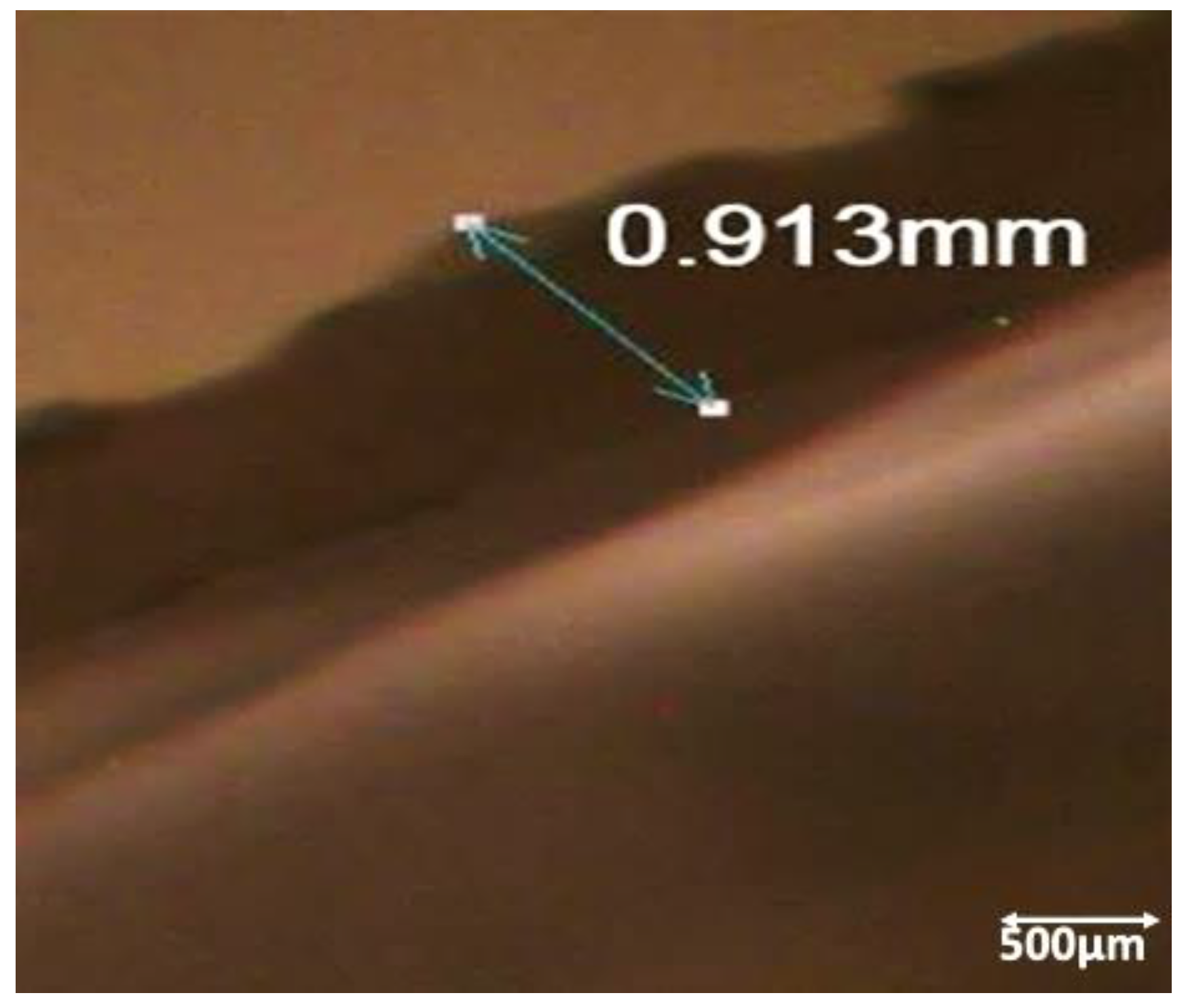
3.3. Surface Characterization of Ag-NP-PDMS Thin-Film

Figure 7a depicts the results of the stereomicrograph of the surface of the thin-film; the black spots show the silver particles and the grey background shows the substrate. The undesirable agglomeration phenomenon can be observed, confirmed by the presence of large black spots, such as in the lower left of the micrograph shown in Figure 7a. Figure 7b presents a high magnification of the surface. The zones in the red boxes in the images capture notable agglomeration [29]. Figure 7c reveals an important property of Ag-NPs, i.e., shining and brightening in the presence of light from the microscope.
The stereomicroscope technique is used to measure line spacing, as well as sample thickness. The results obtained from this test are shown below in Figure 8. There was great variation in the thicknesses of the samples when a mean of four readings was calculated. The variation ranged from 0.914 to 1.312 mm, with the thicknesses of the coated layers differing.
Substrates with different roughness patterns were prepared and silver nanoparticles were synthesized. Then, in this step, the substrates were coated with silver nanoparticles, followed by a sintering process. Figure 9a shows a stereomicrograph of the prepared Ag-NP thin-film on the PDMS substrate sample after sintering. Following this, the sample was subjected to stretching using a homemade apparatus. Next, the electrical resistance of the prepared and stretched samples was measured after applying a suitable load. The sample surface after stretching was investigated using a stereomicroscope, as shown in Figure 9b. It was observed from the micrograph that the sintered Ag-NP thin-film on the PDMS substrate sample (Figure 9a) had a uniform surface morphology. However, the sintered Ag-NP thin-film on the PDMS substrate sample (Figure 9b), after stretching by applying a suitable load, had a deformed surface morphology with some areas marred by grooves, potentially due to the loss of some coated materials from the surface of the substrate during stretching [30].

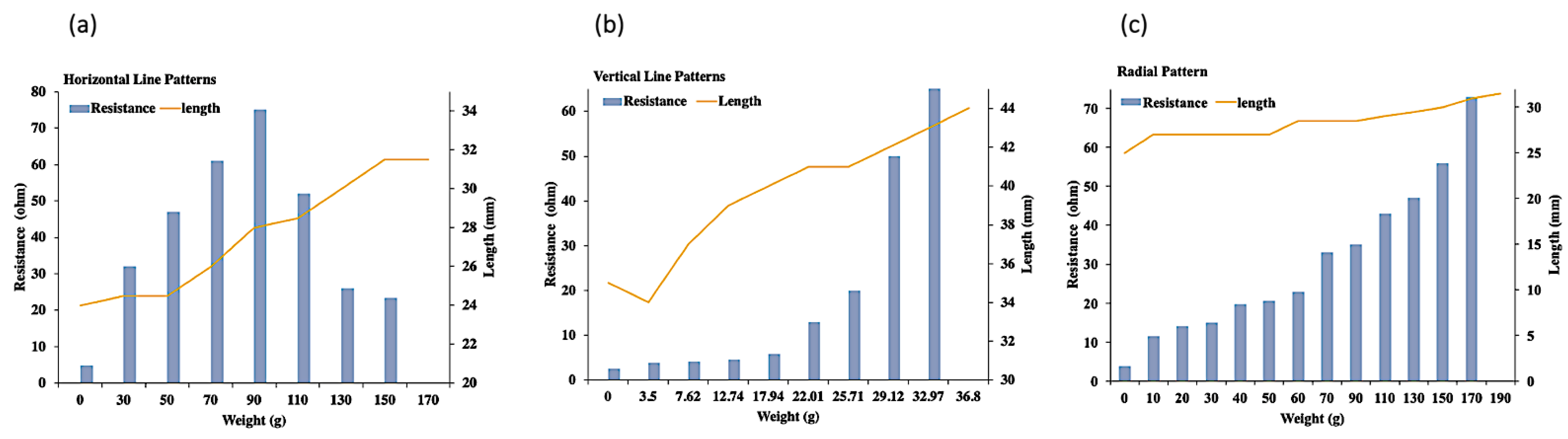
3.4. Electrical Conductivity of the Sintered Ag-NP-PDMS Thin-Film

After successful sintering of silver nanoparticles on the surface of the PDMS substrate, the coated samples were subjected to a measurement of their electrical conductivity under an applied load. Figure 10 shows the electrical resistance values of the thin-film under an applied load with respect to changes in the length of the thin-film. In both unstretched and stretched sample conditions, the electrical resistance of the sample in the horizontal directions was found to be the best, with the lowest electrical resistance [31]. For instance, when the unstretched sample was 2.4 cm in length, the electrical resistance was found to be 1.6 ohms. The sample was stretched slowly, and increases in electrical resistance were observed at higher values, with the highest electrical resistance value of 3.15 ohms at the length of 3.15 cm. In comparison, when we consider the electrical resistance measurements in the vertical and radial directions, the resistances of the unstretched samples were 2.5 and 3.8 ohms at sample lengths of 3.5 and 2.5 cm, respectively. When stretching the sample vertically, the electrical resistance value was observed to be 65 ohms for a maximum stretch of 3.1 cm [32]. Meanwhile, when stretched radially, the maximum electrical resistance was observed to be 73 ohms [33].

4. Conclusions

Silver nanoparticles were prepared chemically with a green reduction method using glucose-gluconic acid oxidation-reduction in diethylamine, with silver nitrate as a source of silver ions. The synthesized silver nanoparticles had an average particle size of 108.3–189.6 nm. Their presence was confirmed by UV-Vis spectrophotometry, indicating an intense peak at a wavelength of 400 nm. PDMS was used as a substrate in the liquid form, and it was solidified via sintering. The prepared silver nanoparticles were successfully deposited as a thin-film on the prepared PDMS substrates by inkjet printing, followed by sintering at 100 °C for 30 min. The electrical resistance of the obtained sintered samples was measured before and after stretching by applying a suitable load in different directions. The samples stretched horizontally showed the best electrical properties. Based on the obtained results, the prepared silver nanoparticle/PDMS stretchable conductor has effective electrical and mechanical properties to be used in bio-sensing and electronic devices. Future studies will be conducted to develop the technique and the properties of the obtained nanocomposites.

Author Contributions

Conceptualization, A.R.J., J.B., W.K., F.A., M.B., R.B. and W.M.D.; methodology, A.R.J., J.B., W.K., F.A., M.B., R.B. and W.M.D.; validation, A.R.J., J.B., W.K., F.A., M.B., R.B. and W.M.D.; formal analysis, A.R.J., J.B., W.K., F.A., M.B., R.B. and W.M.D.; investigation, A.R.J., J.B., W.K., F.A., M.B., R.B. and W.M.D.; resources, A.R.J., J.B., W.K., F.A., M.B., R.B. and W.M.D.; data curation, A.R.J., J.B., W.K., F.A., M.B., R.B. and W.M.D.; writing—original draft preparation A.R.J., J.B., W.K., F.A., M.B., R.B. and W.M.D.; writing—review and editing, A.R.J., J.B., W.K., F.A., M.B., R.B. and W.M.D.; visualization, A.R.J., J.B., W.K., F.A., M.B., R.B. and W.M.D.; supervision, W.M.D.; project administration, A.R.J. All authors have read and agreed to the published version of the manuscript.

Funding

This research received no external funding.

Institutional Review Board Statement

Not applicable.

Informed Consent Statement

Informed consent was obtained from all subjects involved in the study.

Data Availability Statement

Not applicable.

Conflicts of Interest

The authors declare no conflict of interest.

References

  1. Abu-Khalaf, J.M.; Al-Ghussain, L.; Al-Halhouli, A.A. Fabrication of stretchable circuits on polydimethylsiloxane (PDMS) pre-stretched substrates by inkjet printing silver nanoparticles. Materials 2018, 11, 2377. [Google Scholar] [CrossRef] [PubMed] [Green Version]
  2. Feng, P.; Ji, H.; Zhang, L.; Luo, X.; Leng, X.; He, P.; Zhao, W. Highly stretchable patternable conductive circuits and wearable strain sensors based on polydimethylsiloxane and silver nanoparticles. Nanotechnology 2019, 30, 185501. [Google Scholar] [CrossRef] [PubMed]
  3. Soe, H.M.; Abd Manaf, A.; Matsuda, A.; Jaafar, M. Development and fabrication of highly flexible, stretchable, and sensitive strain sensor for long durability based on silver nanoparticles–polydimethylsiloxane composite. J. Mater. Sci. Mater. Electron. 2020, 31, 11897–11910. [Google Scholar] [CrossRef]
  4. Min, S.H.; Lee, G.Y.; Ahn, S.H. Direct printing of highly sensitive, stretchable, and durable strain sensor based on silver nanoparticles/multi-walled carbon nanotubes composites. Composites Part B Eng. 2019, 161, 395–401. [Google Scholar] [CrossRef]
  5. Guo, Y.; Yu, J.; Li, C.; Li, Z.; Pan, J.; Liu, A.; Zhang, C. SERS substrate based on the flexible hybrid of polydimethylsiloxane and silver colloid decorated with silver nanoparticles. Opt. Express 2018, 26, 21784–21796. [Google Scholar] [CrossRef]
  6. Al-Halhouli, A.A.; Al-Ghussain, L.; El Bouri, S.; Liu, H.; Zheng, D. Fabrication and evaluation of a novel non-invasive stretchable and wearable respiratory rate sensor based on silver nanoparticles using inkjet printing technology. Polymers 2019, 11, 1518. [Google Scholar] [CrossRef] [Green Version]
  7. Zhang, S.; Zhang, H.; Yao, G.; Liao, F.; Gao, M.; Huang, Z.; Lin, Y. Highly stretchable, sensitive, and flexible strain sensors based on silver nanoparticles/carbon nanotubes composites. J. Alloys Compd. 2015, 652, 48–54. [Google Scholar] [CrossRef]
  8. Soe, H.M.; Abd Manaf, A.; Matsuda, A.; Jaafar, M. Performance of a silver nanoparticles-based polydimethylsiloxane composite strain sensor produced using different fabrication methods. Sens. Actuators A Phys. 2021, 329, 112793. [Google Scholar] [CrossRef]
  9. Choi, Y.I.; Hwang, B.U.; Meeseepong, M.; Hanif, A.; Ramasundaram, S.; Trung, T.Q.; Lee, N.E. Stretchable and transparent nanofiber-networked electrodes based on nanocomposites of polyurethane/reduced graphene oxide/silver nanoparticles with high dispersion and fused junctions. Nanoscale 2019, 11, 3916–3924. [Google Scholar] [CrossRef]
  10. Tavakoli, M.; Malakooti, M.H.; Paisana, H.; Ohm, Y.; Green Marques, D.; Alhais Lopes, P.; Majidi, C.E. GaIn-Assisted Room-Temperature Sintering of Silver Nanoparticles for Stretchable, Inkjet-Printed, Thin-Film Electronics. Adv. Mater. 2018, 30, 1801852. [Google Scholar] [CrossRef]
  11. Shankar, A.; Salcedo, E.; Berndt, A.; Choi, D.; Ryu, J.E. Pulsed light sintering of silver nanoparticles for large deformation of printed stretchable electronics. Adv. Compos. Hybrid Mater. 2018, 1, 193–198. [Google Scholar] [CrossRef]
  12. Chen, J.; Zheng, J.; Gao, Q.; Zhang, J.; Zhang, J.; Omisore, O.M.; Li, H. Polydimethylsiloxane (PDMS)-based flexible resistive strain sensors for wearable applications. Appl. Sci. 2018, 8, 345. [Google Scholar] [CrossRef] [Green Version]
  13. Huang, Q.; Al-Milaji, K.N.; Zhao, H. Inkjet printing of silver nanowires for stretchable heaters. ACS Appl. Nano Mater. 2018, 1, 4528–4536. [Google Scholar] [CrossRef]
  14. Htwe, Y.Z.N.; Hidayah, I.N.; Mariatti, M. Performance of inkjet-printed strain sensor based on graphene/silver nanoparticles hybrid conductive inks on polyvinyl alcohol substrate. J. Mater. Sci. Mater. Electron. 2020, 31, 15361–15371. [Google Scholar] [CrossRef]
  15. Wang, T.; Wang, R.; Cheng, Y.; Sun, J. Quasi in situ polymerization to fabricate copper nanowire-based stretchable conductor and its applications. ACS Appl. Mater. Interfaces 2016, 8, 9297–9304. [Google Scholar] [CrossRef] [PubMed]
  16. Abu-Khalaf, J.; Al-Ghussain, L.; Nadi, A.; Saraireh, R.; Rabayah, A.; Altarazi, S.; Al-Halhouli, A.A. Optimization of geometry parameters of inkjet-printed silver nanoparticle traces on pdms substrates using response surface methodology. Materials 2019, 12, 3329. [Google Scholar] [CrossRef] [Green Version]
  17. Xin, Z.; Liu, Y.; Li, X.; Liu, S.; Fang, Y.; Deng, Y.; Li, L. Conductive grid patterns prepared by microcontact printing silver nanoparticles ink. Mater. Res. Express 2017, 4, 015021. [Google Scholar] [CrossRef]
  18. Lee, J.Y.; Shin, D.; Park, J. Fabrication of silver nanowire-based stretchable electrodes using spray coating. Thin Solid Film. 2016, 608, 34–43. [Google Scholar] [CrossRef]
  19. Duan, S.; Yang, K.; Wang, Z.; Chen, M.; Zhang, L.; Zhang, H.; Li, C. Fabrication of highly stretchable conductors based on 3D printed porous poly (dimethylsiloxane) and conductive carbon nanotubes/graphene network. ACS Appl. Mater. Interfaces 2016, 8, 2187–2192. [Google Scholar] [CrossRef]
  20. Zhang, K.; Shi, X.; Chen, J.; Xiong, T.; Jiang, B.; Huang, Y. Self-healing and stretchable PDMS-based bifunctional sensor enabled by synergistic dynamic interactions. Chem. Eng. J. 2021, 412, 128734. [Google Scholar] [CrossRef]
  21. Martinez, V.; Stauffer, F.; Adagunodo, M.O.; Forro, C.; Vörös, J.; Larmagnac, A. Stretchable silver nanowire–elastomer composite microelectrodes with tailored electrical properties. ACS Appl. Mater. Interfaces 2015, 7, 13467–13475. [Google Scholar] [CrossRef] [PubMed]
  22. Feng, P.; Zhong, M.; Zhao, W. Stretchable multifunctional dielectric nanocomposites based on polydimethylsiloxane mixed with metal nanoparticles. Mater. Res. Express 2019, 7, 015007. [Google Scholar] [CrossRef] [Green Version]
  23. Zou, Q.; He, K.; Ou-Yang, J.; Zhang, Y.; Shen, Y.; Jin, C. Highly Sensitive and Durable Sea-Urchin-Shaped Silver Nanoparticles Strain Sensors for Human-Activity Monitoring. ACS Appl. Mater. Interfaces 2021, 13, 14479–14488. [Google Scholar] [CrossRef] [PubMed]
  24. Al-Milaji, K.N.; Huang, Q.; Li, Z.; Ng, T.N.; Zhao, H. Direct embedment and alignment of silver nanowires by inkjet printing for stretchable conductors. ACS Appl. Electron. Mater. 2020, 2, 3289–3298. [Google Scholar] [CrossRef]
  25. Duan, S.; Wang, Z.; Zhang, L.; Liu, J.; Li, C. Three-dimensional highly stretchable conductors from elastic fiber mat with conductive polymer coating. ACS Appl. Mater. Interfaces 2017, 9, 30772–30778. [Google Scholar] [CrossRef]
  26. Zhang, S.; Li, Y.; Tian, Q.; Liu, L.; Yao, W.; Chi, C.; Wu, W. Highly conductive, flexible and stretchable conductors based on fractal silver nanostructures. J. Mater. Chem. C 2018, 6, 3999–4006. [Google Scholar] [CrossRef]
  27. Zhou, W.; Yu, Y.; Bai, S.; Hu, A. Laser direct writing of waterproof sensors inside flexible substrates for wearable electronics. Opt. Laser Technol. 2021, 135, 106694. [Google Scholar] [CrossRef]
  28. Chou, N.; Kim, Y.; Kim, S. A method to pattern silver nanowires directly on wafer-scale PDMS substrate and its applications. ACS Appl. Mater. Interfaces 2016, 8, 6269–6276. [Google Scholar] [CrossRef]
  29. Sun, J.; Wang, Q.; Luo, G.; Meng, W.; Cao, M.; Li, Y.; Lang, M.F. A novel flexible Ag/AgCl quasi-reference electrode based on silver nanowires toward ultracomfortable electrophysiology and sensitive electrochemical glucose detection. J. Mater. Res. Technol. 2020, 9, 13425–13433. [Google Scholar] [CrossRef]
  30. Hu, J.; Yu, J.; Li, Y.; Liao, X.; Yan, X.; Li, L. Nano carbon black-based high-performance wearable pressure sensors. Nanomaterials 2020, 10, 664. [Google Scholar] [CrossRef] [Green Version]
  31. Zhao, C.; Xia, Z.; Wang, X.; Nie, J.; Huang, P.; Zhao, S. 3D-printed highly stable flexible strain sensor based on silver-coated-glass fiber-filled conductive silicon rubber. Mater. Des. 2020, 193, 108788. [Google Scholar] [CrossRef]
  32. Choi, S.; Kim, S.; Kim, H.; Lee, B.; Kim, T.; Hong, Y. 2-D Strain Sensors Implemented on Asymmetrically Bi-Axially Pre-Strained PDMS for Selectively Switching Stretchable Light-Emitting Device Arrays. IEEE Sens. J. 2020, 20, 14655–14661. [Google Scholar] [CrossRef]
  33. Xiang, D.; Zhang, X.; Harkin-Jones, E.; Zhu, W.; Zhou, Z.; Shen, Y.; Wang, P. Synergistic effects of hybrid conductive nanofillers on the performance of 3D printed highly elastic strain sensors. Compos. Part A Appl. Sci. Manuf. 2020, 129, 105730. [Google Scholar] [CrossRef]
Figure 1. Synthesis process of Ag-NPs; (a) yellow color appeared during the mixing of silver nitrate and glucose with diethylamine (b) yellow color of the solution change to orange and (c) the orange color of the solution is turned to black indicating the formation of Ag-NPs
Crystals 12 01098 g001
Figure 2. Mechanism of using PVP stabilizer for silver nanoparticles.
Crystals 12 01098 g002
Figure 3. Ag-NPs after centrifugations (left). Silver nanoparticle ink dispersed in ethylene glycol (right).
Crystals 12 01098 g003
Figure 4. Thin-layer coats of Ag-NP ink on the PDMS substrate.
Crystals 12 01098 g004
Figure 5. SEM images with different magnifications, where (a) 6000×, (b) 10,000×, (c) 12,015× and (d) 19,971× of the prepared Ag-NPs. The triangular shape of particles can be seen at different magnification.
Crystals 12 01098 g005
Figure 6. UV spectrum of the AgNPs dispersed in ethanol solution.
Crystals 12 01098 g006
Figure 7. (ac) Stereomicrographs with different magnifications of the fabricated thin-film.
Crystals 12 01098 g007
Figure 8. Stereomicrograph of the cross-sectional area of the deposited Ag-NP thin-film on the PDMS substrate.
Crystals 12 01098 g008
Figure 9. Stereophotographs of the sintered thin-film of Ag-NPs supported on the PDMS substrate, (a) before and (b) after stretching.
Crystals 12 01098 g009
Figure 10. Electrical resistance vs. stretching length of (a) horizontal, (b) vertical and (c) radial patterns.
Crystals 12 01098 g010
Publisher’s Note: MDPI stays neutral with regard to jurisdictional claims in published maps and institutional affiliations.

