Article

Systematic Study on the Synthesis and Magnetism Properties of Manganese Ferrite MnFe2O4 by an Oxidation Roasting Process

1
School of Materials and Metallurgy, University of Science and Technology Liaoning, Anshan 114051, China
2
Key Laboratory for Chemical Metallurgy Engineering of Liaoning Province, University of Science and Technology Liaoning, Anshan 114051, China
*
Author to whom correspondence should be addressed.
Crystals 2023, 13(10), 1509; https://doi.org/10.3390/cryst13101509
Submission received: 18 September 2023 / Revised: 13 October 2023 / Accepted: 15 October 2023 / Published: 17 October 2023
(This article belongs to the Special Issue Fabrication and Properties of Magnetic Materials)

Abstract

:
A low-cost and high-efficiency solid reaction method has been reported as an effective technology to synthesize manganese ferrite MnFe2O4 with a spinel crystal structure. This work clarified the underlying reason for the influence mechanism of SiO2 and Al2O3 on the synthesis of MnFe2O4. Synthetic MnFe2O4 polyhedral microparticles with a saturated magnetization of 71.19 emu/g, a ratio of saturation magnetization to residual magnetization (Ms/Mr) of 0.062 and a coercivity (Hc) of 6.50 Oe were successfully obtained at an oxidization roasting temperature of 1100 °C for 60 min. The experimental results indicate that the tetrahedral Mn2+ ions and octahedral Mn3+ ions in the crystal structure of manganese ferrite MnFe2O4 were replaced by tetrahedral Si2+ ions and octahedral Al3+ ions from (Mn2+)x(Fe2+)y(Si2+)1−x−y[Fe3+]2O4 and (Mn2+)[Fe3+]2−x[Al3+]xO4, respectively. In addition, hercynite FexMn1−xAl2O4 with a spinel crystal structure and olivine MnxFe2−xSiO4 with an orthorhombic crystal structure were partially formed in the synthesis of manganese ferrite MnFe2O4, in which some Fe2+ ions were easily replaced by Mn2+ ions to form stable hercynite MnAl2O4 and olivine Mn2SiO4 in these crystal structures. The current research work provides comprehensive insights for synthesizing manganese ferrite MnFe2O4 and continuously advances its technical progress.

1. Introduction

Spinel-type manganese ferrite (MnFe2O4) combines the advantages of iron-based and manganese-based materials and has many excellent physical and chemical properties. Manganese ferrites have been used in microwave adsorption [1,2,3], biomedicine [4,5], magnetic recording materials [6], electrode materials [7] and environmental remediation [8,9]. In the MnFe2O4 mixed spinel structure, the metal cations Mn2+ and Fe3+ are distributed in the A (tetrahedral) and B (octahedral) positions, and the oxygen anions are formed by the face-centered densely packed [Mn2+1−iFe3+i]A[Mn2+iFe3+2−i]BO4 [10,11,12], where i is the inversion parameter as a reference for the inversion degree. When the manganese cation is on the regular tetrahedron (i = 0), it is characterized as a normal spinel structure, and when the manganese cation is in the octahedron position (i = 1), it is characterized as an inverse spinel structure [10]. Due to their strong surface energy and magnetic properties, nanometer ferrite functional magnetic particles are extremely prone to aggregation during the preparation process [13]. To solve the problem of particle aggregation, researchers have used different techniques to synthesize ferrite functional materials, such as hydrometallurgical technologies [13,14,15,16], solid-state reaction methods [17,18,19,20,21,22,23,24,25], combined electrochemical and chemical methods [26,27] and combined pyro- and hydrometallurgical metallurgical technology [11,28,29].
By comparison with the above synthesis methods, hydrometallurgy and combined methods are reported for synthesizing high-purity manganese ferrites. However, the abovementioned methods have high cost, high energy consumption and complex technology, which seriously restrict their development. Solid-state synthesis has been widely considered a cost-effective way to synthesize manganese ferrite [18,28,29]. As we all know, magnetism is an important index for characterizing manganese ferrite, and the purity of raw materials and preparation conditions will directly affect its magnetism. As studied by Lin et al. [15], MnFe2O4 with different morphologies was successfully synthesized by a solvothermal method using cetyltrimethylammonium bromide as the raw material, and the minimum coercivity was 11.9 Oe. Rashad et al. [23] successfully synthesized MnFe2O4 with a saturation magnetization of 27.24 emu/g by acid leaching with low-grade manganese ore at pH 10, a calcination temperature of 1000 °C and a calcination time of 120 min. Ahmed et al. [24] found that manganese ferrite with a maximum saturation magnetization of 62 emu/g was successfully synthesized by sintering for 2 h at 1300 °C using mill scale and fine particles of manganese ore sinter as the source of ferromanganese spinel. Ding et al. [25] showed that the maximum saturation magnetization of MnFe2O4 samples formed after annealing at 600~700 °C increased from 54 emu/g to 79 emu/g. Reddy et al. [22] performed spark plasma sintering (SPS) of the hydrothermally pretreated material to obtain a soft magnetic material MnFe2O4 with a maximum saturation magnetization of 82.5 emu/g. To date, much research has been carried out on the preparation of MnFe2O4 with the solid-phase reaction method [18,30,31], and the experiments have proven that the magnetic properties of manganese ferrite are closely related to the structure and morphology of the crystal [18,19,20,21]. Since the influence of SiO2 as well as Al2O3 on the asynchronous reaction behavior and product properties of ferromanganese oxides in the solid-phase process is not clear, the study of silicon and aluminum elements is of great significance in elucidating their impact on the synthesis of manganese ferrite, providing theoretical support for the solid-state reaction method of preparing manganese ferrite from ferromanganese ore. The clarification of the interfacial reaction process and the control of the interfacial reaction products is one of the urgent problems in this field.
In this paper, high-purity manganese ferrite MnFe2O4 was prepared by a solid-reaction method in which the evolution of its phase transition, magnetic properties and morphological characteristics were investigated by scanning electron microscopy (SEM), energy dispersive X-ray spectroscopy (EDS), X-ray diffraction (XRD), vibrating sample magnetometer (VSM) and Mössbauer spectroscopy (MBS) analyses. Some impurity components have a certain impact on the synthesis process of MnFe2O4. This work clarified the underlying reason for the influence mechanism of SiO2 and Al2O3 on the synthesis of MnFe2O4 in SiO2-MnO-Fe2O3 systems and Al2O3-MnO-Fe2O3 systems. Therefore, the phase formation and synthesis mechanism of SiO2-MnO-Fe2O3 systems and Al2O3-MnO-Fe2O3 systems were also investigated by X-ray diffraction (XRD) and scanning electron microscopy (SEM) and energy dispersive X-ray spectroscopy (EDS) analysis. Based on these findings, the current research work provides comprehensive insights for synthesizing manganese ferrite MnFe2O4 and continuously advances its technical progress.

2. Materials and Methods

2.1. Raw Materials

The purity of the MnO, Fe2O3, SiO2 and Al2O3 raw materials used in this study was 99.9%, and the reagents were not further purified during the experiment. The gas used was compressed air with a purity of 99.9% or higher. The average particle sizes of the Fe2O3, MnO, SiO2 and Al2O3 purity reagent powders were 56.2 μm, 35.2 μm, 30.6 μm and 26.4 μm, respectively, using a Malvern laser particle size analyzer (Mastersizer 2000, Shanghai, China).

2.2. Synthesis Procedure

In this paper, three kinds of samples were used to assess oxidative competition behavior and synthesis mechanism in the synthesis process of manganese ferrite MnFe2O4. Sample A was Fe2O3-MnO powder used to obtain optimized process parameters for this technology. Sample B was a mixture of pure SiO2, MnO and Fe2O3 powders used to clarify the effect of SiO2 on the synthesis mechanism of MnFe2O4. Finally, sample C was a mixture of pure MnO, Fe2O3 and Al2O3 powders used to elucidate the effect of Al2O3 on the synthesis mechanism of MnFe2O4. Next, the prepared powder sample was mixed evenly by the ball star mixer for 2 h. Then, the mixed samples with diameters of 15 mm and heights of 20 mm were placed into cylindrical molds with briquette equipment operated at a pressure of 15 MPa. The briquettes were placed on a corundum substrate (size: 60 mm × 60 mm) and roasted in air atmosphere to synthesize MnFe2O4. The oxidization roasting temperature was also measured by a Pt-Rh thermocouple and controlled using a digital temperature controller (accuracy ± 1 °C). After the equipment reached the set temperature during roasting, the samples placed in a square corundum crucible were placed in the constant temperature zone of the furnace chamber, and at the same time, a mass flow rate of 2.0 dm3/min was passed into the reaction equipment in air atmosphere. Then, following a certain time of oxidative roasting at the set temperature, the roasted product was placed in the cooling apparatus and cooled to room temperature at a cooling rate of 22 °C/min for subsequent characterization tests. In the same process as above, different mass fractions of SiO2 and Al2O3 were added to the MnO-Fe2O3 system with a molar ratio of 1:1 by oxidative roasting, and the products were later used for characterization tests. Other detailed descriptions of the above equipment were reported in our previous studies [31].

2.3. Characterization Methods

The samples were cooled after oxidative roasting in a muffle furnace and ground with 200-mesh (<0.074 μm) fine particles. X-ray diffraction (XRD) was performed on the samples using RIGAKU D/Max 2500 with a scanning angle of 5°~85°, step size of 0.02° (2θ) and scanning speed of 12°/min. The scanning results were analyzed using X’Pert HighScore Plus software 5.2 to assess the phase transformation and microstructure of the specimens.
The microscopic morphological characteristics of the intermediate products and the synthesized manganese ferrite particles were observed by scanning electron microscopy (SEM) and energy dispersive X-ray spectroscopy (EDS), and the interfacial reaction behaviors during the synthesis process were analyzed by point scanning and line scanning. The oxidization reaction behavior and formation mechanism of MnO and Fe2O3 powder were analyzed by a diffusion coupling model.
The magnetic properties of the synthesized manganese ferrite (MnFe2O4) particles were examined at room temperature using a JDAW-2000 C&D vibrating sample magnetometer (VSM). Since different magnetic materials have different magnetization properties, in considering the magnetic properties of the materials for comparison, the magnetic properties of the reaction samples will be compared by saturation magnetization intensity.
To further determine the occupancy of trivalent ferric ions in manganese ferrite, the experimental samples were analyzed by Mössbauer spectroscopy at room temperature. In the study of magnetic properties of solids, Mössbauer spectroscopy can be used to determine the magnetic ordering temperature, type of magnetic ordering and whether the ferromagnetic solid is antiferromagnetic or ferromagnetic. And this device can analyze the population of magnetic ions between various sublattices and study magnetic or spin structures. In the study of microcrystals, Mössbauer spectroscopy can provide information on the relaxation process of magnetic microcrystals, the number of magnetic anisotropy constants, the size and distribution of microcrystals and other aspects.

3. Results and Discussion

3.1. Discussion on the Oxidization Behavior of MnO-Fe2O3 System

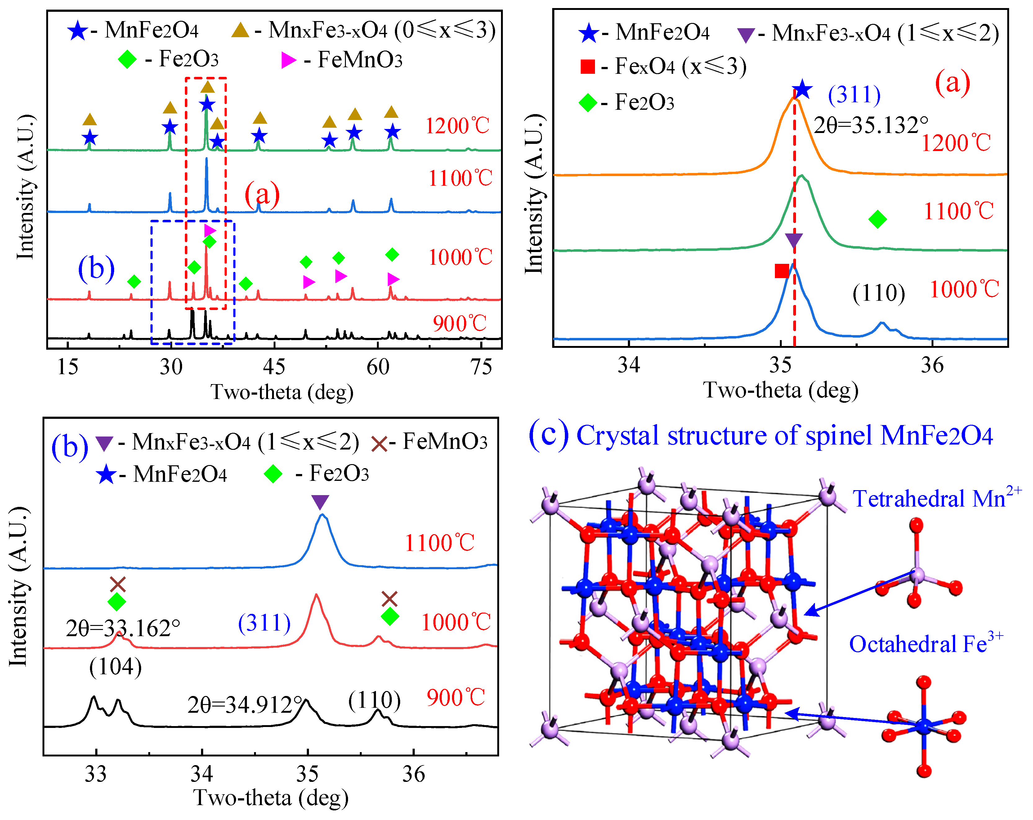
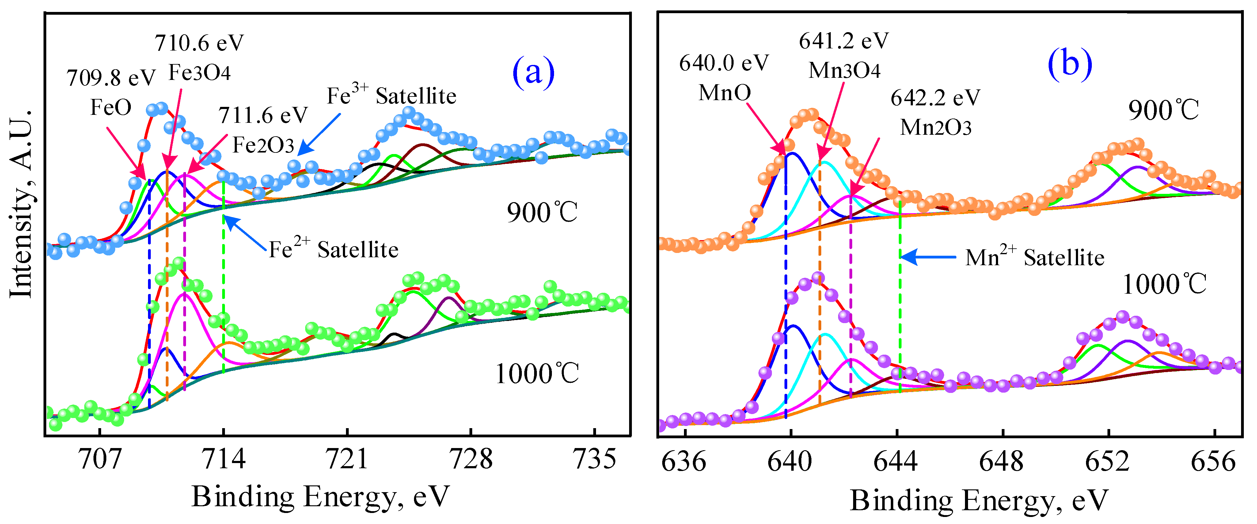
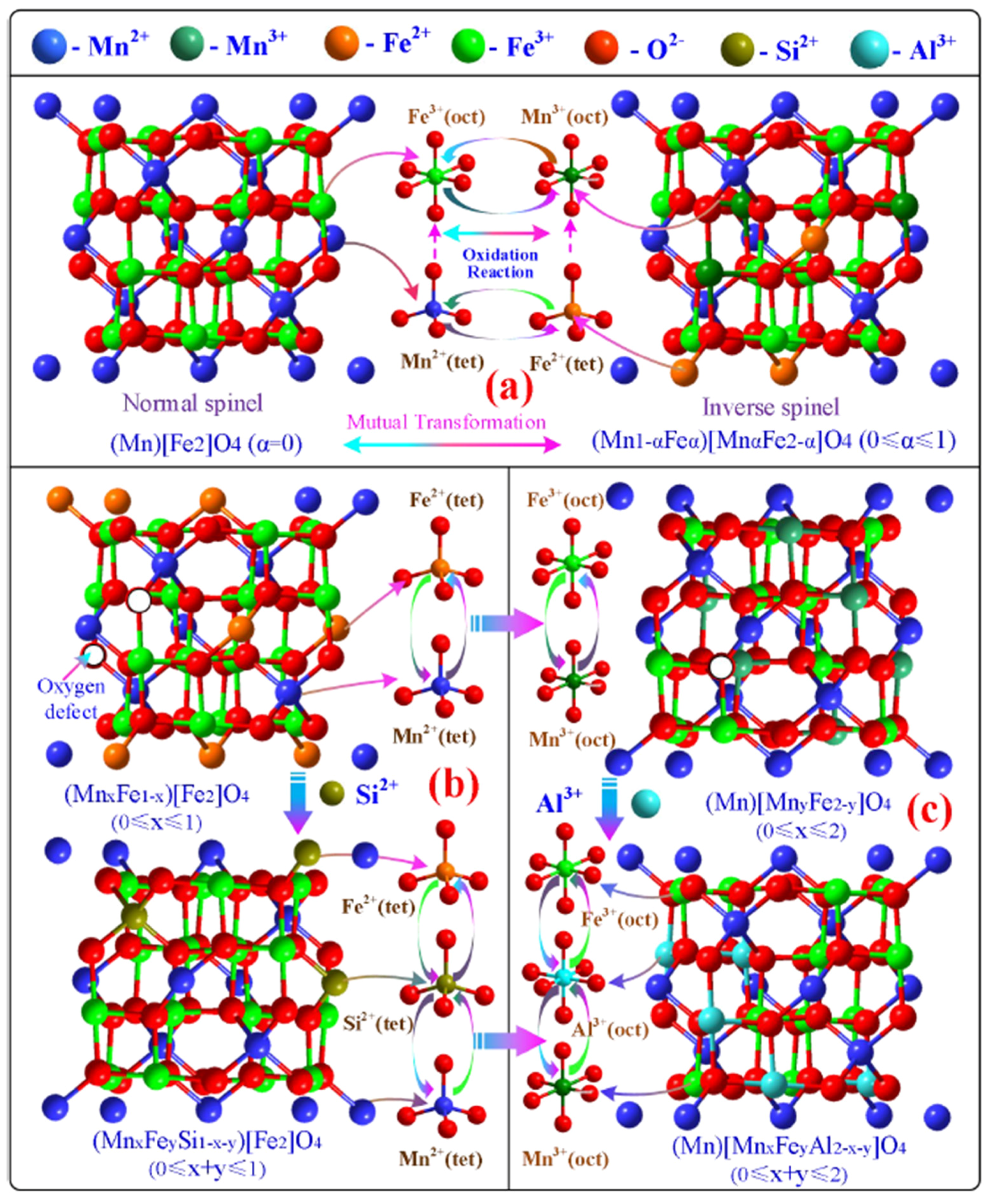
3.1.1. Phase Transition and Synthesis Mechanism in MnO-Fe2O3 System

Figure 1 displays the XRD patterns of the MnO-Fe2O3 system oxidized and roasted at 900 to 1200 °C for 60 min. As shown in Figure 1a, the characteristic diffraction peaks of Mn2O3 and Fe2O3 gradually decreased and disappeared. The oxidization roasting temperature increased from 900 °C to 1000 °C. In this temperature range, the characteristic diffraction peak intensities of MnFe2O4 and MnxFe3−xO4 increased, which indicated that Mn2O3 decomposed into MnO and Mn3O4 and then combined with Fe3O4 to form MnxFe3−xO4. The diffraction peaks of Fe2O3 were not detected with increasing oxidization roasting temperature from 1000 °C to 1100 °C. Nevertheless, the diffraction peak intensity of MnxFe3−xO4 was enhanced and approached the standard diffraction peak of (2 2 0), which indicated that the purity and crystallinity of the synthesized MnFe2O4 increased with increasing roasting temperature. As observed in Figure 1b, the characteristic diffraction peak intensities of FexO4 and Fe2O3 were detected when the oxidization roasting temperature was below 1000 °C, while the intensity of the characteristic peak of MnFe2O4 showed an initial increasing trend followed by stabilization with increasing roasting temperature. In addition, the 2θ value of the diffraction peak (3 1 1) of MnxFe3−xO4 (1 ≤ x ≤ 2) was 35.132°. These results demonstrated that the higher oxidization roasting temperature facilitated the synthesis of manganese ferrite, improved its crystal structure and optimized the atomic distribution in the crystal structure of spinel MnFe2O4. Figure 1c displays the crystal structure of MnFe2O4, which is able to verify the transformation of MnxFe3−xO4 to MnFe2O4 in the mixture with increasing roasting temperature. In the MnFe2O4 mixed spinel structure, the metal cations Mn2+ and Fe3+ are distributed in the A (tetrahedral) and B (octahedral) positions, and the oxygen anions are formed by the face-centered densely packed [Mn2+1−iFe3+i]A[Mn2+iFe3+2−i]BO4 [10,11,12]. The above analysis demonstrates that MnFe2O4 cannot be stabilized at lower stabilization temperatures. However, with increasing roasting temperature, the spinel manganese ferrite MnFe2O4 can exist stably. In addition, PDF standard card (MnFe2O4, PDF#74-2403, cubic, Fd-3m (2 2 7), 8.511 Å × 8.511 Å × 8.511 Å) and then the XRD Rietveld refinement indicated that the synthesized MnFe2O4 had a cubic structure, and the cell parameter was 8.505 Å × 8.505 Å × 8.505 Å, which was close to the abovementioned PDF standard card for MnFe2O4.
Figure 2 illustrates the scanning electron micrographs of the cross-sectional layers of the synthesized manganese ferrite samples at 900 °C and 1100 °C for 60 min. Figure 2a clearly shows that the synthesized ferromanganese oxides have a regular polyhedral structure at a roasting temperature of 900 °C. From the overall appearance morphology, it can be seen that the polyhedral particles have obvious accumulation and gaps of different sizes exist. However, the sample particles have obvious boundaries. The corresponding energy spectrum scan lines reveal that the Mn, Fe and O contents in the sample are basically constant at a ratio of 1:1:3, indicating a relatively uniform distribution of the products. This result precisely coincides with the characteristic XRD peak at 900 °C in Figure 1. The cross-sectional SEM images of the sample roasted at 1100 °C for 60 min are shown in Figure 2b,c. The particles in the sample exhibit a flat spherical shape, and the gaps between the particles are relatively uniform with obvious boundaries. The energy spectrum analysis revealed that the average contents of integrated Mn, Fe and O were 13.67%, 26.51% and 59.83%, respectively, which were close to the theoretical values of the atomic ratio of pure MnFe2O4. The Line 2 scan observed that the atomic ratios and contents of Mn, Fe and O were constant on individual particles, indicating that the distribution of these elements in the sample product was uniform. The experimental results do not correspond to the theoretical values due to the rapid oxidation rate of Mn2+, and the related reasons can be further determined comprehensively by magnetic analysis.

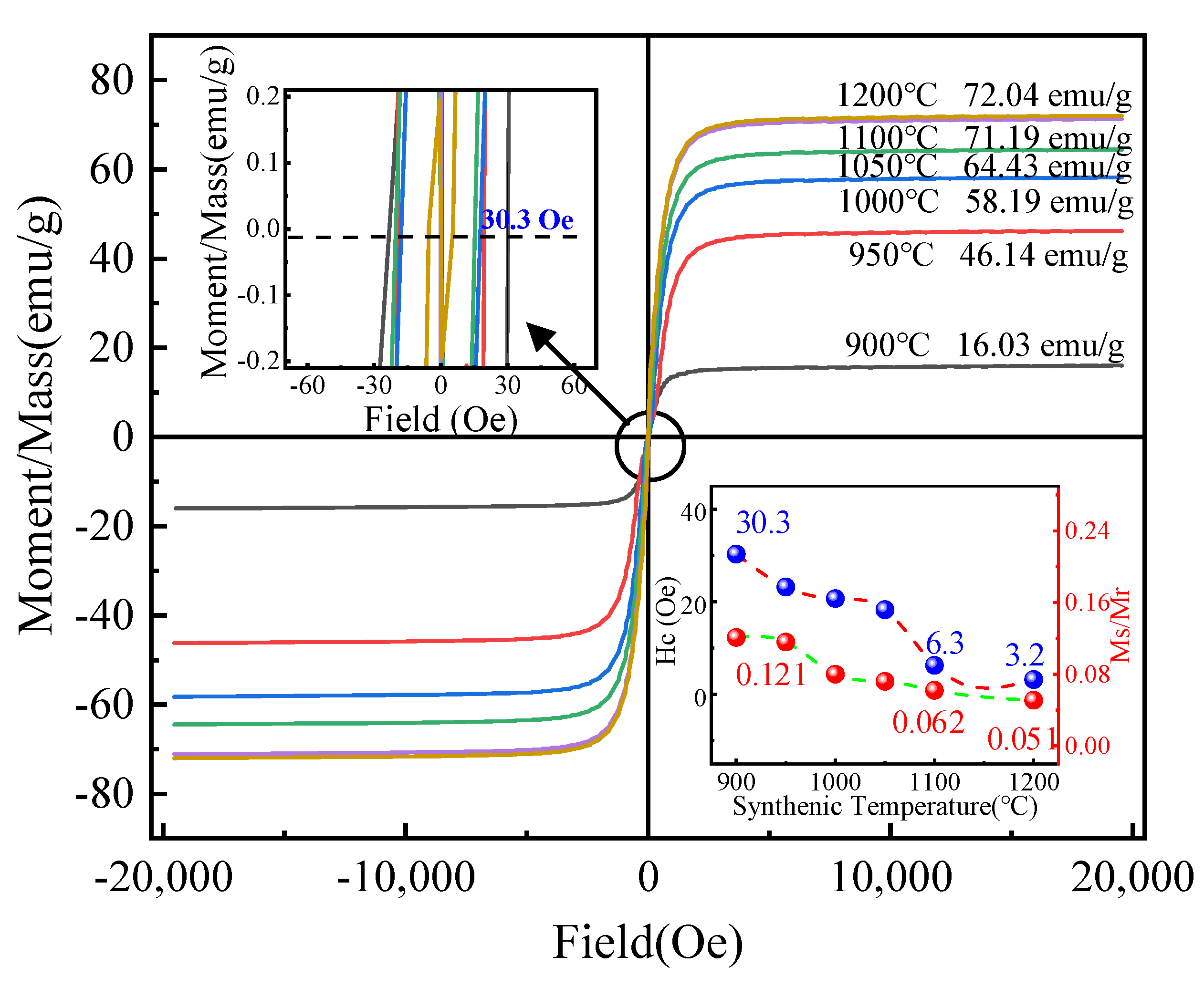
3.1.2. Magnetic Transformation

The magnetization hysteresis loops of the prepared MnxFe3−xO4 products at 900–1200 °C were investigated with VSM, as illustrated in Figure 3. The saturation magnetization intensity Ms of the samples at different temperatures increased from 16.03 emu/g to 72.04 emu/g with increasing roasting temperature. The saturation magnetization intensity was 16.03 emu/g at 900 °C. The explanation for this result combined with Figure 3 shows that a large amount of MnFeO3 could be generated from MnO and Fe2O3 at lower temperature, which leads to the decline of magnetism. Researchers have found that the saturation magnetization strength is proportional to the grain size of the magnetic material [24,32,33], and increasing the temperature will benefit the growth of MnxFe3−xO4 [18,24]. In addition, the saturation magnetic strength of MnFe2O4 involves the distribution of cations on the A or B sites of the spinel crystal structure sites. Therefore, the variation pattern of Ms of manganese ferrite as affected by temperature is also attributed to the variation in Mn2+ or Fe2+ (Fe3+) concentration in the MnxFe3−xO4 sample. The coercivity (Hc) and the ratio of saturation magnetization intensity (Ms) to residual magnetization intensity (Mr) at room temperature and oxidation roasting temperature are shown in Figure 3. The hysteresis lines of the roasted samples show a narrow hysteresis behavior of the ferromagnetic material. As the roasting temperature increased from 900 °C to 1200 °C, the coercivity of the samples decreased from 30.3 Oe to 3.2 Oe, and Ms/Mr decreased from 0.121 to 0.051. The saturation magnetization intensity of the pure MnFe2O4 sample at 1100 °C was 71.8 emu/g [34] and 71.19 emu/g in this study. The crystalline particles of the samples are more complete, and the magnetic properties are closer to those of the pure MnFe2O4 sample, which has better magnetic properties.

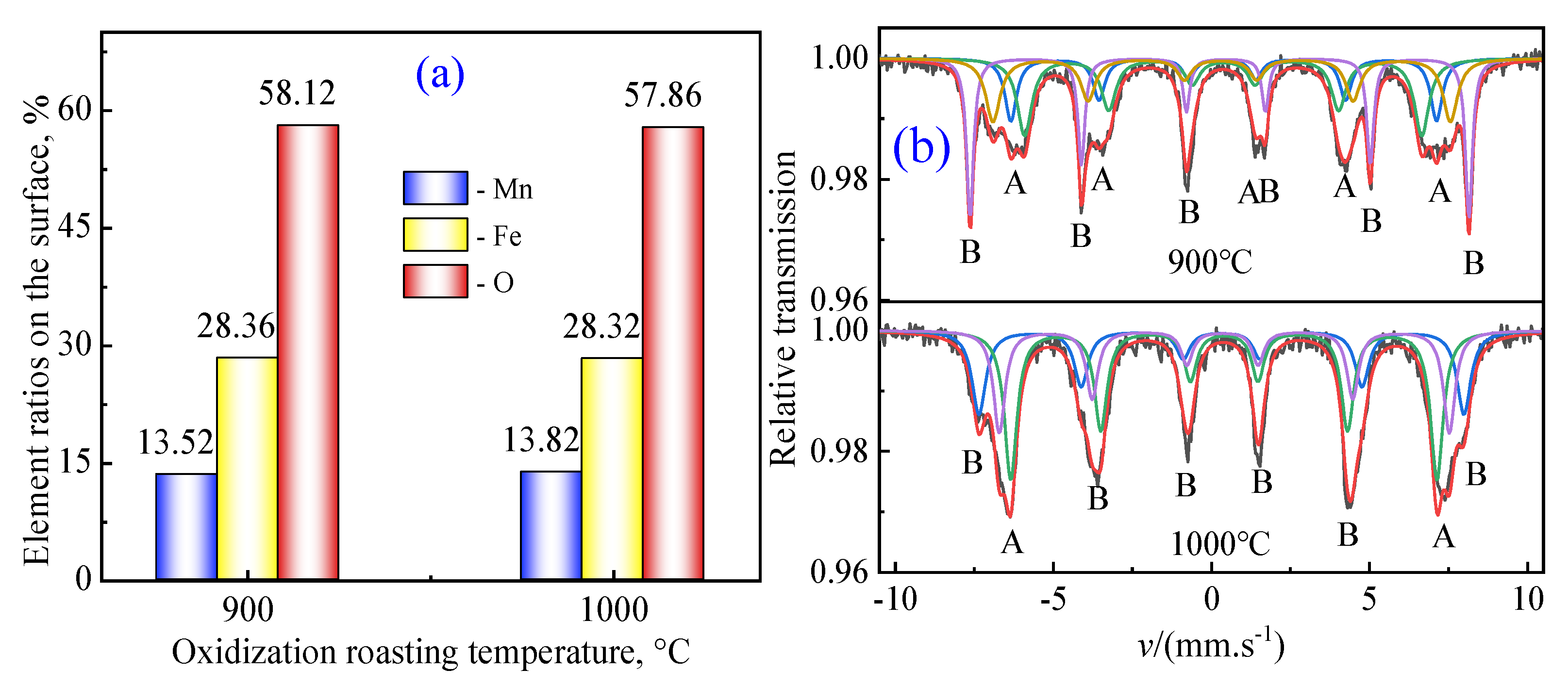
3.1.3. Oxidative Competition Behavior of MnO-Fe2O3 System in Oxidization Roasting Process

X-ray photoelectron spectroscopy (XPS) was used to detect and assess the chemical states of Fe and Mn to clarify the oxidization behavior and consolidation mechanism of manganese ferrite MnFe2O4. The XPS spectra in the Fe 2P regions and Mn 2P regions are illustrated in Figure 4a,b. XPS data are systematically analyzed and evaluated through XPS database XPS Peak software 4.2. In the preparation process of MnFe2O4, the valuable metal ions could occupy the tetra-coordination and octa-coordination in the spinel crystal structure, resulting in different valence states of Fe cations (Fe2+ and Fe3+) and Mn cations (Mn2+ and Mn3+) for different binding energies of the Fe 2p electrons and Mn 2p electrons. In addition, the binding energies of the Fe 2p3/2 peaks for FeO, Fe3O4 and Fe2O3 appeared at 709.8 eV, 710.6 eV and 711.6 eV, respectively. The binding energies of the Mn 2p3/2 peaks for MnO, Mn3O4 and Mn2O3 appeared at 640.0 eV, 641.2 eV and 642.2 eV, respectively [34,35]. It was found that the intergrade area of Fe2p3/2 for FeO at a binding energy of 709.8 eV gradually decreased and that of Fe3O4 at 710.6 eV also decreased, while that of Fe2O3 at 711.6 eV gradually increased with increasing oxidization roasting temperature from 900 °C to 1000 °C. In the meantime, the intergrade area of Mn2p3/2 for MnO at a binding energy of 709.8 eV and Mn3O4 at a binding energy of 641.2 eV changed slightly, and that of Mn2O3 at a binding energy of 642.2 eV instantly decreased. These results showed that the higher temperature could effectively enhance the oxidation of Fe2+ ions in composite oxides to Fe3+ ions and improve the dissociation of Mn2O3 in the crystal spinel structure of MnFe2O4.
Figure 5a illustrates the element ratios on the reaction surface. In theory, the atomic ratios of Fe, Mn and O should be 14.29% 28.57% and 57.14%, respectively, in the crystal structure of MnFe2O4. The error between the experimental data and the theoretical calculation value is small. However, there are differences in the occupancy of metal ions in the crystal structure of spinel manganese ferrite, which demonstrated that the vacancy oxygen in the crystal structure of MnxFe3−xO4 was gradually filled to form lattice oxygen in the crystal structure of MnFe2O4. The higher oxidization roasting temperature helps to adjust the atomic ordering of the spinel structure. Figure 5b illustrates the Mössbauer spectrum of the MnO-Fe2O3 system at different temperatures. The Mössbauer spectrum of each sample is clearly composed of asymmetric magnetic fine splitting hexagonal peaks. Moreover, each sample is superimposed with two different sets of characteristic diffraction peaks of spinel-type manganese ferrite, representing the Mössbauer spectra of the tetrahedral site (A-site) and octahedral site (B-site) [36,37]. Based on the characteristic that the oxygen atoms in the A-site are closer to the metal ions (≈0.67 Å) than in the B-site (≈0.72 Å), both the values of the homogeneous anomeric shift of Fe in the B-site are larger than those in the A-site. It is evident that the percentage of ortho-spinel increases in the shift from the A-site to the B-site as the roasting temperature of the sample is transformed from 900 °C to 1000 °C, which demonstrated that the single bridging oxygen of tetra-coordination and hexa-coordination in composite oxides were transformed into the double bridging oxygen of hexa-coordination in the crystal spinel structure of MnFe2O4 under the action of high temperature.

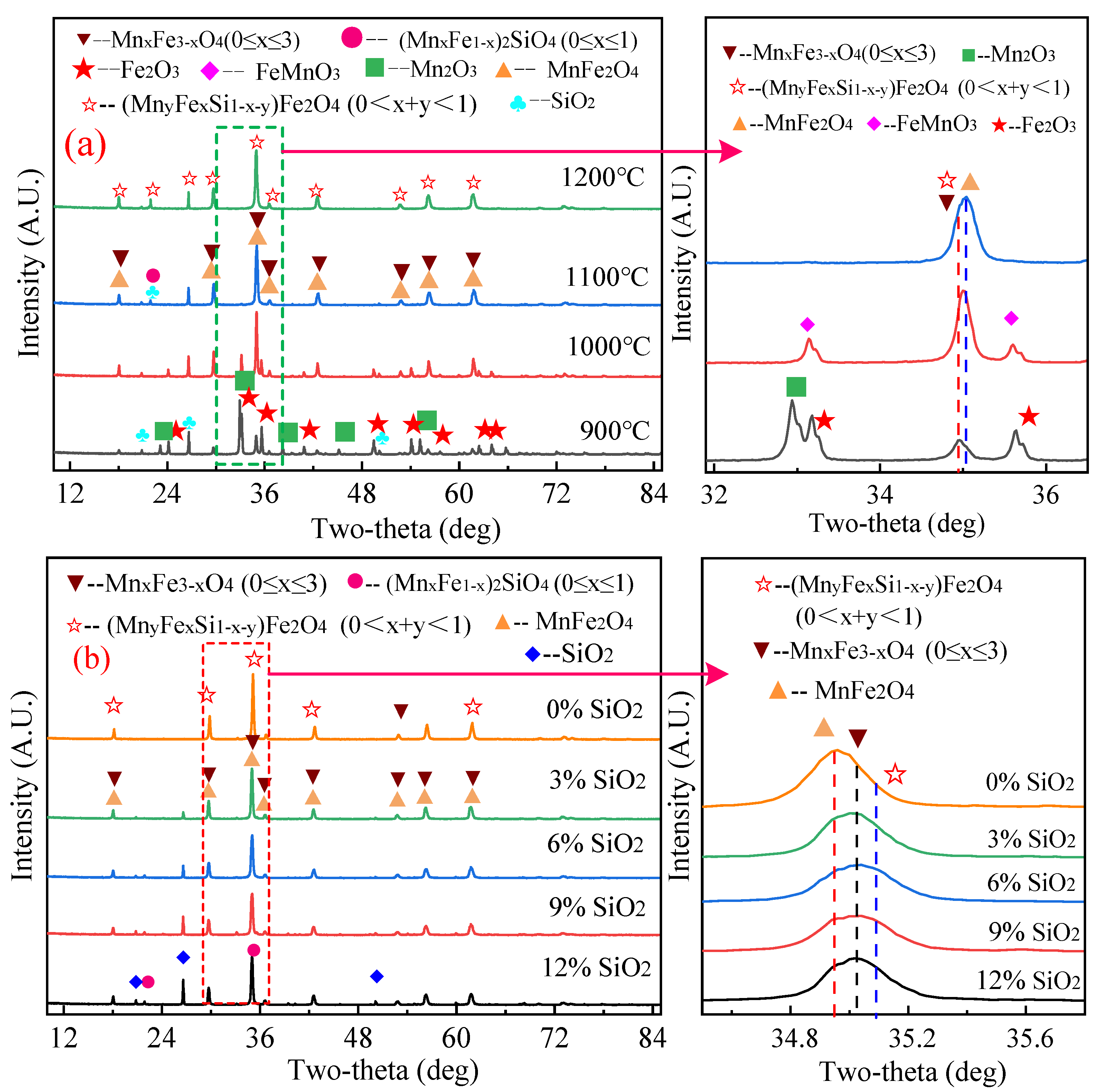
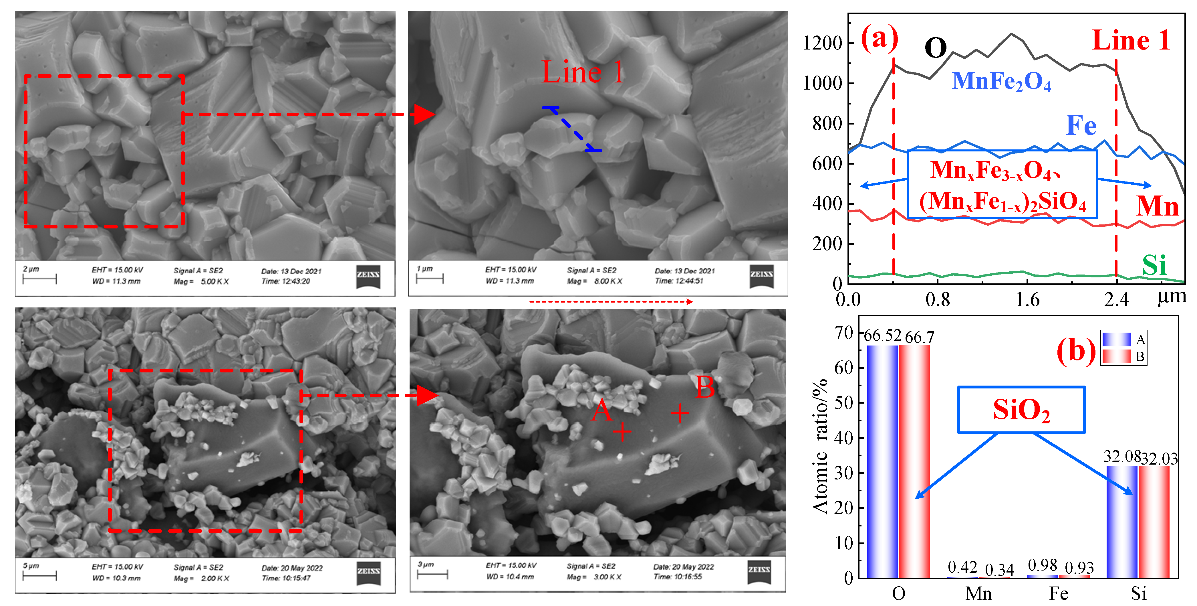
3.2. Discussion on the Oxidization Behavior of MnO-Fe2O3-SiO2 System

Figure 6 illustrates the XRD patterns of the MnO-Fe2O3-SiO2 system at temperatures from 900 to 1200 °C with different SiO2 contents. The XRD patterns at different roasting temperatures from 900 °C to 1200 °C are shown in Figure 6a. The main phases detected are Mn2O3, Fe2O3, SiO2 and a small amount of MnxFe3−xO4 at 900 °C, indicating that Mn2O3 and Fe2O3 are poorly synthesized MnxFe3−xO4 at lower temperatures. It can be seen from Figure 6a,b that the diffraction peaks of Mn2O3 and Fe2O3 disappear, the intensity of the characteristic peak of MnxFe3−xO4 constantly increases and the main diffraction peak approaches the main characteristic peak of MnFe2O4 (PDF#10-0319) with 2θ = 34.981° as the roasting temperature increases from 900 °C to 1100 °C. SiO2 exists mainly as free SiO2 and a very small amount of manganese–iron olivine phase (MnxFe1−x)2SiO4, corresponding to the card information (PDF#65-0466, 2θ = 26.636°) in this temperature range. This demonstrated that the influence of SiO2 on the oxidative roasting of the MnO-Fe2O3 system for the synthesis of MnFe2O4 is small when the roasting temperature is within 900–1100 °C. As the temperature increases to 1200 °C, the intensity of the characteristic peaks of SiO2 and MnFe2O4 slightly decreases, while (MnxFe1−x)2SiO4 is relatively enhanced, indicating that the excessive temperature will force SiO2 to react with some of the Fe and Mn oxides to form a small amount of manganese–iron olivine phase (MnxFe1−x)2SiO4. XRD patterns of SiO2 with different contents were roasted at 1100 °C for 120 min in the MnO-Fe2O3 system. The intensity of the characteristic diffraction peaks of SiO2 and (MnxFe1−x)2SiO4 in the product increased with increasing SiO2 content, the intensity of the characteristic peak of MnFe2O4 decreased and then increased and the main characteristic peak shifted from 2θ = 34.936° to 2θ = 35.022°. It can be inferred that the formation of (MnyFexSi1−x−y)Fe2O4 is related to both temperature and SiO2 content. The formation of (MnyFexSi1−x−y)Fe2O4 mainly originated from MnxFe3−xO4, in which the tetrahedral Fe2+ ions in the crystal structure of MnxFe3−xO4 were replaced by tetrahedral Si2+ ions in the crystal structure of silicates to form (MnyFexSi1−x−y)Fe2O4 at higher oxidation roasting temperatures. Next, the tetrahedral Fe2+ ions and Si2+ ions in the crystal structure of (FexSi1−x)Fe2O4 were replaced by tetrahedral Mn2+ and Fe2+ ions to form (MnyFexSi1−x−y)Fe2O4 (0 < x + y < 1) at higher oxidization roasting temperatures. The replaced Mn2+ ions and Fe2+ ions were combined with the replaced Si2+ ions to form MnxFe2−xSiO4 (0 ≤ x ≤ 2) by the oxidation roasting process. In summary, higher oxidization roasting temperatures are recommended for improving the recrystallization process of MnFe2O4.
Figure 7 shows the scanning electron micrographs of the cross-sectional layers of the MnO-Fe2O3-SiO2 sample when roasted at 1100 °C for 120 min. The sample cross-section was found to have an irregular polyhedral structure and pores of different sizes, whereas there were obvious boundaries between individual particles and obvious precipitates at the edges of individual particles. Figure 7a presents the results of the line-scan energy spectrum, which shows that the content of Mn, Fe and O in the precipitates is close to 1:2:4, while the content of Si is almost nonexistent. This shows that Si cannot enter the crystal lattice of MnFe2O4 and that SiO2 does not react with the MnO-Fe2O3 system under the experimental conditions. The reason for the presence of free SiO2 in Figure 5 was further verified. The results of Figure 7b were obtained by energy spectrum scanning of points A and B. The apparent Si to O atomic ratio was approximately 1:2, which again verified that SiO2 exists in the free state and that its morphology is obviously different from that of manganese ferrite. Combined with the presence of Si in the XRD spectrum of Figure 6, it can be inferred that free SiO2 and (MnxFe1−x)2SiO4 are mainly attached to the surface layer of the product and have similar polyhedral structures. The presence of SiO2 mainly affects the microstructure of manganese ferrite compared with the SEM image of the MnO-Fe2O3 system.

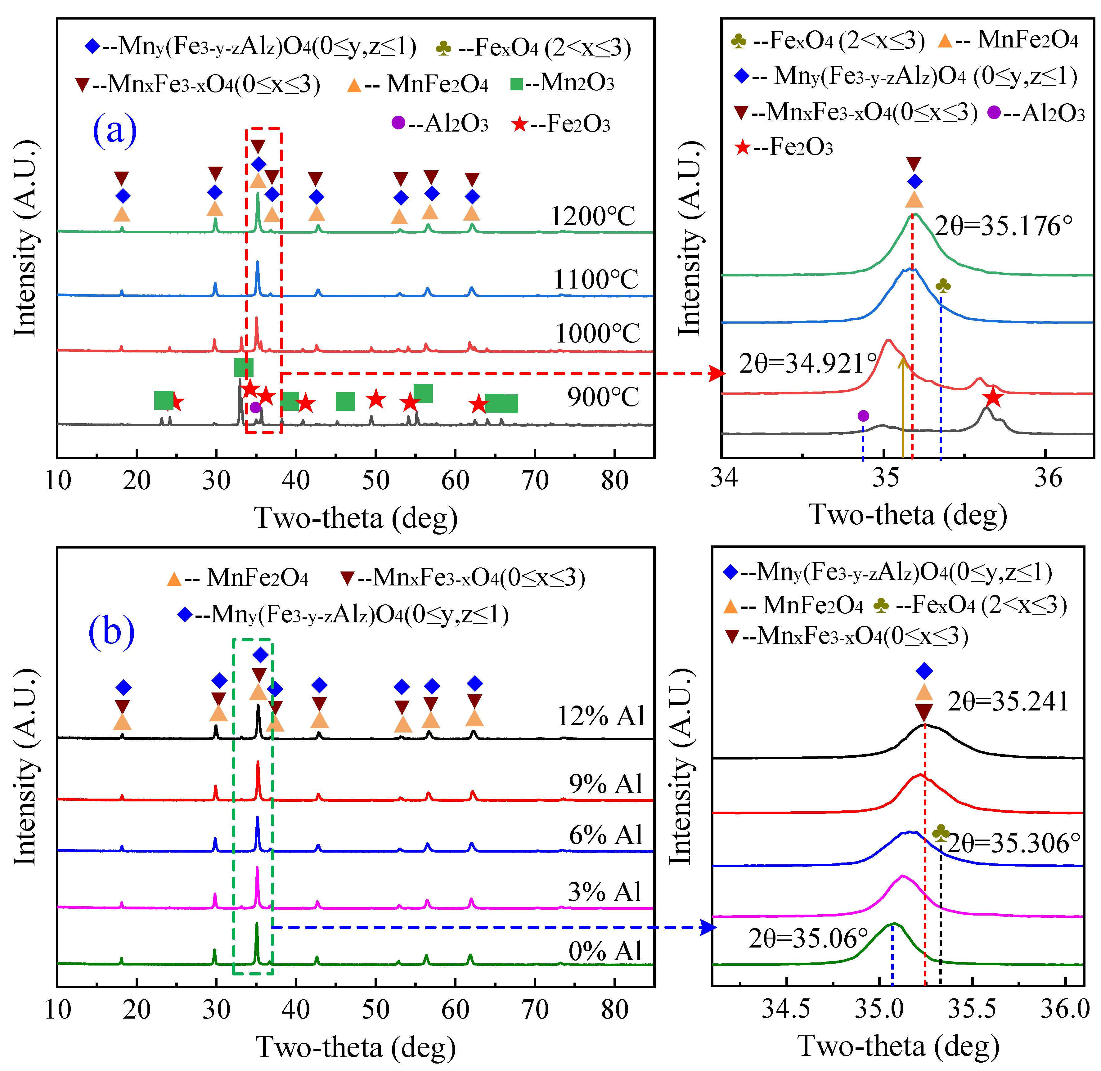
3.3. Discussion on the Oxidization Behavior of MnO-Fe2O3-Al2O3 System

Figure 8 illustrates the XRD patterns of the MnO-Fe2O3-Al2O3 system at 900~1200 °C and at 1100 °C with different Al2O3 contents. Figure 8a shows the XRD patterns at different roasting temperatures from 900 °C to 1200 °C. The main phases detected are Mn2O3, Fe2O3 and a small amount of MnxFe3−xO4 at 900 °C, indicating that MnxFe3−xO4 is not easily synthesized by Mn2O3 and Fe2O3 at low temperatures. Al was present in the form of Al2O3, and other than a small amount of Al2O3 characteristic peaks, no other forms of characteristic peaks were obviously identified. As the roasting temperature increases from 900 °C to 1100 °C, the diffraction peaks of Mn2O3 and Fe2O3 disappear, and the intensity of the characteristic peak of MnxFe3−xO4 increases continuously. The intensity of the characteristic peak of MnxFe3−xO4 increases continuously and moves toward the main characteristic peak of MnFe2O4 (PDF#10-0319) at 2θ = 34.981°. When the temperature increases from 1000 °C to 1100 °C, the diffraction peak of MnFe2O4 shifts from 2θ = 34.921° to 2θ = 35.176°. At the same time, the presence of spinel phases Mny(Fe3−y−zAlz)O4 and FexO4 were detected, however in small amounts. The above results indicate, on one hand, that some MnxFe3−xO4 was converted to Mny(Fe3−y−zAlz)O4 due to the continuous entry of some Al ions into the spinel-type manganese ferrite lattice to occupy the octahedral B-site during the reaction [35]. On the other hand, it demonstrates that the oxides of Fe ions replaced by Al ions were decomposed at high temperatures to form some low-valent Fe-oxides, leading to an increase of Fe-oxides FexO4 in the material phase. Figure 8b shows the XRD patterns of Al2O3 with different contents roasted at 1100 °C for 120 min in the MnO-Fe2O3-Al2O3 system. The diffraction peaks of MnxFe3−xO4, MnFe2O4 and Mny(Fe3−y−zAlz)O4 were detected from Figure 8b, whereas the diffraction peaks of Mn2O3 and Fe2O3 were not detected. This demonstrates that the content of Al2O3 has less influence on the reaction process of MnO and Fe2O3 under the experimental conditions. The position of the characteristic peak (3 1 1) of MnxFe3−xO4 shifts from 2θ = 35.06° to 2θ = 35.241° with increasing Al2O3 content, and the characteristic peaks of FexO4 and Mny(Fe3−y−zAlz)O4 are detected. This indicates that the amount of high content of Al2O3 in the spinel phase increases at 1100 °C and increases the decomposition of iron high-valent oxidation.
Figure 9 displays the SEM image of the cross-sectional layers of the sample roasted at 1100 °C with a mass content of 6% Al2O3. It can be clearly seen that the synthesized sample presents a regular polyhedral structure with a relatively uniform distribution. There are obvious boundaries between the particles, and the morphology of individual particles is mainly flat and spherical. The results obtained by the energy spectral line scan of the physical phase are shown in Figure 9b, which illustrates that the O, Fe and Mn contents are basically stable and the contents are close to 4:2:1, indicating that the composition of the synthesized sample is relatively pure. Combined with the analysis in Figure 9b, only a very small amount of Mny(Fe3−y−zAlz)O4 was present, which further proves the slow solid-phase reaction of Al2O3 with manganese and iron oxides. From Figure 9a, it is found that the contents of Fe and Mn at different positions are relatively evenly distributed, showing an overall ratio of 2:1. There is an obvious gradient of Al, which indicates that Al2O3 enters the spinel-type manganese ferrite more slowly. In summary, the presence of a low content of Al2O3 in the MnO-Fe2O3 system has less effect on manganese ferrite, but a synthesis temperature above 1100 °C and Al2O3 content above 6% will promote the conversion of MnxFe3−xO4 to Mny(Fe3−y−zAlz)O4. This led to a decrease in the amount of manganese ferrite produced, which in turn affected the purity of the product.

3.4. Oxidative Competition Behavior and Formation Mechanism in Synthesis Process of MnFe2O4

Based on the abovementioned results and analysis, the schematic diagram for preparation is illustrated in Figure 10a during the oxidative roasting process. In summary, the three stages were roughly divided for synthesizing manganese ferrite MnFe2O4 in Equations (1)–(7). In the initial stages (Equations (1)–(3)), magnetite (Fe)x[Fe]2O4 (0.878 ≤ x ≤ 1) was easily oxidized to hematite, which contained both hexagonal and cubic crystal structures. The existence of magnetite (Fe)x[Fe]2O4 and maghemite γ-Fe2O3 have a spinel-type crystal structure with a face-center cubic (FCC) configuration, in which both of them combine with MnO to form (MnxFe1−x)Fe2O4 (0 ≤ x ≤ 1) with a similar crystal structure [38,39]. Primarily because the tetrahedral Fe2+ ions in magnetite were partially replaced by octahedral Fe3+ ions, the octahedral coordination occupied by the Fe2+ ions generated vacancies, providing an advantageous environment for the Mn2+ ions to form (MnxFe1−x)Fe2O4 (0 ≤ x ≤ 1) during the oxidization roasting process of converting from magnetite to maghemite. In the second stage (Equations (4)–(6)), the replaced Fe2+ ions were directly oxidized to Fe3+ ions, while the other Fe2+ ions combined with Mn2+ ions to form an intermediate wustite phase (MnyFe1−yO), which was ultimately oxidized to (MnxFe1−x)Fe2O4 (0 ≤ x ≤ 1). In the final stage (Equation (7)), (MnxFe1−x)Fe2O4 (0 ≤ x ≤ 1) was characterized as (Mn1−αFeα)tet.[MnαFe2−α]oct. O4 (0 ≤ x ≤ 1) with the face-centered cubic (FCC) structure of the spinel crystal structure, where α is called the inversion parameter [40,41]. The higher temperature and oxidation environment promoted the mutual transformation between the normal and inverse spinel structure, in which the vacancy requirements for the oxidation reaction were not easily provided during this process, thereby inhibiting the recrystallization process of pellets, resulting in lower metallurgical properties of magnesia flux pellets.
(Fe2+)x[Fe3+]2[O2−]4 (0.878 ≤ x ≤ 1) + O2 → [Fe3+]2[O2−]3
[Fe3+]2[O2−]3 + [Mn2+][O2−] → [Mn2+][Fe3+]2[O2−]4
[Fe2+][Fe3+]2[O2−]4 + x[Mn2+][O2−] → [Mn2+]x[Fe2+]1−x[Fe3+]2[O2−]4 + x[Fe2+][O2−]
[Mn2+]y[Fe2+]1−y[O2−](0 ≤ y ≤ 0.33) + O2 → [Mn2+]x[Fe2+]1−x[Fe3+]2[O2−]4 (0 ≤ x ≤ 1)
[Mn2+]y[Fe2+]1−y[O2−](0.33 < y ≤ 1) + O2 → [Mn2+][Fe3+]2[O2−]4 + x[Mn2+][O2−]
[Mn2+]x[Fe2+]1−x[Fe3+]2[O2−]4 (0 ≤ x ≤ 1) + O2 → [Fe3+]2[O2−]3 + x[Mn2+][O2−]
(Mn1−αFeα)[MnαFe2−α]O4 (0 < α ≤ 1) → (Mn1−αFeα)[MnαFe2−α]O4 (α = 0)
The schematic diagram for the effect of Si on synthesizing manganese ferrite MnFe2O4 is illustrated in Figure 10b during the oxidative roasting process. The two stages in Equations (8)–(10) were roughly divided for synthesizing manganese ferrite MnFe2O4 during the oxidative roasting process. In the initial stages (Equation (8)), these crystal structures of olivine-type Fe2SiO4 and MnxFe2−xSiO4 had similar olivine-type crystal structures. The Fe2+ ions in olivine Fe2SiO4 are easily replaced by Mn2+ ions to form olivine MnxFe2−xSiO4, in which a portion of the replaced Mn2+ ions and Fe2+ ions combine from wustite (MnxFe1−xO), and another portion of the replaced Fe2+ ions is directly oxidized to MnxFe3−xO4 (0 ≤ x ≤ 3). In the second stage (Equations (9)–(10)), it is worth noting that the formation of (FexSi1−x)Fe2O4 mainly originated from (MnxFe1−x)Fe2O4, in which the tetrahedral Fe2+ ions and Mn2+ in the crystal structure of magnetite were replaced by tetrahedral Si2+ ions in the crystal structure of silicates to form (FexSi1−x)Fe2O4 under higher oxidization roasting temperatures and the presence of SiO2.
The schematic diagram for the effect of Al on synthesizing manganese ferrite MnFe2O4 is illustrated in Figure 10c. The two stages in Equations (11)–(13) were roughly divided for synthesizing manganese ferrite MnFe2O4. In the initial stages, the crystal structures of hercynite FeAl2O4 and FexMn1−xAl2O4 were similar. Some of the Fe2+ ions in hercynite FeAl2O4 are easily replaced by Mn2+ ions to form hercynite FexMn1−xAl2O4 (0 ≤ x ≤ 1) and further form MnAl2O4 after the displacement reaction. In the second stage (Equations (12)–(13)), the formation of (Mn2+)[Fe3+]2−x[Al3+]xO4 mainly originated from (Mn2+)[Fe3+]2O4, in which the octahedral Fe3+ ions and octahedral Mn3+ ions in the crystal structure of manganese ferrite MnFe2O4 were replaced by octahedral Al3+ ions to form (Mn2+)[Fe3+]2−x[Al3+]xO4 under higher oxidization roasting temperatures and the presence of Al2O3. In summary, the tetrahedral Mn2+ ions and octahedral Mn3+ ions in the crystal structure of manganese ferrite MnFe2O4 were replaced by tetrahedral Si2+ ions and octahedral Al3+ ions from (Mn2+)x(Fe2+)y(Si2+)1−x−y[Fe3+]2O4 and (Mn2+)[Fe3+]2−x[Al3+]xO4, respectively.
[Fe2+]2[Si4+][O2−]4 + x[Mn2+][O2−] → [Mn2+]x[Fe2+]2−x[Si4+][O2−]4 + x[Fe2+][O2−]
(Fe2+)[Fe3+]2[O2−]4 + x[Si2+][O2−] → (Si2+)x(Fe2+)1−x[Fe3+]2[O2−]4 + x[Fe2+][O2−]
(Si2+)x(Fe2+)1−x[Fe3+]2[O2−]4 + x[Mn2+][O2−] → (Mn2+)x(Fe2+)1−x[Fe3+]2[O2−]4+ x[Si2+][O2−]
[Fe2+][Al3+]2[O2−]4 + x[Mn2+][O2−] → [Mn2+]x[Fe2+]1−x[Al3+]2 [O2−]4 + x[Fe2+][O2−]
(Mn2+)[Fe3+]2[O2−]4 +x[Al3+]2[O2−]3 → (Mn2+)[Fe3+]2−x[Al3+]x][O2−]4 + x[Fe2+][O2−]
(Mn2+)[Fe3+]2−x[Al3+]x][O2−]4 + 2-x[Al3+]2[O2−]3 → (Mn2+) [Al3+]2[O2−]4 + 2-x[Fe2+][O2−]

4. Conclusions and Future Prospects

In this work, a new understanding of the synthesis and magnetism properties of manganese ferrite MnFe2O4 with a spinel crystal structure was proposed to elucidate the oxidization behavior and consolidation mechanism in the oxidization roasting process. The synthetic MnFe2O4 polyhedral microparticle samples with a saturated magnetization of 71.19 emu/g, a ratio of the saturation magnetization to residual magnetization (Ms/Mr) of 0.062 and a coercivity (Hc) of 6.50 Oe can be successfully obtained at an oxidization roasting temperature of 1100 °C for 60 min in the MnO-Fe2O3 system.
This work clarified the underlying reason for the influence mechanism of SiO2 and Al2O3 on the synthesis of MnFe2O4. The experimental results indicate that the tetrahedral Mn2+ ions and octahedral Mn3+ ions in the crystal structure of manganese ferrite MnFe2O4 were replaced by tetrahedral Si2+ ions and octahedral Al3+ ions from (Mn2+)x(Fe2+)y(Si2+)1−x−y[Fe3+]2O4 and (Mn2+)[Fe3+]2−x[Al3+]xO4, respectively. In addition, hercynite FexMn1−xAl2O4 with a spinel crystal structure and olivine MnxFe2−xSiO4 with an orthorhombic crystal structure were partially formed in the synthesis of manganese ferrite MnFe2O4, in which some Fe2+ ions were easily replaced by Mn2+ ions to form stable hercynite MnAl2O4 and olivine Mn2SiO4 in these crystal structures. The current research work provides comprehensive insights for synthesizing manganese ferrite MnFe2O4 and continuously advances its technical progress.

Author Contributions

S.W.: Writing—review & editing, Methodology. B.C.: Methodology, Funding acquisition. J.Z.: Funding acquisition, Methodology, Investigation, Visualization. W.Z.: Software, Funding acquisition. Z.H.: Methodology, Funding acquisition. L.G.: Writing—original draft, Methodology, Funding acquisition. All authors have read and agreed to the published version of the manuscript.

Funding

The work was supported by Doctoral research Foundation of Liaoning Province (2023-BS-181), National Natural Science Foundation of China (52104332, 52074150, 52374339), University of Science and Technology Liaoning Talent Project Grants (601011507-05).

Data Availability Statement

Not applicable.

Acknowledgments

The authors especially thank Y. Liang, H.Y. Sheng and X.Y. Yi for their help in this work.

Conflicts of Interest

The authors declare no conflict of interest.

References

  1. Liu, F.; Liu, J.; Yang, Y.; Wang, X. A mechanistic study of CO oxidation over spinel MnFe2O4 surface during chemical-looping combustion. Fuel 2018, 230, 410–417. [Google Scholar] [CrossRef]
  2. Guo, W.; Zhu, H.; Ren, Q.; Chen, S.; Ding, Y.; Xiong, C.; Chen, J.; Jia, X. MnFe2O4/ZnO/diatomite composites with electromagnetic wave absorption and antibacterial bifunctions. Solid State Sci. 2023, 138, 107152. [Google Scholar] [CrossRef]
  3. Mounkachi, O.; Lamouri, R.; Salmani, E.; Hamedoun, M.; Benyoussef, A.; Ez-Zahraouy, H. Origin of the magnetic properties of MnFe2O4 spinel ferrite: Ab initio and Monte Carlo simulation. J. Magn. Magn. Mater. 2021, 533, 168016. [Google Scholar] [CrossRef]
  4. Qian, Z.; Ting, Y.; Guo, G.; Shapter, G.J.; Lai, W.E.; Huang, P.; Qi, W.; Song, J.; Cui, D.X. Multifunctional Core@Shell Magnetic Nanoprobes for Enhancing Targeted Magnetic Resonance Imaging and Fluorescent Labeling In Vitro and In Vivo. J. Appl. Mater. Interfaces 2017, 21, 17777–17785. [Google Scholar]
  5. Martin, L.; Stigler, J.; Elia, G.; Lynch, I.; Tommy, C.; Dawson, A.K. Nanoparticle size and surface properties determine the protein corona with possible implications for biological impacts. Appl. Surf. Sci. 2008, 38, 14265–14270. [Google Scholar]
  6. Marcela, S.; Eliza, M.; Cornelia, P.; Ciprian, M. Thermal behavior of MnFe2O4 and MnFe2O4/C nanocomposite synthesized by a solvothermal method. Thermochim. Acta 2017, 652, 1–8. [Google Scholar]
  7. Peng, E.; Choo, E.S.G.; Chandrasekharan, P.; Yang, C.; Ding, J.; Chuang, K.; Xue, J.M. Synthesis of manganese ferrite/graphene oxide nanocomposites for biomedical applications. Small 2012, 8, 3620–3630. [Google Scholar] [CrossRef]
  8. Rajalakshmi, R.; Ponpandian, N. Morphological design of MnFe2O4 facets (cube, flakes and capsules) for their role in electrical, magnetic and photocatalytic activity. Mater. Res. Bull. 2023, 164, 112242. [Google Scholar] [CrossRef]
  9. Wu, X.F.; Ding, Z.; Wang, W.; Song, N.N.; Khaimanov, S.; Tsidaeva, N. Effect of polyacrylic acid addition on structure, magnetic and adsorption properties of manganese ferrite nanoparticles. Powder Technol. 2016, 295, 59–68. [Google Scholar] [CrossRef]
  10. Mondal, D.K.; Borgohain, C.; Paul, N.; Borah, J.P. Tuning hyperthermia efficiency of MnFe2O4/ZnS nanocomposites by controlled ZnS concentration. J. Mater. Res. Technol. 2019, 8, 5659–5670. [Google Scholar] [CrossRef]
  11. Neda, A.; Ghasem, N. A Manganese ferrite (MnFe2O4) Nanoparticles: From synthesis to application—A review. J. Ind. Eng. Chem. 2021, 103, 292–304. [Google Scholar]
  12. Masrour, R. Magnetic properties of the spinel systems ACr2X4 (A = Zn, Cd, Hg; X = S, Se). J. Alloys Compd. 2010, 489, 441–444. [Google Scholar] [CrossRef]
  13. Leal, M.P.; Rivera-Fernández, S.; Franco, J.M. Long-circulating PEGylated manganese ferrite nanoparticles for MRI-based molecular imaging. Nanoscale 2015, 7, 2050–2059. [Google Scholar] [CrossRef] [PubMed]
  14. Misra, R.D.K.; Gubbala, S.; Kale, A.; Egelhoff, W.F.E., Jr. A comparison of the magnetic characteristics of nanocrystalline nickel, zinc, and manganese ferrites synthesized by reverse micelle technique. Mater. Sci. Eng. B 2004, 111, 164–174. [Google Scholar] [CrossRef]
  15. Lin, X.M.; Lv, X.; Wang, L.M.; Zhang, F.F.; Duan, L.F. Preparation and characterization of MnFe2O4 in the solvothermal process: Their magnetism and electrochemical properties. Mater. Res. Bull. 2013, 48, 2511–2516. [Google Scholar] [CrossRef]
  16. Carpenter, E.E.; O’Connor, C.J.; Harris, V.G. Atomic structure and magnetic properties of MnFe2O4 nanoparticles produced by reverse micelle synthesis. J. Appl. Phys. 1999, 85, 5175–5177. [Google Scholar] [CrossRef]
  17. Lazarova, T.; Kovacheva, D.; Georgieva, M.; Tzankov, D.; Tyuliev, G.; Spassova, I.; Naydenov, A. Tunable nanosized spinel manganese ferrites synthesized by solution combustion method. Appl. Surf. Sci. 2019, 496, 143571. [Google Scholar] [CrossRef]
  18. Liu, B.B.; Zhang, Y.B.; Su, Z.J.; Lu, M.M.; Peng, Z.W.; Li, G.H.; Jiang, T. Formation mechanism of MnxFe3-xO4 by solid-state reaction of MnO2 and Fe2O3 in air atmosphere Morphologies and properties evolution. Powder Technol. 2017, 313, 201–209. [Google Scholar] [CrossRef]
  19. Ahmad, S.; Ali, S.; Ullah, I.; Zobaer, M.S.; Albakri, A.; Muhammad, T. Synthesis and characterization of manganese ferrite from low grade manganese ore through solid state reaction route. Sci. Rep. 2021, 11, 16190. [Google Scholar] [CrossRef]
  20. Liu, B.B.; Zhang, Y.B.; Wang, J.; Wang, J.; Su, Z.J.; Li, G.H.; Jiang, T. A further investigation on the MnO2-Fe2O3 system roasted under CO-CO2 atmosphere. Adv. Powder Technol. 2019, 30, 302–310. [Google Scholar] [CrossRef]
  21. Wang, G.; Zhao, D.; Ma, Y.; Zhang, Z.; Che, H.; Mu, J.; Zhang, X.; Zhang, Z. Synthesis and characterization of polymer-coated manganese ferrite nanoparticles as controlled drug delivery. Appl. Surf. Sci. 2018, 428, 258–263. [Google Scholar] [CrossRef]
  22. Reddy, M.P.; Mohamed, A.M.A.; Raman, M.V.; Zhou, X.B.; Huang, Q. Spark plasma sintering and microwave electromagnetic properties of MnFe2O4 ceramics. J. Magn. Magn. Mater. 2015, 395, 185–189. [Google Scholar] [CrossRef]
  23. Rashad, M.M. Synthesis and magnetic properties of manganese ferrite from low grade manganese ore. Mater. Sci. Eng. B 2006, 127, 123–129. [Google Scholar] [CrossRef]
  24. Ahmed, Y.M.Z. Synthesis of manganese ferrite from non-standard raw materials using ceramic technique. Ceram. Int. 2010, 36, 969–977. [Google Scholar] [CrossRef]
  25. Makridis, A.; Tziomaki, M.; Topouridou, K.; Yavropoulou, M.P.; Yovos, J.G.; Kalogirou, O.; Samaras, T.; Angelakeris, M. A novel strategy combining magnetic particle hyperthermia pulses with enhanced performance binary ferrite carriers for effective in vitro manipulation of primary human osteogenic sarcoma cells. Int. J. Hyperth. 2016, 32, 778–785. [Google Scholar] [CrossRef]
  26. Chen, D.; Zhang, Y.Z.; Kang, Z.T. A low temperature synthesis of MnFe2O4 nanocrystals by microwave-assisted ball-milling. Chem. Eng. J. 2013, 215–216, 235–239. [Google Scholar] [CrossRef]
  27. Neda, A.; Ghasem, N.; Younesi, H. Facile and green synthesis of cobalt oxide nanoparticles using ethanolic extract of Trigonella foenumgraceum (Fenugreek) leaves. Adv. Powder Technol. 2020, 31, 3562–3569. [Google Scholar]
  28. Aslibeiki, B.; Kameli, P. Magnetic properties of MnFe2O4 nano-aggregates dispersed in paraffin wax. J. Magn. Magn. Mater. 2015, 385, 308–312. [Google Scholar] [CrossRef]
  29. Ba-Abbad, M.M.; Chai, P.V.; Takriff, M.S.; Benamor, A.; Mohammad, A.W. Optimization of nickel oxide nanoparticle synthesis through the sol-gel method using Box-Behnken design. Mater. Des. 2015, 86, 948–956. [Google Scholar] [CrossRef]
  30. Yan, Z.; Chaluvadi, A.; FitzGerald, S.; Spence, S.; Bleyer, C.; Zhu, J.; Crawford, T.M.; Getman, R.B.; Watt, J.; Huber, D.L.; et al. Effect of manganese substitution of ferrite nanoparticles on particle grain structure. Nanoscale Adv. 2022, 4, 3957–3965. [Google Scholar] [CrossRef]
  31. Gao, L.H.; Liu, Z.G.; Ge, Y.; Feng, C.; Chu, M.S.; Tang, J. Synthesis and characterization of manganese ferrite MnxFe3-xO4 from ferruginous manganese ores by multi-step roasting and magnetic separation. Powder Technol. 2019, 356, 373–382. [Google Scholar] [CrossRef]
  32. Devan, R.S.; Ma, Y.; Chougule, B.K. Effective dielectric and magnetic properties of (Ni–Co–Cu) ferrite/BTO composites. Mater. Chem. Phys. 2009, 115, 263–268. [Google Scholar] [CrossRef]
  33. Shekhar, D.B.; Pattayil, A.J. Effect of sintering conditions and microstructure on the magnetostrictive properties of cobalt ferrite. J. Am. Ceram. Soc. 2008, 91, 1976–1980. [Google Scholar]
  34. Gao, L.H.; Liu, Z.G.; Yang, Z.C.; Feng, L.G.C.; Chu, M.S.; Tang, J. Synthesis and magnetism property of manganese ferrite MnFe2O4 by selective reduction and oxidization roasting process. Appl. Surf. Sci. 2020, 508, 145292–145301. [Google Scholar] [CrossRef]
  35. Zhao, X.R.; Wang, W.; Zhang, Y.J.; Wu, S.Z.; Li, F.; Liu, J.P. Synthesis and characterization of gadolinium doped cobalt ferrite nanoparticles with enhanced adsorption capability for Congo Red. Chem. Eng. J. 2014, 250, 164–174. [Google Scholar] [CrossRef]
  36. Wang, Z.; Chen, H.; Han, X.; Gao, L.; Zhan, W.; Zhang, J.; He, Z. Preparation and characterization of MnFe2O4 by a microwave-assisted oxidative roasting process. Adv. Powder Technol. 2023, 34, 104040. [Google Scholar] [CrossRef]
  37. Šepelák, V.; Baabe, D.; Mienert, D.; Schultze, D.; Krumeich, F.; Litterst, F.J.; Becker, K.D. Evolution of structure and magnetic properties with annealing temperature in nanoscale high-energy-milled nickel ferrite. J. Magn. Magn. Mater. 2003, 257, 377–386. [Google Scholar] [CrossRef]
  38. Gao, L.H.; Liu, P.; Zhan, W.L.; Zhang, J.H.; He, Z.J.; Hou, X. New understanding on formation mechanism of CaFe2O4 in Fe2O3-Fe3O4-CaO-SiO2 system during sintering Process: Phase transformation and morphologies evolution. Adv. Powder Technol. 2022, 33, 103712. [Google Scholar] [CrossRef]
  39. Cheng, F.Y.; Shen, J.; Peng, B.; Pan, Y.D.; Tao, Z.L.; Chen, J. Rapid room-temperature synthesis of nanocrystalline spinels as oxygen reduction and evolution electrocatalysts. Nat. Chem. 2011, 3, 79–84. [Google Scholar] [CrossRef]
  40. Allen, G.C.; Hallam, K.R. Characterisation of the spinels MxCo1-xFe2O4 (M=Mn, Fe or Ni) using X-ray photoelectron spectroscopy. Appl. Surf. Sci. 1996, 93, 25–30. [Google Scholar] [CrossRef]
  41. Mirza, M.; Muhammad, A.; Philips, O.; Muhammad, A.; Imran, S.; Muhammad, F. Optimization of different wet chemical routes and phase evolution studies of MnFe2O4 nanoparticles. Ceram. Int. 2019, 45, 12682–12690. [Google Scholar]
Figure 1. XRD patterns of the synthetic MnFe2O4 samples at a temperature from 900 to 1200 °C with the atomic ratio of MnO: Fe2O3 = 1:1 for 60 min in air atmosphere and the crystal structure of spinel MnFe2O4, (a) the oxidization roasting temperature of 900 °C to 1200 °C, (b) Partial enlargement of Figure with two-theta range from 33.5° to 36.5°, (c) Partial enlargement of Figure with two-theta range from 32.5° to 36.7°, (c) Crystal structure of spinel MnFe2O4.
Crystals 13 01509 g001
Figure 2. SEM-EDS micrograph of the surface layer in the section of the synthetic manganese ferrite samples in air atmosphere, for 60 min (a) Samples roasted at 900 °C (b) Samples roasted at 1000 °C and Samples roasted at 1100 °C. (c) The atomic ratios and contents of Mn, Fe and O on individual particles, A and B.
Crystals 13 01509 g002
Figure 3. Magnetic hysteresis loops of MnFe2O4 samples roasted at temperatures in the range of 900 °C–1200 °C for 60 min in an air atmosphere.
Crystals 13 01509 g003
Figure 4. XPS spectra of the synthetic MnFe2O4 with (a) Spectra in Fe2p region and (b) Spectra in Mn2p region.
Crystals 13 01509 g004
Figure 5. Element ratios on the surface (a) and Mössbauer spectrum (b) at different oxidation roasting temperature for 30 min in air atmosphere, A—tetrahedral site and B—octahedral site.
Crystals 13 01509 g005
Figure 6. XRD patterns of MnFe2O4 samples calcined in MnO-Fe2O3-SiO2 system in air atmosphere; (a) 900~1200 °C with 6% SiO2 content for 60 min; (b) The different SiO2 contents at 1100 °C for 60 min.
Crystals 13 01509 g006
Figure 7. SEM-EDS micrograph of the surface layer in the section of the synthetic manganese ferrite samples in air atmosphere, (a) Samples roasted at 1100 °C for 60 min, (b) Samples roasted at 1100 °C for 120 min.
Crystals 13 01509 g007
Figure 8. XRD patterns of MnFe2O4 samples calcined in MnO-Fe2O3-Al2O3 system in air atmosphere; (a) XRD patterns of MnFe2O4 samples with a MnO to Fe2O3 ratio of 1:1 and the Al2O3 mass fraction of 6% calcined at 900~1200 °C for 60 min; (b) XRD patterns of different Al2O3 contents roasted at 1100 °C for 60 min.
Crystals 13 01509 g008
Figure 9. SEM-EDS micrograph of the surface layer in the section of the synthetic manganese ferrite samples in air atmosphere, (a) Samples roasted at 1100 °C for 60 min, (b) Samples roasted at 1100 for 120 min.
Crystals 13 01509 g009
Figure 10. Schematic diagram of phase transformation mechanism and oxidation behavior in synthesizing process of manganese ferrite MnFe2O4, (a) Fe2O3-MnO system, (b) Fe2O3-MnO-SiO2 system and (c) Fe2O3-MnO-Al2O3 system.
Crystals 13 01509 g010
Disclaimer/Publisher’s Note: The statements, opinions and data contained in all publications are solely those of the individual author(s) and contributor(s) and not of MDPI and/or the editor(s). MDPI and/or the editor(s) disclaim responsibility for any injury to people or property resulting from any ideas, methods, instructions or products referred to in the content.

