Article

Transverse Microradiography Evidence on the Effect of Phosphoryl Oligosaccharides of Calcium (POs-Ca) in Toothpaste on Decalcified Enamel

Department of Cariology and Operative Dentistry, Division of Oral Health Sciences, Graduate School of Medical and Dental Sciences, Tokyo Medical and Dental University (TMDU), 1-5-45 Yushima, Bunkyo-ku, Tokyo 113-8549, Japan
*
Author to whom correspondence should be addressed.
Crystals 2023, 13(2), 206; https://doi.org/10.3390/cryst13020206
Submission received: 20 December 2022 / Revised: 18 January 2023 / Accepted: 20 January 2023 / Published: 23 January 2023

Abstract

:
In the current study, we sought to evaluate the effects of phosphoryl-oligosaccharides of calcium (POs-Ca) and/or fluoride-containing toothpaste on enamel. Six groups of experimental toothpaste were prepared by adding various concentrations of POs-Ca and/or fluoride. A 5 × 10-mm window on the enamel of the bovine incisor was immersed in resin and divided into three areas for “sound”, “demineralized (DEM)”, and “after pH cycle (aft. pH cycle)”. All specimens were subjected to pH cycling, including soaking in a slurry with toothpaste diluted threefold for 5 min. Transverse microradiography was employed to evaluate the mineral recovery, and the pH values of each demineralization solution were measured. The high fluoride concentration group showed a significantly better mineral recovery rate than the others, with no statistical differences between before and after pH cycling among the POs-Ca with low fluoride, POs-Ca only, low fluoride only, and control groups. In the low-concentration fluoride groups, the group containing POs-Ca tended to have a higher remineralization effect than the non-POs-Ca group. After pH cycling, the demineralization solution showed no pH changes in any group. The addition of POs-Ca and fluoride to toothpaste may recover the mineral density in enamel subsurface lesions at low-fluoride concentrations, but the high-fluoride concentrations did not show a meaningful difference in the two groups with and without POs-Ca.

1. Introduction

Recently, the number of remaining teeth has been increasing with longer life expectancy [1]. It has been estimated that having over 20 teeth remaining may facilitate masticatory function and significantly reduce the risk of all-cause mortality [2,3,4]. Teeth are always hanging by a thread due to dental caries or dental erosion. Unhealthy dietary habits (especially including sugar and carbonated drink overconsumption) and neglect of oral care are the biggest factors that cause these diseases. Several studies have investigated preventative treatment [5,6,7]. Still, dental care in person is more effective and more important than any other treatment [8]. Therefore, the recognition of oral care has increased, and the demand for new care methods/agents has grown. Fluoride-containing dentifrices are among the convenient care methods to prevent dental caries throughout a person’s lifetime. Increasing reports have demonstrated that a high concentration of fluoride is effective in preventing caries [9,10], with prevention rates of 41% for coronal caries [11] and 67% for root caries [11,12]. In 2017, the Japanese Ministry of Health approved dentifrices containing a maximum limit of 1500 ppm concentrate of fluoride with a minimum of 1450 ppm in the current market. However, because high-concentration fluoride use carries a high risk of dental fluorosis in children and given that fluorine itself can be an addictive substance, the use of high-level fluoride-containing dentifrices has recently been reconsidered [13,14]. Some studies have suggested that low and slightly elevated levels of fluoride caused by fluoride-containing products help prevent caries by restraining demineralization, exhibiting antimicrobial activity, and enhancing remineralization [15,16,17]. However, overall, there remains a lack of understanding of the efficacy and optimal applications of fluoride dosing adjustment.
Calcium is an indispensable mineral for strengthening the tooth structure. During demineralization, much calcium from the teeth is dissolved irreversibly. The combination of fluorine and calcium is a key factor in conducting remineralization effectively [18]. Some studies have indicated that calcium pretreatment enhances fluoride’s reactivity in both the enamel and dentin [19]. On the contrary, fluoride can be highly reactive to calcium and becomes less effective when it meets with a free calcium ion. Therefore, artificially constructed fluoride and calcium are easily ionically bonded before the materials enter the tooth. To counter this problem, some researchers have contrived bioavailable fluoride and calcium in various forms [20,21,22,23].
Phosphoryl-oligosaccharides of calcium (POs-Ca) is a highly water-soluble calcium material made from potato starch that can dissolve in the presence of phosphoric acid and fluorine ions without forming calcium-phosphate precipitates [24]. POs-Ca provides calcium ions in a bioavailable form and is not assimilated by cariogenic microorganisms, such as Streptococcus mutans, which prevents plaque formation and retains the pH buffering capacity [25]. Previous studies have identified several significant effects of POs-Ca on enamel remineralization [24], including effects induced by chewing gum containing POs-Ca on enamel remineralization [21] and dentin remineralization [26]. However, few studies have focused on the effect of dentifrice containing POs-Ca and fluoride on remineralization. Therefore, in this study, we investigated the optimal dose of POs-Ca and fluoride to enhance the mineralization of enamel in vitro using transverse microradiography (TMR). The null hypothesis was that no difference would exist between groups with and without the addition of POs-Ca or the concentration of fluoride in toothpaste.

2. Materials and Methods

2.1. Materials and Study Design

The study was based on a randomized controlled in vitro design with six toothpastes that were prepared solely for this examination (six experimental groups):
Group 1 (Control): Toothpaste not containing POs-Ca and fluoride;
Group 2 (F3): Low fluoride concentration toothpaste without POs-Ca;
Group 3 (F1450): High fluoride concentration toothpaste without POs-Ca;
Group 4 (P): With POs-Ca, non-fluoride toothpaste;
Group 5 (P + F3): With POs-Ca, low fluoride concentration toothpaste;
Group 6 (P + F1450): With POs-Ca, high fluoride concentration toothpaste.
The proportions of each POs-Ca (POs-Ca®, Ezaki Glico, Osaka, Japan) and NaF are listed in Table 1. POs-Ca was prepared as described by To-o et al. [27]. All toothpastes were received in code-labeled packages, stored at room temperature, and then dissolved in remineralization solution at a ratio of 1:3 for use (toothpaste slurry).

2.2. Specimen Preparation

Specimen preparation was illustrated in Figure 1. Thirty-six fresh bovine incisors were used in this study. The remaining tissue on each tooth was removed, and the crown portion was dissected from the root using a low-speed diamond saw (Isomet, Buehler, Lake Bluff, IL, USA) under a running water coolant. Enamel specimens, trimmed from the crown portion, were embedded in acrylic resin (Unifast Trad; GC, Tokyo, Japan). The specimens left behind a 5 mm × 10 mm × 3 mm (width × length × depth) window before being embedded. The enamel surfaces were sequentially ground flat with a series of silicon carbide papers (SiC) (800, 1000, and 2000 grit lapping papers, 3M, St. Paul, MN, USA) under running water. The polished enamel surface of each specimen comprised three zones, the “sound”, “demineralized (DEM)”, and “after pH cycle (aft. pH cycle)” zones, which were defined as follows. First, one-third of the surface of each specimen was covered by nail varnish (Nail POP, Chamon, Kyonggi, South Korea) to serve as the “sound” range; subsequently, subsurface lesions were formed using the two-layer demineralization method with 8% methylcellulose gel (Methocel MC, Fluka, Everett, WA, USA) and 0.1 M lactate buffer (pH 4.6) at 37 °C for 96 h [28]. After demineralization, another one-third of the surface was covered by nail varnish (Revlon nail enamel 161, REVLON, Tokyo, Japan) to constitute the DEM zone, and the remaining exposed one-third of the enamel surface was saved as the aft. pH cycle zone. Finally, the specimens were randomly allocated into six experimental groups (n = 6 per group).

2.3. pH-Cycling

The pH-cycling conditions were depicted as the pH fluctuation during human “meal” and “toothbrushing” times, with a chosen daily schedule of three cycles for 8 days. Each 0.5 h of demineralization and 3 h of remineralization was conducted at 37 °C. Between the DEM and REM periods, the specimens were soaked in the toothpaste slurry with each type of diluted toothpaste for 5 min. During the night, the specimens were kept in a preserved solution.
The demineralization solution contained 1.5 mM CaCl2, 0.9 mM KH2PO4, and 50 mM acetate adjusted to pH 4.5 with KOH; the remineralization solution contained 1.5 mM CaCl2, 0.9 mM KH2PO4, 130 mM KCl, and 20 mM HEPES at pH 7.0; and the preserved solution contained 1.5 mM CaCl2, 0.9 mM KH2PO4, and 130 mM KCl at pH 7.0 (unbuffered) [29]. All solutions were refreshed daily, and each specimen was cycled in 14 mL aliquots.

2.4. TMR Analysis

The treated enamel specimens in each group (n = 36) were cut into sections approximately 150 µm thick from four equal parts per block using a low-speed diamond saw. The section surface was carefully polished by monitoring the thickness of each section with a micrometer (Mitutoyo, Tokyo, Japan). Following these steps, each specimen was obtained from three zones (sound, DEM, aft. pH cycle). All sections were placed with a 15-step aluminum step-wedge on an X-ray glass plate (High Precision Photo plate, Konica Minolta Photo, Tokyo, Japan). The slabs were then microradiographed with a soft X-ray generator (SOFTEX CMR-2, Softex, Kanagawa, Japan) under the conditions of a 20 kV tube voltage and a 2.5 mA tube current with an exposure time of 9 min. TMR images were digitally photographed using an optical microscope (CKX41, Olympus Optical Co., Tokyo, Japan) and a CCD camera (DP74, Olympus Optical Co., Tokyo, Japan). The digitized images were analyzed by image analysis software (Image J, version 2.1.0, Wayne Rasband, NIH, Bethesda, MD, USA), and the mineral loss was customized and calculated (ΔZ: vol%·µm), where ΔZ was defined as the integrated mineral loss from the sound surface to the bottom of the lesion. The mineral recovery vol% (%R) was calculated according to the following equation: (ΔZ baseline − ΔZ after pH cycling)/ΔZ baseline × 100.

2.5. pH Measurement

The pH value in the demineralization solution (after soaking the specimens, excluding the control group) was measured directly using a pH meter (LAQUA F-52 pH meter, Horiba Ltd., Kyoto, Japan) calibrated by standard solutions of pH 4.0, 7.0, and 9.0 (500-F-SH, Horiba Ltd., Kyoto, Japan). Each demineralization solution was measured once, and the mean value of three cycles per day was calculated.
The values were recorded to two decimal places. After each analysis, the electrodes were washed with deionized water and dried with paper towels. All evaluations were performed 1, 3, 5, and 7 days after specimen preparation.

2.6. Data Analysis

All statistical tests were performed with a statistical program (SPSS ver. 26.0, IBM, Chicago, IL, USA). The data distribution was analyzed by the Shapiro–Wilk test. The TMR data were analyzed using one-way ANOVA with the Games–Howell post hoc test, and the pH values were analyzed using the Kruskal–Wallis test. The significance level was defined as p = 0.05.

3. Results

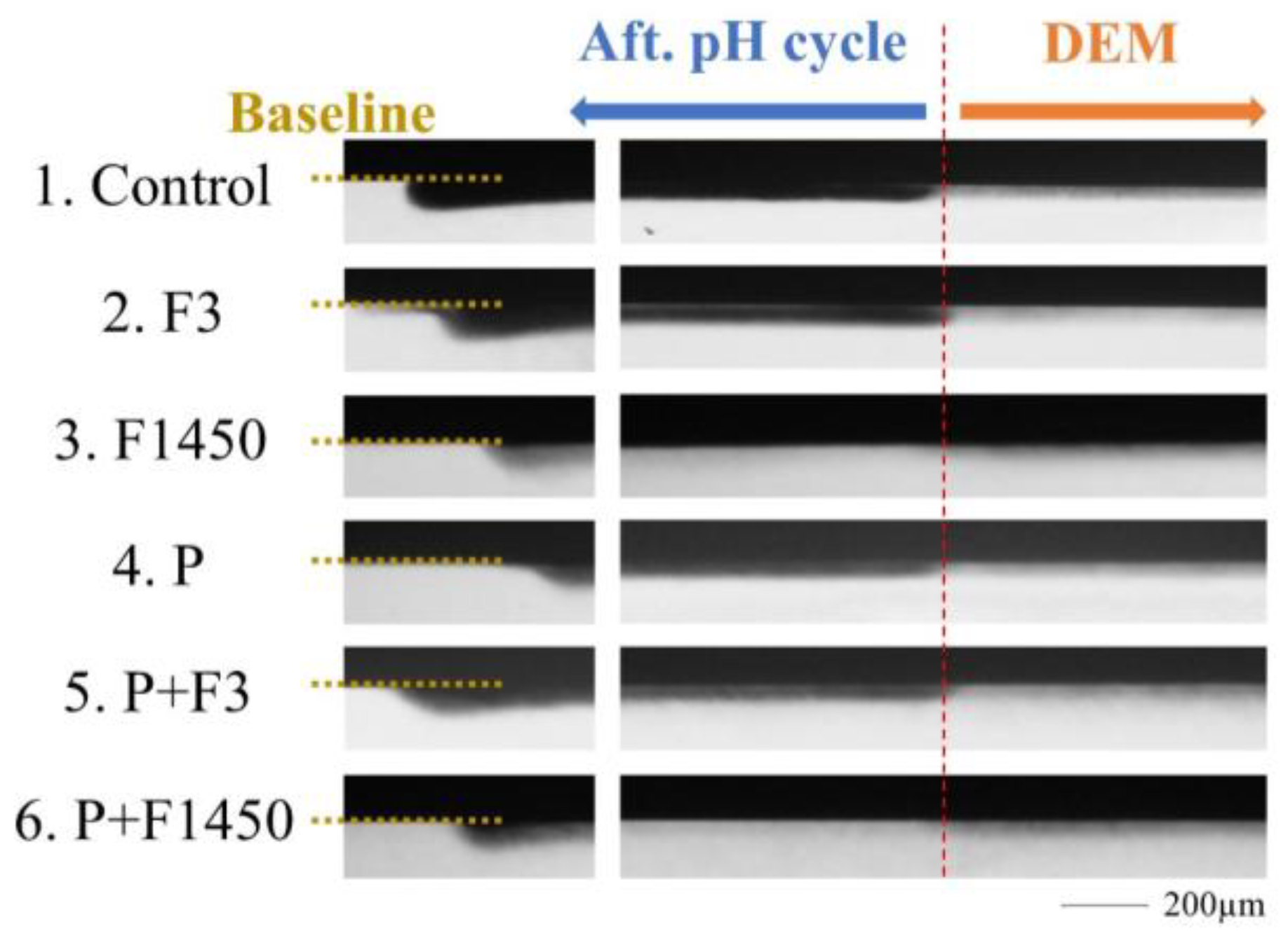
The TMR microradiographs of each group are summarized in Figure 2. All DEM areas represent increased radiolucency with a clear surface line. The results showed that the demineralization after 96 h generated a subsurface delimiting layer. Comparing each post-treatment area (aft. pH cycle area), F1450 and P + F1450 showed higher radiopacity than the other four groups. Figure 3 also shows that the aft. pH cycle and DEM mineral profiles were reversed in the F1450 and P + F1450 groups.
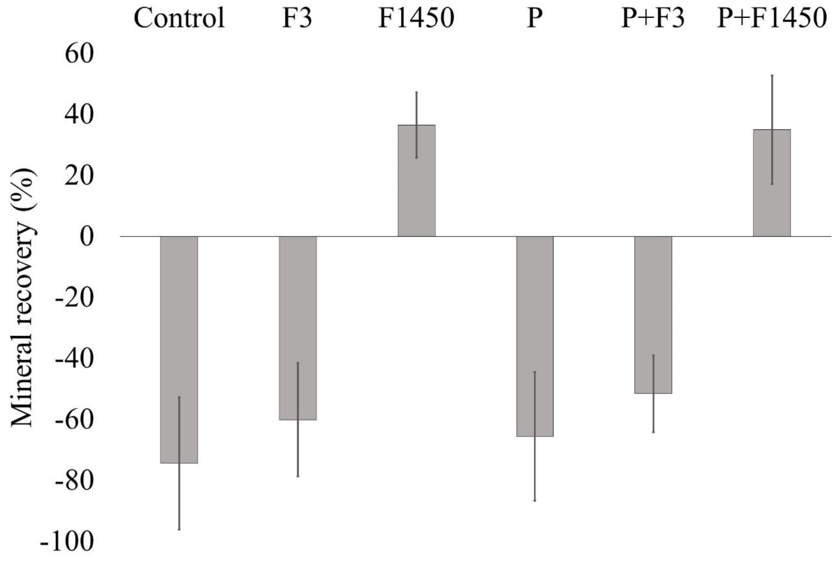
The mineral recovery rates of each group are reported in Table 2 and Figure 4. With the exception of the control group, all groups showed remineralization reactions in the aft. pH cycle area. Both F1450 and P + F1450 demonstrated significantly higher recovery rates compared with the other four groups (p < 0.001); P + F1450 had almost the same recovery rate as F1450, although it was not significantly different (p > 0.05); and P + F3 showed a tendency toward a higher rate than F3.
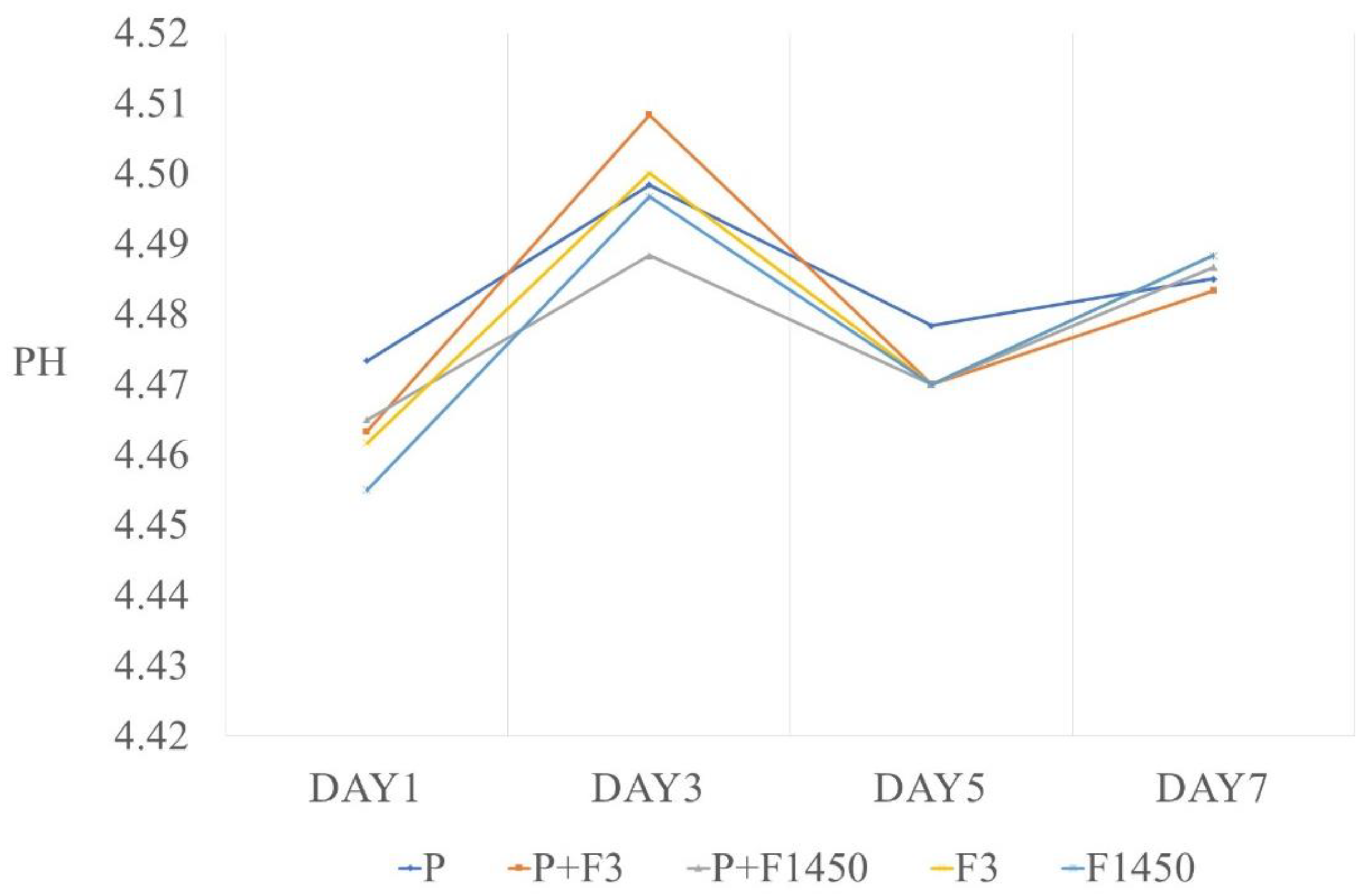
Figure 5 demonstrates the changes in the pH values of all groups except for the control group. The fluctuation range settled between 4.46 and 4.51. All groups exhibited a mild increase over time, but there was no significant difference among the six groups or the data points collected on days 1, 3, 5, and 7 (p > 0.05).

4. Discussion

Dental caries and dental erosion are common, slowly progressive diseases, which, at the serious stage, proceed to irreversible substantial defects over time. This may result in tooth extraction, which may ultimately affect a person’s general health. Therefore, it is important to prevent the onset of the disease or to suppress progression in the early stage. POs-Ca is a substance whose recalcification potential has been proven in previous research [24]. In 2013, the Japanese government validated the remineralization effect of POs-Ca gum and gave it approval as a food for specified health uses. Now, this gum is generally accepted as a dental hygiene product. In the context of these circumstances, the present study provides a new POs-Ca product for dental prevention. Here, we highlight the remineralization enhancement using POs-Ca dentifrices with different concentrations of fluoride.
The most notable finding of our study was that the groups with high fluoride concentrations achieved better mineral recoveries (%R). Both the POs-Ca-containing and non-POs-Ca-containing groups showed a significantly higher %R as the fluoride concentration increased. During the demineralization period in pH cycling, the pH dropped below 5.5, at which point hydroxyapatite (HA) was dissolved and fluorhydroxyapatite (FA) was precipitated at the surface simultaneously. FA crystallites are more difficult to dissolve than HA crystallites due to their high crystallinity [30], and with respect to the remineralization period in pH cycling (pH rises above 5.5), both HA and FA are precipitated at the surface [31]. Calcium fluoride can be generated when the fluoride concentration in the saliva is more than 1000 ppm [32]. This substance becomes temporary storage for fluoride, which gradually releases active ions. On the basis of the above investigations, it is evident that the higher the fluoride concentration, the more calcium fluoride is generated, and the higher the possibility of forming FA, the stronger acid resistance the tooth will gain.
Although the low fluoride concentration groups did not show significant mineral recovery, they demonstrated a tendency toward higher recovery than the groups without fluoride (control and only POs-Ca). A previous paper concluded that even with low concentrations of fluoride (<0.1 ppm) in the saliva interface, FA would be formed under slightly acidic conditions (pH down to 4.0) [9]. Another study ascertained that POs-Ca produced HA with a similar structure as sound enamel [33]. They also verified FA crystals being formed on the lesion after POs-Ca and fluoride at 0.5 or 1.0 ppm under the μXANE observation [34]. The lesions in our experiment were shallow, and only subsurface demineralization occurred. Previous in situ studies have shown that the net de-/remineralization balance can affect the initial lesion size [29]. We conclude that it is difficult to confirm a difference between low fluoride concentrations and the absence of fluoride.
According to all our results, POs-Ca seems to provide an auxiliary effect during the remineralization process. Compared with each low fluoride concentration group and the groups without fluoride, POs-Ca-containing groups tended to have a higher %R. The effective remineralization of subsurface lesions requires an agent to diffuse across the pellicle-covered enamel surface before entering the subsurface lesion area [22]. POs-Ca has unique potential, as it is soluble in the presence of phosphoric acid and fluorine ions even under non-acidic conditions (pH 6.5) [24]. Thus, the Pos-Ca solution can provide bioavailable calcium in the saliva without forming an insoluble salt, which allows the saliva to retain a stable supply of calcium or phosphate ions. Indeed, POs concentrations of 0.07–4% were shown to have a statistically equal remineralization effect as a 2 ppm NaF solution [35], while the %R did not increase at high fluoride concentrations. We believe that this can be explained by the fact that the remineralization effect was sufficiently strong at high fluoride concentrations to camouflage the potential of POs-Ca. Alternatively, the number of calcium fluoride-like crystals generated by high fluoride concentrations may inhibit calcium intake and disrupt the action of POs-Ca. A previous study indicated that treatment with high fluoride concentrations or daily fluoride addition markedly suppressed mineral deposition [36]. Another report proposed that the transformation to calcium fluoride during acidulated phosphate fluoride application (a topical fluoride treatment) may have destroyed the porous structure of the enamel surface, making it less permeable to calcium and phosphate ions [37]. Clinically, high fluoride concentrations should be avoided for children due to the risk of dental fluorosis. Therefore, for the sake of long-term caries prevention, a low fluoride concentration is considered better than a high concentration, and POs-Ca may enhance the remineralization effect of low concentrations.
Groups 1, 2, 4, and 5 showed a negative %R (Figure 4), which may have been caused by the start order of pH cycling. pH-cycling models are designed to simulate the dynamics of mineral losses and gains involved in caries formation [38] and can be used to approximate the pH balance during a daily diet. We strived to create a condition that replicated daily life, which put the specimens into a demineralization solution at the start as a “meal”. It is possible that the demineralization solution diminished the action of the dentifrice on the subsurface layer first, which may have made it difficult for the low fluoride concentration groups and groups with no fluoride to gain mineral recovery.
The null hypothesis was partially rejected. The dentifrice containing POs-Ca is applicable to prevent dental caries or erosion. Our findings highlight the huge potential of POs-Ca dentifrice with low fluoride concentrations. However, our research is limited to the measurement of the fluoride concentration, and further studies are needed to evaluate the treatment time or periods between high and low fluoride concentrations under POs-Ca.

5. Conclusions

High-concentration fluoride dentifrices with/without POs-Ca showed the highest remineralization effect. In the low-concentration fluoride groups, POs-Ca containing dentifrice tended to lead to a higher remineralization effect. These results can be applied to caries prevention in children.

Author Contributions

Conceptualization, G.I. and X.C.; methodology, G.I.; software, X.C.; validation, G.I., X.C. and Y.S.; formal analysis, X.C.; investigation, G.I. and X.C.; resources, G.I. and X.C.; data curation, X.C.; writing—original draft preparation, G.I.; writing—review and editing, X.C. and Y.S.; visualization, X.C.; supervision, Y.S.; project administration, G.I. All authors have read and agreed to the published version of the manuscript.

Funding

This research received no external funding.

Data Availability Statement

Not applicable.

Acknowledgments

We wish to thank M Tanaka from the Institute of Health Sciences, Ezaki Glico Co. Ltd., for offering the experimental agents. The authors declare no potential conflicts of interest with respect to the authorship and/or publication of this article.

Conflicts of Interest

The authors declare no conflict of interest.

References

  1. Tanji, F.; Komiyama, T.; Ohi, T.; Hattori, Y.; Watanabe, M.; Lu, Y.; Tsuji, I. The Association between Number of Remaining Teeth and Maintenance of Successful Aging in Japanese Older People: A 9-Year Longitudinal Study. Tohoku J. Exp. Med. 2020, 252, 245–252. [Google Scholar] [CrossRef] [PubMed]
  2. Dibello, V.; Lobbezoo, F.; Lozupone, M.; Sardone, R.; Ballini, A.; Berardino, G.; Mollica, A.; Coe-lho-Júnior, H.J.; de Pergola, G.; Stallone, R.; et al. Oral Frailty Indicators to Target Major Adverse Health-Related Outcomes in Older Age: A Systematic Review; Springer: Berlin/Heidelberg, Germany, 2022. [Google Scholar] [CrossRef]
  3. Singh, K.A.; Brennan, D.S. Chewing disability in older adults attributable to tooth loss and other oral conditions. Gerodontology 2012, 29, 106–110. [Google Scholar] [CrossRef] [PubMed]
  4. Ishikawa, S.; Konta, T.; Susa, S.; Ishizawa, K.; Togashi, H.; Ueno, Y.; Yamashita, H.; Kayama, T.; Iino, M. Association between presence of 20 or more natural teeth and all-cause, cancer-related, and cardiovascular disease-related mortality: Yamagata (Takahata) prospective observational study. BMC Oral Health 2020, 20, 353. [Google Scholar] [CrossRef] [PubMed]
  5. Deari, S.; Wegehaupt, F.J.; Tauböck, T.T.; Attin, T. Influence of Different Pretreatments on the Microtensile Bond Strength to Eroded Dentin. J. Adhes. Dent. 2017, 19, 147–155. [Google Scholar]
  6. Wegehaupt, F.J.; Tauböck, T.T.; Attin, T. Durability of the Anti-Erosive Effect of Surfaces Sealants under Erosive Abrasive Conditions. Acta Odontol. Scand. 2013, 71, 1188–1194. [Google Scholar] [CrossRef] [PubMed] [Green Version]
  7. Attin, T.; Becker, K.; Wiegand, A.; Tauböck, T.T.; Wegehaupt, F.J. Impact of Laminar Flow Velocity of Different Acids on Enamel Calcium Loss. Clin. Oral Investig. 2013, 17, 595–600. [Google Scholar] [CrossRef] [Green Version]
  8. Yamato, M.; Matsuyama, S.; Murakami, Y.; Aida, J.; Lu, Y.; Sugawara, Y.; Tsuji, I. Association between the number of remaining teeth and disability-free life expectancy, and the impact of oral self-care in older Japanese adults: A prospective cohort study. BMC Geriatr. 2022, 22, 820. [Google Scholar] [CrossRef]
  9. Ekstrand, K.R. High Fluoride Dentifrices for Elderly and Vulnerable Adults: Does It Work and if So, Then Why? Caries Res. 2016, 50, 15–21. [Google Scholar] [CrossRef] [Green Version]
  10. Marinho, V.; Higgins, J.; Logan, S.; Sheiham, A. Fluoride toothpastes for preventing dental caries in children and adolescents. Cochrane Database Syst. Rev. 2003, 2003, CD002278. [Google Scholar] [CrossRef]
  11. Jesen, M.K.; Kohout, F. The effect of a fluoridated dentifrice on root and coronal caries in an older adult population. J. Am. Dent. Assoc. 1988, 117, 829–832. [Google Scholar] [CrossRef]
  12. Gluzman, R.; Katz, R.V.; Frey, B.J.; McGowan, R. Prevention of Root Caries: A Literature Review of Primary and Secondary Preventive Agents. Spec. Care Dent. 2013, 33, 133–140. [Google Scholar] [CrossRef] [PubMed] [Green Version]
  13. Pretty, I.R. High Fluoride Concentration Toothpastes for Children and Adolescents. Caries Res. 2016, 50, 9–14. [Google Scholar] [CrossRef] [PubMed]
  14. Walsh, T.; Worthington, H.V.; Glenny, A.M.; Marinho, V.C.C.; Jeroncic, A. Fluoride toothpastes of different concentrations for preventing dental caries. Cochrane Database Syst. Rev. 2019, 3, CD007868. [Google Scholar] [CrossRef]
  15. Featherstone, J.D. Prevention and reversal of dental caries: Role of low level fluoride. Community Dent. Oral Epidemiol. 1999, 27, 31–40. [Google Scholar] [CrossRef] [PubMed]
  16. Shellies, R.; Duckworth, R. Studies on the cariostatic mechanisms of fluoride. Int. Dent. J. 1994, 44, 263–273. [Google Scholar]
  17. Reddy, D.; Selvan, A.; Paul, S.T.; Azher, U. Antimicrobial Efficacy of Commercially Available Low-Fluoride and Fluoride-Free Dentifrices for Children. Int. J. Clin. Pediatr. Dent. 2021, 14, 183–186. [Google Scholar] [CrossRef] [PubMed]
  18. Kantrong, N.; Khongkhaphet, K.; Sitornsud, N.; Lo-Apirukkul, P.; Phanprom, W.; Rojviriya, C.; Amonpat-taratkit, P.; Ariyakriangkai, W. Synchrotron Radiation Analysis of Root Dentin: The Roles of Fluoride and Calcium Ions in Hydroxyapatite Remineralization. J. Synchrotron Rad. 2022, 29, 496–504. [Google Scholar] [CrossRef]
  19. da Silva Spinola, M.; Tenuta, L.M.A. Calcium pretreatment enhances fluoride reactivity with enamel and dentine. Arch. Oral Biol. 2022, 134, 105338. [Google Scholar] [CrossRef]
  20. Cochrane, N.J.; Cai, F.; Huq, N.L.; Burrow, M.F.; Reynold, E.C. New approaches to enhanced remineralization of tooth enamel. J. Dent. Res. 2010, 89, 1187–1197. [Google Scholar] [CrossRef]
  21. Kitasasko, Y.; Tanaka, M.; Sadr, A.; Hamba, H.; Ikeda, M.; Tagami, J. Effects of a chewing gum containing phosphoryl oligosaccharides of calcium (POs-Ca) and fluoride on remineralization and crystallization of enamel subsurface lesions in situ. J. Dent. 2011, 39, 771–779. [Google Scholar] [CrossRef]
  22. Reynolds, E.C.; Cai, F.; Shen, P.; Walker, G.D. Retention in plaque and remineralization of enamel lesions by various forms of calcium in a mouthrinse or sugar-free chewing gum. J. Dent. Res. 2003, 82, 206–211. [Google Scholar] [CrossRef] [PubMed]
  23. Shen, P.; Manton, D.J.; Cochrane, N.J.; Walker, G.D.; Yuan, Y.; Reynolds, C.; Reynolds, E.C. Effect of added calcium phosphate on enamel remineralization by fluoride in a randomized controlled in situ trial. J. Dent. 2011, 39, 518–525. [Google Scholar] [CrossRef] [PubMed]
  24. Tanaka, T.; Kobayashi, T.; Takii, H.; Kamasaka, H.; Ohta, N.; Matsuo, T.; Yagi, N.; Kuriki, T. Optimization of calcium concentration of saliva with phosphoryl oligosaccharides of calcium (POs-Ca) for enamel remineralization in vitro. Arch. Oral Biol. 2013, 58, 174–180. [Google Scholar] [CrossRef] [PubMed]
  25. Kamasaka, H.; Imai, S.; Nishimura, T.; Kuriki, T.; Nishizawa, T. Effect of phosphoryl oligosaccharides from potato starch on acid fermentation by mutans streptococci. J. Dent. Health 2002, 52, 66–71. [Google Scholar]
  26. Inaba, D.; Minami, K.; Kamasaka, H.; Yonemitsu, M. Remineralization of enamel and dentin by a chewing gum containing Phosphryl-Oligosaccharide calcium (POs-Ca) in situ. Dent. J. Iwate Med. Univ. 2002, 27, 203–209. [Google Scholar]
  27. To-o, K.; Kamasaka, K.; Nishimura, T.; Kuriki, T.; Saeki, S.; Nakabou, Y. Bioavailability of calcium-bound phosphoryl oligosaccharides in rats. J. Appl. Glicosci. 2002, 49, 159–165. [Google Scholar] [CrossRef]
  28. ten Cate, J.M.; Dundon, K.A.; Vernon, P.G.; Damato, F.A.; Huntington, E.; Exterkate, R.A.M.; Wefel, J.S.; Jordan, T.; Stephen, K.W.; Roberts, A.J. Preparation and measurement of artificial enamel lesions, a four-laboratory ring test. Caries Res. 1996, 30, 400–407. [Google Scholar] [CrossRef]
  29. ten Cate, J.M.; Exterkate, R.A.M.; Buijs, M.J. The relative efficacy of fluoride toothpastes assessed with pH cycling. Caries Res. 2006, 40, 136–141. [Google Scholar] [CrossRef]
  30. Pajor, K.; Pajchel, L.; Kolmas, J. Hydroxyapatite and Fluorapatite in Conservative Dentistry and Oral Im-plantology-a Review. Materials 2019, 12, 2683. [Google Scholar] [CrossRef] [Green Version]
  31. Twentman, S.; Ekstrand, K.R. Caries management by influencing mineralization. In Caries Management—Science and Clinical Practice, 1st ed.; Meyer-Lueckel, H., Paris, S., Ekstrand, K.R., Eds.; Thieme: Stuttgart, Germany, 2013; pp. 177–190. [Google Scholar]
  32. Bruun, C.; Givskov, H. Formation of CaF2 on sound enamel and caries like lesions after different forms of fluoride application in vitro. Caries Res. 1991, 25, 96–100. [Google Scholar] [CrossRef]
  33. Tanaka, T.; Yagi, N.; Ohta, T.; Matsuo, Y.; Terada, H.; Kamasaka, K.; To-o, K.; Kometani, T.; Kuriki, T. Evaluation of the distribution and orientation of remineralized enamel crystallites in subsurface lesions by X-ray diffraction. Caries Res. 2010, 44, 253–259. [Google Scholar] [CrossRef] [PubMed]
  34. Tanaka, T.; Kobayashi, T.; Tamenori, Y.; Sakanaka, A.; Kuriki, T.; Amano, A. Phosphoryl oligosaccha-rides of calcium enhance mineral availability and fluorapatite formation. Arch. Oral Biol. 2019, 10, 135–141. [Google Scholar] [CrossRef]
  35. Inaba, D.; Minami, K.; Kamasaka, H.; Yonemitsu, M. Effects of phosphoryl-oligosaccharides(POs) on remineralization of enamel lesions in vitro. Dent. J. Iwate Med. Univ. 2002, 27, 197–202. [Google Scholar]
  36. ten Cate, J.M.; Jongebloed, W.L.; Arends, J. Remineralization of artificial enamel lesions in vitro—IV. Influence of fluorides and diphosphonates on short-and long-term remineralization. Caries Res. 1981, 15, 60–69. [Google Scholar] [CrossRef] [PubMed]
  37. Boddé, H.E.; Koops, P.G.; Arends, J. Effect of an APF pretreatment on in vitro remineralization of initial enamel lesions. Caries Res. 1984, 18, 344–347. [Google Scholar] [CrossRef]
  38. Buzalaf, M.A.R.; Hannas, A.R.; Magalhaes, A.C.; Rios, D.; Honorio, H.M.; Delbem, A.C.B. PH-cycling models for in vitro evaluation of the efficacy of fluoridated dentifrices for caries control: Strengths and limitations. J. Appl. Oral Sci. 2010, 18, 316–334. [Google Scholar] [CrossRef]
Figure 1. Illustration of specimen preparation. (a) Three-millimeter enamel samples obtained from bovine incisor teeth. (b) Embedding (epoxy resin) and making a window (5 × 10 mm) using nil varnish. Divided into two areas (made “sound”). (c) Soaking demineralization solution for 96 h at 37 °C. (d) Application of nail varnish to 1/3 of the window (made “DEM”). (e) Performing pH cycling by 3 times/day for 8 days. Measuring the pH of demineralized solution. (f) TMR analysis.
Crystals 13 00206 g001
Figure 2. Transverse microradiographs of the DEM and aft. pH cycle zones including the treated enamel surface of the six groups.
Crystals 13 00206 g002
Figure 3. Relative mineral density of DEM and aft. pH cycle for the six groups. The mineral density was normalized to 87% (v/v) in sound enamel by Image J quantifying.
Crystals 13 00206 g003
Figure 4. Mineral recovery rate of the six groups.
Crystals 13 00206 g004
Figure 5. pH value of the demineralization solution after pH cycling. The x-axis indicates the elapsed time in days of the experiment.
Crystals 13 00206 g005
Table 1. Composition of the toothpaste used in the study.
GroupF in Formulation (ppm)F in Solution (ppm)Ca from POs-Ca in Formulation (mM)Ca from POs-Ca in Solution (mM)
1. Control0000
2. F33100
3. F1450145048000
4. P0013.24.3
5. P + F33113.24.3
6. P + F1450145048013.24.3
Table 2. Mineral recovery of enamel subsurface lesions assessed by TMR (mean ± SD). Values marked by the same superscript letters denote no significant differences. MLDEM: Mineral loss of demineralized enamel (vol% µm), MLREM: Mineral loss of remineralized enamel after pH cycling (vol% µm), %R: Mineral recovery ((MLDEM − MLREM/MLDEM)/100).
GroupMLDEMMLREMMLDEM − MLREM%R
1. Control19.56 ± 4.4733.67 ± 6.80−14.11 ± 3.43−74.39 ± 21.68 a
2. F320.75 ± 2.9032.85 ± 3.51−12. 10 ± 3.37−60.09 ± 18.64 a
3. F145021.95 ± 4.8013.71 ± 2.788.24 ± 3.6936.54 ± 10.76 b
4. P18.95 ± 3.2831.03 ± 4.99−12.07 ± 4.06−65.56 ± 21.05 a
5. P + F322.36 ± 3.8533.62 ± 5.17−11.27 ± 2.76−51.55 ± 12.59 a
6. P + F145018.26 ± 6.4311.80 ± 5.246.46 ± 3.6934.93 ± 17.81 b
Disclaimer/Publisher’s Note: The statements, opinions and data contained in all publications are solely those of the individual author(s) and contributor(s) and not of MDPI and/or the editor(s). MDPI and/or the editor(s) disclaim responsibility for any injury to people or property resulting from any ideas, methods, instructions or products referred to in the content.