Article

Monte Carlo Simulation for Investigating the Sintering Temperatures Effects on Radiation Shielding Performances of Lead-Free ABO3 Perovskite Ceramic

1
Department of Nuclear Medicine Research, Institute for Research and Medical Consultations (IRMC), Imam Abdulrahman bin Faisal University (IAU), P.O. Box 1982, Dammam 31441, Saudi Arabia
2
Department of Nuclear Power Plants and Renewable Energy, Ural Federal University, St. Mira, 19, 620002 Yekaterinburg, Russia
3
Department of Biophysics, Institute for Research and Medical Consultations (IRMC), Imam Abdulrahman bin Faisal University (IAU), P.O. Box 1982, Dammam 31441, Saudi Arabia
4
Department of Physics, Faculty of Science, Isra University, Amman 11622, Jordan
5
College of Engineering and Technology, American University of the Middle East, Eqaila 54200, Kuwait
6
University of Tunis El Manar, Tunis 2092, Tunisia
*
Author to whom correspondence should be addressed.
Crystals 2023, 13(2), 230; https://doi.org/10.3390/cryst13020230
Submission received: 8 January 2023 / Revised: 21 January 2023 / Accepted: 23 January 2023 / Published: 28 January 2023

Abstract

:
In this study, a series of barium titanate ceramics of the chemical composition BaTiO3 was prepared. The solid-state reaction route was adopted to synthesize the ceramic samples at various sintering temperatures of 1100–1300 °C. X-ray diffraction and FTIR spectroscopy were utilized to examine the structure of the fabricated ceramics. The UV–Vis–reflectance data were recorded to guess the optical bandgap energy of the synthesized ceramics. The ability of the synthesized ceramics to attenuate ionizing radiation was qualified using a Monte Carlo simulation (MCNP code) in the γ-energy interval ranging between 59 keV and 1408 keV. Shielding parameters, including LAC, TF, and RPE, were evaluated. The XRD and FTIR analyses showed the formation of a tetragonal BaTiO3 perovskite structure with the Pmmm space group. The crystallite size and the relative density increased, whereas the porosity decreased, with increasing sintering temperatures. Optical bandgap energy (Eg) values decreased as the sintering temperatures increased. The radiation shielding results depicted that raising the sintering temperature between 1100 °C and 1300 °C resulted in a slight increase in the µ values by a factor of ≈8 %. The mentioned increase in the µ values caused a reduction in the Δeq and Δ0.5, and TF values for the fabricated BaTiO3 ceramic samples, while the RPE values increased with increasing sintering temperatures between 1100 °C and 1300 °C.

1. Introduction

Over the years, nuclear technologies have found their track in diverse domains, especially in medical science, such as imaging nuclear fuels, treating cancer diseases, producing thermal energy, etc. [1,2,3,4]. Nuclear technology necessitates the radionuclides’ emission, such as X-rays, neutrons, and gamma rays. These radionuclides induce a grave danger to all living organisms, especially humans. It has a miraculous ability to destroy living cells, and therefore, care must be taken to protect against its destructive effect [5]. The protection of people, structures, and equipment from harmful radiation impacts is of interest to researchers working in the field of nuclear engineering. To minimize the radiation effect, physicians, technicians, and patients must track some key rules [6]. Primarily, the period of contact must be maintained to the lowest possible, and the sources of radiation must be maintained as far away from persons; in addition, protective material must be utilized, specifically in radiation absorption situations. Therefore, there is a serious requirement for “protection” [7]. The choice of matter for radiation protection mainly depends on the source of the emissions, their type, and the weight of the material [8]. Several materials have been searched for protection from ionizing radiation, including lead, polyethylene, iron, graphite, and concrete [9]. For many years, lead, with its elevated atomic number and density, has been a prime radiation shield, but it has noxious impacts on humans. Therefore, scientists have concentrated on finding new protective materials that are non-toxic and cheap. A number of studies demonstrated that dense and heavy concretes have the potential to considerably enhance protection performance [9,10,11]. Glass is also one of the new and suitable radiation-protective materials. Glass was employed for diverse applications in actuality, such as optoelectronics, actuators, transparent electrodes, optical materials, lasers, phototransistors, diodes, amplifiers, and so on. Many researchers proposed various glass materials as radiation-shielding systems [12,13,14,15]. For example, Acikgoz et al. [16] reported the radiation protection of borate glasses doped with cerium and erbium oxides. The results of radiation shielding showed good performance for the glasses doped with erbium oxide. A.M. Madbouly et al. [17] evaluated the radiation-protecting performance of bismuth boron phosphate glasses. S. Alzahrani et al. made a comparative study on the radiation-protecting properties of tellurite and borate glasses, and their results showed that tellurite glasses offer higher gamma-ray shielding performance than borate glass [18].
Recently, much interest has been paid to ceramic materials. From the point of view of shielding, these materials provide good radiation-protective properties. Ceramics are characterized by their stability, high resistance to oxidation, low thermal expansion, hardness, toughness, environmental friendliness, and tunable physical and chemical properties [19,20,21]. Efficient shielding materials should possess important characteristics, such as durability, high density, high atomic number, and non-toxicity. Generally, density has a deep effect on the structural elements, leading to a significant minimization in the thickness of the protective elements while preserving the shielding performances. Dense materials can be achieved by adopting special preparation methods, changing the synthesis conditions (temperature, pressure, atmosphere, etc.), using additives or dopants in the ceramic material matrix, and so on [22,23,24]. For example, many studies have engrossed on the shielding performance of several types of ceramic materials, such as perovskites-based ceramics [25,26,27], peridot, ruby, silicon nitride, and magnesium silicate ceramic samples [28]. Other works have been done to study the impacts of additives or dopants on the physical features of some ceramics in order to further enhance their shielding performances [29,30,31]. It is agreed that a certain quantity of additives can lead to the production of ceramic materials with high density and good protective properties.
In this work, we propose to prepare ABO3 (where A = Ba, B = Ti) perovskites-based ceramic using the simple solid-state reaction route (SSR). The proposed ceramics were sintered at different sintering temperatures. Emphasis was placed on the influence of the sintering temperatures on the radiation-protecting performances of such material.

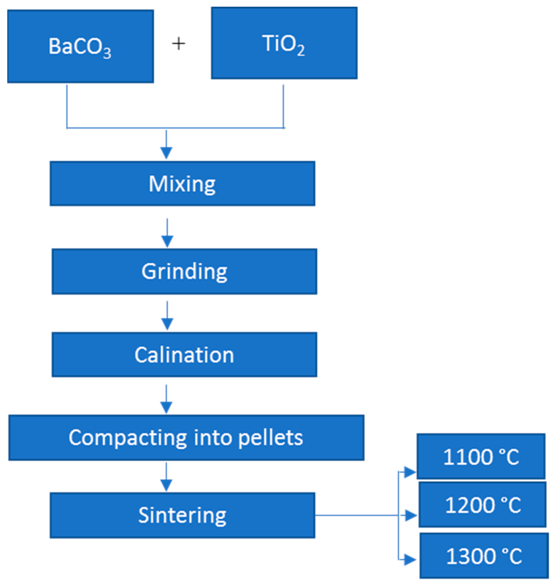
2. Experimental Details

2.1. Chemicals and Method

BaTiO3 ceramics were fabricated via the SSR route. BaCO3 (AR, >99%) and TiO2 (AR, 99.9%), supplied by Sigma Aldrich (Darmstadt, Germany), were exploited as the initial reagents. Proper quantities of the powders were weighed and mixed thoroughly with a mortar and pestle, and then further ground using the ball milling technique to get a homogenous mixture. The resultant was sited in crucible alumina for calcination at 1100 °C for 2 h. After that, the powders were ground again by adding the proper amount of binder (PVA; polyvinyl alcohol) and then compacted into pellets. Finally, the obtained pellets were sintered at temperatures of 1100 °C, 1200 °C, and 1300 °C in a furnace with a rate of heating of 4 °C min−1 and left to cool to room temperature. Figure 1 shows a schematic illustration for the synthesis protocol of the ceramic samples.

2.2. Ceramics Characterizations

The ceramics fabricated using SSR were examined using Fourier transform infrared spectroscopy (FTIR), UV-vis spectroscopy, and X-ray powder diffraction. An X-ray diffractometer (Rigaku Benchtop Miniflex; Tokyo, Japan; 2θ range: 20°–70°) with CuKα radiation and a FT-IR spectrometer (Bruker alpha-II; USA; wavenumber range: 2000–400 cm−1) were adopted to assess the structural traits of the ceramics. The optical bandgap energy of the different ceramics was evaluated with a JASCO V-780 UV–vis spectrometer (JASCO, Easton, PA, USA).

2.3. Radiation Protection Qualifications

The γ-ray shielding properties of the fabricated ceramic samples were qualified using the Monte Carlo simulation code (MCNP-5) in a γ-photon energy region (Eγ, MeV) ranging from 59 keV to 1408 keV [32]. The MCNP simulation code uses the ENDF/B-VI.8 nuclear library code to elicit the cross-section of the γ-photon in the mentioned energy interval. Furthermore, the input file required for running the simulation process should accurately describe all the information about the geometry and the simulation components, such as source, samples, and detector. The present MCNP-5 input file contains the geometry composed of an outer lead cylinder (out protection cover), and the height of the protection cylinder is 35 cm, and its thickness is 5 cm. This outer protection cylinder of lead contains many important components, such as a source, collimators, ceramic samples, and detectors. All required information about the radioactive source was introduced in the SDEF card, where the radioactive source emits photons along the Z directions. The emitted photon energies varied between 59 keV and 1408 keV. The source also has a dimension of 1 cm in diameter and 0.5 cm in height, and it is placed in the center of the outer cylindrical shielding. Moreover, the material card contains all information about the materials used in the geometry, such as elemental chemical composition and density. In order to control the emission of gamma photons, the cut-off card was used in the current input file, where the cut-off card is set to kill the photons after the emission of 106 historical. The detector is assumed to be an F4 tally to qualify the average track length (ATL) of the γ-photons inside the fabricated materials [33,34].
The ATL was transferred to the other shielding properties according to Equations (1)–(4) [35,36]:
μ   ( cm 1 ) = I x ln ( I o I t )
The thickness required to absorb half of the applied γ- photons is known as the half-value thickness (Δ0.5, cm). It is reversely proportional to the µ value as the following:
Δ 0.5   ( cm ) = ln ( 2 ) μ  
The transmission factor (TF, %) is the ratio of the transmitted number of photons (It) to the total number of emitted photons (Io), while the radiation-protection efficiency (RPE, %) represents the absorbed number of photons (Ia) to the total number of emitted photons (Io):
T F   ( % ) = I t I o × 100
R P E   ( % ) = I a I o × 100  

3. Results and Discussion

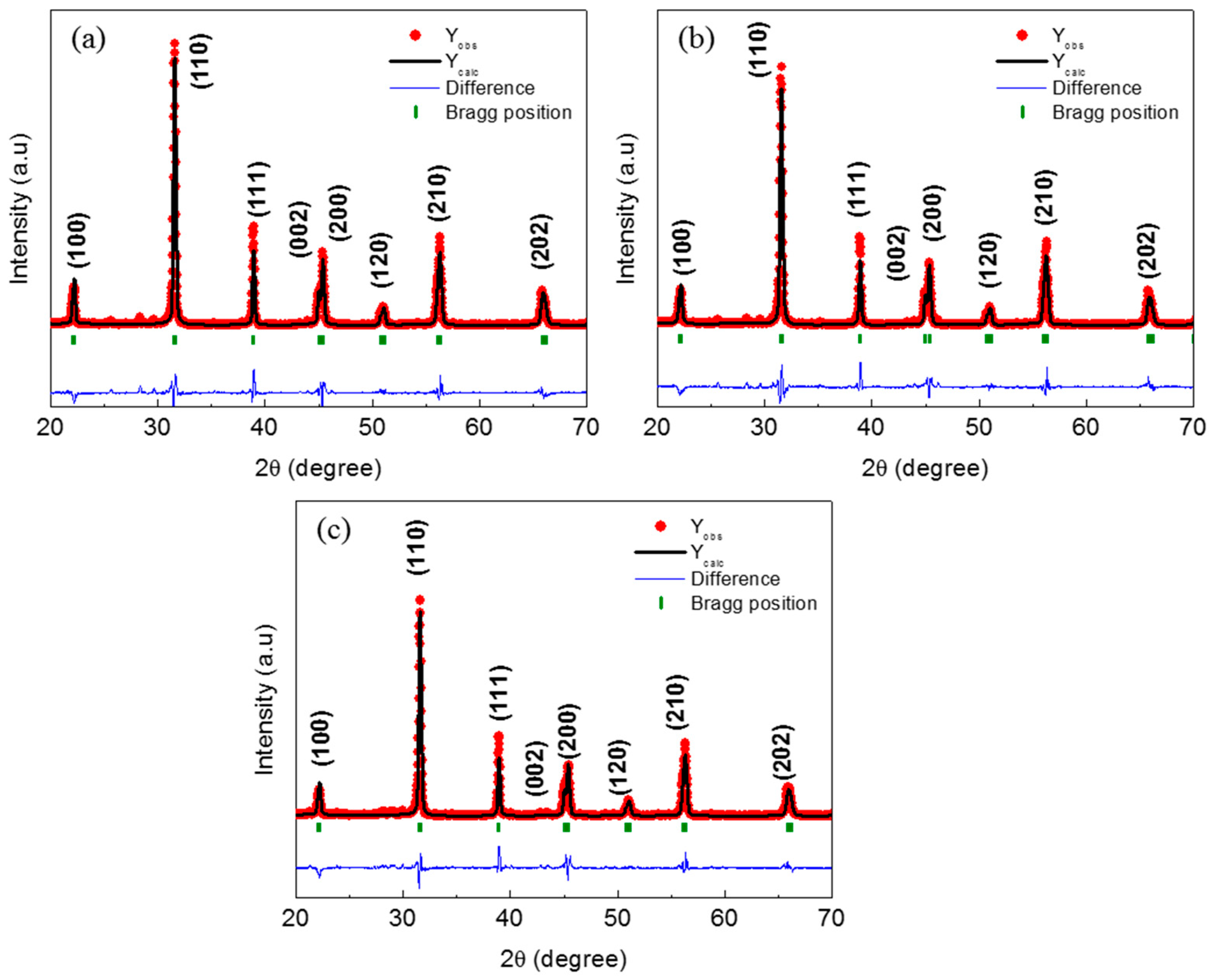
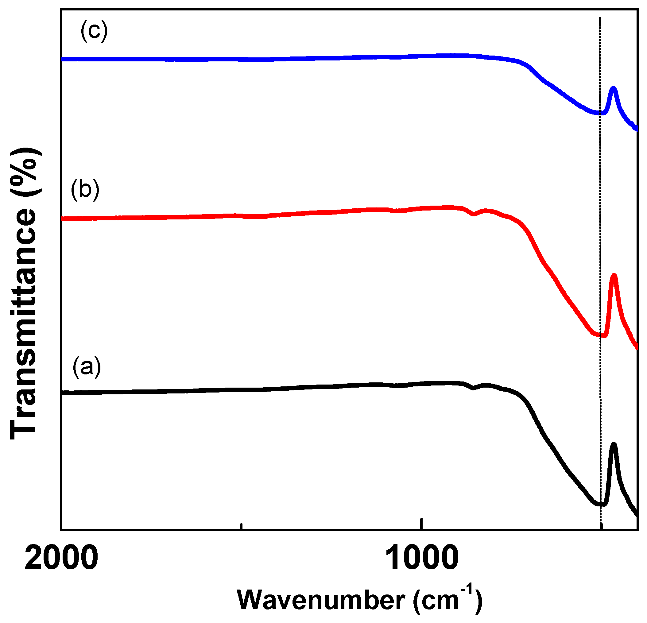
3.1. Structural Analyses

Figure 2 depicts the XRD patterns of the BaTiO3 ceramic samples sintered at different temperatures. For the first look, it is noticed that all the peaks are indexed to a tetragonal structure with space group P4mm [37].
The tetragonal structure is well identified by the two distinct split (002)/(200) peaks at 2θ~45°. This splitting was detected for all the prepared samples. This indicates that there is no tetragonal to cubic or other phase transition that occurred when increasing the temperatures. Our results are consistent with previous reports studying the effect of sintering temperature on the structural properties of BaTiO3-based ceramics. For instance, Ruizhao Liu et al. showed that a BaTiO3 ceramic sample sintered at 1180 °C displayed a tetragonal structure [38]. A. Rani et al. [39] prepared a BaTiO3 sample via a solid-state reaction route at various sintering temperatures between 1200 °C and 1350 °C. Their XRD analysis revealed a perovskite structure with a tetragonal phase except for the samples sintered at 1350 °C where both a tetragonal and a hexagonal phase were observed in this case. Besides, all the reflections are sharp, indicating that the prepared ceramics are well-crystallized. The lattice parameters were refined using the Rietveld method. The variations of the lattice parameters with the sintering temperatures are reported in Figure 3a. The lattice parameters are a = b = 3.9956 Å and c = 4.0262 Å, a = b = 3.9977Å and c = 4.0290 Å, and a = b = 3.9951 Å and c = 4.0242 Å for the BaTiO3 ceramics sintered at 1100 °C, 1200 °C, and 1300 °C, respectively. The lattice constants (a and c) show a slight variation with the change in the sintering temperatures. This variation reflects a slight expansion in the unit cell volume V for the BaTiO3 sintered at 1200 °C as obviously observed in Figure 3b. The variation of the c/a ratio is dependent on the sintering temperature. The c/a ratio as a function of sintering temperatures is also shown in Figure 3b. The tetragonality of the BaTiO3 sintered at 1100 °C is 1.0076. The increase in temperatures results in a rise in the tetragonality of the structure (c/a = 1.0078) for the ceramic prepared at 1200 °C, then a decrease (c/a = 1.0072) with a further increase of the sintering temperature. The upward then downward trend of the tetragonality degree (c/a) observed in Figure 3b signposts that the temperature of 1200 °C is optimal for stabilizing and maintaining the tetragonal structure of the BaTiO3 ceramic. The influence of the sintering temperatures on the crystallite size (Cs) was determined with Scherrer's equation ( C s = k   λ   β Cos θ ), where β is the full width at half maximum, and k is the shape factor equal approximately to unity [40]. The calculated Cs values using this equation are 33.25 nm, 34.51 nm, and 41.06 nm corresponding to 1100 °C, 1200 °C, and 1300 °C, respectively. The sintering temperatures apparently have an obvious influence on the crystallite/grain size. Similar results were reported previously in ref [41]. The determination of the density and porosity values of the material is important for the study of its radiation-shielding performance. It is desired to increase the value of the density or decrease the porosity in order to be able to reach the highest shielding capacity of the material. Therefore, the porosity (Φ, %) and the relative density (RD, %) of the BaTiO3 ceramics are investigated in this study. The relative density of the ceramic increases from 87.30%, for the ceramic sintered at 1100 °C, to 95.27% and 94.57% for the ceramics prepared at 1200 °C and 1300 °C, respectively. Similar results were obtained by W. Li et al. for BaTiO3 samples prepared with the sol-gel method [41]. In our case, the ceramic sintered at 1200 °C displays the maximum relative density of 95.27% and the minimum porosity Φ of 4.72%. The enhancement in the RD for the ceramic sintered at 1200 °C can be accredited to the growth in grain size and a reduction in porosity [42].
The FTIR results of the ceramics are presented in Figure 4. The FTIR spectra of all prepared ceramics are not complex. The absorption band at wavenumbers smaller than 800 cm−1 is associated with BaTiO3 [43]. Indeed, a strong band at ~490 cm–1 is owing to metal–oxygen bonds [20] and is resulting from the Ti-O-Ti and Ti-O bonds. It is important to note that the Ti-O vibration bond is complex. Indeed, there exist other bands in addition to those detected in Figure 4; however, our measurement did not encompass their regions [44]. For example, an absorption band at low frequency (<400 cm−1) is due to TiO6 cation vibration. Another band very close to 400 cm−1 is due to the Ti-OII bending normal vibration. Our obtained FTIR results support the XRD analysis and prove the successful formation of the BaTiO3 ceramics.

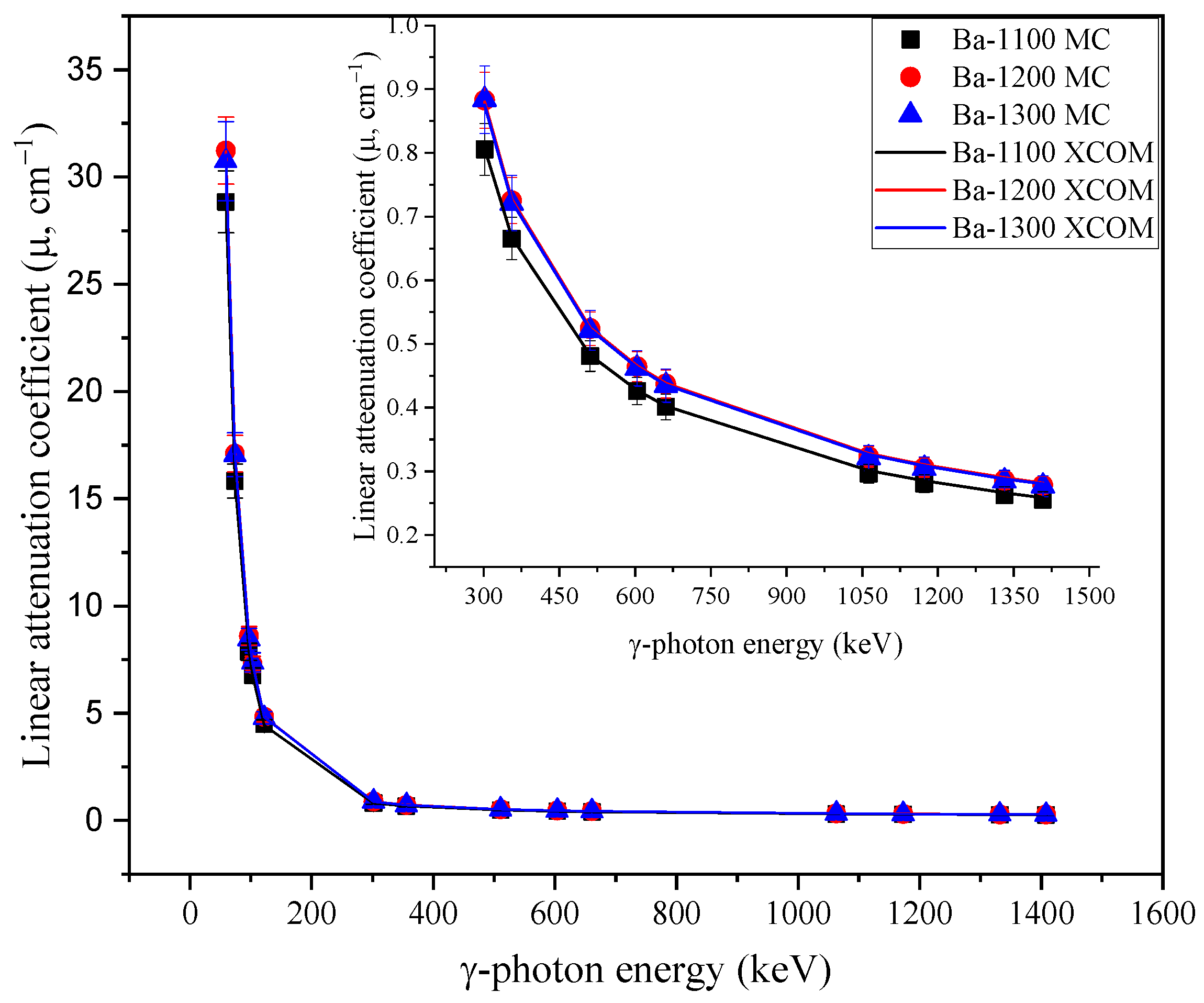
3.2. Radiation-protecting Parameters

Figure 5 depicts the influence of emitted γ-photon energy on the fabricated ceramic samples’ µ values. Among the considered γ-photon energy intervals, the maximum µ values are achieved at low γ-energy (i.e., 59 keV < Eγ < 122 keV) where the photoelectric interaction (PE) is predominant. In the mentioned Eγ interval, the µ values suffer a high decrease by a factor of approximately 84.4 % for all fabricated samples when the Eγ increased from 59 keV to 122 keV. In this regard, the µ values dropped from 28.837 to 4.478 cm−1 with an average of 12.76 cm−1 for sample Ba-1100, decreased from 31.227 to 4.831 cm−1 with an average of 13.81 cm−1 for sample Ba-1200, and decreased from 30.7844 to 4.783 cm−1 with an average of 13.68 cm−1 for sample Ba-1300. In the second energy interval where Eγ expanded from 302 to 1408 keV, the values of µ suffer a moderate decrease with a factor of 68% for all samples. This moderate reduction occurred under the Compton scattering interaction effect (Cs). In the second Cs interval, the µ values reduced from 0.805 to 0.255 cm−1 with an average of 0.430 cm−1 for sample Ba-1100, reduced from 0.882 to 0.278 cm−1 with an average of 0.278 cm−1 for sample Ba-1200, and reduced from 0.884 to 0.277 cm−1 with an average of 0.468 cm−1 for sample Ba-1300. The µ values also showed a strong dependence on the ceramic’s sintering temperature, where the temperature affects the particle size and porosity (Φ, %) of the fabricated samples.
Figure 6 shows the influence of the ceramic’s porosity (Φ, %) on the µ values at some fixed Eγ. Increasing the temperature induces a slight rise in the crystallite size, which is associated with a significant diminution in the Φ values. Thus, the fabricated materials became more compacted, and the density of the prepared samples increased, supplemented by a boost in the µ values. For example, raising the temperature from 1100 °C to 1300 °C caused an increase in the crystallite size from 33.25 nm to 41.06 nm accompanied by a decrease in the Φ values from 12.69 to 4.72%. For example, decreasing the Φ values from 12.69 to 4.72% caused an increase in µ from 28.83 to 31.22 cm−1 (at Eγ of 59 keV). The obtained results depict that µ values were enhanced by a factor of ≈ 9% by raising the preparing temperature from 1100 °C to 1300 °C and decreasing the Φ values from 12.69 to 4.72%.
Unlike the µ values, the half-value thickness (Δ0.5, cm) and mean-free path (λ, cm) are found to decrease with rising temperatures and decreasing Φ values, as illustrated in Figure 7. For example, at an Eγ of 661 keV, the Δ0.5 values increased from 1.59 to 1.73 cm, and the λ values increased from 2.30 to 2.49 cm with rising Φ values from 4.72 to 12.79%, respectively. The remarkable increase in the Δ0.5 and λ is accredited to the number of pores, which intensified with decreasing temperature. Thus, the γ-photons can flow easily from these pores with minimal collisions with the neighboring atoms. Accordingly, the µ values decreased with increasing Δ0.5 and λ values.
The thickness of the prepared ceramics that corresponded to 1 cm of pure lead (Δeq, cm) was estimated based on the MTL of both the prepared ceramic samples and the pure lead. The influence of Eγ on the estimated values of Δeq was studied, as shown in Figure 8.
The estimated findings indicate that Δeq has high values at an Eγ between 97 keV and 122 keV due to the K-absorption edges of lead that induce a high enhancement in the µ values of lead compared to those obtained for the prepared ceramics. For example, the Δeq values diminished from 8.6 to 8.5 cm with rising Eγ values between 97 and 122 keV for ceramic sample Ba-1100, respectively. After that, the Δeq values decreased exponentially with Eγ values higher than 302 keV. This reduction in the Δeq values is attributed to the high decrease in the pure lead’s µ value compared to the decrease achieved in the prepared ceramics’ µ values with rising Eγ values. For instance, the Δeq values diminished from 5.6 to 2.4 cm for ceramic sample Ba-1100 when the Eγ increased between 302 and 1408 keV, respectively. Also, the Δeq values were affected by the porosity of the samples; the Δeq decreased from 2.1 to 1.9 cm with a decrease in the Φ values from 12.69% to 4.72% (at Eγ of 59 keV, for example). The reduction in the Δeq values is accredited to the decrease in the pore volume, as illustrated in the previous section.
The TF and RPE were calculated for the prepared samples, and the results depict that they are dependent on three main factors: Eγ, material thickness, and Φ value. The dependencies of the TF and RPE on the emitted Eγ are reported in Figure 9.
The TF values increased with rising Eγ, while the RPE decreased with rising Eγ values. For example, the TF values for the prepared ceramic sample Ba-1200 increased from 0 to 75.7%, while the RPE decreased from 100 to 24.3% by raising the Eγ values between 59 and 1408 keV, respectively. This behavior is correlated with the emitted photons' power penetration, where the photons’ penetration power increased with rising Eγ. Thus, the emitted photons penetrate the prepared material with a relatively small number of collisions, so the Ia decreased and the It increased. As a result, the TF increased accompanied by a reduction in the RPE of the prepared ceramics. The second parameter affecting the TF and RPE is thickness. The influence of the ceramic thickness on the TF and RPE is shown in Figure 10.
The TF values dropped, which was associated with an increase in the RPE of the prepared ceramic samples’ thickness. For example, at an Eγ of 511 MeV, the TF values reduced from 87.7 to 35.1% for Ba-1200, while the RPE enhanced from 12.3 to 64.9%, raising the prepared ceramic thickness between 0.25 and 2 cm. The illustrated behavior is attributed to the path length (PL) of the γ-photons in the prepared sample, where raising the thickness of the material can cause a PL increase of the γ-photons in the prepared ceramics. As a result, the number of photon–electron interactions increased accompanied by an increase in the Ia and a decrease in the It. Thus, the RPE increased followed by a remarkable decay in the TF values by raising the prepared ceramic thickness. The porosity of the fabricated samples is considered the third parameter affecting the values of TF and RPE. Figure 11 shows the dependences of the TF and RPE on the Φ value; the TF values somewhat dropped from 61.8% to 59.2%, whereas the RPE values increased from 38.2% to 40.7% (at Eγ of 511 keV) with decreasing Φ values between 12.69% to 4.72%, respectively. This behavior is related to the pore volume, which has a direct proportionality with material porosity, where the pore volume decreased with decreasing Φ values. As a result, the prepared sample became more compact, leading to an increase in the collisions between the atoms and emitted photons. Thus, the Ia increase is accompanied by a relative decrease in the It values. Therefore, the RPE increased, and the TF decreased by decreasing the Φ values.

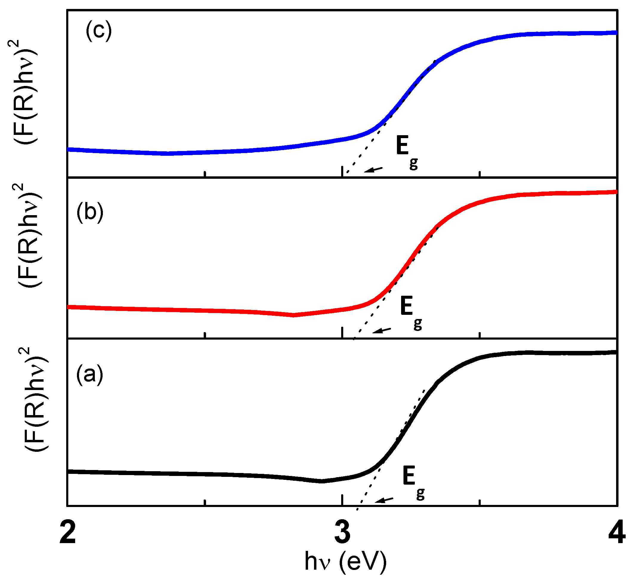
3.3. Optical Bandgap Energy

To study the optical characteristics of the samples, the UV–visible reflectance data were recorded. The optical bandgap energy could be determined by taking into consideration the direct transition between the conduction and valance bands using the Tauc procedure. This method correlates the absorption Kubelka–Munk function F(R) (computed from the reflectance data) with photon energy   h v and the bandgap energy E g   by the following expression [45]:
F ( R ) h v = C ( h v E g ) n
where C is a constant, and n is an exponent that can take values of 0.5, 2, 1.5, and 3 for allowed direct, allowed indirect, forbidden direct, and forbidden indirect transitions, respectively. Extrapolating a linear segment (F(R))2 against (hv) plot to F(R) = 0 gives the direct bandgap energy value (Figure 12). The optical bandgap decreased with increasing temperature. The values of E g are found to be 3.07, 3.05, and 3.01 eV for 1100 °C, 1200 °C, and 1300 °C, respectively, signifying the red shifting in the E g . Many factors can account for changes in the bandgap energy, such as the preparation method, the sintering temperature, and the doping effect. [26,46,47]. In our case, the increase in the sintering temperature caused an increase in the crystallite size. This growth caused a reduction in the density of the grain boundaries leading to a reduction in the barrier’s height at the grain boundaries and, as a consequence, a reduction in the bandgap energy. This reduction in the bandgap energy may be owed to a reduction in the effects of quantum wells or confinements of electrons [46]. Smaller crystals resulted in quantum confinement, that is, spatial confinement of the carriers’ charge by the surface potential barrier or by the potential well of the quantum box [47]. Therefore, the Eg decreased when the average crystallite size increased, and vice versa.

4. Conclusions

The results of the present work showed the role of sintering temperatures in the structural, optical, physical, and radiation-protective traits of BaTiO3 perovskite ceramic. The XRD analysis showed that the samples crystallized into a tetragonal structure. The structural parameters were affected by the sintering temperature changes. The crystallite size increased from 33.25 nm, for the ceramic sintered at 1100 °C, to 34.51 nm and 41.06 nm, corresponding to sintering temperatures of 1200 °C and 1300 °C, respectively. The sintering temperatures had a noteworthy influence on the physical parameters, such as the porosity φ and the relative density RD. The maximum RD (95.27%) and the minimum φ (4.72%) were achieved for the BaTiO3 ceramic sintered at 1200 °C. This led to an enhanced shielding competence against radiation. The optical bandgap energy values were estimated using Tauc plots and were equal to 3.07 eV, 3.05 eV, and 3.01 eV for the ceramics sintered at 1100 °C, 1200 °C, and 1300 °C, respectively. The decreasing trend of Eg was correlated with grain size growth. The impact of the sintering temperatures on the radiation-protecting competence was evaluated using Monte Carlo simulation. The results showed that the µ values enhanced from 28.84 cm−1 to 30.74 cm−1 by raising the sintering temperature between 1100 °C and 1300 °C at an Eγ of 59 keV. The slight enhancement in the µ values was observed to enhance the Δ0.5 values as well as the Δeq. In the present study, the Δ0.5 values decreased from 0.024 to 0.022 cm, and the Δeq decreased at the same energy from 2.06 to 1.90 cm by raising the sintering temperature between 1100 °C and 1300 °C. The increase in the sintering temperature led also to an increase in the RPE and a decrease in the TF of the fabricated ceramic samples. The current work concluded that the fabricated BaTiO3 ceramic samples under various sintering temperatures (especially under sintering temperatures of 1200 °C) have good shielding properties suitable for shielding gamma photons with low and intermediate Eγ values.

Author Contributions

Conceptualization, E.H.; Methodology, E.H. and Y.S.; Software, M.I.S.; Validation, K.G.M.; Formal analysis, E.H. and M.I.S.; Investigation, E.H., K.G.M. and Y.S.; Resources, Y.S.; Writing—original draft, E.H., K.G.M., Y.S. and M.I.S.; Writing—review and editing, E.H. and M.I.S.; Supervision, E.H. and M.I.S.; Funding acquisition, J.A. and Y.M. All authors have read and agreed to the published version of the manuscript.

Funding

This research received no external funding.

Institutional Review Board Statement

Not applicable.

Informed Consent Statement

Not applicable.

Data Availability Statement

Not applicable.

Acknowledgments

The authors acknowledge the Institute for Research and Medical Consultations (IRMC) at Imam Abdulrahman Bin Faisal University (Dammam, Saudi Arabia) for using the Lab facilities.

Conflicts of Interest

The authors declare no conflict of interest.

References

  1. Du, W.; Zhang, L.; Li, X.; Ling, G.; Zhang, P. Nuclear Targeting Subcellular-delivery Nanosystems for Precise Cancer Treatment. Int. J. Pharm. 2022, 619, 121735. [Google Scholar] [CrossRef] [PubMed]
  2. Tanaka, S.; Hosokawa, M.; Tatsumi, A.; Asaumi, S.; Imai, R.; Ogawara, K.-I. Improvement of resistance to oxaliplatin by vorinostat in human colorectal cancer cells through inhibition of Nrf2 nuclear translocation. Biochem. Biophys. Res. Commun. 2022, 607, 9–14. [Google Scholar] [CrossRef]
  3. Pinsky, R.; Sabharwall, P.; Hartvigsen, J.; O’Brien, J. Comparative review of hydrogen production technologies for nuclear hybrid energy systems. Prog. Nucl. Energy 2020, 123, 103317. [Google Scholar] [CrossRef]
  4. Parker, H.M.O.; Joyce, M.J. The use of ionising radiation to image nuclear fuel: A review. Prog. Nucl. Energy 2015, 85, 297–318. [Google Scholar] [CrossRef] [Green Version]
  5. Golden, A.P.; Cohen, S.S.; Chen, H.; Ellis, E.D.; Boice, J.D., Jr. Evaluation of statistical modeling approaches for epidemiologic studies of low-dose radiation health effects. Int. J. Radiat. Biol. 2022, 98, 572–579. [Google Scholar] [CrossRef]
  6. DiCarlo, A.L.; Bandremer, A.C.; Hollingsworth, B.A.; Kasim, S.; Laniyonu, A.; Todd, N.F.; Wang, S.-J.; Wertheimer, E.R.; Rios, C.I. Cutaneous radiation injuries: Models, assessment and treatments. Radiat. Res. 2020, 194, 315–344. [Google Scholar] [CrossRef]
  7. Kaewjaeng, S.; Kothan, S.; Chaiphaksa, W.; Chanthima, N.; Rajaramakrishna, R.; Kim, H.J.; Kaewkhao, J. High transparency La2O3-CaO-B2O3-SiO2 glass for diagnosis X-rays shielding material application. Radiat. Phys. Chem. 2019, 160, 41–47. [Google Scholar] [CrossRef]
  8. Ahmad, I.; Shahzada, K.; Ahmad, M.I.; Khan, F.; Badrashi, Y.I.; Khan, S.W.; Muhammad, N.; Ahmad, H. Densification of Concrete using Barite as Fine Aggregate and its Effect on Concrete Mechanical and Radiation Shielding Properties. J. Eng. Res. 2019, 7, 81–95. [Google Scholar]
  9. Amin, M.N.; Ahmad, I.; Iqbal, M.; Abbas, A.; Khan, K.; Faraz, M.I.; Alabdullah, A.A.; Ullah, S. Computational AI Models for Investigating the Radiation Shielding Potential of High-Density Concrete. Materials 2022, 15, 4573. [Google Scholar] [CrossRef]
  10. Khalaf, M.A.; Ban, C.C.; Ramli, M.; Ahmed, N.M.; Sern, L.J.; Khaleel, H.A. Physicomechanical and gamma-ray shielding properties of high-strength heavyweight concrete containing steel furnace slag aggregate. J. Build. Eng. 2020, 30, 101306. [Google Scholar] [CrossRef]
  11. Mostofinejad, D.; Reisi, M.; Shirani, A. Mix design effective parameters on γ-ray attenuation coefficient and strength of normal and heavyweight concrete. Constr. Build. Mater. 2012, 28, 224–229. [Google Scholar] [CrossRef]
  12. Al-Harbi, N.; Sayyed, M.; Al-Hadeethi, Y.; Kumar, A.; Elsafi, M.; Mahmoud, K.; Khandaker, M.U.; Bradley, D. A novel CaO–K2O–Na2O–P2O5 glass systems for radiation shielding applications. Radiat. Phys. Chem. 2021, 188, 109645. [Google Scholar] [CrossRef]
  13. Kamislioglu, M. Research on the effects of bismuth borate glass system on nuclear radiation shielding parameters. Results Phys. 2021, 22, 103844. [Google Scholar] [CrossRef]
  14. Katubi, K.M.; Ibraheem, A.A.; Alwadai, N.; Alrowaili, Z.; Olarinoye, I.; Sriwunkum, C.; Al-Buriahi, M. Enhancement on radiation shielding performance of B2O3 + Li2O + ZnO + Na2O glass system. Radiat. Phys. Chem. 2022, 201, 110457. [Google Scholar] [CrossRef]
  15. Sayyed, M.; Kumar, A.; Albarzan, B.; Jecong, J.; Kurtulus, R.; Almuqrin, A.H.; Kavas, T. Investigation of the optical, mechanical, and radiation shielding features for strontium-borotellurite glass system: Fabrication, characterization, and EPICS2017 computations. Optik 2021, 243, 167468. [Google Scholar] [CrossRef]
  16. Acikgoz, A.; Demircan, G.; Yılmaz, D.; Aktas, B.; Yalcin, S.; Yorulmaz, N. Structural, mechanical, radiation shielding properties and albedo parameters of alumina borate glasses: Role of CeO2 and Er2O3. Mater. Sci. Eng. B 2022, 276, 115519. [Google Scholar] [CrossRef]
  17. Madbouly, A.; Sallam, O.; Issa, S.A.; Rashad, M.; Hamdy, A.; Tekin, H.; Zakaly, H.M. Experimental and FLUKA evaluation on structure and optical properties and γ-radiation shielding capacity of bismuth borophosphate glasses. Prog. Nucl. Energy 2022, 148, 104219. [Google Scholar] [CrossRef]
  18. Alzahrani, J.S.; Eke, C.; Alrowaili, Z.A.; Boukhris, I.; Mutuwong, C.; Bourham, M.A.; Al-Buriahi, M.S. A theoretical study on the radiation shielding performance of borate and tellurite glasses. Solid State Sci. 2022, 129, 106902. [Google Scholar] [CrossRef]
  19. Mhareb, M.; Slimani, Y.; Alajerami, Y.; Sayyed, M.; Lacomme, E.; Almessiere, M. Structural and radiation shielding properties of BaTiO3 ceramic with different concentrations of Bismuth and Ytterbium. Ceram. Int. 2020, 46, 28877–28886. [Google Scholar] [CrossRef]
  20. Slimani, Y.; Selmi, A.; Hannachi, E.; Almessiere, M.A.; Baykal, A.; Ercan, I. Impact of ZnO addition on structural, morphological, optical, dielectric and electrical performances of BaTiO3 ceramics. J. Mater. Sci. Mater. Electron. 2019, 30, 9520–9530. [Google Scholar] [CrossRef]
  21. Slimani, Y.; Selmi, A.; Hannachi, E.; Almessiere, M.A.; Mumtaz, M.; Baykal, A.; Ercan, I. Study of tungsten oxide effect on the performance of BaTiO3 ceramics. J. Mater. Sci. Mater. Electron. 2019, 30, 13509–13518. [Google Scholar] [CrossRef]
  22. Hannachi, E.; Sayyed, M.; Slimani, Y.; Elsafi, M. Experimental investigation on the physical properties and radiation shielding efficiency of YBa2Cu3Oy/M@ M3O4 (M= Co, Mn) ceramic composites. J. Alloys Compd. 2022, 904, 164056. [Google Scholar] [CrossRef]
  23. Rashid, R.S.M.; Salem, S.M.; Azreen, N.M.; Voo, Y.L.; Haniza, M.; Shukri, A.A.; Yahya, M.-S. Effect of elevated temperature to radiation shielding of ultra-high performance concrete with silica sand or magnetite. Constr. Build. Mater. 2020, 262, 120567. [Google Scholar] [CrossRef]
  24. Hannachi, E.; Mahmoud, K.; Sayyed, M.; Slimani, Y. Effect of sintering conditions on the radiation shielding characteristics of YBCO superconducting ceramics. J. Phys. Chem. Solids 2022, 164, 110627. [Google Scholar] [CrossRef]
  25. Slimani, Y.; Hamad, M.K.; Olarinoye, I.O.; Alajerami, Y.S.; Sayyed, M.I.; Almessiere, M.A.; Mhareb, M.H.A. Determination of structural features of different Perovskite ceramics and investigation of ionizing radiation shielding properties. J. Mater. Sci. Mater. Electron. 2021, 32, 20867–20881. [Google Scholar] [CrossRef]
  26. Hannachi, E.; Mahmoud, K.; Sayyed, M.; Slimani, Y. Structure, optical properties, and ionizing radiation shielding performance using Monte Carlo simulation for lead-free BTO perovskite ceramics doped with ZnO, SiO2, and WO3 oxides. Mater. Sci. Semicond. Process. 2022, 145, 106629. [Google Scholar] [CrossRef]
  27. Şakar, E.; Alim, B.; Özpolat, Ö.F.; Şakar, B.C.; Baltakesmez, A.; Akbaba, U. A surveying of photon and particle radiation interaction characteristics of some perovskite materials. Radiat. Phys. Chem. 2021, 189, 109719. [Google Scholar] [CrossRef]
  28. Kacal, M.R.; Akman, F.; Sayyed, M.I. Investigation of radiation shielding properties for some ceramics. Radiochim. Acta 2019, 107, 179–191. [Google Scholar] [CrossRef]
  29. Hannachi, E.; Sayyed, M.; Slimani, Y.; Almessiere, M.; Baykal, A.; Elsafi, M. Synthesis, characterization, and performance assessment of new composite ceramics towards radiation shielding applications. J. Alloys Compd. 2022, 899, 163173. [Google Scholar] [CrossRef]
  30. Sayyed, M.; Hannachi, E.; Slimani, Y.; Khandaker, M.U.; Elsafi, M. Radiation shielding properties of bi-ferroic ceramics added with CNTs. Radiat. Phys. Chem. 2022, 200, 110096. [Google Scholar] [CrossRef]
  31. Hannachi, E.; Sayyed, M.I.; Mahmoud, K.A.; Slimani, Y.; Akhtar, S.; Albarzan, B.; Almuqrin, A.H. Almuqrin. Impact of tin oxide on the structural features and radiation shielding response of some ABO3 perovskites ceramics (A = Ca, Sr, Ba; B = Ti). Appl. Phys. A 2021, 127, 970. [Google Scholar] [CrossRef]
  32. X-5 Monte Carlo Team, MCNP—A General Monte Carlo N-Particle Transport Code; Version 5, La-Ur-03-1987. II; Los Alamos National Laboratory: Los Alamos, NM, USA, 2003.
  33. Hannachi, E.; Sayyed, M.I.; Almuqrin, A.H.; Mahmoud, K.G. Study of the structure and radiation-protective properties of yttrium barium copper oxide ceramic doped with different oxides. J. Alloys Compd. 2021, 885, 161142. [Google Scholar] [CrossRef]
  34. Naseer, K.; Marimuthu, K.; Mahmoud, K.; Sayyed, M. The concentration impact of Yb3+ on the bismuth boro-phosphate glasses: Physical, structural, optical, elastic, and radiation-shielding properties. Radiat. Phys. Chem. 2021, 188, 109617. [Google Scholar] [CrossRef]
  35. Sayyed, M.; Zaid, M.H.M.; Effendy, N.; Matori, K.A.; Sidek, H.A.A.; Lacomme, E.; Mahmoud, K.; Al Shammari, M.M. The influence of PbO and Bi2O3 on the radiation shielding and elastic features for different glasses. J. Mater. Res. Technol. 2020, 9, 8429–8438. [Google Scholar] [CrossRef]
  36. Abouhaswa, A.; I Sayyed, M.; Altowyan, A.S.; Al-Hadeethi, Y.; Mahmoud, K.A. Synthesis, structural, optical and radiation shielding features of tungsten trioxides doped borate glasses using Monte Carlo simulation and phy-X program. J. Non-Cryst. Solids 2020, 543, 120134. [Google Scholar] [CrossRef]
  37. Varshney, D.; Dar, M.A. Structural and magneto-transport properties of (1 − x) La0. 67Sr0. 33MnO3 (LSMO)+(x) BaTiO3 (BTO) composites. J. Alloys Compd. 2015, 619, 122–130. [Google Scholar] [CrossRef]
  38. Liu, R.; Chen, Z.; Lu, Z.; Wang, X. Effects of sintering temperature and Bi2O3, Y2O3 and MgO co-doping on the dielectric properties of X8R BaTiO3-based ceramics. Ceram. Int. 2022, 48, 2377–2384. [Google Scholar] [CrossRef]
  39. Rani, A.; Kolte, J.; Gopalan, P. Influence of sintering temperature on structural, electrical, and magnetoelectric properties of multiferroic Fe-substituted BaTiO3 ceramics. Appl. Phys. A 2022, 128, 442. [Google Scholar] [CrossRef]
  40. Patterson, A.L. The Scherrer formula for X-ray particle size determination. Phys. Rev. 1939, 56, 978. [Google Scholar] [CrossRef]
  41. Li, W.; Xu, Z.; Chu, R.; Fu, P.; Hao, J. Structure and electrical properties of BaTiO3 prepared by sol–gel process. J. Alloys Compd. 2009, 482, 137–140. [Google Scholar] [CrossRef]
  42. Ying, K.-L.; Hsieh, T.-E. Sintering behaviors and dielectric properties of nanocrystalline barium titanate. Mater. Sci. Eng. B 2007, 138, 241–245. [Google Scholar] [CrossRef]
  43. Singh, M.; Yadav, B.; Ranjan, A.; Kaur, M.; Gupta, S. Synthesis and characterization of perovskite barium titanate thin film and its application as LPG sensor. Sens. Actuators B Chem. 2017, 241, 1170–1178. [Google Scholar] [CrossRef]
  44. Ashiri, R. Detailed FT-IR spectroscopy characterization and thermal analysis of synthesis of barium titanate nanoscale particles through a newly developed process. Vib. Spectrosc. 2013, 66, 24–29. [Google Scholar] [CrossRef]
  45. Kumari, A.; Kumari, K.; Ahmed, F.; Alshoaibi, A.; Alvi, P.; Dalela, S.; Ahmad, M.M.; Aljawfi, R.N.; Dua, P.; Vij, A.; et al. Influence of Sm doping on structural, ferroelectric, electrical, optical and magnetic properties of BaTiO3. Vacuum 2021, 184, 109872. [Google Scholar] [CrossRef]
  46. Rawat, M.; Yadav, K.L. Study of structural, electrical, magnetic and optical properties of 0.65 BaTiO3–0.35 Bi0.5Na0.5TiO3–BiFeO3 multiferroic composite. J. Alloys Compd. 2014, 597, 188–199. [Google Scholar] [CrossRef]
  47. Sahana, M.; Sudakar, C.; Dixit, A.; Thakur, J.; Naik, R.; Naik, V. Quantum confinement effects and band gap engineering of SnO2 nanocrystals in a MgO matrix. Acta Mater. 2012, 60, 1072–1078. [Google Scholar] [CrossRef]
Figure 1. Schematic illustration of the synthesis protocol of the BaTiO3 ceramics.
Crystals 13 00230 g001
Figure 2. Powder X-ray diffraction powders for the BaTiO3 ceramics sintered at (a) 1100 °C, (b) 1200 °C, and (c) 1300 °C.
Crystals 13 00230 g002
Figure 3. Variations of (a) lattice parameters and (b) unit volume cell and c/a ratio as a function of sintering temperatures.
Crystals 13 00230 g003
Figure 4. FTIR spectra of the BaTiO3 ceramics sintered at (a) 1100 °C, (b) 1200 °C, and (c) 1300 °C.
Crystals 13 00230 g004
Figure 5. Influence of the incident γ-photon energy on the µ values of the fabricated ceramic samples.
Crystals 13 00230 g005
Figure 6. Dependence of the µ values on the porosity of the samples (Φ, %).
Crystals 13 00230 g006
Figure 7. Influence of the porosity on both the half-value thickness (Δ0.5, cm) and mean-free path (λ, cm) at a fixed Eγ of 661 keV.
Crystals 13 00230 g007
Figure 8. Dependence equivalent thickness (Δeq, cm) on the emitted γ-photon energy (Eγ, keV) for the prepared samples.
Crystals 13 00230 g008
Figure 9. Influence of the emitted γ-photon energy (Eγ,keV) on the transmission factor (TF,%) and radiation-protection efficiency (RPE,%) of the prepared samples.
Crystals 13 00230 g009
Figure 10. Influence of the thickness of the prepared samples on both the transmission factor (TF, %) and radiation-protection efficiency (RPE, %).
Crystals 13 00230 g010
Figure 11. Dependence of both the transmission factor (TF, %) and radiation-protection efficiency (RPE, %) on the fabricated ceramic samples’ porosity (Φ, %).
Crystals 13 00230 g011
Figure 12. Tauc plots of the BaTiO3 ceramics sintered at (a) 1100 °C, (b) 1200 °C, and (c) 1300 °C.
Crystals 13 00230 g012
Disclaimer/Publisher’s Note: The statements, opinions and data contained in all publications are solely those of the individual author(s) and contributor(s) and not of MDPI and/or the editor(s). MDPI and/or the editor(s) disclaim responsibility for any injury to people or property resulting from any ideas, methods, instructions or products referred to in the content.

